red blood cells/ erythrocyte. this red blood cell carries oxygen from the lungs to tissues and...
TRANSCRIPT

Red Blood Cells/ Erythrocyte

• This red blood cell carries oxygen from the lungs to tissues and exchanges it for carbon dioxide to carry back to the lungs for exhalation. Unlike most cells, red blood cells have a nucleus only when they are first formed and lose it as they mature and pack themselves with red, oxygen- rich hemoglobin molecules. They must be pliable and fold so they can enter the tiny capillaries that extend to the far reaches of the body. Unsaturated fatty acids in the diet are important for this pliablity. [Red blood cells are red, platlets are green]

Amoeba

• The amoeba proteus, a single-celled organism named for the Greek god Proteus, has extensions to the cell, known as pesudopodia, which allow it to move and capture prey.

Adreanal cell

• In this portaion of an adreanal cell, the dark oval shapes are lysosomes. A lysosome is a relatively large vesicle which contains enzymes involved in breaking down proteins, polysaccharids, and lipids. They may contain forty or more different hydrolytic enzymes.

Green-algae

• The Pandorina, a green-algae protist, is made up of 32 cells, held together by a jullylike substance, whose outlines you can see. Each of these cells can survive independently of the others. To reproduce, each cell divides, producing a new cell inside, and then the parent colony breaks apart.

Starch molecules

• These starch molecules are important carbohydrates used in cells for mainly storage. Chemically, starch is a polysaccharide, formed by combining many units of glucose.

Poplar Tree Cell

• A single living cell from the leaf of a poplar tree.

Escherichia coli

• Shown are cells of Escherichia coli, the heterotrophic prokaryote that is most thoroughly studied of all living organisms. The DNA of the E. coli is the lighter appearing area in the center of each cell; this region, which is not enclosed by a membrane, in known as the nucloid. The two main cells in the center of the picture have just finished dividing and have not yet separated completely.

Chlamydomonas

• An electron micrograph of Chlamydomonas, a photosynthetic eukaryotic cell. Some of the organelles which can be seen from the micrograph are the nucleus (center), mitochondrion, and chloroplasts (dark, irregularly shaped). The large circle near the bottom of the cell is the organism's food reserve, in the form of starch granules.

Chlamydomonas
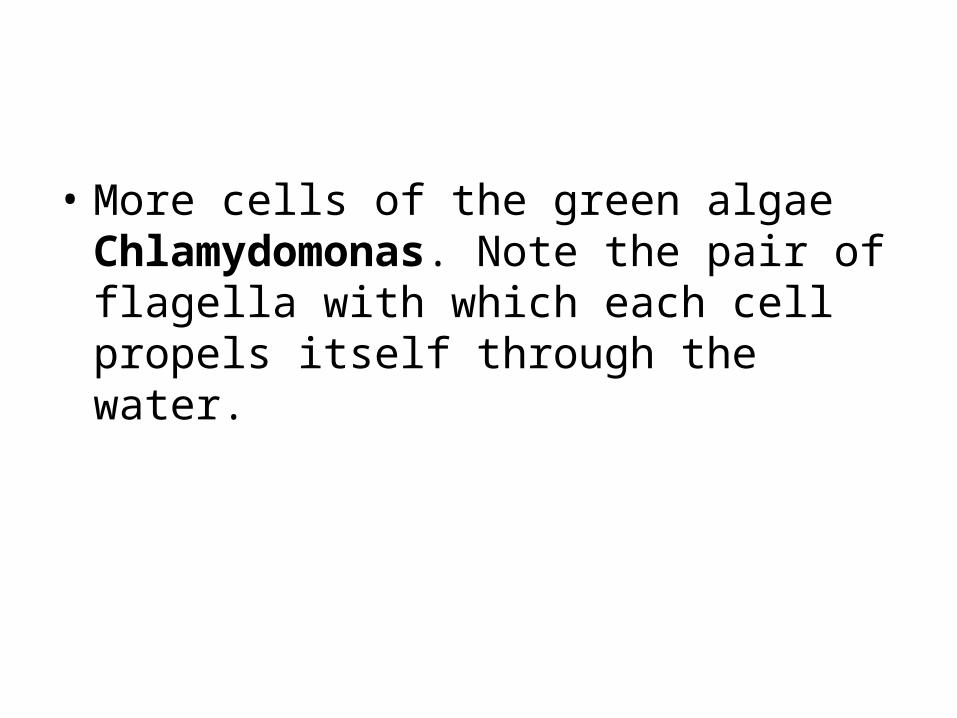
• More cells of the green algae Chlamydomonas. Note the pair of flagella with which each cell propels itself through the water.

Corn plant

• This is an electron micrograph of cells from the leaf of a corn plant. The nucleus is the large circle on the right side of the central cell. Diagonal to the nucleus is a vacuole, quite characteristic of plant cells. There are also many mitochondria and chloroplasts (the dark, membrane-enclosed objects).

Embryo of a sea urchin

• Shown is the embryo of a sea urchin at the two-celled stage of deveopment. Within each cell is a nucleus that carries all the genetic information needed for every cell in the mature sea urchin.

Peroxisome

• Shown here is a peroxisome. The crystalline material in the center is a peroxide-producing enzume involved in the break down of purines.

Cells from the cerebral cortex

• These cells from the cerebral cortex are the most highly organized structures on earth. The actions of these cells and the interconnections among them are respondible for consciousness, intellignece, dreams, and memory.

Mitochondria

• Shown here is an electron micrograph of a mitochondrion. Mitochondria are surrounded by two membranes; the inner membrane folds inward to make a series of shelves called cristae. These cristae play important roles in the energy-releasing chemical reactions of cellular respiration that occur in the mitochondria.

Chloroplast

• Shown here is a mature chloroplast. Like a mitochondrion, it is surrounded by two membranes. Additionally, it contains an elaborate internal membrane system where the light-capturing reactions of photosynthesis occur.

White blood cell

• In this electron micrograph, a human white blood cell is trapping bacterial cells. This type of cell defends the body against pathogens and other harmful particles by engulfing foreign objects and destroying them with the help of enzymes from the cell's lysosomes.

Pancreas cell

• Shown here is an electron micrograph of a portion of a cell from the pancreas. The cell has a large, central nucleus with scattered chromatin, many mitochondria, large quantities of rough ER, and many small vesicles. Not only is the cell itself enclosed by a membrane, but the nucleus is surrounded by a double membrane system called the nuclear envelope, and certain organelles are also membrane-enclosed.