reduction of patient motion artifacts in digital ... · patient motion artifacts are a major cause...
TRANSCRIPT
Reduction of Patient Motion Artifacts in Digital
Subtraction Angiography: Evaluation of a Fast
and Fully Automatic Technique
Erik H. W. Meijering, Wiro J. Niessen, Jeannette Bakker,Aart J. van der Molen, Gerard A. P. de Kort, Rob T. H. Lo,
Willem P. Th. M. Mali, Max A. Viergever
Radiology, vol. 219, no. 1, April 2001, pp. 288–293
Abstract—The performance of an automatic technique for reduction of patient motion artifactsin digital subtraction angiography (DSA) was evaluated. Four observers assessed the image qualityof 104 cerebral DSA images, which were processed by both the automatic technique and manualpixel shifting. The automatic technique was found to result in better image quality and to beconsiderably less time consuming.
Keywords—Cerebral angiography, digital subtraction angiography, image artifacts, image pro-cessing, image registration, image quality.
INTRODUCTION
Patient motion artifacts are a major cause of image quality degradation in digital subtrac-tion angiography (DSA). Although several techniques have been proposed over the pasttwo decades to improve the acquisition of DSA images in relation to this problem [6], mo-tion artifacts cannot be entirely avoided. Currently, the only post-processing techniquesavailable on clinical DSA devices are manual remasking and pixel shifting, which allowfor reduction of artifacts caused by uniform translational motion only [5, 6]. Generally,however, patient movements have a more complex nature, which limits the effectivenessof these reduction techniques. This problem has been recognized by researchers in thefield of image processing and has been the incentive to the development of a number ofsemi- or even full-automatic, nonlinear retrospective registration techniques [6]. However,apart from two exceptions [4, 8], clinical evaluations of these techniques have never beenreported. Another major problem with these techniques is that they are generally tootime consuming for use in clinical practice.
Recently, a new, fully automatic registration technique was developed, which is capableof aligning pairs of images nonlinearly within less than a second [7]. In this study we evalu-ated the effectiveness of this technique in reducing patient motion artifacts, by comparisonwith manual pixel shifting. The study was carried out on cerebral DSA images.

PP-2 Evaluation of a Technique for Motion Artifact Reduction in DSA
MATERIALS AND METHODS
Images and Equipment
During a five-month period, 104 cerebral X-ray angiography runs from 21 patients (13men and 8 women, age range 28–82 years) were archived digitally. From each run, werandomly selected one mask-contrast image pair, of which the corresponding DSA imagehad been printed on film by the radiologists at the time of the initial examination. Allimages had been acquired on an Integris V3000 C-arm imaging system (Philips MedicalSystems, Best, the Netherlands), with a 20cm or 25cm image intensifier, a matrix size ofeither 512 × 512 pixels (31 images) or 1024 × 1024 pixels (73 images), and a grey-levelresolution of 10 bits per pixel.
Post-processing operations as well as image quality assessments were carried out onan Octane® workstation (Silicon Graphics, De Meern, the Netherlands) with one 195MHzMIPS R10000 processor, 256MB main memory (instruction and data cache size both32KB), and an IMPACTSR graphics board with 4MB texture memory. All images weredisplayed in a window of 700×700 pixels on a 19-inch monitor (Silicon Graphics, De Meern,the Netherlands), which had a resolution of 1280 × 1024 pixels (refresh rate 75Hz). Byusing this window, images were displayed with the same effective diameter (≈ 11.5 inch) asthey are usually displayed on the 15-inch progressive display monitor of the Integris V3000.The contrast and brightness settings of the window were fixed during the evaluation.
Manual and Automatic Registration
Manual correction for motion artifacts in the DSA images corresponding to the 104 mask-contrast pairs was obtained by using a special pixel-shifting tool that could be executedon the Octane® workstation and that exactly mimiced the pixel-shifting facility on theviewing console of the Integris V3000 used in daily practice. Automatic correction wasobtained by using the algorithm by Meijering et al. [7], briefly described below (see alsoFigure 1). First, the algorithm applies edge detection to the mask image in order to ex-tract regions that have a high potential for showing artifacts. Next, control points for thewarping operation are automatically selected at local maxima of the gradient magnitude,while constraining the minimum and maximum distance between these points. The localdisplacements of image structures at the control points are then computed by means ofa template matching procedure based on the energy similarity measure [2], followed bydetection and correction of inconsistent displacement vectors by comparison with neigh-boring vectors. Finally, the mask image is warped according to the displacement vectorfield resulting from linear interpolation of the local displacements at the control points.This is done very efficiently by using a triangulation of the control points in combinationwith hardware accelerated texture-mapping. For the present study, all parameters of thealgorithm were fixed to the proposed values [7].
Method of Evaluation
Four observers (three radiologists and a resident) participated in the evaluation, whichconsisted of two parts. In the first part, manual registrations of the 104 mask-contrastimage pairs were carried out separately and independently by the four observers, using thepixel-shifting tool. Since optimal manual registration of an image pair is task dependent,the observers were provided with the clinical indication for acquisition of the images,which was either a cerebral aneurysm (41 images; 7 patients), a stenosis in the carotid

Evaluation of a Technique for Motion Artifact Reduction in DSA PP-3
Figure 1. Example of the several stages in the automatic registration algorithm evaluatedin this study. Top-left: mask image of a lateral cerebral DSA image. Top-right: output ofthe edge detection algorithm (grey regions) and control-point selection mechanism (whitedots). Bottom-left: live image overlayed with the automatically computed local displace-ment vectors, indicating the correspondence with the mask image. From the vector field itis clear that in this example the patient’s movement was of a rotational nature. Bottom-right: triangulation of the set of control points used for interpolation of the displacementvectors and final warping of the mask image.
arteries (34 images; 8 patients), a tumor (20 images; 4 patients), or vasculitis (9 images; 2patients). However, the images were presented in random order. The final horizontal andvertical mask shifts for all mask-contrast pairs as indicated by each of the observers werestored automatically by the computer, together with the time it took for each observerto carry out the manual registration of each pair. The resulting manually corrected DSAimages were also stored. The DSA images resulting from automatic registration of allmask-contrast pairs were computed and stored separately.
PP-4 Evaluation of a Technique for Motion Artifact Reduction in DSA
The second part of the study concerned the comparison of the quality of the auto-matically and manually corrected DSA images. To this end, the following three DSAimage pairs were formed for each of the 104 original DSA images: (i) the automaticallycorrected DSA image and the original DSA image, (ii) the manually corrected DSA imageand the original DSA image, and (iii) the automatically corrected DSA image and themanually corrected DSA image. This resulted in a total of 312 DSA image pairs, whichwere presented to the observers. Although the original and automatically corrected DSAimages were the same for all four observers, each of the observers was confronted with hisor her own manual corrections resulting from the first part. For each of the DSA imagepairs, the differences between the two images (denoted “Image A” and “Image B”) couldbe assessed by alternating the image that was displayed.
The observers were given the clinical information of all images and were asked to ratethe relative quality of the two images by choosing one of the following: (AB) Image Aand Image B are similar (i.e., the number of artifacts and the magnitude of the artifactsis the same in the diagnostically relevant parts or in the entire images), (A+) Image Ais better than, or (A++) much better than Image B (i.e., the number of artifacts or themagnitude of the artifacts in Image A is smaller, or much smaller than in Image B, inthe diagnostically relevant parts or in the entire image), (B+) Image B is better than, or(B++) much better than Image A (i.e., the number of artifacts or the magnitude of theartifacts in Image B is smaller, or much smaller than in Image A, in the diagnosticallyrelevant parts or in the entire image).
Similar to the first part, the second part of the study was carried out separately andindependently by the four observers. However, prior to this part, there was a meetingbetween the observers in order to obtain consensus regarding the rating of relative imagequality. For this consensus meeting, 10 sample cerebral DSA image pairs were used, whichwere not included in the actual study.
To avoid bias in the ratings, the images were presented to the observers in a completelyrandomized and blinded fashion; not only were the 312 DSA image pairs randomized, butalso the order of the images within each pair was randomized, and the observers wereignorant of the type of correction (no, manual, or automatic correction) that was appliedto the images. Furthermore, to reduce the possibility of observers recognizing their ownmanual corrections, the time period between the first and the second part of the studywas at least three weeks for each of the observers.
Statistical Analyses
Inter-observer agreement for the image quality ratings resulting from the second part ofthe study was assessed by using a kappa (κ) test. In order to take account of the degreeof disagreement, we used the weighted kappa (κw) test, the weights for discrepancies of 0,1, 2, 3, and 4 categories in the ratings being 1, 0.75, 0.5, 0.25, and 0, respectively [1, 3].Six κw values were computed, based on a comparison of the ratings of two observers at atime. A κw value of 1.0 indicates that the agreement is perfect and a value of 0.0 that it isnot different from chance agreement. For the interpretation of κw values in between theseextremes, we used the Landis-Koch guidelines [1].
The ratings resulting from the second part allowed us to make both implicit and explicitcomparisons of the effectiveness of the automatic and the manual registration technique inreducing motion artifacts. For this purpose, the ratings of the 312 DSA image pairs weredivided into three groups: (i) ratings expressing the quality of automatically corrected DSAimages relative to corresponding original (uncorrected) DSA images (or vice versa), (ii)

Evaluation of a Technique for Motion Artifact Reduction in DSA PP-5
Original rating
Relative image quality A++ A+ AB B+ B++
Image A compared to Image B ++ + 0 − −−Image B compared to Image A −− − 0 + ++
Table 1. Rules for conversion of the original ratings resulting from the second part of thestudy, necessary in order to be able to express the quality of any one of the DSA imagesin a given pair in terms of the other.
ratings expressing the quality of manually corrected DSA images relative to correspondingoriginal (uncorrected) DSA images (or vice versa), and (iii) ratings expressing the qualityof automatically corrected DSA images relative to corresponding manually corrected DSAimages (or vice versa). Since the images of each pair were presented in random order, theoriginal ratings were converted by using the rules presented in Table 1 in order to be ableto express the quality of any of the images in a given pair in terms of the other. Implicitcomparison of the performance of the automatic and manual registration technique wasthen obtained by constructing a frequency table of the converted ratings from groups (i)and (ii). Explicit comparison was obtained by analyzing the ratings from group (iii). Thesecomparisons were carried out separately for the results of each observer. A comparisonbased on the average frequencies was also carried out.
The statistical significance of the possible improvement of the automatic registrationtechnique over manual pixel shifting was assessed by using a Chi-squared (χ2) test [1, 3]applied to the frequency tables containing the ratings from groups (i) and (ii). Since oneof the variables in these tables (viz., relative image quality) represents ordered categories,we did not use the ordinary χ2 test, but the more powerful χ2 test for linear trend [1,3].For this test, we used uniform spacing of the categories. The null hypothesis was thatthe automatic and manual registration technique would be equally effective in reducingmotion artifacts. A probability of p < 0.05 for this hypothesis was chosen to indicate astatistically significant difference between the two techniques.
RESULTS
In the first part of the study, the observers found that most of the 104 original cerebralDSA images could be improved to some extent by manual correction, since on average88% of the mask shift parameters differed from zero. The maximum shift recorded ineither direction was 8.0 pixels, while the average length of the shift vectors of all fourobservers was 1.2 pixels. From this it may be concluded that, although in some casespatient motion was quite severe, in most cases the motion artifacts were due to relativelysmall displacements only. The timing information stored along with the shift parametersrevealed that manual correction required on average about 12 sec. (median: 7 sec.) perDSA image. In contrast, the automatic registration algorithm required on average onlyabout 1 sec. (median: 1 sec.) per DSA image.
The κw values computed from the ratings in the second part ranged from 0.60 to 0.71.According to the Landis-Koch guidelines, this indicates substantial agreement. Therefore,we restrict ourselves to presenting averages. The average frequencies of the ratings fromgroups (i) and (ii), as described in Section , are presented in Table 2. From the implicitcomparison based on the results in this table it follows that, overall, the automatic regis-

PP-6 Evaluation of a Technique for Motion Artifact Reduction in DSA
Relative image quality
Comparison −− − 0 + ++
Manually corrected versus original 0% 4% 25% 62% 9%Automatically corrected versus original 0% 0% 15% 69% 16%
Table 2. Average frequencies of the ratings resulting from the comparison of correctedDSA images and their corresponding original (uncorrected) DSA images. Manual correc-tions were obtained by using the pixel shifting technique, and automatic corrections byusing the technique described by Meijering et al. [7].
Relative image quality
Comparison −− − 0 + ++
Automatically versus manually corrected 0% 5% 44% 48% 3%
Table 3. Average frequencies of the ratings resulting from the explicit comparison of auto-matically corrected DSA images and their corresponding manually corrected DSA images.Manual corrections were obtained by using the pixel shifting technique, and automaticcorrections by using the technique described by Meijering et al. [7].
tration technique resulted in better image quality than manual pixel shifting. In order tobe able to apply the χ2 test for trend, the frequencies in the columns “−−” and “−” hadto be combined, since this test does not allow rows or columns to be entirely filled withzeroes. The χ2 test for trend applied to the modified frequency table showed that theprobability for the null hypothesis of equal effectiveness to be true is p < 0.05, from whichit can be concluded that the automatic correction technique is statistically significantlybetter than manual pixel shifting in reducing motion artifacts. We note that the sameconclusion was found with this test applied to the results of the observers separately. Theaverage frequencies of the ratings from group (iii), representing the results of the explicitcomparison of the quality of automatically and manually corrected images, are presentedin Table 3. These results support the conclusion drawn from the implicit comparison.
Two examples of cases in which the automatic registration technique was found tobe superior to manual pixel shifting are given in Figures 2 and 3, where the artifacts inthe original DSA images are located primarily in the lower part of the image, around themain vessels. Although the artifacts could be removed to some extent by manual pixelshifting, it was not possible to completely remove them using this technique. In the caseof Figure 2, pixel shifting even resulted in a deterioration of artifacts in the lower rightpart of the image. Application of the automatic registration technique, on the other hand,resulted in overall correction and improved vessel visibility.
DISCUSSION
Of the 104 DSA images included in this study, 38 (37%) had been manually correctedby means of pixel shifting before being printed on film and stored in the archive of ourhospital after initial examination. From the fact that, in the first part of the study, theobservers found that no less than 92 (88%) of the images could be improved to some extent

Evaluation of a Technique for Motion Artifact Reduction in DSA PP-7
Figure 2. First example of a case in which the automatic registration technique wasfound to be superior compared to manual pixel shifting. Top-left: original lateral cerebralDSA image. Top-right and bottom-left: resulting DSA image after manual registration bymeans of pixel shifting, for two of the four observers. Due to the rotational nature of thepatient’s movement, it was not possible to obtain an overall optimal correction of motionartifacts by means of this technique. From these two images it is clear that a reductionof artifacts in one part of the image (in this example the lower left part) may result in adeterioration of artifacts elsewhere (in this example the lower right part). bottom-right:DSA image resulting from application of the automatic registration technique.
by this technique, we conclude that in practice more images contain motion artifacts thanare usually corrected. Manual correction of all images by means of pixel shifting is alabor intensive operation: the results of our study indicated that, on average, 12 sec. perDSA image are required to apply this technique optimally. Apart from resulting in betteroverall image quality than manual pixel shifting, the automatic technique is considerablyfaster. On average, it requires only 1 sec. per DSA image. Moreover, the technique doesnot require any effort from the radiologist.
PP-8 Evaluation of a Technique for Motion Artifact Reduction in DSA
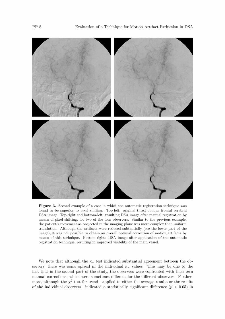
Figure 3. Second example of a case in which the automatic registration technique wasfound to be superior to pixel shifting. Top-left: original tilted oblique frontal cerebralDSA image. Top-right and bottom-left: resulting DSA image after manual registration bymeans of pixel shifting, for two of the four observers. Similar to the previous example,the patient’s movement as projected in the imaging plane was more complex than uniformtranslation. Although the artifacts were reduced subtantially (see the lower part of theimage), it was not possible to obtain an overall optimal correction of motion artifacts bymeans of this technique. Bottom-right: DSA image after application of the automaticregistration technique, resulting in improved visibility of the main vessel.
We note that although the κw test indicated substantial agreement between the ob-servers, there was some spread in the individual κw values. This may be due to thefact that in the second part of the study, the observers were confronted with their ownmanual corrections, which were sometimes different for the different observers. Further-more, although the χ2 test for trend—applied to either the average results or the resultsof the individual observers—indicated a statistically significant difference (p < 0.05) in
Evaluation of a Technique for Motion Artifact Reduction in DSA PP-9
image quality after application of the automatic correction technique and manual pixelshifting, the outcome of an ordinary χ2 test would have been somewhat less persuasive:for two of the observers, this test would have given p < 0.1 for the null hypothesis tobe true. However, the χ2 test for trend is more appropriate, since we are dealing withordered categories.
As clearly illustrated by the examples in Figures 2 and 3, manual pixel shifting oftenresults in improved image quality in and near the diagnostically relevant parts of animage, but may sometimes result in deterioration of artifacts in other parts. This is adirect consequence of the fact that with this technique, patient motion as projected inthe imaging plane is assumed to be uniform translational. One may argue that this isnot really a problem in practice as long as artifacts can be reduced in the diagnosticallyrelevant parts of the image, and that it is therefore sufficient to use manual pixel shiftingrather than a more sophisticated automatic correction technique. However, even if therewould be no difference in performance from the point of view of image quality, it wouldstill be advantageous to use the automatic technique evaluated in this paper, since it isconsiderably less time consuming.
The fact that the automatic registration technique performed statistically significantlybetter than manual pixel shifting does not imply that in practice the former techniquewill always be better than the latter. The average results from the explicit comparison ofthe two techniques (Table 3) indicated that in 5% of all cases, the corrected DSA imageresulting from manual pixel shifting was found to be better than the corresponding auto-matically corrected DSA image. We observed that in these cases the automatic techniquedid not introduce new artifacts, but it was unable to reduce some of the artifacts at theborders of the images. This may be caused by the lack of image content in those regions,which reduces the possibilities for any template matching procedure to find the correctlocal displacement vectors.
In most reports on reduction of motion artifacts in DSA images, the evaluation ofnewly developed techniques involved only one or at most a few clinical DSA images, orphantoms, and the quality of the resulting corrected images was assessed by the same per-sons that developed the algorithm. To the best of our knowledge, the only more elaborateand objective evaluation studies are the ones by Takahashi et al. [8] and Hayashi et al. [4].In the former study, three techniques were evaluated: manual remasking, manual pixelshifting, and an automatic registration technique. It was concluded that remasking wasmost effective. It was also found that, after having applied remasking, remaining artifactswere reduced equally well by manual pixel shifting and their automatic registration tech-nique. In the study of Hayashi et al. [4], the authors compared the performance of twotechniques: manual pixel shifting and an automatic registration technique developed bysome of the co-authors of that study. In 14 out of 16 cerebral DSA image series, the imagesresulting from the automatic registration technique were found to have better quality. Inthe other two cases, the techniques were found to perform comparably.
Due to the lack of detailed information provided by the authors of these papers, itis difficult to explicitly compare their findings to ours. We note, however, that the au-tomatic registration technique evaluated in our study is based on approaches which havebeen shown to yield faster and more accurate registrations compared to their techniques(see Meijering et al. [6,7] for more technical details). Hayashi et al. [4] reported that theiralgorithm required about eight minutes of computation time. In contrast, the algorithmevaluated in our study requires on average only about 1 sec. per DSA image, which cer-tainly makes it more suitable for use in clinical practice. Furthermore, owing to the useof better similarity measures in the template matching procedure, the images processed
PP-10 Evaluation of a Technique for Motion Artifact Reduction in DSA
by our algorithm are either comparably to, better than, or even much better than thoseresulting from manual pixel shifting in 95% of all cases.
Finally, we mention the fact that our study involved only images that were alreadyconsidered clinically useful. Frequently it occurs that, during acquisition, the patient’smovements are too severe to result in diagnostically useful DSA images, even when usingpixel shifting afterwards, and in such cases the run is repeated. In some cases, the auto-matic registration technique might help avoid a second DSA run. On-line availability ofthe automatically corrected DSA images would offer the radiologist the possibility to checkdirectly whether a new run must be acquired, thereby avoiding the need to go back to theconsole to check it manually by means of pixel shifting. We also note that in our study,we were only interested in overall image quality improvement, without relation to specificdiagnostic tasks, such as the grading of stenoses or the detection of small aneurysms. Itmay be that the automatic registration technique also implies an improvement in thatrespect compared to manual pixel shifting. Confirmation of these claims is the goal offuture studies.
ACKNOWLEDGMENTS
The research described in this paper was carried out at the University Medical CenterUtrecht (UMCU), the Netherlands, and was financially supported by the NetherlandsMinistry of Economic Affairs. The authors are grateful to Philips Medical Systems (Best,the Netherlands) for making available their clinical Octane® workstation (Silicon Graph-ics, De Meern, the Netherlands) on which the evaluation was carried out. Gerard vanHoorn, Tineke Kievit, Wilma Pauw, Koen Vincken, Theo van Walsum, Remko van derWeide, and Onno Wink (all with the UMCU) are acknowledged for their assistance in thestart-up phase of this study.
REFERENCES
[1] D. G. Altman, Practical Statistics for Medical Research, Chapman & Hall, London, UK, 1991.
[2] T. M. Buzug & J. Weese, “Image Registration for DSA Quality Enhancement”, Computerized MedicalImaging and Graphics, vol. 22, no. 2, 1998, pp. 103–113.
[3] J. L. Fleiss, Statistical Methods for Rates and Proportions, 2nd ed., Wiley Series in Probability andMathematical Statistics, Wiley, New York, USA, 1981.
[4] N. Hayashi, T. Sakai, M. Kitagawa, R. Inagaki, N. Sadato, Y. Ishii, Y. Nishimoto, M. Tanaka,T. Fukushima, H. Komuro, H. Ogura, H. Kobayashi, T. Kubota, “Nonlinear Geometric Warpingof the Mask Image: A New Method for Reducing Misregistration Artifacts in Digital SubtractionAngiography”, CardioVascular and Interventional Radiology, vol. 21, no. 2, 1998, pp. 138–141.
[5] D. C. Levin, R. M. Shapiro, L. M. Boxt, L. Dunham, D. P. Harrington, D. L. Ergun, “Digital Sub-traction Angiography: Principles and Pitfalls of Image Enhancement Techniques”, American Journalof Roentgenology, vol. 143, no. 3, 1984, pp. 447–454.
[6] E. H. W. Meijering, W. J. Niessen, M. A. Viergever, “Retrospective Motion Correction in DigitalSubtraction Angiography: A Review”, IEEE Transactions on Medical Imaging, vol. 18, no. 1, 1999,pp. 2–21.
[7] E. H. W. Meijering, K. J. Zuiderveld, M. A. Viergever, “Image Registration for Digital SubtractionAngiography”, International Journal of Computer Vision, vol. 31, no. 2/3, 1999, pp. 227–246.
[8] M. Takahashi, J. Shinzato, Y. Korogi, K. Fukui, S. Ueno, I. Horiba, N. Suzumura, “AutomaticReregistration for Correction of Localized Misregistration Artifacts in Digital Subtraction Angiographyof the Head and Neck”, Acta Radiologica (Supplementum), vol. 369, 1986, pp. 281–284.