research article acceptance:01/19/2018
TRANSCRIPT

Asian Pacific Journal of Cancer Prevention, Vol 19 667
DOI:10.22034/APJCP.2018.19.3.667 Expression and Regulation of IGFBP7 in Gastric Cancer
Asian Pac J Cancer Prev, 19 (3), 667-675
Introduction
A multistep process of genetic and epigenetic alterations results in gastric cancer tumorigenesis and progression (Yasui et al., 2000; Tamura, 2002). Gastric cancer incidence exhibits complex process, including oncogene activation and tumor suppressor gene inactivation; however, the molecular pathogenesis for gastric cancer remains largely unknown and therapeutic targets are limited. Aberrant DNA hypermethylation within CpG island regions results in chromatin compaction or down-regulation of tumor suppressor genes (Tamura, 2002; Lo et al., 2010; Takada et al., 2010), and alterations have been recognized as an important mechanism involved in gastric carcinogenesis.
The insulin-like growth factor (IGF) signaling pathway has a key role in regulating cell proliferation, differentiation, and apoptosis (Fürstenberger and Senn, 2002). The IGF system is composed of IGFs, IGF receptors, and IGF-binding proteins (IGFBPs) with high- and low-affinity for IGFs (Kim et al., 1997; Khandwala et al., 2000; Pollak, 2012). Disruption or inhibition of IGF receptor type 1 (IGF-1R) activity has been shown to inhibit the growth and motility of cancer cells (Khandwala et al., 2000; Pollak, 2012). IGF-1R binding with its ligand, IGF-1, results in receptor phosphorylation and activation of MAPK and PI3K/Akt
Abstract
Background: Insulin-like growth factor-binding protein 7 (IGFBP7) has been found to be a tumor suppressor in several human cancers, but the role of IGFBP7 in gastric cancer has not yet been fully investigated. Herein, we examined the epigenetic downregulation of IGFBP7 expression in gastric cancer. Methods: Expression and methylation of IGFBP7 in gastric cancer cells and primary gastric cancer patients were determined using qRT-PCR, western blot, immunohistochemistry, and methylation specific-PCR, respectively. The effects of IGFBP7 on gastric cancer cells were investigated by various experimental conditions, such as proliferation, colony formation, apoptosis, invasion, and migration assay. Results: IGFBP7 methylation was inversely correlated with IGFBP7 expression in gastric cancer. Univariate and multivariate analysis showed that IGFBP7 expression and tumor stage were independent prognostic factors. IGFBP7 knockdown increased gastric cancer cell growth, invasion, and migration, whereas IGFBP7 overexpression in gastric cancer cells induced cell growth inhibition and apoptosis. Conclusion: Our data suggest that IGFBP7 functions as a tumor suppressor in gastric cancer via an epigenetic pathway.
Keywords: IGFBP7- gastric cancer- tumor suppressor- downregulation- methylation
RESEARCH ARTICLE
Epigenetic Downregulation and Growth Inhibition of IGFBP7 in Gastric CancerJin Kim1,2, Woo Ho Kim1,2, Sun-Ju Byeon2, Byung Lan Lee3, Min A Kim2*
signaling (Samani et al., 2007; Pollak, 2012). Ligand bioavailability is regulated in part by the IGFBP family, which can bind to IGFs and regulate interactions of ligands with their receptors (Kim et al., 1997; Pollak, 2012). IGFBP7 shares 30% structural homology with IGFBP1 to IGFBP6 at its N-terminal domain; however, IGFBP7 binds to IGFs (IGF-1 and IGF-2) with low affinity (Oh et al., 1996; Burger et al., 2005), indicating that its individual characteristics differ from these other IGFBP family members (IGFBP1 to IGFBP6). IGFBP7 is silenced by DNA methylation in various types of tumors (Lin et al., 2007; Sullivan et al., 2012; Suzuki et al., 2013). However, the function of IGFBP7 in gastric carcinogenesis is not fully understood to date. Aberrant expression of IGFBP7 has been associated with the biological b e h a v i o r o f t u m o r s a n d c l i n i c a l o u t c o m e (An et al., 2012; Tomimaru et al., 2012). Accordingly, investigation of regulatory mechanisms underlying IGFBP7 expression is crucial to improve understanding and treatment of gastric cancer.
Materials and Methods
Cell lines and tissues Ten gastric cancer cell lines (SNU-1, -5, -16, -216,
-484, -601, -620, -638, -668, -719) were obtained from the Korean Cell Line Bank (http://cellbank.snu.ac.kr, Seoul,
1Cancer Research Institute, 2Department of Pathology, 3Department of Anatomy, Seoul National University College of Medicine, Seoul, Korea. *For Correspondence: [email protected]
Editorial Process: Submission:04/28/2017 Acceptance:01/19/2018

Jin Kim et al
Asian Pacific Journal of Cancer Prevention, Vol 19668
Korea). All cell lines were maintained in RPMI-1640 (HyClone, Logan, UT, USA) supplemented with 10% fetal bovine serum (HyClone), 100 units/ml penicillin, and 100 µg/ml streptomycin (HyClone) at 37°C and 5% CO2. Surgically resected formalin-fixed paraffin-embedded human gastric cancer tissues (n=393), and fresh gastric cancer tissues and paired normal tissues (n=37) were obtained during surgery at the Seoul National University Hospital in 2004. Clinicopathological parameters were evaluated by reviewing medical charts, pathology reports, and glass slides. Patients lost due to follow-up and deaths attributed to causes other than gastric cancer were censored during survival analysis. This retrospective study design was approved by the Ethics Committee of the Institutional Review Board at Seoul National University Hospital under the condition of anonymization (No. H-1611-008-803).
Quantitative reverse transcription polymerase chain reaction (qRT-PCR)
Total RNA was extracted using TRIZOL reagent (Invitrogen, Carlsbad, CA, USA). cDNA was synthesized using SuperScriptTM III RT (Invitrogen). TaqMan gene expression assays were performed for IGFBP7: Hs00944482_m1 and Glyceraldehyde-3-phosphate dehydrogenase (GAPDH) : Hs99999905_m1 (both Applied Biosystems, Foster City, CA, USA). qRT-PCR assays were performed in triplicate using a 7500 Real Time PCR System (Applied Biosystems), and data were evaluated using SDS (Ver. 1.4) software. After reaction, relative mRNA expression was normalized with the endogenous control gene GAPDH.
Western blot analysis C e l l u l a r p r o t e i n s w e r e e x t r a c t e d w i t h
Pro-Prep™ for cell/tissue protein extraction solution (iNtRON Biotechnology, Seongnam, Korea), and protein levels were determined using BCA protein assay kits (Pierce, Rockford, IL, USA). Anti-IGFBP7 (1:500; R&D systems, Minneapolis, MN, USA), anti-phospho-Akt (1:500; Cell Signaling, Danvers, MA, USA), anti-AKT (1:500; Epitomics, Burlingame CA, USA), anti-phospho-ERK (1:500; Cell Signaling), anti-ERK (1:500; Cell Signaling), anti-CDK2 (1:200; Santa Cruz Biotechnology, Dallas, Texas, USA), anti-Cyclin D1 (1:200; Santa Cruz Biotechnology), anti-cleaved caspase 3 (1:500; Cell Signaling), anti-phospho-IGF-1R (1:500; Santa Cruz Biotechnology), anti-IGF-1R (1:100; Santa Cruz Biotechnology), and anti-β-actin (1:5000; Sigma Aldrich, St. Louis, MO, USA) were used as primary antibodies. Following overnight incubation at 4°C, blots were incubated with secondary antibodies and visualized using ECL kits (Pierce).
5-Aza-2’-deoxycytidine (5-aza-dc) and Trichostatin A (TSA) treatment
Gastric cancer cells were treated with 5 μM 5-aza-dc for 4 days and/or 0.3 μM TSA for 24 h (both Sigma Aldrich). To combine treatment, cells were exposed to 5-aza-dc for 3 days and were subsequently treated with TSA for 24 h.
Methylation specific-PCR (MSP) G e n o m i c D N A w a s i s o l a t e d b y
Che lex -100 (S igma Ald r i ch ) and mod i f i ed us ing the EZ DNA Methyla t ion-Gold™ Ki t (Zymo Research, Orange, CA, USA). Bisulfite-modified DNA was amplified using specific primer sets that discriminate between unmethylated and methylated promoter regions of IGFBP7. The sequences of the unmethylated DNA-specific primers were: forward 5’-TTTAGGTTAGTAAGGGTATTTGTGA-3’ and reverse 5’-CAATAAACAACACAAAACAAA-3’ and the sequences of the methylated DNA-specific primers were: forward 5’-TTTAGGTTAGTAAGGGTATTTGCGA-3’ and reverse 5’-CAATAAACAACGCGAAACGA-3’. All experiments were carried out three times.
Immunohistochemistry (IHC) We immunostained tissue microarray slides, which
represented 393 gastric cancer cases collected from Seoul National University College of Medicine. An adequate case was defined as a tumor occupying more than 10% of the core area. All IHC processing was operated by Leica Bond-max autostainer using Bond polymer detection kit (both Leica microsystems, Wetzlar, Germany). Cytoplasmic staining was scored in terms of intensity as 0 (negative), 1 (weak), 2 (moderate), and 3 (strong). Tumors with scores of 0 and 1 were considered to have low protein expression, and tumors with scores of 2 and 3 as having high expression.
Small hairpin RNA (shRNA) lentiviral particle transduction Small hairpin RNAs constructed in lentiviral particles
were purchased from Santa Cruz Biotechnology. Cells were transduced with control shRNA or IGFBP7 shRNA lentiviral particles with 10 µg/ml Polybrene (Santa Cruz Biotechnology) according to the manufacturer’s instructions. After incubation with lentiviral particles for 24 h, transduced cells were selected with puromycin (Santa Cruz Biotechnology).
Expression vector transfection IGFBP7 human cDNA clones were obtained from
Origene Technologies, Inc (Rockville, MD, USA). Cells were transfected with IGFBP7 expression cDNA construct or empty vector using Lipofectamine 2000 transfection reagent (Invitrogen), according to the manufacturer’s instructions. After transfection, cells were subjected to G418 (Gibco-BRL, Grand Island, NY, USA) selection.
Proliferation assay Cells were seeded in 96-well plates and incubated
for 24 h at 37°C. Cells were treated with Cell Counting Kit-8 reagent (Dojindo, Tokyo, Japan) and incubated for 2 h at 37°C; absorbance was determined using a spectrophotometer (Thermo Labsystems, Beverly, MA, USA). All assays were carried out in triplicate and experiments were performed three times.
Colony formation assay Cells were seeded in 6-well plates and maintained
in complete RPMI-1640 medium for 2 weeks. Surviving
Asian Pacific Journal of Cancer Prevention, Vol 19 669
DOI:10.22034/APJCP.2018.19.3.667 Expression and Regulation of IGFBP7 in Gastric Cancer
incubation for 48 h, wound closure was photographed using a microscope.
Statistical analysis The Pearson’s chi-square test and Fisher’s exact
test (two-sided) were used to assess significance of association. Results were considered statistically significant when p-values were <0.05. All statistical calculations were conducted using SPSS 18.0 software (IBM SPSS, Chicago, IL, USA). Survival curves were estimated using the Kaplan-Meier product-limit method, and significant difference between survival curves were determined using the log-rank test.
Results
Epigenetic silencing of IGFBP7 in gastric cancer cells We performed qRT-PCR to determine the relative
mRNA levels of IGFBP7 expression, 8 gastric cancer cell lines showed markedly reduced expression of
colonies were fixed with 100% methanol and stained with 0.5% crystal violet (Sigma Aldrich). After washing dye, stained colonies were counted.
Apoptosis analysisApoptosis was validated using an annexin V-fluorescein
isothiocyanate apoptosis detection kit (BD Biosciences, San Diego, CA, USA), according to the manufacturer’s instructions. Cells in binding buffer and FITC-conjugated annexin V/propidium iodide (PI) double staining reagent were incubated for 15 min at room temperature. After adding binding buffer, flow cytometry was conducted using FACScalibur (BD Biosciences).
IGF-1 treatment Powdered human IGF-1 (Sigma Aldrich) was
dissolved in sterile water and 100 ng/ml was used to treat cells for 10 and 20 min. Cells were incubated at 37°C in a humidified atmosphere containing 5% CO2.
Cell invasion and migration assay The cell migration and invasion assay was analyzed
with BD BioCoat Control Cell Culture Inserts and BD BioCoat Matrigel Invasion Chamber (both BD Biosciences), according to the manufacturer’s instructions. After 48 h incubation, cells were wiped off from the upper surface of the insert. Cells on the lower surface were fixed with methanol, stained with hematoxylin-eosin, and counted by microscope.
Wound healing assay Cells were grown to confluence in 60 mm culture
dishes and wounded using a sterile pipette tip. After
IGFBP7 Protein expression
Low High P value
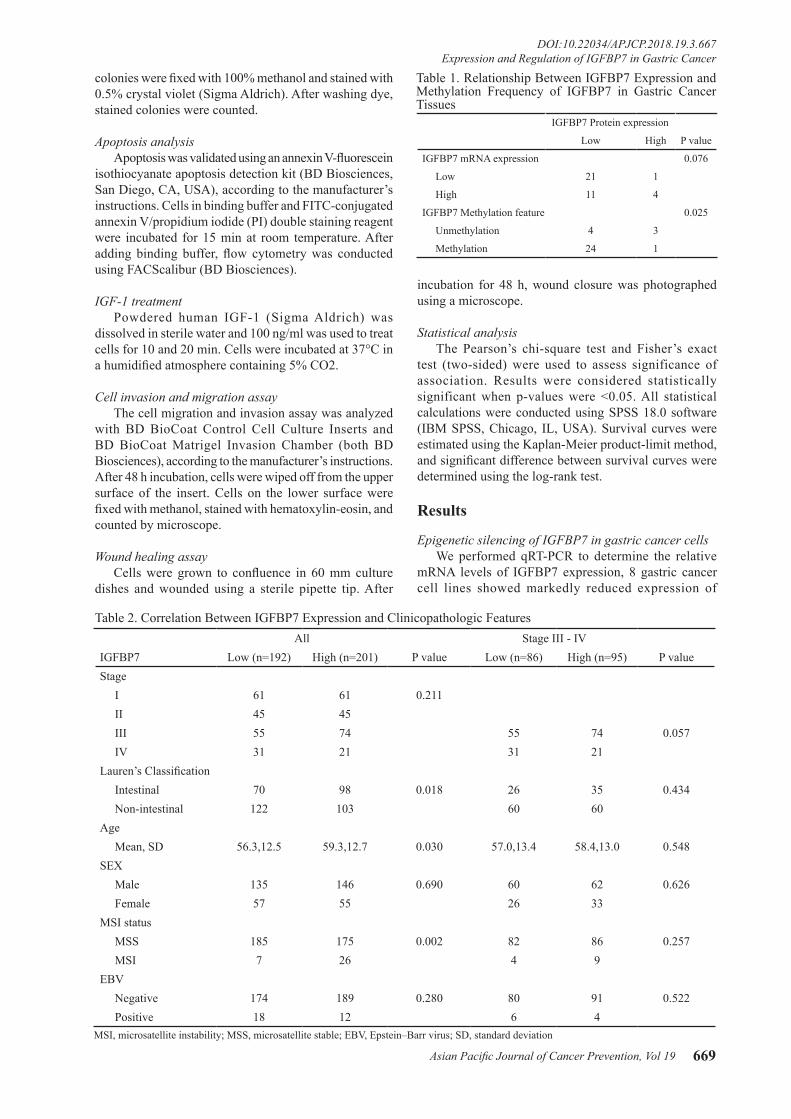
IGFBP7 mRNA expression 0.076
Low 21 1
High 11 4
IGFBP7 Methylation feature 0.025
Unmethylation 4 3
Methylation 24 1
Table 1. Relationship Between IGFBP7 Expression and Methylation Frequency of IGFBP7 in Gastric Cancer Tissues
All Stage III - IVIGFBP7 Low (n=192) High (n=201) P value Low (n=86) High (n=95) P valueStage I 61 61 0.211 II 45 45 III 55 74 55 74 0.057 IV 31 21 31 21Lauren’s Classification Intestinal 70 98 0.018 26 35 0.434 Non-intestinal 122 103 60 60Age Mean, SD 56.3,12.5 59.3,12.7 0.030 57.0,13.4 58.4,13.0 0.548SEX Male 135 146 0.690 60 62 0.626 Female 57 55 26 33MSI status MSS 185 175 0.002 82 86 0.257 MSI 7 26 4 9EBV Negative 174 189 0.280 80 91 0.522 Positive 18 12 6 4
Table 2. Correlation Between IGFBP7 Expression and Clinicopathologic Features
MSI, microsatellite instability; MSS, microsatellite stable; EBV, Epstein–Barr virus; SD, standard deviation

Jin Kim et al
Asian Pacific Journal of Cancer Prevention, Vol 19670
IGFBP7 (Figure 1A). To investigate whether IGFBP7 is epigenetically silenced in gastric cancer, we evaluated the methylation status of CpG islands in the exon 1 region of
the IGFBP7 gene using MSP. We found that IGFBP7 was partially or fully methylated in 8 gastric cancer cell lines, which show reduced expression of IGFBP7 (Figure 1B).
Figure 1. Expression and Methylation of IGFBP7 in Gastric Cancer Cells and Tissues. (A) Expression of IGFBP7 mRNA was detected by qRT-PCR. (B) The methylation status of IGFBP7 exon 1 was examined by MSP. Distilled water (DW), negative loading control; UM, unmethylated DNA band; M, methylated DNA band. (C) By qRT-PCR, treatment with 5-aza-dc, TSA, or combination restored IGFBP7 mRNA expression. (D) IGFBP7 protein expression in gastric cancer (C) tissues compared to paired normal (N) gastric tissues was assessed by western blot. (E) A representative result of methylation was measured using MSP. (F) Immunohistochemical expression of IGFBP7. Left, negative staining for IGFBP7. Middle, weakly positive staining for IGFBP7. Right, strongly positive staining for IGFBP7. Original magnification, 100X. (G) Kaplan-Meier analysis of overall survival for different IGFBP7 expression in gastric cancer patients.
MSI, microsatellite instability; MSS, microsatellite stable; EBV, Epstein–Barr virus; HR, hazard ratio; CI, confidence interval
Parameter Univariate P value Multivariate P valueHR (95% CI) HR (95% CI)
IGFBP7 (High vs. Low) 1.823 <0.001 1.704 0.004(1.275-2.606) (1.186-2.447)
Stage (III vs. IV) 3.520 <0.001 3.708 <0.001(2.425-5.110) (2.514-5.470)
Lauren’s Classification 1.231 0.282 1.075 0.712(Intestinal vs. Non-intestinal) (0.843-1.797) (0.732-1.579)Age (≤60 vs. >60) 1.202 0.310 1.527 0.024
(0.843-1.715) (1.057-2.206)MSI (MSS vs. MSI) 0.450 0.080 0.592 0.259
(0.184-1.101) (0.238-1.473)EBV (Negative vs. Positive) 1.347 0.416 1.080 0.836
(0.657-2.760) (0.522-2.233)
Table 3. Univariate and Multivariate Analysis of IGFBP7 Expression in Gastric Cancer

Asian Pacific Journal of Cancer Prevention, Vol 19 671
DOI:10.22034/APJCP.2018.19.3.667 Expression and Regulation of IGFBP7 in Gastric Cancer
To test the epigenetic regulation of IGFBP7 we selected 6 gastric cancer cell lines which show reduced expression of IGFBP7. Using qRT-PCR, IGFBP7 mRNA expression was restored after treatment with 5-aza-dc and/or TSA in all tested cells (Figure 1C). These results indicate that the silencing of IGFBP7 in gastric cancer cells is mainly caused by aberrant methylation, and histone deacetylation may also participate in transcriptional silencing.
Expression and methylation of IGFBP7 in gastric cancerTo further examine the role of IGFBP7 in gastric
carcinogenesis, we investigated its expression in gastric cancer tissues and paired adjacent normal tissues by qRT-PCR and western blot. Of the 37 gastric cancer tissues, 22 (59.5%) showed lower IGFBP7 mRNA expression than paired normal tissues (Table 1). Low expression of IGFBP7 protein in gastric cancer tissues was observed in 32 of 37 (86.5%) samples (Figure 1D and Table 1). Next, we assessed the methylation status of IGFBP7 by MSP. As shown in Figure 1E, IGFBP7 methylation frequency was higher in gastric cancer tissues than in normal tissues. IGFBP7 methylation was detected in 78.1% (25/32) of tumors. Furthermore, low expression of IGFBP7 was significantly correlated with hypermethylation of IGFBP7 in gastric cancer tissues (p=0.025) (Table 1). In primary gastric cancer tissues, expression of IGFBP7 protein was assessed by immunohistochemistry (Figure 1F). The correlation between IGFBP7 expression and clinicopathologic features in gastric cancer is shown
in Table 2. Among high stage gastric cancer tissues, 86 out of 181 samples (47.5%) showed low expression by IGFBP7 staining. Patients with low IGFBP7 protein expression had significantly worse survival than those with high IGFBP7 expression in stage III and IV patients (p<0.001) (Figure 1G). Univariate analysis indicated that IGFBP7 expression and stage were significantly correlated with poor survival in patients with stage III and IV gastric cancer. In multivariate analysis with the Cox proportional hazard model, IGFBP7 expression (low expression) and tumor stage (IV) were identified as independent predictors of unfavorable prognosis (p<0.05) )(Table 3).
Effect of IGFBP7 knockdown in gastric cancer cells To analyze the potential role of IGFBP7 in gastric
carcinogenesis, SNU484 and SNU668 cells were selected for further experiments. Expression of IGFBP7 mRNA and protein was suppressed by IGFBP7 shRNA transduction, as confirmed by qRT-PCR and western blot (Figure 2A). Cell growth analysis showed that cell proliferation was significantly increased in IGFBP7 shRNA-transduced cells (Figure 2B). Moreover, colony numbers were higher in shIGFBP7-transduced cells than shControl-transduced cells (Figure 2C). This result suggested that IGFBP7 plays an important role in the regulation of gastric cancer cell growth. As previous study has shown that downregulation of IGFBP7 increases cell proliferation through AKT and ERK signaling (Verhagen et al., 2014), we determined the levels of phospho-AKT (p-AKT) and
Figure 2. Inhibition of IGFBP7 Expression Promotes Gastric Cancer Cell Growth. (A) Knock-down of IGFBP7 was determined using western blot and qRT-PCR. (B) Inhibition of IGFBP7 expression increased cell proliferation. (C) Colony formation was significantly promoted in IGFBP7 suppressed cells. (D) Western blot analysis was performed with the indicated antibodies on shRNA transduced cells. (E) Quantification of apoptosis by flow cytometry. Data presented mean ± SD and experiments were performed three times in triplicate. *p<0.05.

Jin Kim et al
Asian Pacific Journal of Cancer Prevention, Vol 19672
phospho-extracellular signal-regulated kinases (p-ERK) in shRNA transduced cells. Expression of p-AKT and p-ERK was increased in the shIGFBP7-transduced cells (Figure 2D). In addition, we found that the expression of genes involved in cell cycle progression, such as cyclin-dependent kinase 2 (CDK2) and Cyclin D1, were increased in shIGFBP7-transduced cells (Figure 2D). In order to assess apoptosis in relation to IGFBP7 knockdown, the expression of apoptosis-related proteins was assessed. The results showed that cleaved caspase 3 (c-caspase3) was reduced in shIGFBP7-transduced cells (Figure 2D). Furthermore, we examined apoptosis by flow cytometry, and observed slightly decreased early (lower right) and late (upper right) apoptotic cells in shIGFBP7-transduced cells as compared with shControl-transduced cells (Figure 2E). These results demonstrate that inactivation of IGFBP7 can increase cell growth in gastric cancer cells.
Effect of IGFBP7 induction on cell growth To clarify functions of IGFBP7 in gastric cancer, we
determined the effect of IGFBP7 in gastric cancer cells with the stable transfection of IGFBP7. Analysis by qRT-PCR
and western blot showed that IGFBP7 mRNA and protein levels in IGFBP7-transfected SNU601 and SNU638 cells were increased (Figure 3A). The proliferation and colony formation ability of IGFBP7-overexpressed cells showed a significantly inhibited growth rate (Figure 3B and C). Additionally, the expression of p-AKT, p-ERK, CDK2, and Cyclin D1 were suppressed, whereas c-caspase3 was increased in IGFBP7-induced cells (Figure 3D). Using flow cytometry, a slight increase in the percentage of early and late apoptotic cells were presented in IGFBP7-induced SNU601 cells compared to control cells (Figure 3E). These results indicate that IGFBP7 negatively regulates gastric cancer cell growth. Next, we expected that IGFBP7-induced growth suppression may be regulated by the ability of IGFBP7 to interfere with IGF-1 signaling. Previously, we observed that phospho-IGF-1R (p-IGF-1R) levels were upregulated in shIGFBP7-transduced cells and downregulated in IGFBP7-overexpressed cells (Figure 2D and 3D). To examine the effect of IGFBP7 on gastric cancer cell growth through IGF-1, we cultured IGFBP7-induced cells with treatment of IGF-1 (100 ng/ml) each for 10 or 20 min. As shown in Figure 3F, SNU601 and SNU638 cells showed phosphorylation
Figure 3. Effect of IGFBP7 Overexpression on Gastric Cancer Cells. (A) Upregulation of IGFBP7 was proved by western blot and qRT-PCR. (B) Overexpression of IGFBP7 inhibited cell proliferation. (C) Colonies in transfected cells were visualized by staining and counted. (D) Western blot analysis was performed with the indicated antibodies on IGFBP7 overexpressed cells. (E) Apoptosis was detected by flow cytometry. (F) Detection of p-IGF-1R after IGF-1 ligand stimulation by western blot. (G) Cell growth rate was measured using proliferation assay. Data presented mean ± SD and experiments were performed three times in triplicate. *p<0.05.

Asian Pacific Journal of Cancer Prevention, Vol 19 673
DOI:10.22034/APJCP.2018.19.3.667 Expression and Regulation of IGFBP7 in Gastric Cancer
of IGF-1R after IGF-1 treatment in both IGFBP7-overexpressed cells and control cells. Treatment of IGF-1 promoted the growth rate of IGFBP7-induced and control cells. However, IGFBP7-induced cells were still growth-suppressed after IGF-1 treatment (Figure 3G.) This indicates that IGFBP7-induced growth inhibition was not significantly influenced by IGF-1 stimulation in gastric cancer. Overall, these results suggest that IGFBP7 inhibits gastric cancer cell growth in an IGF-1/IGF-1R-independent manner, which has also been reported for other IGFBP family members (Oh, 1998).
IGFBP7 stimulates invasion and migration of gastric cancer cells
We assessed whether the effects of IGFBP7 induction in gastric cancer cells could be related to invasion and migration. Invasion and migration abilities of the IGFBP7-suppressed cells were significantly higher than those of control cells (Figure 4A). In contrast, the invasion and migration rate of the IGFBP7-induced cells was significantly lower than that of the control cells (Figure 4B). We next confirmed the cell motility and migration capability of IGFBP7 by wound healing assay. The wound closure rate was observed at 48 h after scratch in SNU601 and SNU638 cells. Wounds of control cells were almost healed, whereas wounds were still opened in IGFBP7-overexpressed cells (Figure 4C).
Discussion
IGFBP7 expression has been reported in several human cancers and the levels of expression of IGFBP7 differ among various tumor types (Lin et al., 2007; Jiang et al., 2008; Vizioli et al., 2010; An et al., 2012; Benatar et al., 2012; Sullivan et al., 2012; Tomimaru et al., 2012). As regulatory mechanisms for the aberrant expression of IGFBP7 in cancers are not fully understood, more studies are needed to further elucidate the mechanistic and functional roles of IGFBP7 in gastric cancer.
Aberrant DNA methylation is a common molecular event in human carcinogenesis. Additionally, the role of histone deacetylation and chromatin remodeling is integral to tumor suppressor gene silencing (Tamura, 2002; Lo et al., 2010; Takada et al., 2010). Indeed, inactivation of IGFBP7 expression through epigenetic regulation has been studied in various types of cancers (Lin et al., 2007; Heesch et al., 2011; Sullivan et al., 2012; Suzuki et al., 2013). A previous study has reported the relationship between aberrant methylation of different CpG sites and gene transcription, and found that methylation of different CpG regions had different effects on gene transcription (Lin et al., 2007; Heesch et al., 2011). In this study, we tried to determine the methylation status of the IGFBP7 promoter region using MSP; however, it could not be discriminated in IGFBP7-expressed and silenced
Figure 4. IGFBP7 Inhibits Gastric Cancer Cell Invasion and Migration. (A) The number of invading and migrating cells in IGFBP7 suppressed cells. (B) The number of invading and migrating cells in IGFBP7 overexpressed cells. (C) Cell migration was confirmed by a wound healing assay. Dotted lines indicate wound edges. Experiments were performed three times in triplicate, and data presented means ± SD. *p<0.05.

Jin Kim et al
Asian Pacific Journal of Cancer Prevention, Vol 19674
gastric cancer cells. We therefore examined the methylation status of the 5′ CpG island and found that IGFBP7 expression was downregulated by hypermethylation of exon 1 of IGFBP7. In this regard, studies have also found other genes for which methylation at the CpG island of the exon 1 region affects gene expression in the same way (Hattori et al., 2001; Chen et al., 2005; Lin et al., 2007). These findings support the fact that site- or region-specific methylation can downregulate gene expression and suggests that the exon 1 region of IGFBP7 may be critical for the regulation of IGFBP7 mRNA transcription by DNA methylation in gastric cancer.
To investigate whether IGFBP7 expression is associated with gastric cancer patient prognosis, we assessed results from protein expression of IGFBP7 with clinical outcome of gastric cancer patients. Our analysis found that low expression of IGFBP7 in patients with stage III and IV gastric cancer was significantly associated with an unfavorable prognosis. In addition, IGFBP7 expression was an independent significant prognostic factor for gastric cancer patients. These data suggest that assessing the IGFBP7 expression status of patients with gastric cancer could potentially improve the prediction of prognosis.
IGFBP7 is involved in several cellular processes, including proliferation and apoptosis (Lin et al., 2007; Benatar et al., 2012; Sullivan et al., 2012). Although IGFBP7 has shown tumor suppressive properties in a number of cancers (Lin et al., 2007; Vizioli et al., 2010; An et al., 2012; Benatar et al., 2012; Sullivan et al., 2012; Tomimaru et al., 2012), little is known about its inhibitory effect in gastric cancer. From these findings, we speculate that IGFBP7 plays a contributory role in gastric carcinogenesis by influencing cancer cell growth. In order to investigate this hypothesis and to gain further insights into the functional role of IGFBP7 in gastric carcinoma, we used IGFBP7 shRNA transduction or stable transfection with an IGFBP7 expression vector in gastric cancer cells. Our results suggest that IGFBP7 has a suppressive effect on gastric cancer development.
To the present, many studies have provided evidence that IGFBPs exert IGF-dependent and/or -independent biological actions (Oh, 1998; Han et al., 2011; Verhagen et al., 2014). In this study, we show that IGF-1R is activated in IGFBP7-silenced cells and upon stimulation with IGF-1. These results demonstrate that increased apoptosis and inhibited growth by IGFBP7 in gastric cancer cells is in part independent from the activation of the IGF-1R/IGF-1 axis. To our knowledge, this is the first in vitro study to investigate the functional role of IGFBP7 in gastric cancer.
In summary, we found that aberrant IGFBP7 expression was regulated by DNA methylation and silencing of IGFBP7 protein expression was significantly correlated with unfavorable clinical outcome in gastric cancer. Altogether our results demonstrate that IGFBP7 acts as a tumor suppressor in gastric carcinogenesis and could be a novel therapeutic target in gastric cancer patients with poor prognosis.
Acknowledgements
This work was supported by the National Research Foundation of Korea (NRF-2016R1D1A1B03931235).
References
An W, Ben QW, Chen HT, et al (2012). Low expression of IGFBP7 is associated with poor outcome of pancreatic ductal adenocarcinoma. Ann Surg Oncol, 19, 3971-8.
Benatar T, Yang W, Amemiya Y, et al (2012). IGFBP7 reduces breast tumor growth by induction of senescence and apoptosis pathways. Breast Cancer Res Treat, 133, 563-73.
Burger AM, Leyland-Jones B, Banerjee K, Spyropoulos DD, Seth AK (2005). Essential roles of IGFBP-3 and IGFBP-rP1 in breast cancer. Eur J Cancer, 41, 1515-27.
Chen WD, Han ZJ, Skoletsky J, et al (2005). Detection in fecal DNA of colon cancer-specific methylation of the nonexpressed vimentin gene. J Natl Cancer Inst, 97, 1124-32.
Fürstenberger G, Senn HJ (2002). Insulin-like growth factors and cancer. Lancet Oncol, 3, 298-302.
Han J, Jogie-Brahim S, Harada A, Oh Y (2011). Insulin-like growth factor-binding protein-3 suppresses tumor growth via activation of caspase-dependent apoptosis and cross-talk with NF-κB signaling. Cancer Lett, 307, 200-10.
Hattori M, Sakamoto H, Satoh K, Yamamoto T (2001). DNA demethylase is expressed in ovarian cancers and the expression correlates with demethylation of CpG sites in the promoter region of c-erbB-2 and survivin genes. Cancer Lett, 169, 155-64.
Heesch S, Bartram I, Neumann M, et al (2011). Expression of IGFBP7 in acute leukemia is regulated by DNA methylation. Cancer Sci, 102, 253-9.
Jiang W, Xiang C, Cazacu S, Brodie C, Mikkelsen T (2008). Insulin-like growth factor binding protein 7 mediates glioma cell growth and migration. Neoplasia, 10, 1335-42.
Khandwala HM, McCutcheon IE, Flyvbjerg A, Friend KE (2000). The effects of insulin-like growth factors on tumorigenesis and neoplastic growth. Endocr Rev, 21, 215-44.
Kim HS, Nagalla SR, Oh Y, et al (1997). Identification of a family of low-affinity insulin-like growth factor binding proteins (IGFBPs): characterization of connective tissue growth factor as a member of the IGFBP superfamily. Proc Natl Acad Sci U S A, 94, 12981-6.
Lin J, Lai M, Huang Q, et al (2007). Methylation patterns of IGFBP7 in colon cancer cell lines are associated with levels of gene expression. J Pathol, 212, 83-90.
Lo PK, Lee JS, Liang X, et al (2010). Epigenetic inactivation of the potential tumor suppressor gene FOXF1 in breast cancer. Cancer Res, 70, 6047-58.
Oh Y (1998). IGF-independent regulation of breast cancer growth by IGF binding proteins. Breast Cancer Res Treat, 47, 283-93.
Oh Y, Nagalla SR, Yamanaka Y, et al (1996). Synthesis and characterization of insulin-like growth factor-binding protein (IGFBP)-7. Recombinant human mac25 protein specifically binds IGF-I and –II. J Biol Chem, 271, 30322-5.
Pollak M (2012). The insulin and insulin-like growth factor receptor family in neoplasia, an update. Nat Rev Cancer, 12, 159-69.
Samani AA, Yakar S, LeRoith D, Brodt P (2007). The role of the IGF system in cancer growth and metastasis: overview and recent insights. Endocr Rev, 28, 20-47.
Sullivan L, Murphy TM, Barrett C, et al (2012). IGFBP7 promoter methylation and gene expression analysis in prostate cancer. J Urol, 188, 1354-60.

Asian Pacific Journal of Cancer Prevention, Vol 19 675
DOI:10.22034/APJCP.2018.19.3.667 Expression and Regulation of IGFBP7 in Gastric Cancer
Suzuki M, Shiraishi K, Eguchi A, et al (2013). Aberrant methylation of LINE-1, SLIT2, MAL and IGFBP7 in non-small cell lung cancer. Oncol Rep, 29, 1308-14.
Takada H, Wakabayashi N, Dohi O, et al (2010). Tissue factor pathway inhibitor 2 (TFPI2) is frequently silenced by aberrant promoter hypermethylation in gastric cancer. Cancer Genet Cytogenet, 197, 16-24.
Tamura G (2002). Genetic and epigenetic alterations of tumor suppressor and tumor-related genes in gastric cancer. World J Gastroenterol, 17, 323-9.
Tomimaru Y, Eguchi H, Wada H, et al (2012). IGFBP7 downregulation is associated with tumor progression and clinical outcome in hepatocellular carcinoma. Int J Cancer, 130, 319-27.
Verhagen HJ, de Leeuw DC, Roemer MG, et al (2014). IGFBP7 induces apoptosis of acute myeloid leukemia cells and synergizes with chemotherapy in suppression of leukemia cell survival. Cell Death Dis, 5, e1300.
Vizioli MG, Sensi M, Miranda C, et al (2010). IGFBP7: an oncosuppressor gene in thyroid carcinogenesis. Oncogene, 29, 3835-44.
Yasui W, Yokozaki H, Fujimoto J, et al (2000). Genetic and epigenetic alterations in multistep carcinogenesis of the stomach. J Gastroenterol, 35, 111-5.
This work is licensed under a Creative Commons Attribution-Non Commercial 4.0 International License.