research article morphometric analysis of lateral masses...
TRANSCRIPT
Research ArticleMorphometric Analysis of Lateral Masses of Axis Vertebrae inNorth Indians
Monika Lalit1 Sanjay Piplani2 J S Kullar3 and Anupama Mahajan1
1 Department of Anatomy Sri Guru Ram Das Institute of Medical Science and Research Vallah Amritasr 143001 Punjab India2Department of Pathology Sri Guru Ram Das Institute of Medical Science and Research Vallah Amritasr 143001 Punjab India3 Department of Anatomy Government Medical College Amritsar Punjab India
Correspondence should be addressed to Monika Lalit monikalalityahoocom
Received 28 May 2014 Accepted 30 July 2014 Published 24 August 2014
Academic Editor Udo Schumacher
Copyright copy 2014 Monika Lalit et al This is an open access article distributed under the Creative Commons Attribution Licensewhich permits unrestricted use distribution and reproduction in any medium provided the original work is properly cited
Background and ObjectiveThe lateral masses of axis have good cancellous bone quality beneath the articular surface of facets thatmake this area a good site for the insertion of an internal fixation deviceMethods 60 dry axis vertebrae were obtained for anatomicevaluation focused on pedicle superior and inferior articular facets and foramen transversarium Based upon linear and angularparameters the mean range and standard deviation were calculated Results The mean length width and height of the pediclewere 2161 plusmn 237mm 882 plusmn 243mm and 563 plusmn 206mmThe mean pedicle superior angle and median angle were 233 and 322degrees The mean superior articular facet length width and external and internal height were 1634 plusmn 156mm 1435 plusmn 175mm898 plusmn 136mm and 423 plusmn 081mm Depth of vertebral artery was 472 plusmn 083mm Mean inferior articular facet length and widthwere 1113 plusmn 143mm and 789 plusmn 130mm The mean foramen transversarium length and width were 511 plusmn 091mm and 506 plusmn123mm ConclusionsThe study may provide information for the surgeons to determine the safe site of entry and trajectory for thescrew implantation and also to avoid injuries to vital structures while operating around axis
1 Introduction
Axis the second cervical vertebra forms a pivot on whichthe atlas rotates carrying the head to allow greater range ofmotion at the atlantoaxial joints [1 2] The lateral masses ofaxis have good cancellous bone quality beneath the articularsurface of facets that makes this area a good site for insertionof an internal fixation device [3]
The elements of pedicle and pedicle axis are critical to thestructural anatomy of axis vertebra which are important tonormal function and also for cases of pathology or fractureswhen surgical intervention is required [4 5] The superiorarticular facets (SAF) of axis differ from other vertebralfacets which make this region more prone to vertebral arteryinjury during screw fixation [6] In axis vertebrae the SAFlies in proximity to the body and medial aspect of the axisof the pedicle whereas SAF of other vertebrae are lying inproximity to the junction of pedicle and lamina and also thevertebral artery foramen is lying partially or completely in theundersurface of axis while in other vertebrae the vertebral
artery foramen is located entirely in relation to foramentransversarium [7 8] This unusual location of vertebralartery foramen makes the vertebral artery more prone toinjury during screw fixation [9]
The present study may also assist with placement ofscrews into pedicle or lateral mass For this reason theposterior point of projection of the pedicle axis has beendefined in terms of reference points visible in a postsurgicalapproach to the cervical spine and the two angles representthe safe bounds for transpedicular screw fixationplacement[4 10] Thus if there is any variation in the lateral mass ofaxis or associated anomalous vertebral artery it may lead tothinning of lateral mass and pedicle would prevent adequatefixation of transpedicular screw and vertebral artery will alsobe at risk [2]
Therefore the present study was designed to know thedimensions of lateral masses of axis that are important toanalyze their relationship with the vertebral artery and alsoto determine the ideal drill angle for accurate placementof a screw in the area resulting from fracture or partial
Hindawi Publishing CorporationAnatomy Research InternationalVolume 2014 Article ID 425868 9 pageshttpdxdoiorg1011552014425868
2 Anatomy Research International
E
F
e fE998400
F998400
(a)
IJ
K
L
(b)
i
jk
ll
(c)
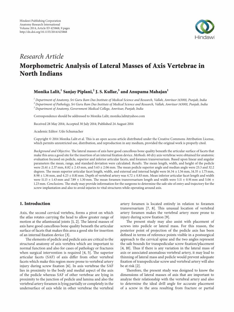
Figure 1 (a) Superior view of axis showing pedicle length (PL = EF) pedicle height (PH = E1015840F1015840) and pedicle width (PW = ef) (b) Lateralview of axis showing superior articular facet length (SAFL = IJ) and width (SAFW = KL) (c) Anterior view of axis showing inferior articularfacet length (IAFL = ij) and width (IAFW = kl)
S
T
(a)
s
t
(b)
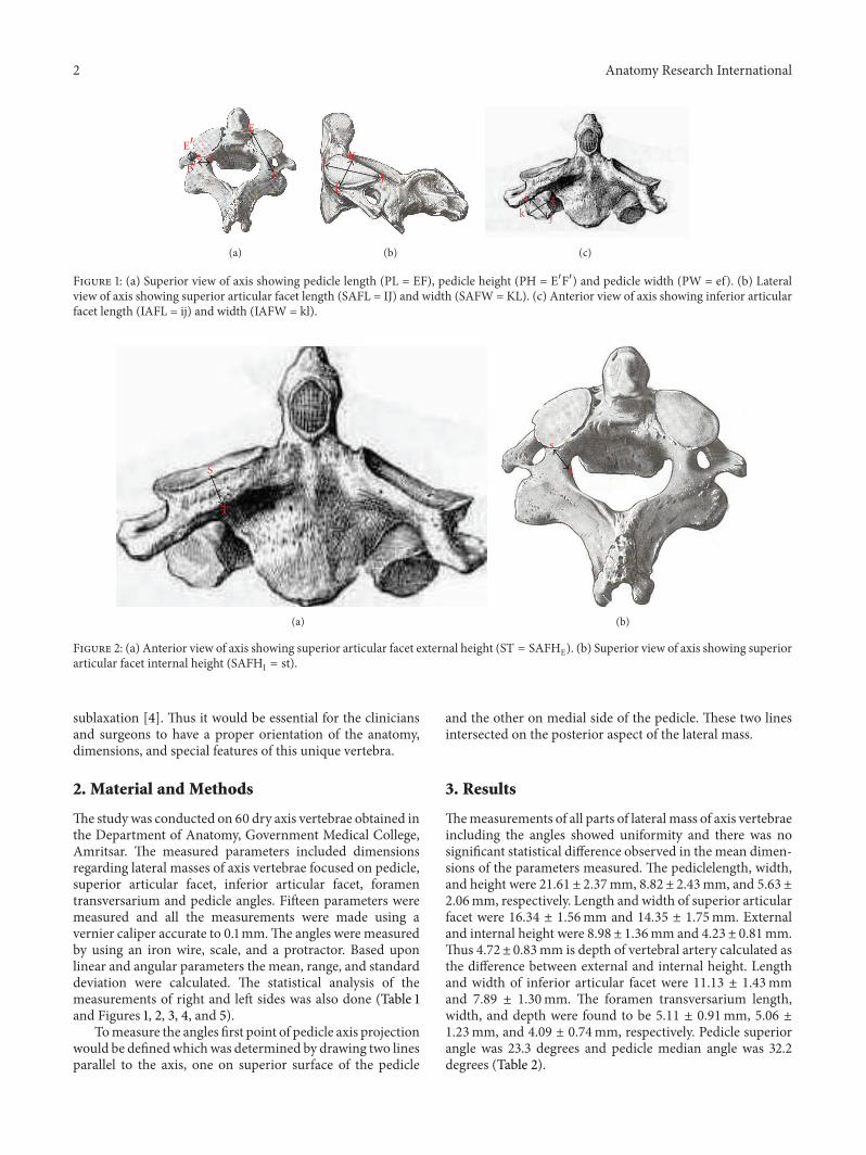
Figure 2 (a) Anterior view of axis showing superior articular facet external height (ST = SAFHE) (b) Superior view of axis showing superiorarticular facet internal height (SAFHI = st)
sublaxation [4] Thus it would be essential for the cliniciansand surgeons to have a proper orientation of the anatomydimensions and special features of this unique vertebra
2 Material and Methods
The study was conducted on 60 dry axis vertebrae obtained inthe Department of Anatomy Government Medical CollegeAmritsar The measured parameters included dimensionsregarding lateral masses of axis vertebrae focused on pediclesuperior articular facet inferior articular facet foramentransversarium and pedicle angles Fifteen parameters weremeasured and all the measurements were made using avernier caliper accurate to 01mmThe angles were measuredby using an iron wire scale and a protractor Based uponlinear and angular parameters the mean range and standarddeviation were calculated The statistical analysis of themeasurements of right and left sides was also done (Table 1and Figures 1 2 3 4 and 5)
Tomeasure the angles first point of pedicle axis projectionwould be definedwhichwas determined by drawing two linesparallel to the axis one on superior surface of the pedicle
and the other on medial side of the pedicle These two linesintersected on the posterior aspect of the lateral mass
3 Results
Themeasurements of all parts of lateral mass of axis vertebraeincluding the angles showed uniformity and there was nosignificant statistical difference observed in the mean dimen-sions of the parameters measured The pediclelength widthand height were 2161 plusmn 237mm 882 plusmn 243mm and 563 plusmn206mm respectively Length and width of superior articularfacet were 1634 plusmn 156mm and 1435 plusmn 175mm Externaland internal height were 898 plusmn 136mm and 423 plusmn 081mmThus 472plusmn 083mm is depth of vertebral artery calculated asthe difference between external and internal height Lengthand width of inferior articular facet were 1113 plusmn 143mmand 789 plusmn 130mm The foramen transversarium lengthwidth and depth were found to be 511 plusmn 091mm 506 plusmn123mm and 409 plusmn 074mm respectively Pedicle superiorangle was 233 degrees and pedicle median angle was 322degrees (Table 2)
Anatomy Research International 3
Table 1 The measured parameters
S number Parameters of lateral mass of axis vertebrae (pedicle SAF IAF FT and angles)
1 Pedicle length (PL) Length was measured from anterior most point of the pedicle axis to theposterior point of pedicle axis projection EF = PL (Figure 1(a))
2 Pedicle width (PW) It was taken from internal surface of pedicle to its external surface at thelevel of transverse foramen ef = PW (Figure 1(a))
3 Pedicle height (PH) It was measured from its superior surface to inferior surface with in theforamen transversarium E1015840F1015840 = PH (Figure 1(a))
4-5 Superior articular facet length(SAFL) and width (SAFW)
It is the maximum anteroposterior and transverse diameter of articularsurface of superior facet (IJ = SAFL) (KL = SAFW) (Figure 1(b))
6 External height (SAFHE)It was taken from upper midpoint on the superior articular surface to thelower midpoint on the inferior surface ST = SAFHE (Figure 2(a))
7 Internal height (SAFHI)It was taken from midpoint of articular surface to the nearest point on theinferior surface st = SAFHI (Figure 2(b))
8 Depth of vertebral artery It was taken as the difference between external and internal height
9-10 Inferior articular facet length(IAFL) and width (IAFW)
It is the maximum anteroposterior and transverse diameter of articularsurface of Inferior facet (ij = IAFL) (kl = IAFW) (Figure 1(c))
11ndash13Foramen transversarium length(FTL) width (FTW) and depth(FTD)
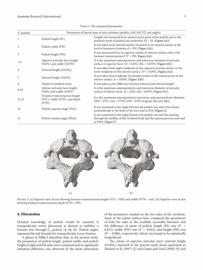
It is the maximum anteroposterior transverse and superoinferior diameter(MN = FTL) (mn = FTW) (OP = FTD) (Figures 3(a) and 3(b))
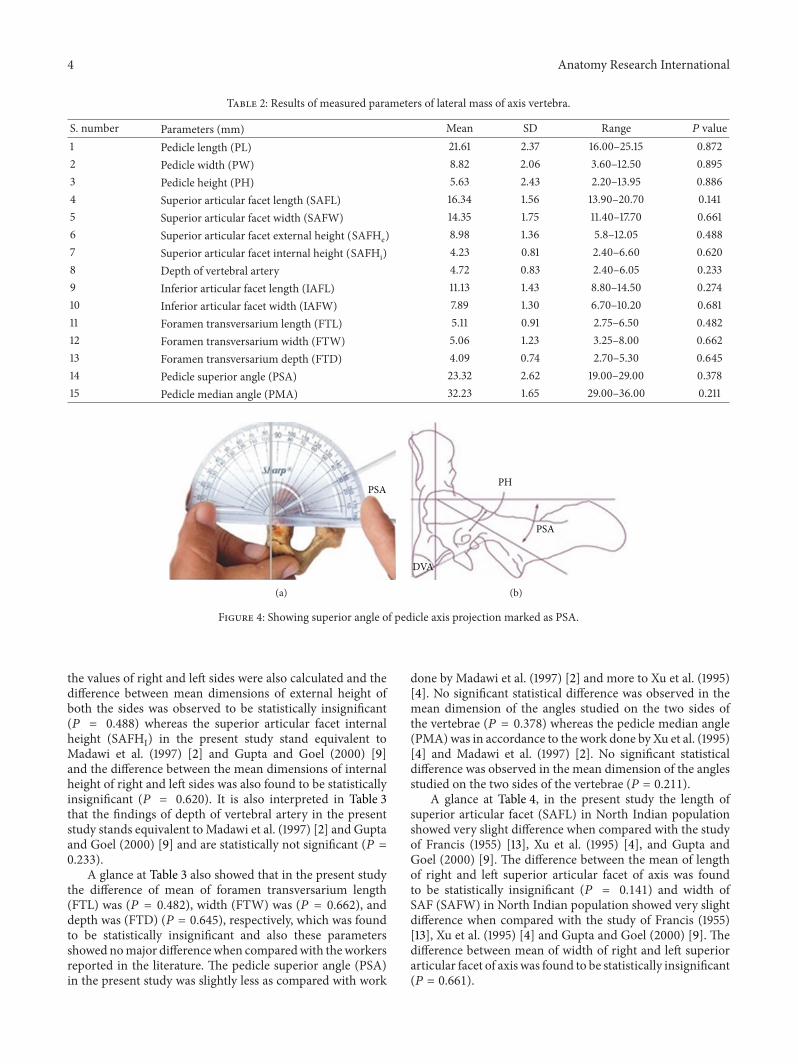
14 Pedicle superior angle (PSA) It was measured as the angle between the pedicle axis and a line drawnperpendicular to the body of the axis read as PSA (Figure 4)
15 Pedicle median angle (PMA)It was measured as the angle between the pedicle axis and line passingthrough the midline of the vertebral body and the spinous process and readas PMA (Figure 5)
M
Nm n
(a)
O
P
(b)
Figure 3 (a) Superior view of axis showing foramen transversarium length (FTL = MN) and width (FTW = mn) (b) Superior view of axisshowing foramen transversarium depth (FTD = OP)
4 Discussion
Detailed knowledge of pedicle would be essential iftranspedicular screw placement is desired to stabilize afracture line through 119862
2pedicle [4 14 15] Pedicle angles
represent the safe bounds for transpedicular screw fixationA glance at Table 3 elucidates that in the present study
the parameters of pedicle length pedicle width and pedicleheight of right and left sideswere compared andno significantstatistical difference was observed in the mean dimension
of the parameters studied on the two sides of the vertebraeNone of the earlier authors have compared the parametersof both the sides in the available accessible literature andthe difference of mean of pedicle length (PL) was (119875 =0872) width (PW) was (119875 = 0895) and height (PH) was(119875 = 0886) respectively which was found to be statisticallyinsignificant
The values of superior articular facet external height(SAFHE) reported in the present study stood equivalent toMadawi et al (1997) [2] and Gupta and Goel (2000) [9] and
4 Anatomy Research International
Table 2 Results of measured parameters of lateral mass of axis vertebra
S number Parameters (mm) Mean SD Range 119875 value1 Pedicle length (PL) 2161 237 1600ndash2515 08722 Pedicle width (PW) 882 206 360ndash1250 08953 Pedicle height (PH) 563 243 220ndash1395 08864 Superior articular facet length (SAFL) 1634 156 1390ndash2070 01415 Superior articular facet width (SAFW) 1435 175 1140ndash1770 06616 Superior articular facet external height (SAFHe) 898 136 58ndash1205 04887 Superior articular facet internal height (SAFHi) 423 081 240ndash660 06208 Depth of vertebral artery 472 083 240ndash605 02339 Inferior articular facet length (IAFL) 1113 143 880ndash1450 027410 Inferior articular facet width (IAFW) 789 130 670ndash1020 068111 Foramen transversarium length (FTL) 511 091 275ndash650 048212 Foramen transversarium width (FTW) 506 123 325ndash800 066213 Foramen transversarium depth (FTD) 409 074 270ndash530 064514 Pedicle superior angle (PSA) 2332 262 1900ndash2900 037815 Pedicle median angle (PMA) 3223 165 2900ndash3600 0211
PSA
(a)
PSA
PH
DVA
(b)
Figure 4 Showing superior angle of pedicle axis projection marked as PSA
the values of right and left sides were also calculated and thedifference between mean dimensions of external height ofboth the sides was observed to be statistically insignificant(119875 = 0488) whereas the superior articular facet internalheight (SAFHI) in the present study stand equivalent toMadawi et al (1997) [2] and Gupta and Goel (2000) [9]and the difference between the mean dimensions of internalheight of right and left sides was also found to be statisticallyinsignificant (119875 = 0620) It is also interpreted in Table 3that the findings of depth of vertebral artery in the presentstudy stands equivalent toMadawi et al (1997) [2] and Guptaand Goel (2000) [9] and are statistically not significant (119875 =0233)
A glance at Table 3 also showed that in the present studythe difference of mean of foramen transversarium length(FTL) was (119875 = 0482) width (FTW) was (119875 = 0662) anddepth was (FTD) (119875 = 0645) respectively which was foundto be statistically insignificant and also these parametersshowed nomajor differencewhen comparedwith theworkersreported in the literature The pedicle superior angle (PSA)in the present study was slightly less as compared with work
done by Madawi et al (1997) [2] and more to Xu et al (1995)[4] No significant statistical difference was observed in themean dimension of the angles studied on the two sides ofthe vertebrae (119875 = 0378) whereas the pedicle median angle(PMA) was in accordance to the work done by Xu et al (1995)[4] and Madawi et al (1997) [2] No significant statisticaldifference was observed in the mean dimension of the anglesstudied on the two sides of the vertebrae (119875 = 0211)
A glance at Table 4 in the present study the length ofsuperior articular facet (SAFL) in North Indian populationshowed very slight difference when compared with the studyof Francis (1955) [13] Xu et al (1995) [4] and Gupta andGoel (2000) [9] The difference between the mean of lengthof right and left superior articular facet of axis was foundto be statistically insignificant (119875 = 0141) and width ofSAF (SAFW) in North Indian population showed very slightdifference when compared with the study of Francis (1955)[13] Xu et al (1995) [4] and Gupta and Goel (2000) [9] Thedifference between mean of width of right and left superiorarticular facet of axis was found to be statistically insignificant(119875 = 0661)
Anatomy Research International 5
Table3Com
paris
onof
results
repo
rted
inthep
resent
study
andserie
sreportedin
theliterature
Parameters
Lalit
etal(present
study)2
014
North
Indian
60
Xuetal1995[4]
Ohio50
Madaw
ietal1997
[2]
England50
Gup
taandGoel
2000
[9]
Maharashtria
n50
How
ington
etal2001
[11]
American
10
Taitz
etal1978[12]
Israel32
Mean(m
m)a
ndSD
range(mm)
Mean(m
m)a
ndSD
range(mm)
Mean(m
m)a
ndSD
range(mm)
Mean(m
m)a
ndSD
range(mm)
Mean(m
m)a
ndSD
range(mm)
Mean(m
m)a
ndSD
range(mm)
PL2161plusmn
237
1600ndash
2515
Male(30)
256plusmn18
23ndash31
Female(20)
255plusmn14
23ndash29
287plusmn22
22ndash36
mdash166
mdash
PW882plusmn206
360ndash1250
Male(30)
77plusmn14
5ndash10
Female(20)
9plusmn11
4ndash10
79plusmn15
47ndash124
79 48ndash145
91mdash
PH563plusmn243
220ndash1395
Male(30)
86plusmn12
6ndash12
Female(20)
79plusmn14
6ndash10
78plusmn13
34ndash
122
77 45ndash12
79mdash
SAFH
E
Rt 887plusmn16
355ndash1390
Lt 90
8plusmn15
156ndash
1330
Mean 898plusmn13
658ndash1205
mdash86plusmn14
53ndash123
84
55ndash115
mdashmdash
SAFH
i
Rt 427plusmn088
270ndash6
50
Lt 4
21plusmn086
220ndash710
Mean 423plusmn081
240
ndash660
mdash44plusmn18
08ndash90
462
15ndash85
mdashmdash
6 Anatomy Research International
Table3Con
tinued
Parameters
Lalit
etal(present
study)2
014
North
Indian
60
Xuetal1995[4]
Ohio50
Madaw
ietal1997
[2]
England50
Gup
taandGoel
2000
[9]
Maharashtria
n50
How
ington
etal2001
[11]
American
10
Taitz
etal1978[12]
Israel32
Mean(m
m)a
ndSD
range(mm)
Mean(m
m)a
ndSD
range(mm)
Mean(m
m)a
ndSD
range(mm)
Mean(m
m)a
ndSD
range(mm)
Mean(m
m)a
ndSD
range(mm)
Mean(m
m)a
ndSD
range(mm)
Depth
ofvertebral
artery
Rt 4
61plusmn10
9260ndash740
Lt 4
87plusmn095
220ndash6
60
Mean 472plusmn083
240
ndash605
mdash42plusmn16
10ndash94
436
20ndash
82
mdashmdash
FTL
Rt 503plusmn10
7300ndash700
Lt 518plusmn10
915
0ndash70
0Mean 511plusmn091
275ndash6
50
mdashmdash
mdashmdash
Rt 585plusmn13
939ndash
113
Lt 576plusmn071
40ndash
73
FTW
Rt 512plusmn13
2300ndash800
Lt 500plusmn15
3200ndash800
Mean 506plusmn12
3325ndash800
mdash56plusmn09
43ndash80
mdashmdash
Rt 477plusmn070
36ndash
62
Lt 4
99plusmn066
34ndash
60
FTD
Rt 413plusmn087
250ndash580
Lt 4
05plusmn089
240
ndash580
Mean 409plusmn074
270ndash530
mdash57plusmn10
39ndash
90mdash
mdashmdash
Anatomy Research International 7
Table3Con
tinued
Parameters
Lalit
etal(present
study)2
014
North
Indian
60
Xuetal1995[4]
Ohio50
Madaw
ietal1997
[2]
England50
Gup
taandGoel
2000
[9]
Maharashtria
n50
How
ington
etal2001
[11]
American
10
Taitz
etal1978[12]
Israel32
Mean(m
m)a
ndSD
range(mm)
Mean(m
m)a
ndSD
range(mm)
Mean(m
m)a
ndSD
range(mm)
Mean(m
m)a
ndSD
range(mm)
Mean(m
m)a
ndSD
range(mm)
Mean(m
m)a
ndSD
range(mm)
PSA
Rt 23
40plusmn263
1900ndash
2900
Lt 23
23plusmn271
1900ndash
2900
Mean 2332plusmn262
1900ndash
2900
Male(30)
204plusmn40
14ndash30
Female(20)
200plusmn38
13ndash25
240plusmn42
13ndash34
mdashmdash
mdash
PMA
Rt 32
10plusmn19
52900ndash
3700
Lt 32
37plusmn15
22900ndash
3600
Mean 3223plusmn16
52900ndash
3600
Male(30)
333plusmn29
26ndash4
0Female(20)
327plusmn34
28ndash4
1
324plusmn40
24ndash4
3mdash
mdashmdash
8 Anatomy Research International
PMA
(a)
PMA
(b)
Figure 5 Showing median angle of pedicle axis marked as PMA
Table 4 Comparison of superior and inferior articular facets length and width of axis vertebrae
Worker andyear Population
SAFLmean (mm) and SDrange (mm)
SAFWmean (mm) and SDrange (mm)
IAFLmean (mm) and SDrange (mm)
IAFWmean (mm) and SDrange (mm)
Francis 1955[13]
White males 109
Rt188 plusmn 1615ndash23
Lt187 plusmn 1515ndash23
Rt167 plusmn 1614ndash22
Lt183 plusmn 1715ndash23
Rt110 plusmn 178ndash18
Lt113 plusmn 159ndash16
Rt119 plusmn 149ndash16
Lt121 plusmn 159ndash17
White females27
Rt180 plusmn 1814ndash20
Lt174 plusmn 1414ndash20
Rt151 plusmn 1313ndash18
Lt167 plusmn 1712ndash20
Rt116 plusmn 139ndash14
Lt111 plusmn 149ndash14
Rt107 plusmn 119ndash13
Lt114 plusmn 1210ndash15
Negro males 135
Rt190 plusmn 1316ndash22
Lt189 plusmn 1516ndash22
Rt177 plusmn 1415ndash21
Lt179 plusmn 1415ndash21
Rt120 plusmn 179ndash16
Lt114 plusmn 188ndash16
Rt113 plusmn 168ndash15
Lt118 plusmn 149ndash15
Negro females57
Rt176 plusmn 1315ndash20
Lt177 plusmn 1515ndash21
Rt157 plusmn 1114ndash18
Lt161 plusmn 1413ndash19
Rt112 plusmn 119ndash13
Lt110 plusmn 129ndash14
Rt107 plusmn 119ndash14
Lt111 plusmn 119ndash13
Xu et al 1995[4]
Ohio males 30 182 plusmn 1516ndash23
176 plusmn 1316ndash21 mdash mdash
Ohio females 20 171 plusmn 1115ndash19
169 plusmn 1314ndash19 mdash mdash
Gupta andGoel 2000 [9]
Maharashtrian50
16513ndash205
15912ndash195
10597ndash15
9586ndash15
Lalit et al(PresentStudy) 2014
North Indians60
Rt1649 plusmn 1611390ndash2050
Lt1618 plusmn 1701350ndash2090
Mean634 plusmn 1561390ndash2070
Rt1439 plusmn 2011100ndash1820
Lt1431 plusmn 1621140ndash1730
Mean1435 plusmn 1751140ndash1770
Rt1126 plusmn 146870ndash1400
Lt1099 plusmn 168780ndash1500
Mean1113 plusmn 143880ndash1450
Rt783 plusmn 127580ndash1180
Lt794 plusmn 164480ndash1240
Mean789 plusmn 130670ndash1020
Anatomy Research International 9
Values of inferior articular facet length of axis (IAFL) inthe present study stood equivalent whereas inferior articularfacet width (IAFW) was found to be less when comparedwith work done by Francis (1955) [13] and Gupta and Goel(2000) [9] But the comparison of right and left sides wasnot specified by them No significant statistical difference wasobserved in themean dimension of the IAFL (119875 = 0274) andIAFW (119875 = 0681) studied on the two sides of the vertebrae
5 Conclusions
The study may provide information for the surgeons todetermine the safe site of entry and trajectory for the screwimplantation and also toavoid injuries to vital structureswhile operating around axis Dimensions of axis vertebralforamen transversarium are important and act as a usefulguide in the estimation of dilation of vertebral artery Thevertebral artery and the basilar artery contribute bloodsupply not only to the brain but to inner ear also and theircompression may lead to irritation of sympathetic plexusmanifested not only by neurological symptoms but also bylabyrinthine or hearing disturbancesThus dimensions of axisvertebral foramen transversarium are important and act as auseful guide in the estimation of dilation of vertebral artery[12 16 17] SAFof axis has a crucial relationshipwith vertebralartery that makes the vertebral artery more prone to injuryAsymmetry of articular processes in particular hypertrophyof articular processes might have caused torticollis withsevere constriction of cervical mobility [18] To determineaccurate placement of a screw in the area of any deformityresulting from fracture or partial sublaxation ideal drill anglefor transpedicular screw placement is required Thereforecareful anatomic reduction is essential [4 19]
Conflict of Interests
The authors declare that there is no conflict of interestsregarding the publication of this paper
References
[1] M William R L M Newell and P Collin ldquoThe back cervicalvertebraerdquo in Grayrsquos Anatomy S Standring H Ellis J CHaely and A Williams Eds pp 742ndash746 Elsevier ChurchillLivingstone London UK 39th edition 2005
[2] A A Madawi G Solanki A T H Casey and H A CrockardldquoVariation of the groove in the axis vertebra for the vertebralarteryrdquo Journal of Bone and Joint Surgery B vol 79 no 5 pp820ndash823 1997
[3] M H Heggeness and B J Doherty ldquoThe trabecular anatomy ofthe axisrdquo Spine vol 18 no 14 pp 1945ndash1949 1993
[4] R Xu M C Nadaud N A Ebraheim and R A YeastingldquoMorphology of the second cervical vertebra and the posteriorprojection of the C
2pedicle axisrdquo Spine vol 20 no 3 pp 259ndash
263 1995[5] L Rogers ldquoSpine and spinal cordrdquo in Surgical Applied Anatomy
pp 505ndash513 Cassell and Company London UK 11th edition1947
[6] L M Overton and J W Grossman ldquoAnatomical variationsin the articulation between the second and third cervicalvertebraerdquo Journal of Bone and Joint Surgery vol 36 pp 155ndash161 1952
[7] C Taitz and B Arensburg ldquoVertebral artery tortuosity withconcomitant erosion of the foramen of the transverse processof the axis Possible clinical implicationsrdquo Acta Anatomica vol141 no 2 pp 104ndash108 1991
[8] B J Doherty and M H Heggeness ldquoThe quantitative anatomyof the atlasrdquo Spine vol 19 no 22 pp 2497ndash2500 1994
[9] S Gupta and A Goel ldquoQuantitative anatomy of the lateralmasses of the atlas and axis vertebraerdquo Neurology India vol 48no 2 pp 120ndash125 2000
[10] S T Dull R M Toselli E C Benzel and P R Cooper ldquoPreop-erative oblique axial computed tomographic imaging for C
1-C2
transarticular screw fixation Technical noterdquoNeurosurgery vol37 no 1 pp 150ndash152 1995
[11] J U Howington J J Kruse and D Awasthi ldquoSurgical anatomyof the C-2 pediclerdquo Journal of Neurosurgery vol 95 no 1 pp88ndash92 2001
[12] C Taitz H Nathan and B Arensburg ldquoAnatomical obser-vations of the foramina transversariardquo Journal of NeurologyNeurosurgery and Psychiatry vol 41 no 2 pp 170ndash176 1978
[13] C C Francis ldquoVariations in the articular facets of the cervicalvertebraerdquoThe Anatomical Record vol 122 no 4 pp 589ndash6021955
[14] R E Anderson and C N Shealy ldquoCervical pedicle erosionand rootlet compression caused by a tortuous vertebral arteryrdquoRadiology vol 96 no 3 pp 537ndash538 1970
[15] D F Cooper ldquoBone erosion of the cervical vertebrae secondaryto tortuosity of the vertebral artery case reportrdquo Journal ofNeurosurgery vol 53 no 1 pp 106ndash108 1980
[16] G I Wickbom and M R Williamson ldquoAnomalous foramentransversarium of C
2simulating erosion of bonerdquo Neuroradi-
ology vol 19 no 1 pp 43ndash45 1980[17] F Cacciola U Phalke and A Goel ldquoVertebral artery in rela-
tionship to C1-C2vertebrae an anatomical studyrdquo Neurology
vol 52 no 2 pp 178ndash184 2004[18] M Kawashima N Tanriover A L Rhoton A J Ulm and T
Matsushima ldquoExtreme lateral variants of the atlanto occipitaltransarticular approach to anterior extradural lesions of thecranio vertebral junctionmdashanatomic reportsrdquo Neurosurgeryvol 53 no 3 pp 662ndash675 2003
[19] M I Yusof L K Ming and M S Abdullah ldquoComputed tomo-graphic measurement of cervical pedicles for transpedicularfixation in a Malay populationrdquo Journal of Orthopaedic Surgeryvol 15 no 2 pp 187ndash190 2007
Submit your manuscripts athttpwwwhindawicom
Hindawi Publishing Corporationhttpwwwhindawicom Volume 2014
Anatomy Research International
PeptidesInternational Journal of
Hindawi Publishing Corporationhttpwwwhindawicom Volume 2014
Hindawi Publishing Corporation httpwwwhindawicom
International Journal of
Volume 2014
Zoology
Hindawi Publishing Corporationhttpwwwhindawicom Volume 2014
Molecular Biology International
GenomicsInternational Journal of
Hindawi Publishing Corporationhttpwwwhindawicom Volume 2014
The Scientific World JournalHindawi Publishing Corporation httpwwwhindawicom Volume 2014
Hindawi Publishing Corporationhttpwwwhindawicom Volume 2014
BioinformaticsAdvances in
Marine BiologyJournal of
Hindawi Publishing Corporationhttpwwwhindawicom Volume 2014
Hindawi Publishing Corporationhttpwwwhindawicom Volume 2014
Signal TransductionJournal of
Hindawi Publishing Corporationhttpwwwhindawicom Volume 2014
BioMed Research International
Evolutionary BiologyInternational Journal of
Hindawi Publishing Corporationhttpwwwhindawicom Volume 2014
Hindawi Publishing Corporationhttpwwwhindawicom Volume 2014
Biochemistry Research International
ArchaeaHindawi Publishing Corporationhttpwwwhindawicom Volume 2014
Hindawi Publishing Corporationhttpwwwhindawicom Volume 2014
Genetics Research International
Hindawi Publishing Corporationhttpwwwhindawicom Volume 2014
Advances in
Virolog y
Hindawi Publishing Corporationhttpwwwhindawicom
Nucleic AcidsJournal of
Volume 2014
Stem CellsInternational
Hindawi Publishing Corporationhttpwwwhindawicom Volume 2014
Hindawi Publishing Corporationhttpwwwhindawicom Volume 2014
Enzyme Research
Hindawi Publishing Corporationhttpwwwhindawicom Volume 2014
International Journal of
Microbiology
2 Anatomy Research International
E
F
e fE998400
F998400
(a)
IJ
K
L
(b)
i
jk
ll
(c)
Figure 1 (a) Superior view of axis showing pedicle length (PL = EF) pedicle height (PH = E1015840F1015840) and pedicle width (PW = ef) (b) Lateralview of axis showing superior articular facet length (SAFL = IJ) and width (SAFW = KL) (c) Anterior view of axis showing inferior articularfacet length (IAFL = ij) and width (IAFW = kl)
S
T
(a)
s
t
(b)
Figure 2 (a) Anterior view of axis showing superior articular facet external height (ST = SAFHE) (b) Superior view of axis showing superiorarticular facet internal height (SAFHI = st)
sublaxation [4] Thus it would be essential for the cliniciansand surgeons to have a proper orientation of the anatomydimensions and special features of this unique vertebra
2 Material and Methods
The study was conducted on 60 dry axis vertebrae obtained inthe Department of Anatomy Government Medical CollegeAmritsar The measured parameters included dimensionsregarding lateral masses of axis vertebrae focused on pediclesuperior articular facet inferior articular facet foramentransversarium and pedicle angles Fifteen parameters weremeasured and all the measurements were made using avernier caliper accurate to 01mmThe angles were measuredby using an iron wire scale and a protractor Based uponlinear and angular parameters the mean range and standarddeviation were calculated The statistical analysis of themeasurements of right and left sides was also done (Table 1and Figures 1 2 3 4 and 5)
Tomeasure the angles first point of pedicle axis projectionwould be definedwhichwas determined by drawing two linesparallel to the axis one on superior surface of the pedicle
and the other on medial side of the pedicle These two linesintersected on the posterior aspect of the lateral mass
3 Results
Themeasurements of all parts of lateral mass of axis vertebraeincluding the angles showed uniformity and there was nosignificant statistical difference observed in the mean dimen-sions of the parameters measured The pediclelength widthand height were 2161 plusmn 237mm 882 plusmn 243mm and 563 plusmn206mm respectively Length and width of superior articularfacet were 1634 plusmn 156mm and 1435 plusmn 175mm Externaland internal height were 898 plusmn 136mm and 423 plusmn 081mmThus 472plusmn 083mm is depth of vertebral artery calculated asthe difference between external and internal height Lengthand width of inferior articular facet were 1113 plusmn 143mmand 789 plusmn 130mm The foramen transversarium lengthwidth and depth were found to be 511 plusmn 091mm 506 plusmn123mm and 409 plusmn 074mm respectively Pedicle superiorangle was 233 degrees and pedicle median angle was 322degrees (Table 2)
Anatomy Research International 3
Table 1 The measured parameters
S number Parameters of lateral mass of axis vertebrae (pedicle SAF IAF FT and angles)
1 Pedicle length (PL) Length was measured from anterior most point of the pedicle axis to theposterior point of pedicle axis projection EF = PL (Figure 1(a))
2 Pedicle width (PW) It was taken from internal surface of pedicle to its external surface at thelevel of transverse foramen ef = PW (Figure 1(a))
3 Pedicle height (PH) It was measured from its superior surface to inferior surface with in theforamen transversarium E1015840F1015840 = PH (Figure 1(a))
4-5 Superior articular facet length(SAFL) and width (SAFW)
It is the maximum anteroposterior and transverse diameter of articularsurface of superior facet (IJ = SAFL) (KL = SAFW) (Figure 1(b))
6 External height (SAFHE)It was taken from upper midpoint on the superior articular surface to thelower midpoint on the inferior surface ST = SAFHE (Figure 2(a))
7 Internal height (SAFHI)It was taken from midpoint of articular surface to the nearest point on theinferior surface st = SAFHI (Figure 2(b))
8 Depth of vertebral artery It was taken as the difference between external and internal height
9-10 Inferior articular facet length(IAFL) and width (IAFW)
It is the maximum anteroposterior and transverse diameter of articularsurface of Inferior facet (ij = IAFL) (kl = IAFW) (Figure 1(c))
11ndash13Foramen transversarium length(FTL) width (FTW) and depth(FTD)
It is the maximum anteroposterior transverse and superoinferior diameter(MN = FTL) (mn = FTW) (OP = FTD) (Figures 3(a) and 3(b))
14 Pedicle superior angle (PSA) It was measured as the angle between the pedicle axis and a line drawnperpendicular to the body of the axis read as PSA (Figure 4)
15 Pedicle median angle (PMA)It was measured as the angle between the pedicle axis and line passingthrough the midline of the vertebral body and the spinous process and readas PMA (Figure 5)
M
Nm n
(a)
O
P
(b)
Figure 3 (a) Superior view of axis showing foramen transversarium length (FTL = MN) and width (FTW = mn) (b) Superior view of axisshowing foramen transversarium depth (FTD = OP)
4 Discussion
Detailed knowledge of pedicle would be essential iftranspedicular screw placement is desired to stabilize afracture line through 119862
2pedicle [4 14 15] Pedicle angles
represent the safe bounds for transpedicular screw fixationA glance at Table 3 elucidates that in the present study
the parameters of pedicle length pedicle width and pedicleheight of right and left sideswere compared andno significantstatistical difference was observed in the mean dimension
of the parameters studied on the two sides of the vertebraeNone of the earlier authors have compared the parametersof both the sides in the available accessible literature andthe difference of mean of pedicle length (PL) was (119875 =0872) width (PW) was (119875 = 0895) and height (PH) was(119875 = 0886) respectively which was found to be statisticallyinsignificant
The values of superior articular facet external height(SAFHE) reported in the present study stood equivalent toMadawi et al (1997) [2] and Gupta and Goel (2000) [9] and
4 Anatomy Research International
Table 2 Results of measured parameters of lateral mass of axis vertebra
S number Parameters (mm) Mean SD Range 119875 value1 Pedicle length (PL) 2161 237 1600ndash2515 08722 Pedicle width (PW) 882 206 360ndash1250 08953 Pedicle height (PH) 563 243 220ndash1395 08864 Superior articular facet length (SAFL) 1634 156 1390ndash2070 01415 Superior articular facet width (SAFW) 1435 175 1140ndash1770 06616 Superior articular facet external height (SAFHe) 898 136 58ndash1205 04887 Superior articular facet internal height (SAFHi) 423 081 240ndash660 06208 Depth of vertebral artery 472 083 240ndash605 02339 Inferior articular facet length (IAFL) 1113 143 880ndash1450 027410 Inferior articular facet width (IAFW) 789 130 670ndash1020 068111 Foramen transversarium length (FTL) 511 091 275ndash650 048212 Foramen transversarium width (FTW) 506 123 325ndash800 066213 Foramen transversarium depth (FTD) 409 074 270ndash530 064514 Pedicle superior angle (PSA) 2332 262 1900ndash2900 037815 Pedicle median angle (PMA) 3223 165 2900ndash3600 0211
PSA
(a)
PSA
PH
DVA
(b)
Figure 4 Showing superior angle of pedicle axis projection marked as PSA
the values of right and left sides were also calculated and thedifference between mean dimensions of external height ofboth the sides was observed to be statistically insignificant(119875 = 0488) whereas the superior articular facet internalheight (SAFHI) in the present study stand equivalent toMadawi et al (1997) [2] and Gupta and Goel (2000) [9]and the difference between the mean dimensions of internalheight of right and left sides was also found to be statisticallyinsignificant (119875 = 0620) It is also interpreted in Table 3that the findings of depth of vertebral artery in the presentstudy stands equivalent toMadawi et al (1997) [2] and Guptaand Goel (2000) [9] and are statistically not significant (119875 =0233)
A glance at Table 3 also showed that in the present studythe difference of mean of foramen transversarium length(FTL) was (119875 = 0482) width (FTW) was (119875 = 0662) anddepth was (FTD) (119875 = 0645) respectively which was foundto be statistically insignificant and also these parametersshowed nomajor differencewhen comparedwith theworkersreported in the literature The pedicle superior angle (PSA)in the present study was slightly less as compared with work
done by Madawi et al (1997) [2] and more to Xu et al (1995)[4] No significant statistical difference was observed in themean dimension of the angles studied on the two sides ofthe vertebrae (119875 = 0378) whereas the pedicle median angle(PMA) was in accordance to the work done by Xu et al (1995)[4] and Madawi et al (1997) [2] No significant statisticaldifference was observed in the mean dimension of the anglesstudied on the two sides of the vertebrae (119875 = 0211)
A glance at Table 4 in the present study the length ofsuperior articular facet (SAFL) in North Indian populationshowed very slight difference when compared with the studyof Francis (1955) [13] Xu et al (1995) [4] and Gupta andGoel (2000) [9] The difference between the mean of lengthof right and left superior articular facet of axis was foundto be statistically insignificant (119875 = 0141) and width ofSAF (SAFW) in North Indian population showed very slightdifference when compared with the study of Francis (1955)[13] Xu et al (1995) [4] and Gupta and Goel (2000) [9] Thedifference between mean of width of right and left superiorarticular facet of axis was found to be statistically insignificant(119875 = 0661)
Anatomy Research International 5
Table3Com
paris
onof
results
repo
rted
inthep
resent
study
andserie
sreportedin
theliterature
Parameters
Lalit
etal(present
study)2
014
North
Indian
60
Xuetal1995[4]
Ohio50
Madaw
ietal1997
[2]
England50
Gup
taandGoel
2000
[9]
Maharashtria
n50
How
ington
etal2001
[11]
American
10
Taitz
etal1978[12]
Israel32
Mean(m
m)a
ndSD
range(mm)
Mean(m
m)a
ndSD
range(mm)
Mean(m
m)a
ndSD
range(mm)
Mean(m
m)a
ndSD
range(mm)
Mean(m
m)a
ndSD
range(mm)
Mean(m
m)a
ndSD
range(mm)
PL2161plusmn
237
1600ndash
2515
Male(30)
256plusmn18
23ndash31
Female(20)
255plusmn14
23ndash29
287plusmn22
22ndash36
mdash166
mdash
PW882plusmn206
360ndash1250
Male(30)
77plusmn14
5ndash10
Female(20)
9plusmn11
4ndash10
79plusmn15
47ndash124
79 48ndash145
91mdash
PH563plusmn243
220ndash1395
Male(30)
86plusmn12
6ndash12
Female(20)
79plusmn14
6ndash10
78plusmn13
34ndash
122
77 45ndash12
79mdash
SAFH
E
Rt 887plusmn16
355ndash1390
Lt 90
8plusmn15
156ndash
1330
Mean 898plusmn13
658ndash1205
mdash86plusmn14
53ndash123
84
55ndash115
mdashmdash
SAFH
i
Rt 427plusmn088
270ndash6
50
Lt 4
21plusmn086
220ndash710
Mean 423plusmn081
240
ndash660
mdash44plusmn18
08ndash90
462
15ndash85
mdashmdash
6 Anatomy Research International
Table3Con
tinued
Parameters
Lalit
etal(present
study)2
014
North
Indian
60
Xuetal1995[4]
Ohio50
Madaw
ietal1997
[2]
England50
Gup
taandGoel
2000
[9]
Maharashtria
n50
How
ington
etal2001
[11]
American
10
Taitz
etal1978[12]
Israel32
Mean(m
m)a
ndSD
range(mm)
Mean(m
m)a
ndSD
range(mm)
Mean(m
m)a
ndSD
range(mm)
Mean(m
m)a
ndSD
range(mm)
Mean(m
m)a
ndSD
range(mm)
Mean(m
m)a
ndSD
range(mm)
Depth
ofvertebral
artery
Rt 4
61plusmn10
9260ndash740
Lt 4
87plusmn095
220ndash6
60
Mean 472plusmn083
240
ndash605
mdash42plusmn16
10ndash94
436
20ndash
82
mdashmdash
FTL
Rt 503plusmn10
7300ndash700
Lt 518plusmn10
915
0ndash70
0Mean 511plusmn091
275ndash6
50
mdashmdash
mdashmdash
Rt 585plusmn13
939ndash
113
Lt 576plusmn071
40ndash
73
FTW
Rt 512plusmn13
2300ndash800
Lt 500plusmn15
3200ndash800
Mean 506plusmn12
3325ndash800
mdash56plusmn09
43ndash80
mdashmdash
Rt 477plusmn070
36ndash
62
Lt 4
99plusmn066
34ndash
60
FTD
Rt 413plusmn087
250ndash580
Lt 4
05plusmn089
240
ndash580
Mean 409plusmn074
270ndash530
mdash57plusmn10
39ndash
90mdash
mdashmdash
Anatomy Research International 7
Table3Con
tinued
Parameters
Lalit
etal(present
study)2
014
North
Indian
60
Xuetal1995[4]
Ohio50
Madaw
ietal1997
[2]
England50
Gup
taandGoel
2000
[9]
Maharashtria
n50
How
ington
etal2001
[11]
American
10
Taitz
etal1978[12]
Israel32
Mean(m
m)a
ndSD
range(mm)
Mean(m
m)a
ndSD
range(mm)
Mean(m
m)a
ndSD
range(mm)
Mean(m
m)a
ndSD
range(mm)
Mean(m
m)a
ndSD
range(mm)
Mean(m
m)a
ndSD
range(mm)
PSA
Rt 23
40plusmn263
1900ndash
2900
Lt 23
23plusmn271
1900ndash
2900
Mean 2332plusmn262
1900ndash
2900
Male(30)
204plusmn40
14ndash30
Female(20)
200plusmn38
13ndash25
240plusmn42
13ndash34
mdashmdash
mdash
PMA
Rt 32
10plusmn19
52900ndash
3700
Lt 32
37plusmn15
22900ndash
3600
Mean 3223plusmn16
52900ndash
3600
Male(30)
333plusmn29
26ndash4
0Female(20)
327plusmn34
28ndash4
1
324plusmn40
24ndash4
3mdash
mdashmdash
8 Anatomy Research International
PMA
(a)
PMA
(b)
Figure 5 Showing median angle of pedicle axis marked as PMA
Table 4 Comparison of superior and inferior articular facets length and width of axis vertebrae
Worker andyear Population
SAFLmean (mm) and SDrange (mm)
SAFWmean (mm) and SDrange (mm)
IAFLmean (mm) and SDrange (mm)
IAFWmean (mm) and SDrange (mm)
Francis 1955[13]
White males 109
Rt188 plusmn 1615ndash23
Lt187 plusmn 1515ndash23
Rt167 plusmn 1614ndash22
Lt183 plusmn 1715ndash23
Rt110 plusmn 178ndash18
Lt113 plusmn 159ndash16
Rt119 plusmn 149ndash16
Lt121 plusmn 159ndash17
White females27
Rt180 plusmn 1814ndash20
Lt174 plusmn 1414ndash20
Rt151 plusmn 1313ndash18
Lt167 plusmn 1712ndash20
Rt116 plusmn 139ndash14
Lt111 plusmn 149ndash14
Rt107 plusmn 119ndash13
Lt114 plusmn 1210ndash15
Negro males 135
Rt190 plusmn 1316ndash22
Lt189 plusmn 1516ndash22
Rt177 plusmn 1415ndash21
Lt179 plusmn 1415ndash21
Rt120 plusmn 179ndash16
Lt114 plusmn 188ndash16
Rt113 plusmn 168ndash15
Lt118 plusmn 149ndash15
Negro females57
Rt176 plusmn 1315ndash20
Lt177 plusmn 1515ndash21
Rt157 plusmn 1114ndash18
Lt161 plusmn 1413ndash19
Rt112 plusmn 119ndash13
Lt110 plusmn 129ndash14
Rt107 plusmn 119ndash14
Lt111 plusmn 119ndash13
Xu et al 1995[4]
Ohio males 30 182 plusmn 1516ndash23
176 plusmn 1316ndash21 mdash mdash
Ohio females 20 171 plusmn 1115ndash19
169 plusmn 1314ndash19 mdash mdash
Gupta andGoel 2000 [9]
Maharashtrian50
16513ndash205
15912ndash195
10597ndash15
9586ndash15
Lalit et al(PresentStudy) 2014
North Indians60
Rt1649 plusmn 1611390ndash2050
Lt1618 plusmn 1701350ndash2090
Mean634 plusmn 1561390ndash2070
Rt1439 plusmn 2011100ndash1820
Lt1431 plusmn 1621140ndash1730
Mean1435 plusmn 1751140ndash1770
Rt1126 plusmn 146870ndash1400
Lt1099 plusmn 168780ndash1500
Mean1113 plusmn 143880ndash1450
Rt783 plusmn 127580ndash1180
Lt794 plusmn 164480ndash1240
Mean789 plusmn 130670ndash1020
Anatomy Research International 9
Values of inferior articular facet length of axis (IAFL) inthe present study stood equivalent whereas inferior articularfacet width (IAFW) was found to be less when comparedwith work done by Francis (1955) [13] and Gupta and Goel(2000) [9] But the comparison of right and left sides wasnot specified by them No significant statistical difference wasobserved in themean dimension of the IAFL (119875 = 0274) andIAFW (119875 = 0681) studied on the two sides of the vertebrae
5 Conclusions
The study may provide information for the surgeons todetermine the safe site of entry and trajectory for the screwimplantation and also toavoid injuries to vital structureswhile operating around axis Dimensions of axis vertebralforamen transversarium are important and act as a usefulguide in the estimation of dilation of vertebral artery Thevertebral artery and the basilar artery contribute bloodsupply not only to the brain but to inner ear also and theircompression may lead to irritation of sympathetic plexusmanifested not only by neurological symptoms but also bylabyrinthine or hearing disturbancesThus dimensions of axisvertebral foramen transversarium are important and act as auseful guide in the estimation of dilation of vertebral artery[12 16 17] SAFof axis has a crucial relationshipwith vertebralartery that makes the vertebral artery more prone to injuryAsymmetry of articular processes in particular hypertrophyof articular processes might have caused torticollis withsevere constriction of cervical mobility [18] To determineaccurate placement of a screw in the area of any deformityresulting from fracture or partial sublaxation ideal drill anglefor transpedicular screw placement is required Thereforecareful anatomic reduction is essential [4 19]
Conflict of Interests
The authors declare that there is no conflict of interestsregarding the publication of this paper
References
[1] M William R L M Newell and P Collin ldquoThe back cervicalvertebraerdquo in Grayrsquos Anatomy S Standring H Ellis J CHaely and A Williams Eds pp 742ndash746 Elsevier ChurchillLivingstone London UK 39th edition 2005
[2] A A Madawi G Solanki A T H Casey and H A CrockardldquoVariation of the groove in the axis vertebra for the vertebralarteryrdquo Journal of Bone and Joint Surgery B vol 79 no 5 pp820ndash823 1997
[3] M H Heggeness and B J Doherty ldquoThe trabecular anatomy ofthe axisrdquo Spine vol 18 no 14 pp 1945ndash1949 1993
[4] R Xu M C Nadaud N A Ebraheim and R A YeastingldquoMorphology of the second cervical vertebra and the posteriorprojection of the C
2pedicle axisrdquo Spine vol 20 no 3 pp 259ndash
263 1995[5] L Rogers ldquoSpine and spinal cordrdquo in Surgical Applied Anatomy
pp 505ndash513 Cassell and Company London UK 11th edition1947
[6] L M Overton and J W Grossman ldquoAnatomical variationsin the articulation between the second and third cervicalvertebraerdquo Journal of Bone and Joint Surgery vol 36 pp 155ndash161 1952
[7] C Taitz and B Arensburg ldquoVertebral artery tortuosity withconcomitant erosion of the foramen of the transverse processof the axis Possible clinical implicationsrdquo Acta Anatomica vol141 no 2 pp 104ndash108 1991
[8] B J Doherty and M H Heggeness ldquoThe quantitative anatomyof the atlasrdquo Spine vol 19 no 22 pp 2497ndash2500 1994
[9] S Gupta and A Goel ldquoQuantitative anatomy of the lateralmasses of the atlas and axis vertebraerdquo Neurology India vol 48no 2 pp 120ndash125 2000
[10] S T Dull R M Toselli E C Benzel and P R Cooper ldquoPreop-erative oblique axial computed tomographic imaging for C
1-C2
transarticular screw fixation Technical noterdquoNeurosurgery vol37 no 1 pp 150ndash152 1995
[11] J U Howington J J Kruse and D Awasthi ldquoSurgical anatomyof the C-2 pediclerdquo Journal of Neurosurgery vol 95 no 1 pp88ndash92 2001
[12] C Taitz H Nathan and B Arensburg ldquoAnatomical obser-vations of the foramina transversariardquo Journal of NeurologyNeurosurgery and Psychiatry vol 41 no 2 pp 170ndash176 1978
[13] C C Francis ldquoVariations in the articular facets of the cervicalvertebraerdquoThe Anatomical Record vol 122 no 4 pp 589ndash6021955
[14] R E Anderson and C N Shealy ldquoCervical pedicle erosionand rootlet compression caused by a tortuous vertebral arteryrdquoRadiology vol 96 no 3 pp 537ndash538 1970
[15] D F Cooper ldquoBone erosion of the cervical vertebrae secondaryto tortuosity of the vertebral artery case reportrdquo Journal ofNeurosurgery vol 53 no 1 pp 106ndash108 1980
[16] G I Wickbom and M R Williamson ldquoAnomalous foramentransversarium of C
2simulating erosion of bonerdquo Neuroradi-
ology vol 19 no 1 pp 43ndash45 1980[17] F Cacciola U Phalke and A Goel ldquoVertebral artery in rela-
tionship to C1-C2vertebrae an anatomical studyrdquo Neurology
vol 52 no 2 pp 178ndash184 2004[18] M Kawashima N Tanriover A L Rhoton A J Ulm and T
Matsushima ldquoExtreme lateral variants of the atlanto occipitaltransarticular approach to anterior extradural lesions of thecranio vertebral junctionmdashanatomic reportsrdquo Neurosurgeryvol 53 no 3 pp 662ndash675 2003
[19] M I Yusof L K Ming and M S Abdullah ldquoComputed tomo-graphic measurement of cervical pedicles for transpedicularfixation in a Malay populationrdquo Journal of Orthopaedic Surgeryvol 15 no 2 pp 187ndash190 2007
Submit your manuscripts athttpwwwhindawicom
Hindawi Publishing Corporationhttpwwwhindawicom Volume 2014
Anatomy Research International
PeptidesInternational Journal of
Hindawi Publishing Corporationhttpwwwhindawicom Volume 2014
Hindawi Publishing Corporation httpwwwhindawicom
International Journal of
Volume 2014
Zoology
Hindawi Publishing Corporationhttpwwwhindawicom Volume 2014
Molecular Biology International
GenomicsInternational Journal of
Hindawi Publishing Corporationhttpwwwhindawicom Volume 2014
The Scientific World JournalHindawi Publishing Corporation httpwwwhindawicom Volume 2014
Hindawi Publishing Corporationhttpwwwhindawicom Volume 2014
BioinformaticsAdvances in
Marine BiologyJournal of
Hindawi Publishing Corporationhttpwwwhindawicom Volume 2014
Hindawi Publishing Corporationhttpwwwhindawicom Volume 2014
Signal TransductionJournal of
Hindawi Publishing Corporationhttpwwwhindawicom Volume 2014
BioMed Research International
Evolutionary BiologyInternational Journal of
Hindawi Publishing Corporationhttpwwwhindawicom Volume 2014
Hindawi Publishing Corporationhttpwwwhindawicom Volume 2014
Biochemistry Research International
ArchaeaHindawi Publishing Corporationhttpwwwhindawicom Volume 2014
Hindawi Publishing Corporationhttpwwwhindawicom Volume 2014
Genetics Research International
Hindawi Publishing Corporationhttpwwwhindawicom Volume 2014
Advances in
Virolog y
Hindawi Publishing Corporationhttpwwwhindawicom
Nucleic AcidsJournal of
Volume 2014
Stem CellsInternational
Hindawi Publishing Corporationhttpwwwhindawicom Volume 2014
Hindawi Publishing Corporationhttpwwwhindawicom Volume 2014
Enzyme Research
Hindawi Publishing Corporationhttpwwwhindawicom Volume 2014
International Journal of
Microbiology
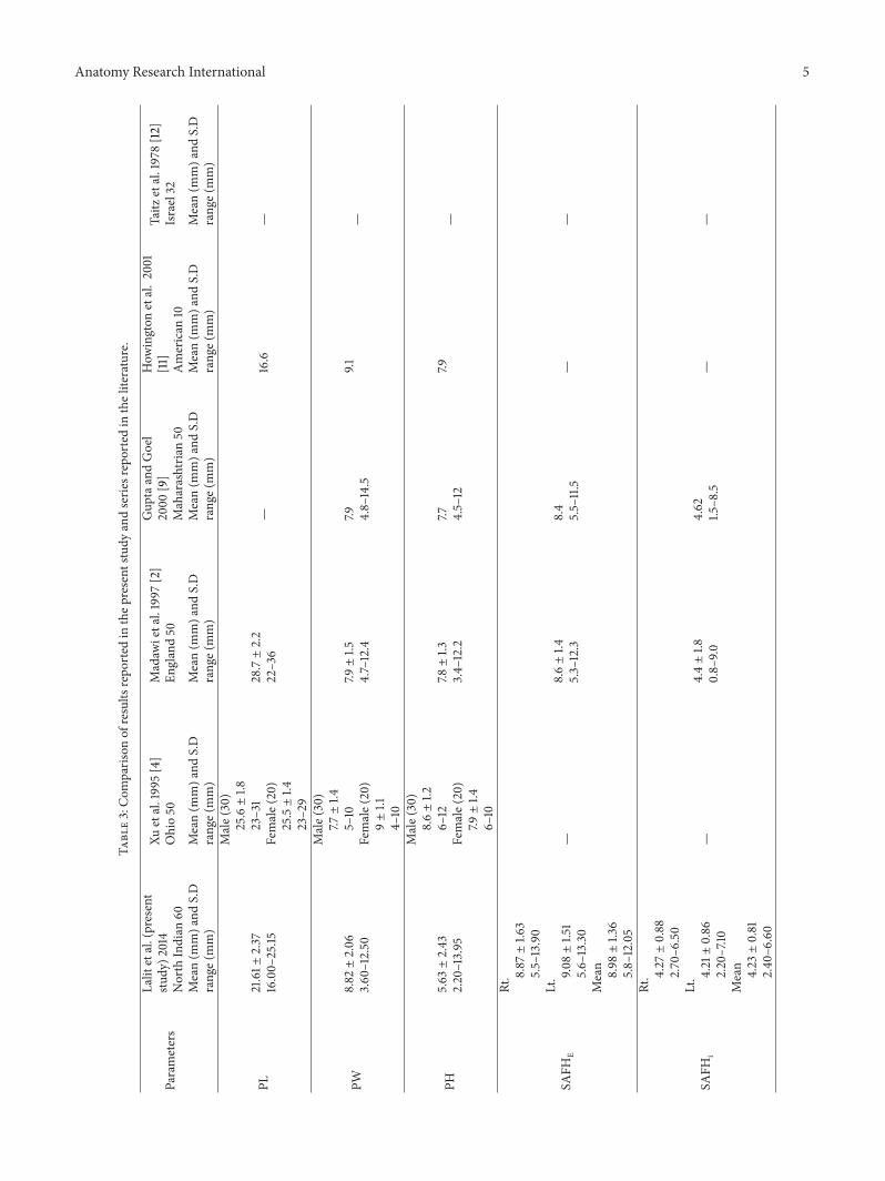
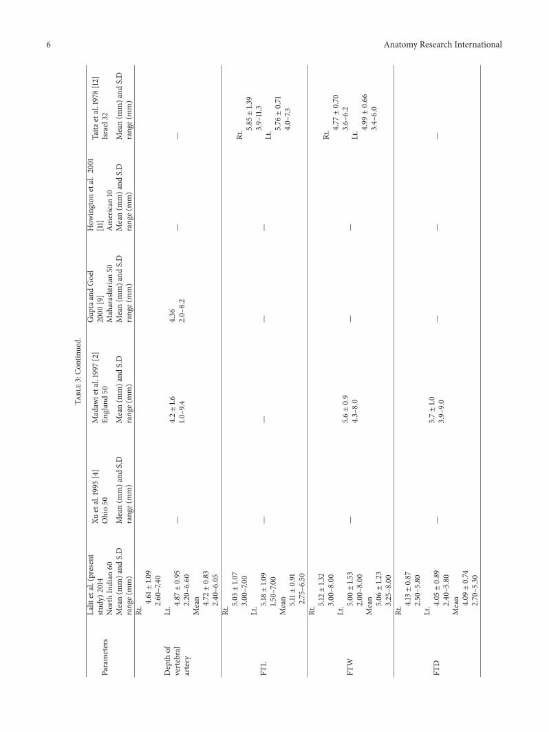
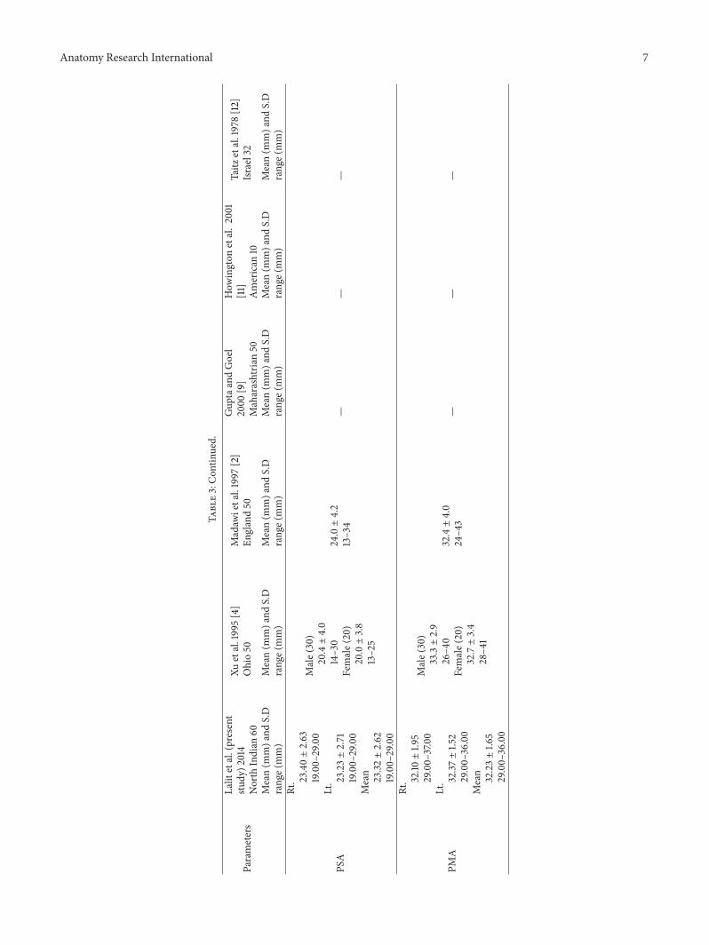
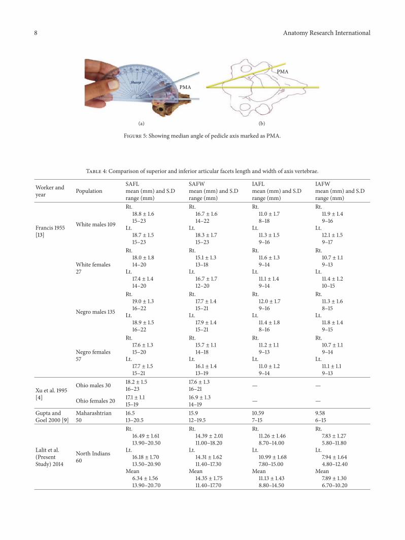