research article risk factors associated with increased...
TRANSCRIPT

Research ArticleRisk Factors Associated with IncreasedMorbidity in Living Liver Donation
Helry L. Candido,1,2 Eduardo A. da Fonseca,1,2 Flávia H. Feier,1,2 Renata Pugliese,1,2
Marcel A. Benavides,1,2 Enis D. Silva,3 Karina Gordon,4 Marcelo Gama de Abreu,5
Jaume Canet,6 Paulo Chapchap,1 and Joao Seda Neto1,2
1Hepatology and Liver Transplantation, Hospital Sırio-Libanes, Rua Barata Ribeiro 414, cj 65, 01308-000 Bela Vista, SP, Brazil2Hepatology and Liver Transplantation, A.C. Camargo Cancer Center, Sao Paulo, SP, Brazil3Department of Anesthesiology, Hospital Sırio-Libanes, Rua Barata Ribeiro 414, cj 65, 01308-000 Bela Vista, SP, Brazil4Department of Anesthesiology, A.C. Camargo Cancer Center, Sao Paulo, Brazil5Department of Anesthesiology and Intensive Care Therapy, Pulmonary Engineering Group, University Hospital Carl Gustav Carus,Technische Universitat Dresden, Dresden, Germany6Department of Anesthesiology, Hospital Universitari Germans Trias i Pujol, Badalona, Barcelona, Spain
Correspondence should be addressed to Joao Seda Neto; [email protected]
Received 16 July 2015; Revised 19 November 2015; Accepted 24 November 2015
Academic Editor: Patrizia Burra
Copyright © 2015 Helry L. Candido et al. This is an open access article distributed under the Creative Commons AttributionLicense, which permits unrestricted use, distribution, and reproduction in any medium, provided the original work is properlycited.
Living donor liver donation (LDLD) is an alternative to cadaveric liver donation.We aimed at identifying risk factors and developinga score for prediction of postoperative complications (POCs) after LDLD in donors. This is a retrospective cohort study in 688donors between June 1995 and February 2014 at Hospital Sırio-Libanes and A.C. Camargo Cancer Center, in Sao Paulo, Brazil.Primary outcome was POC graded ≥III according to the Clavien-Dindo classification. Left lateral segment (LLS), left lobe (LL),and right lobe resections (RL) were conducted in 492 (71.4%), 109 (15.8%), and 87 (12.6%) donors, respectively. In total, 43 (6.2%)developed POCs, which were more common after RL than LLS and LL (14/87 (16.1%) versus 23/492 (4.5%) and 6/109 (5.5%), resp.,𝑝 < 0.001). Multivariate analysis showed that RL resection (OR: 2.81, 95% CI: 1.32 to 3.01; 𝑝 = 0.008), smoking status (OR: 3.2, 95%CI: 1.35 to 7.56; 𝑝 = 0.012), and blood transfusion (OR: 3.15, 95% CI: 1.45 to 6.84; 𝑝 = 0.004) were independently associated withPOCs. RL resection, intraoperative blood transfusion, and smoking were associated with increased risk for POCs in donors.
1. Introduction
In order to mitigate the shortage of cadaveric organs forliver transplantation, Raia et al. [1] and Broelsch et al. [2]introduced the techniques of reduced-size and split livertransplantation and resecting left lateral segments (LLS)from living adults for transplantation into children. Livingdonor liver transplantation (LDLT) was then introduced intoclinical practice and subsequently expanded to adult patientsafter the first right lobe (RL) donation in 1990 [3].
Theprerequisite to performing LDLT, however, is reducedmorbidity and mortality risks of the donor [4]. Providingpotential liver donors accurate and timely information
regarding the risks associated with living donor liver dona-tion (LDLD) is hampered by the lack of standardized report-ing systems [5]. Additionally, under reporting of technicalcomplications, blood and blood product transfusions andaborted donations all contribute to the lack of reliableinformation about the risks involved in LDLD [6].
In the present study, we aimed at identifying risk factorsassociated with postoperative complications (POCs) afterLDLD in donors in two tertiary care center. We hypothesizedthat the risk of postoperative complications in this donorpopulation is influenced by preexisting comorbidities, typeof resection, and intraoperative characteristics.
Hindawi Publishing CorporationJournal of TransplantationVolume 2015, Article ID 949674, 8 pageshttp://dx.doi.org/10.1155/2015/949674

2 Journal of Transplantation
2. Materials and Methods
The sample selection for this manuscript was based in thecollected experience of 697 living donor liver resectionsperformed at Sirio-Libanes Hospital and AC Camargo Can-cer Center between June 1995 and February 2014. It allowedthe assessment of 193 POCs, of which 43 were graded ≥IIIaccording to the Clavien-Dindo classification; 688 donorshad records with complete data, which were retrospectivelyreviewed through patient charts and from a prospectivelycollected database. The hospital’s ethics committee approvedthis study’s protocol (HSL 2011-21).
The variables studied included the following: type of liverresection (LLS, LL, RL), living donor’s age, gender, AmericanSociety of Anesthesiology (ASA) classification, presence ofcomorbidities, body mass index (BMI), and intraoperativepacked red blood cell transfusion (PRBCT). The patientswere evaluated for the development of POCs according tothe Clavien-Dindo classification [7]. The primary outcomewas the development of POCs ≥ grade III according tothe Clavien-Dindo classification among the three types ofliver resections performed. The secondary outcomes wereas follows: intensive care unit (ICU) stay and hospital stay,reoperation, and readmission.
The preoperative, intraoperative, and postoperative pro-tocols changed and evolved over the years. From the begin-ning of the experience until 2004, a Cell Saver (CellSaver) wasroutinely used during the live donor liver resections. Duringthis period, autologous blood was collected a week beforesurgery to be used during the operation. These practiceswere used to ensure donor safety in the early stages of thegroup’s experience with these procedures. After 2004, thisprotocol was abandoned and homologous PRBCT was usedonly when needed during the operation. In the outcomeanalysis, independently of the nature of blood transfusion,patients requiring blood were classified as PRBCT group.
2.1. Classification of Complications. Complications thatoccurred within 3 months from surgery were categorizedaccording to the Clavien-Dindo classification for postop-erative events [7]. Patients who developed more than onecomplication were graded according to the more severe type.
The POCs were divided into the following categories:bile leaks, being infectious (abdominal collection), beinggastrointestinal (prolonged ileus, gastroparesis), liver necro-sis, wound complications (wound infection, incisional her-nia), deep venous thrombosis (DVT), being cardiovascular(atrial flutter, hypertension, endocarditis), pulmonary (pleu-ral effusion, pneumothorax, bronchopneumonia, pulmonarythromboembolism), being hemorrhagic (intra-abdominalbleeding), and others.
2.2. Preoperative Donor Evaluation. The voluntary intentof the donor was first assessed and informed consent wasmandatory. All donors underwent psychological evaluationto rule out any psychological disorders, coercion, or commer-cial motives. The preoperative evaluation of the candidatesincluded routine blood tests, ABO system compatibility,
urine analysis, electrocardiography, and chest X-ray. Theywere also tested for hepatitis A, B, andC serology,HIV,HTLV,CMV, EBV, syphilis, and Chagas disease. Abdominal Dopplerultrasound was performed to evaluate vascular anatomy,liver echogenicity (detection of steatosis and parenchymallesions), and liver volumetry for LLS donation. For LL andRL resection, the anatomical and volumetric evaluation wasperformed with magnetic resonance imaging (MRI) andcholangio-MRI.The ratio of liver weight/recipient weight wasused to estimate the preoperative graft-to-recipient weightratio (GRWR). A GRWR > 1% was aimed when transplantingadolescents and adult recipients. When planning the donoroperation, the accepted remaining liver volume calculated bythe image studies was 30%.
Candidates with significant comorbidities, defined as≥ASA III, were excluded as possible donors. Only donorsyounger than 50 years of age with a BMI less than 28 kg/m2were accepted for surgery. All donors were preoperativelyevaluated by an anesthesiologist and classified according tothe ASA physical status classification [8]. Donors who werecurrent smokers were encouraged to stop smoking 4 weeksbefore surgery, even though they were considered activesmokers in this analysis.
2.3. Operative Techniques. The operative technique for thedonor hepatectomy followed principles described elsewhere[9]. Parenchymal transection was accomplished with a Cav-itron ultrasound surgical aspirator (Cavitron, Stanford, CA)after dissection of the hepatic hilum. The transection line onthe liver varied with the graft type. Intraoperative cholecys-tectomy and cholangiography were performed to determinewhere to cut the donor’s bile duct. Conventional clamping,cutting, and suturing of the major vessels were performedduring graft removal. After the hepatectomy, the graft wasflushed with Euro-Collins solution or histidine-tryptophan-ketoglutarate solution at 4∘C and prepared for implantation.
2.4. Postoperative Management. All the donors recoveredin the ICU during the immediate postoperative period. AJackson-Pratt abdominal drain was routinely inserted at theend of the hepatectomy, close to the cut surface of the liver, tomonitor for bleeding and bile leaks. The drain was removedonly after day five if it showed a low output and serous aspect.The donors routinely receivedDVT prophylaxis with heparin5000UI every 12 hours, which was maintained until theywere discharged from the hospital. The use of proton pumpinhibitors continued for 30 days after surgery. If an ischemicresidual liver segment was detected by clinical inspectionat the end of the liver resection in cases of LLS, a 14-daycourse of ciprofloxacin was administered. This practice wasintroduced after 2007. Before that, the management of thisoccurrence included liver segment resection after completionof the hepatectomy, as previously reported [10].
2.5. Statistical Analysis. Means and medians were calculatedto summarize continuous variables and were compared using𝑡-tests or appropriate nonparametric tests when distribu-tional assumptions were in doubt. Categorical variables were

Journal of Transplantation 3
0
1995
1996
1997
1998
1999
2000
2001
2002
2003
2004
2005
2006
2007
2008
2009
2010
2011
2012
2013
2014
Num
ber o
f don
ors
Year
DonorsASA II (%)
PRBCT (%)
1020304050607080
020406080
100120140160180200220240
Clavien ≥ 3 (%)
ASA
II, P
RBCT
, Cla
vien
≥3
(%)
Figure 1: Distribution of donor operations, use of PRBCT, preva-lence of ASA II donors, and prevalence of complications ≥ gradeIII of the Clavien-Dindo classification, from 1995 to 2013. ASA:American Society of Anesthesiology classification; PRBCT: packedred blood cell transfusion.
expressed as numbers and percentages. Differences betweengroups were assessed by chi-square or Fisher’s exact tests,when needed.
Logistic regression was applied to the data and thosevariables found to be significant at 𝑝 < 0.10 were selectedfor the multivariate modeling. Multivariate analysis wasperformed by stepwise forward logistic regression modeling.Variables were included in the model following a forwardstepwise selection with a statistical critical level set at 𝑝 <0.05.
All analyses were performed using the SPSS 22.0 statisti-cal package (IBM, Inc., Chicago, IL).
3. Results
The number of LDLDs increased from 5 in 1995 to 69 in2013, totaling 688 in the observation period (Figure 1). Fromthese, 492 (71.4%) were LLS resections, 109 (15.8%) were LL,and 87 (12.6%) were RL. Overall, 346 (50.2%) of the donorswere male, the mean BMI was of 23.6 ± 2.68 kg/m2, and 586(86%) were classified as ASA I. Patients whowere classified asASA II (96 patients) presented the habits and comorbiditiesdescribed in Table 1. Reasons for ASA II classification werenot identified in 7 patients. Sixty-six (9.6%) donors receivedintraoperative PRBCT. The characteristics according to thetype of liver resection are shown in Table 1.
When studying the donor’s relationship with the recip-ient, it was observed that 223 (32.4%) were fathers, 240(34.8%) weremothers, 58 (8.5%) were uncles/aunts, 50 (7.3%)were sons/daughters, 8 (1.2%) were grandfathers/grand-mothers, 7 (1%) were husbands/wives, 60 (8.8%) were otherrelatives, and 39 (5.7%) were nonrelated.
A total of 193 out of 688 (28%) of the donors developed atleast one POC; of the 193, 45 (23.3%) developed grade I, 105(54.4%) developed grade II, 5 (2.5%) developed grade IIIa,33 (17%) developed grade IIIb, and 5 (2.5%) developed gradeIVa. Forty-three donors (6.2%) developed at least one POC≥ grade III. Twenty-three patients (4.5%) in the LLS group,
6 (5.5%) patients in the LL group, and 14 (16.1%) patients inthe RL group developed POCs ≥ grade III (𝑝 < 0.001).
A total of 242 POCs were documented in 193 patients.Most of the complications (52.8%) were grade II. The mostfrequently observed complication was bile leak (23.1%), butthe majority (71.4%) was resolved without need of furtherintervention. The most serious complications reported weregrade IVa, which occurred in 5 patients and includedpulmonary thromboembolism, deep venous thrombosis,endocarditis, atrial flutter, and rhabdomyolysis. One patientpresented portal vein thrombosis 6 months following RLdonation, whereas 2 LLS donors committed suicide 5 yearsafter the surgery. The POCs are shown in Table 2.
As shown in Table 3, bile leaks, wound complications,and pulmonary complications were more frequent with RLthan other liver resection types. Also, the occurrence of atleast one complication was more frequent in donors whounderwent RL liver resection (23.9% LLS versus 33.9% LLversus 43.6% RL; 𝑝 < 0.001) (Table 4). Donors submitted toLL and RL resections had longer ICU and hospital stays aswell as readmission and reoperation rates when compared todonors who underwent LLS resection (Table 5).
Donors who smoked were more likely to develop POCsthan nonsmokers (44.9% versus 26.7%; 𝑝 = 0.007). Also, thefrequency of complications ≥ grade III was higher in smokers(16.3% versus 5.5%;𝑝 = 0.008).Themost frequently observedcomplication in this subgroup was bile leak, followed by livernecrosis and pulmonary and gastrointestinal complications(Table 6).
The variables initially included in the multivariate modelwere as follows: type of liver resection, gender, age, PRBCT,ASA classification, and smoking status. RL donation (OR:2.81, 95% CI: 1.32 to 3.01; 𝑝 = 0.008), smoking status (OR:3.2, 95% CI: 1.35 to 7.56; 𝑝 = 0.012), and PRBCT (OR: 3.15,95% CI: 1.45 to 6.84; 𝑝 = 0.004) were retained in the model(Table 7).
Figure 1 shows the prevalence of ASA II patients, theuse of PRBCT, and the incidence of POCs ≥ grade III. Inmore recent years, the need for PRBCT has become minimal(1.8%) and the incidence of complications ≥ grade III has alsodropped from 20–30% in the early years to 1.5–5% in recentyears.
4. Discussion
Themain findings of this retrospective analysis in 688 donorswho underwent liver resection for LDLT were as follows:(1) the risk of severe POCs is higher in patients submittedto RL donation; (2) donors who were active smokers andthose that received blood transfusion during surgery alsohave higher risk of developing severe POCs; (3) the mostfrequently observed complication was bile leaks.
Since the introduction of LDLT into clinical practice,the focus of the medical community has been to define therisk factors and establish procedures to ensure donor safetyduring the process of living donation. It is well known thatthere is a significant difference between the donor risks forRL/extended right donation and LL/LLS donation and thatRL donors have more risk factors than non-RL donors [11].

4 Journal of Transplantation
Table 1: Preoperative and intraoperative variables according to the type of liver resection.
Variables All LLS LL RL 𝑝
𝑁 = 688 𝑁 = 492 𝑁 = 109 𝑁 = 87
Age, mean ± SD 29.8 ± 7.20 28.7 ± 6.7 33.6 ± 8.2 31.2 ± 8.5 <0.001Sex, male, number(%) 346 (50.3) 225 (45.7) 67 (61.5) 55 (63.2) <0.001
BMI, mean ± SD 23.6 ± 2.7 23.4 ± 2.7 24 ± 2.3 23.9 ± 2.7 0.13𝑁 = 682 𝑁 = 492 𝑁 = 106 𝑁 = 84
ASA, number (%) 0.36I 586 (85.9) 427 (86.8) 91 (85.8) 68 (80.9)II 96 (14.1) 65 (13.2) 15 (14.2) 16 (19.1)
𝑁 = 89 𝑁 = 59 𝑁 = 15 𝑁 = 15 0.32Smoker, number (%) 49 (55.0) 32 (54.1) 8 (53.3) 9 (60.0)Anemia, number (%) 7 (7.9) 6 (10.2) 1 (6.7) 0Asthma, number (%) 6 (6.7) 3 (5.1) 1 (6.7) 2 (13.3)Renal lithiasis,number (%) 4 (4.5) 4 (6.8) 0 0
Overweight, number(%) 4 (4.5) 3 (5.1) 1 (6.7) 0
MMVD, number (%) 2 (2.3) 2 (3.4) 0 0Hypertension,number (%) 4 (4.5) 1 (1.7) 2 (13.3) 1 (6.7)
Gastritis, number (%) 2 (2.2) 0 0 2 (13.3)Other, number (%)∗ 11 (12.4) 8 (13.6) 2 (13.3) 1 (6.7)
𝑁 = 688 𝑁 = 492 𝑁 = 109 𝑁 = 87
PRBCT, number (%) 66 (9.6) 29 (5.9) 13 (11.9) 24 (27.6) <0.001Graft weight (g),mean ± SD 288.6 ± 60.9 433.8 ± 100.0 814.2 ± 269.8 <0.001
𝑁 = 608 𝑁 = 432 𝑁 = 99 𝑁 = 77
Number of bile ducts,number (%) <0.001
1 479 (78.7) 354 (82.0) 82 (82.8) 43 (55.8)2 122 (20.1) 74 (17.1) 16 (16.2) 32 (41.6)3 7 (1.2) 4 (0.9) 1 (1.0) 2 (2.6)
LLS, left lateral segment; LL, left lobe; RL, right lobe; BMI, body mass index; ASA, American Society of Anesthesiology physical status classification; PRBCT,packed red blood cell transfusion; MMVD, minor mitral valve dysfunction; SD, standard deviation.∗hypothyroidism, hiatal hernia, dyslipidemia, upper airway disturbances, mood disorders, leucopenia, and arrhythmia.
Table 2: Classification distribution of postoperative complications according to Clavien-Dindo classification.
Grade Bile leak Infectious GI Liver necrosis Wound DVT Cardio Pulm Bleeding Other Total, 𝑛 (%)I 5 12 17 0 11 0 0 3 0 1 49 (20.2)II 35 24 19 8 5 2 2 10 0 23 128 (52.9)IIIa 1 0 0 0 1 0 0 4 0 0 6 (2.5)IIIb 15 10 4 13 5 1 0 4 1 1 54 (22.3)IVa 0 0 0 0 0 1 2 1 0 1 5 (2.1)Total, 𝑛 (%) 56 (23.1) 46 (19.0) 40 (16.5) 21 (8.7) 22 (9.1) 4 (1.7) 4 (1.7) 22 (9.1) 1 (0.4) 26 (10.7) 242Infectious: infectious complications; DVT: deep venous thrombosis; GI: gastrointestinal complications; Cardio: cardiac complications; Pulm: pulmonarycomplications. Some donors experienced two or more complications.
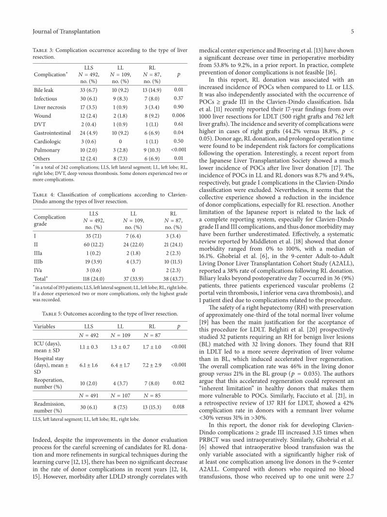
Journal of Transplantation 5
Table 3: Complication occurrence according to the type of liverresection.
Complication∗LLS𝑁 = 492,no. (%)
LL𝑁 = 109,no. (%)
RL𝑁 = 87,no. (%)
𝑝
Bile leak 33 (6.7) 10 (9.2) 13 (14.9) 0.01Infectious 30 (6.1) 9 (8.3) 7 (8.0) 0.37Liver necrosis 17 (3.5) 1 (0.9) 3 (3.4) 0.90Wound 12 (2.4) 2 (1.8) 8 (9.2) 0.006DVT 2 (0.4) 1 (0.9) 1 (1.1) 0.61Gastrointestinal 24 (4.9) 10 (9.2) 6 (6.9) 0.04Cardiologic 3 (0.6) 0 1 (1.1) 0.50Pulmonary 10 (2.0) 3 (2.8) 9 (10.3) <0.001Others 12 (2.4) 8 (7.3) 6 (6.9) 0.01∗in a total of 242 complications; LLS, left lateral segment; LL, left lobe; RL,right lobe; DVT, deep venous thrombosis. Some donors experienced two ormore complications.
Table 4: Classification of complications according to Clavien-Dindo among the types of liver resection.
Complicationgrade
LLS𝑁 = 492,no. (%)
LL𝑁 = 109,no. (%)
RL𝑁 = 87,no. (%)
I 35 (7.1) 7 (6.4) 3 (3.4)II 60 (12.2) 24 (22.0) 21 (24.1)IIIa 1 (0.2) 2 (1.8) 2 (2.3)IIIb 19 (3.9) 4 (3.7) 10 (11.5)IVa 3 (0.6) 0 2 (2.3)Total∗ 118 (24.0) 37 (33.9) 38 (43.7)∗in a total of 193 patients; LLS, left lateral segment; LL, left lobe; RL, right lobe.If a donor experienced two or more complications, only the highest gradewas recorded.
Table 5: Outcomes according to the type of liver resection.
Variables LLS LL RL 𝑝
𝑁 = 492 𝑁 = 109 𝑁 = 87
ICU (days),mean ± SD 1.1 ± 0.3 1.3 ± 0.7 1.7 ± 1.0 <0.001
Hospital stay(days), mean ±SD
6.1 ± 1.6 6.4 ± 1.7 7.2 ± 2.9 <0.001
Reoperation,number (%) 10 (2.0) 4 (3.7) 7 (8.0) 0.012
𝑁 = 491 𝑁 = 107 𝑁 = 85
Readmission,number (%) 30 (6.1) 8 (7.5) 13 (15.3) 0.018
LLS, left lateral segment; LL, left lobe; RL, right lobe.
Indeed, despite the improvements in the donor evaluationprocess for the careful screening of candidates for RL dona-tion and more refinements in surgical techniques during thelearning curve [12, 13], there has been no significant decreasein the rate of donor complications in recent years [12, 14,15]. However, morbidity after LDLD strongly correlates with
medical center experience and Broering et al. [13] have showna significant decrease over time in perioperative morbidityfrom 53.8% to 9.2%, in a prior report. In practice, completeprevention of donor complications is not feasible [16].
In this report, RL donation was associated with anincreased incidence of POCs when compared to LL or LLS.It was also independently associated with the occurrence ofPOCs ≥ grade III in the Clavien-Dindo classification. Iidaet al. [11] recently reported their 17-year findings from over1000 liver resections for LDLT (500 right grafts and 762 leftliver grafts).The incidence and severity of complicationswerehigher in cases of right grafts (44.2% versus 18.8%, 𝑝 <0.05). Donor age, RL donation, and prolonged operation timewere found to be independent risk factors for complicationsfollowing the operation. Interestingly, a recent report fromthe Japanese Liver Transplantation Society showed a muchlower incidence of POCs after live liver donation [17]. Theincidence of POCs in LL and RL donors was 8.7% and 9.4%,respectively, but grade I complications in the Clavien-Dindoclassification were excluded. Nevertheless, it seems that thecollective experience showed a reduction in the incidenceof donor complications, especially for RL resection. Anotherlimitation of the Japanese report is related to the lack ofa complete reporting system, especially for Clavien-Dindograde II and III complications, and thus donormorbiditymayhave been further underestimated. Effectively, a systematicreview reported by Middleton et al. [18] showed that donormorbidity ranged from 0% to 100%, with a median of16.1%. Ghobrial et al. [6], in the 9-center Adult-to-AdultLiving Donor Liver Transplantation Cohort Study (A2ALL),reported a 38% rate of complications following RL donation.Biliary leaks beyond postoperative day 7 occurred in 36 (9%)patients, three patients experienced vascular problems (2portal vein thrombosis, 1 inferior vena cava thrombosis), and1 patient died due to complications related to the procedure.
The safety of a right hepatectomy (RH) with preservationof approximately one-third of the total normal liver volume[19] has been the main justification for the acceptance ofthis procedure for LDLT. Belghiti et al. [20] prospectivelystudied 32 patients requiring an RH for benign liver lesions(BL) matched with 32 living donors. They found that RHin LDLT led to a more severe deprivation of liver volumethan in BL, which induced accelerated liver regeneration.The overall complication rate was 46% in the living donorgroup versus 21% in the BL group (𝑝 = 0.035). The authorsargue that this accelerated regeneration could represent an“inherent limitation” in healthy donors that makes themmore vulnerable to POCs. Similarly, Facciuto et al. [21], ina retrospective review of 137 RH for LDLT, showed a 42%complication rate in donors with a remnant liver volume<30% versus 31% in >30%.
In this report, the donor risk for developing Clavien-Dindo complications ≥ grade III increased 3.15 times whenPRBCT was used intraoperatively. Similarly, Ghobrial et al.[6] showed that intraoperative blood transfusion was theonly variable associated with a significantly higher risk ofat least one complication among live donors in the 9-centerA2ALL. Compared with donors who required no bloodtransfusions, those who received up to one unit were 2.7

6 Journal of Transplantation
Table 6: Classification of postoperative complications in smokers versus nonsmokers.
ComplicationSmokers 𝑛 = 49 Nonsmoker 𝑛 = 639
Grades I + IInumber (%)
≥Grade IIInumber (%)
Grades I + IInumber (%)
≥Grade IIInumber (%)
Bile leak 9 (52.9) 3 (20.0) 31 (19.4) 13 (26.0)Infectious 1 (5.9) 1 (6.7) 35 (21.9) 9 (18.0)Liver necrosis 2 (11.8) 3 (20.0) 6 (3.8) 10 (20.0)Wound 0 1 (6.7) 16 (10.0) 5 (10.0)DVT 0 2 (13.3) 2 (1.3) 0Gastrointestinal 2 (11.8) 1 (6.7) 34 (21.3) 3 (6.0)Cardiologic 0 2 (13.3) 2 (1.3) 0Pulmonary 1 (5.9) 2 (13.3) 12 (7.5) 7 (14.0)Other 2 (11.8) 0 22 (13.8) 3 (6.0)Total∗,𝑁 17 (34.7) 15 (30.6) 160 (25) 50 (7.8)∗in a total of 242 complications; DVT: deep venous thrombosis.
Table 7: Multivariate logistic regression analysis for the develop-ment of complications ≥ grade III.
Variables 𝑝 OR 95% CIGraft type
LLS — — —LL 0.94 1.04 0.41 to 2.67RL 0.008 2.81 1.32 to 6.01
Smoker (no) — 1 —Smoker (yes) 0.012 3.20 1.35 to 7.56PRBCT (no) — 1 —PRBCT (yes) 0.004 3.15 1.45 to 6.84LLS, left lateral segment; RL, right lobe; PRBCT, packed red blood celltransfusion; OR, odds ratio; CI, confidence interval.
times more susceptible to complications. A more recentpublication from the A2ALL [22] in a cohort of 740 LDLDcorroborated the results from the previous report: there wasa significant association of transfusion requirements withthe development of a first complication of any type (HR =1.38 per unit; 𝑝 < 0.0001) and specifically with theoccurrence of bile leak and infection. Indeed, bile leaks,wound complications, and pulmonary complications weremore frequent in RL liver resections in the present report.Lo [23] showed a series of 1508 cases of living liver donorswhere the rate of complications was 15.8%. In these cases,pulmonary complications were uncommon. Conversely, insmaller cohort of live donation, Dondero et al. [24] reportedfourteen major respiratory complications in 11 of 112 donors(9.8%). In that study, pulmonary complications were frequentin living liver donors after the operation and pulmonaryembolism was the most frequent of these complications. Ofnote, pulmonary morbidity was observed mainly followingRH.
Ideally, candidates for living donation should be classifiedas ASA I [8]. In practice, most of the transplant centersallow ASA II candidates to proceed and go through with
the operation because these patients present no functionallimitations. In our cohort, 55% of the donors classified asASA II were active smokers. Indeed, ASA II was found tobe a surrogate marker of the effects of smoking. Donorswho smoke have a risk 3.2 times higher than nonsmokersof developing POCs ≥ grade III, according to our findings.This is similar to the risk presented by the need for intra-operative PRBCT. The effects of smoking on cardiovascularand pulmonary systems as well as the detrimental effects onwound healing have already been described. Smokers haveincreased mucus production and the damage to tracheal ciliaimpairs the clearance of such mucus [25]. The impairmentof the wound healing process can also be responsible forthe higher risk of anastomotic leaks after colorectal surgery,presented by patients who smoke [26]. In our series, themost frequent complication in smokers was bile leaks andliver necrosis, followed by pulmonary complications. In fact,the distribution of types of complications was similar tothose observed in the entire cohort. The effects of preoper-ative smoking cessation are controversial, as is the periodnecessary for this intervention to be beneficial. A decreasein wound complications was demonstrated in nonsmokingpatients [27], but most of the studies show no difference inthis outcome with smoking cessation [25]. In a large review[25] of studies of smoking cessation intervention prior tosurgery, the incidence of cardiopulmonary complications innonsmoking patients compared to current smokers did notdiffer significantly. It has been determined that pulmonaryfunction recovers in 8 weeks after smoking cessation, butthe minimal timing needed to decrease the risk of POCs hasnot been determined yet [25]. Nakagawa et al. [28] foundthat both current smokers and former smokers that stopped4 weeks before pulmonary surgery had a higher risk ofpostoperative pulmonary complications than those whowerenever smokers. Our current practice with donors who smokeis to encourage a 4-week period of abstinence from smokingbefore surgery. The findings in the present paper raise thequestion if we should continue to accept smokers as donors,further prioritizing donor safety. Whether advising donors

Journal of Transplantation 7
to abstain from smoking for a longer period before surgerywould be enough to minimize the risk of complicationsremains to be determined. Interestingly, the presence of asmoking habit has not been reported in previous studiesregarding the evaluation of complications following LDLD.
Limitations. This study has different limitations. First, thiswas a retrospective study, and donors were assessed bythe same medical team. Results are likely affected by localcharacteristics, including the experience of the staff. Second,change in clinical practice during the observation periodmayhave occurred, a fact that would influence the results.
5. Conclusions
POCs were relatively common in this cohort of LDLDs. RLdonation, use of intraoperative PRBCT, and smoking wereindependently associated with the occurrence of POCs ≥grade III in the Clavien-Dindo classification after live dona-tion.
Abbreviations
LLS: Left lateral segment (liver segments II and III)LDLT: Living donor liver transplantationLDLD: Living donor liver donationASA: American Society of AnesthesiologyBMI: Body mass indexPOC: Postoperative complicationPRBCT: Packed red blood cells transfusionICU: Intensive care unitDVT: Deep venous thrombosisMRI: Magnetic resonance imagingGRWR: Graft-to-recipient weight ratioLL: Left lobe (liver segments II, III, and IV)LT: Liver transplantRL: Right lobe (liver segments V, VI, VII, and VIII)RH: Right hepatectomy.
Conflict of Interests
The authors declare no conflict of interests.
Authors’ Contribution
Helry L. Candido, Flavia H. Feier, Eduardo A. da Fonseca,and Joao Seda Neto contributed to the hypothesis forming,collected the data, performed statistical analysis, and pre-pared and reviewed the paper. Renata Pugliese, Marcel A.Benavides, Karina Gordon, and Enis D. Silva contributedto the data collection and paper writing. Marcelo Gama deAbreu, Jaume Canet, and Paulo Chapchap contributed to theresults interpretation and reviewed the paper.
References
[1] S. Raia, J. R. Nery, and S. Mies, “Liver transplantation from livedonors,”The Lancet, vol. 334, no. 8661, p. 497, 1989.
[2] C. E. Broelsch, P. F. Whitington, J. C. Emond et al., “Livertransplantation in children from living related donors. Surgicaltechniques and results,” Annals of Surgery, vol. 214, no. 4, pp.428–439, 1991.
[3] S. Fujita, I.-D. Kim, K. Uryuhara et al., “Hepatic grafts fromlive donors: donor morbidity for 470 cases of live donation,”Transplant International, vol. 13, no. 5, pp. 333–339, 2000.
[4] Y. Soejima, A. Taketomi, T. Yoshizumi et al., “Feasibility of leftlobe living donor liver transplantation between adults: an 8-year, single-center experience of 107 cases,” American Journalof Transplantation, vol. 6, no. 5, part 1, pp. 1004–1011, 2006.
[5] Y. L. Cheah, M. A. Simpson, J. J. Pomposelli, and E. A. Pomfret,“Incidence of death and potentially life-threatening near-missevents in living donor hepatic lobectomy: a world-wide survey,”Liver Transplantation, vol. 19, no. 5, pp. 499–506, 2013.
[6] R.M. Ghobrial, C. E. Freise, J. F. Trotter et al., “Donormorbidityafter living donation for liver transplantation,”Gastroenterology,vol. 135, no. 2, pp. 468–476, 2008.
[7] D. Dindo, N. Demartines, and P.-A. Clavien, “Classificationof surgical complications: a new proposal with evaluation ina cohort of 6336 patients and results of a survey,” Annals ofSurgery, vol. 240, no. 2, pp. 205–213, 2004.
[8] R. D. Dripps, A. Lamont, and J. E. Eckenhoff, “The role ofanesthesia in surgical mortality,” The Journal of the AmericanMedical Association, vol. 178, pp. 261–266, 1961.
[9] J. S. Neto, R. Pugliese, E. A. Fonseca et al., “Four hundred thirtyconsecutive pediatric living donor liver transplants: variablesassociated with posttransplant patient and graft survival,” LiverTransplantation, vol. 18, no. 5, pp. 577–584, 2012.
[10] J. Seda-Neto, A. L. Godoy, E. Carone et al., “Left lateralsegmentectomy for pediatric live-donor liver transplantation:special attention to segment IV complications,”Transplantation,vol. 86, no. 5, pp. 697–701, 2008.
[11] T. Iida, Y. Ogura, F. Oike et al., “Surgery-related morbidity inliving donors for liver transplantation,” Transplantation, vol. 89,no. 10, pp. 1276–1282, 2010.
[12] J. W. Marsh, E. Gray, R. Ness, and T. E. Starzl, “Complicationsof right lobe living donor liver transplantation,” Journal ofHepatology, vol. 51, no. 4, pp. 715–724, 2009.
[13] D. C. Broering, C. Wilms, P. Bok et al., “Evolution of donormorbidity in living related liver transplantation: a single-centeranalysis of 165 cases,”Annals of Surgery, vol. 240, no. 6, pp. 1013–1026, 2004.
[14] D. Azoulay, P. Bhangui, P. Andreani et al., “Short- and long-termdonormorbidity in right lobe living donor liver transplantation:91 consecutive cases in a European center,” American Journal ofTransplantation, vol. 11, no. 1, pp. 101–110, 2011.
[15] N.-J. Yi, K.-S. Suh, J. Y. Cho et al., “Three-quarters of rightliver donors experienced postoperative complications,” LiverTransplantation, vol. 13, no. 6, pp. 797–806, 2007.
[16] S. Hwang, S.-G. Lee, Y.-J. Lee et al., “Lessons learned from 1,000living donor liver transplantations in a single center: how tomake living donations safe,” Liver Transplantation, vol. 12, no.6, pp. 920–927, 2006.
[17] Y. Hashikura, T. Ichida, K. Umeshita et al., “Donor compli-cations associated with living donor liver transplantation inJapan,” Transplantation, vol. 88, no. 1, pp. 110–114, 2009.
[18] P. F. Middleton, M. Duffield, S. V. Lynch et al., “Living donorliver transplantation—adult donor outcomes: a systematicreview,” Liver Transplantation, vol. 12, no. 1, pp. 24–30, 2006.
8 Journal of Transplantation
[19] Y. Kishi, E. K. Abdalla, Y. S. Chun et al., “Three hundredand one consecutive extended right hepatectomies: evaluationof outcome based on systematic liver volumetry,” Annals ofSurgery, vol. 250, no. 4, pp. 540–548, 2009.
[20] J. Belghiti, G. Liddo, V. Raut et al., “‘Inherent limitations’ indonors: control matched study of consequences following aright hepatectomy for living donation and benign liver lesions,”Annals of Surgery, vol. 255, no. 3, pp. 528–533, 2012.
[21] M. Facciuto, A. Contreras-Saldivar, M. K. Singh et al., “Righthepatectomy for living donation: role of remnant liver volumein predicting hepatic dysfunction and complications,” Surgery,vol. 153, no. 5, pp. 619–626, 2013.
[22] M. M. Abecassis, R. A. Fisher, K. M. Olthoff et al., “Com-plications of living donor hepatic lobectomy-a comprehensivereport,” American Journal of Transplantation, vol. 12, no. 5, pp.1208–1217, 2012.
[23] C.-M. Lo, “Complications and long-term outcome of livingliver donors: a survey of 1,508 cases in five Asian centers,”Transplantation, vol. 75, supplement 3, pp. S12–S15, 2003.
[24] F. Dondero, C. Taille, H. Mal et al., “Respiratory complications:a major concern after right hepatectomy in living liver donors,”Transplantation, vol. 81, no. 2, pp. 181–186, 2006.
[25] T. Thomsen, N. Villebro, and A. M. Møller, “Interventionsfor preoperative smoking cessation,” The Cochrane Database ofSystematic Reviews, vol. 3, Article ID CD002294, 2014.
[26] L. T. Sørensen, T. Jørgensen, L. T. Kirkeby, J. Skovdal, B. Vennits,and P. Wille-Jørgensen, “Smoking and alcohol abuse are majorrisk factors for anastomotic leakage in colorectal surgery,” TheBritish Journal of Surgery, vol. 86, no. 7, pp. 927–931, 1999.
[27] A. M. Møller, N. Villebro, T. Pedersen, and H. Tønnesen,“Effect of preoperative smoking intervention on postoperativecomplications: a randomised clinical trial,”The Lancet, vol. 359,no. 9301, pp. 114–117, 2002.
[28] M. Nakagawa, H. Tanaka, H. Tsukuma, and Y. Kishi, “Rela-tionship between the duration of the preoperative smoke-free period and the incidence of postoperative pulmonarycomplications after pulmonary surgery,” Chest, vol. 120, no. 3,pp. 705–710, 2001.

Submit your manuscripts athttp://www.hindawi.com
Stem CellsInternational
Hindawi Publishing Corporationhttp://www.hindawi.com Volume 2014
Hindawi Publishing Corporationhttp://www.hindawi.com Volume 2014
MEDIATORSINFLAMMATION
of
Hindawi Publishing Corporationhttp://www.hindawi.com Volume 2014
Behavioural Neurology
EndocrinologyInternational Journal of
Hindawi Publishing Corporationhttp://www.hindawi.com Volume 2014
Hindawi Publishing Corporationhttp://www.hindawi.com Volume 2014
Disease Markers
Hindawi Publishing Corporationhttp://www.hindawi.com Volume 2014
BioMed Research International
OncologyJournal of
Hindawi Publishing Corporationhttp://www.hindawi.com Volume 2014
Hindawi Publishing Corporationhttp://www.hindawi.com Volume 2014
Oxidative Medicine and Cellular Longevity
Hindawi Publishing Corporationhttp://www.hindawi.com Volume 2014
PPAR Research
The Scientific World JournalHindawi Publishing Corporation http://www.hindawi.com Volume 2014
Immunology ResearchHindawi Publishing Corporationhttp://www.hindawi.com Volume 2014
Journal of
ObesityJournal of
Hindawi Publishing Corporationhttp://www.hindawi.com Volume 2014
Hindawi Publishing Corporationhttp://www.hindawi.com Volume 2014
Computational and Mathematical Methods in Medicine
OphthalmologyJournal of
Hindawi Publishing Corporationhttp://www.hindawi.com Volume 2014
Diabetes ResearchJournal of
Hindawi Publishing Corporationhttp://www.hindawi.com Volume 2014
Hindawi Publishing Corporationhttp://www.hindawi.com Volume 2014
Research and TreatmentAIDS
Hindawi Publishing Corporationhttp://www.hindawi.com Volume 2014
Gastroenterology Research and Practice
Hindawi Publishing Corporationhttp://www.hindawi.com Volume 2014
Parkinson’s Disease
Evidence-Based Complementary and Alternative Medicine
Volume 2014Hindawi Publishing Corporationhttp://www.hindawi.com