research article subtoxic doses of cadmium modulate
TRANSCRIPT
Research ArticleSubtoxic Doses of Cadmium Modulate Inflammatory Propertiesof Murine RAW 264.7 Macrophages
Sina Riemschneider,1 Martin Herzberg,2 and Jörg Lehmann1
1Fraunhofer Institute for Cell Therapy and Immunology (IZI), 04103 Leipzig, Germany2Martin Luther University of Halle-Wittenberg, Institute for Biology/Microbiology, 06120 Halle, Germany
Correspondence should be addressed to Jorg Lehmann; [email protected]
Received 28 November 2014; Revised 13 May 2015; Accepted 27 May 2015
Academic Editor: Petros Gikas
Copyright © 2015 Sina Riemschneider et al. This is an open access article distributed under the Creative Commons AttributionLicense, which permits unrestricted use, distribution, and reproduction in any medium, provided the original work is properlycited.
Cadmium (Cd) is a toxic heavy metal that exhibits various adverse effects in the human and animal organism. Its resemblanceto essential metals such as calcium, iron, and zinc leads to an unintended uptake in cells after intake through inhalation andingestion. In this study we investigated the toxicity and the immunomodulatory potential of Cd in nonactivated and activatedmurine macrophages (i.e., cell line RAW 264.7). Cadmium alone caused a dose-dependent decreased viability of exposed cells.Subtoxic Cd concentrations delayed cell death in macrophages, resulting from cytotoxic storm, producing reactive oxygen species(ROS) and nitric oxide (NO), in response to their stimulation by bacterial antigens via pattern-recognition receptors (PRRs). Inaddition, production of selected pro- and anti-inflammatory cytokines, the chemokine CXCL1 (KC), and NO was determined. Weobserved that proinflammatory IL-1𝛽 and also CXCL1 were highly upregulated whereas anti-inflammatory or regulatory cytokinesIL-6 and IL-10 were suppressed by 10 𝜇M Cd. Also production of antibacterial NO was significantly reduced through exposure to10 𝜇M Cd, maybe explaining better survival of macrophages. Additionally, we could show by analysis via ICP-MS that differenteffects of Cd in nonactivated and activated macrophages definitely did not result from different Cd uptake rates.
1. Introduction
Global industrialization has caused a dramatic contamina-tion of the environment with toxic heavy metals such ascadmium (Cd), lead, or mercury representing a latent dangerformen’s health via the nutrition chain through contaminatedwater or foodproducts [1, 2] ormaternalmilk [3]. Alarmingly,modern production methods in agriculture contribute moreand more to Cd contamination of food products throughphosphate fertilizers or mobilisation of biosolid-borne Cd bychloride ligands in soil solution [4], indiscriminate use ofpesticides such as glyphosate [5], and the agricultural use ofanaerobically digested residues from full-scale biogas plants[6]. Other crucial sources for accumulation of heavy metals,in particular cadmium, in men is cigarette smoking [2] andexposure of workers in industries [7]. Since Cd is capable ofentering cells via Ca2+ channel [8], ZIP-transporters [9, 10]and divalent metal transporters 1 (DMT-1, Nramp2) [11,12] of the cell membrane of many cells and accumulate
intracellularly due to its binding to cytoplasmic and nuclearmaterial constituting a potential threat to human and animalhealth [13, 14].
Although tremendous work has been done to identifydiverse toxicological pathways of heavy metals, such asmutagenic, cancerogenic, teratogenic, reprotoxic, nephro-toxic, or neurotoxic effects [15–22], there is still fragmentaryunderstanding how certain heavy metals affect the innateor adaptive immune system. However, those adverse effectsmay potentially impact human health in terms of suppressingimmunity to infection and control of cancer or supportingthe development of autoimmunity or allergy even at very lowsubtoxic exposure doses (reviewed in [23]).
Data from an in vivo infection model (i.e., SalmonellaEnteritidis) under Cd exposure showed a significant immu-nosuppressive effect of this heavy metal on the early and lateimmune response against infectious agents, suggesting thatCd influences both innate as well as adaptive immune mech-anisms (Hemdan and Lehmann, unpublished). Interestingly,
Hindawi Publishing CorporationBioMed Research InternationalVolume 2015, Article ID 295303, 8 pageshttp://dx.doi.org/10.1155/2015/295303
2 BioMed Research International
the phenotype of this mouse model was associated with ahyperactivation rather than a suppressed immune responsein vivo. A first in vitro study to investigate the underlyingmechanismusing human peripheral bloodmononuclear cells(PBMCs) has shown that the immunomodulating capac-ity of Cd depends significantly on the activation stimulusand the target cell population. Polyclonal activation of Tcells and antigen-presenting cells (APCs) by anti-CD3/anti-CD28 or anti-CD40, respectively, versus activation of APCsvia pattern-recognition receptor (PRR) ligands by heat-killed salmonellae (hk S.E.) resulted in completely differentimmunomodulatory effects [24]. However, the mode ofaction is still elusive. For the complete understanding ofthe immunomodulation by Cd a deeper knowledge of Cd-mediated cellular and molecular effects on individual celltypes orchestrating the innate and adaptive immune responseis essentially required. In the present study we attemptedto characterize the influence of cadmium on the activityof murine macrophages in vitro to get an impression howactivation of macrophages by multiple PRR ligation, inducedby hk S.E., is modulated under cadmium exposure.
2. Materials and Methods
2.1. Cell Culture of Murine Macrophage Cell Line RAW264.7. RAW 264.7 (ATCC TIB-71) represents a murine(BALB/c; H2d) adherent growingmonocyte/macrophage cellline which is transformed by Abelson murine leukemiavirus [25]. RAW 264.7 cells are capable of pinocytosis andphagocytosis, antibody-dependent lysis of tumor cells as wellas nitric oxide (NO) and cytokine production and they areresponsive to LPS [25]. Cells were cultured in 175 cm2 cellculture flasks (Greiner Bio-One, Frickenhausen, Germany) inphenol-red free RPMI 1640 medium supplemented with 10%FBS, 2mM L-glutamine, 10mM HEPES buffer, 100 𝜇g/mLpenicillin/streptomycin (Biochrom, Berlin, Germany), and50𝜇M 𝛽-mercaptoethanol (Sigma Aldrich, Steinheim, Ger-many) at 37∘C, 5% CO
2, and 95% air humidity.
For experiments the cells were activated with heat-killedSalmonella enterica Serovar Enteritidis (SalmoVac SE, IDTBiologika GmbH, Dessau-Rosslau, Germany). The relativeantigen concentration used in this study (ratio: 108 hk S.E.to 107 macrophages) was previously determined (data notshown) and ensures appropriate activation of macrophages.
2.2. Determination of Cadmium Content by Means of Induc-tively Coupled Plasma: Mass Spectrometry Analysis (ICP-MS).Cells were plated at a density of 1 × 10⁶ cells/mL in 58 cm2 cellculture dishes with 10mL of cell culture medium describedabove. Afterwards cells were incubated with 0.01𝜇M, 0.1 𝜇MCdCl2, or 10 𝜇M CdCl
2(Sigma Aldrich) alone, or in the
presence of 1 × 10⁸ hk S.E. After 2 h cells were harvested andwashed twice with phosphate-buffered saline (0.15 M NaCl,pH 7.4; PBS) containing 5 mM EDTA (Sigma Aldrich). Thesupernatant was discarded and the residual liquid carefullyremoved at each step. The pellet was suspended in concen-trated 67% (w/v) HNO
3(trace metal grade; BDH Prolabo,
VWR, Darmstadt, Germany) and mineralized at 70∘C for
2 h. Samples were diluted to a final concentration of 2%(w/v) nitric acid. Indium was added as internal standardat a final concentration of 10 ppb. Elemental analysis wasperformed via inductively ICP-MS using ESI-sampler SC-2 (Elemental Scientific Inc., Omaha, USA) and an X-SeriesII ICP-MS instrument (Thermo Fisher Scientific, Bremen,Germany) operating with a collision/reaction cell and flowrates of 5ml/min of He/H
2(93%/7%), with an Ar carrier flow
rate of 0.76 l/min and an Ar make-up flow rate of 15 l/min.An external calibration curve was recorded with ICP-multielement standard solution XVI (Merck, Darmstadt, Ger-many) in 2% nitric acid. The sample was introduced via aperistaltic pump and analyzed for its metal content. For blankmeasurement and quality/quantity thresholds, calculationsbased on DIN32645 TMM were used. The results weretransformed fromppm, ppb, or ppt viamolar units into atomsper sample and divided by the number of cells per sample.
2.3. Cytokine and Chemokine Detection by Enzyme-LinkedImmunosorbent Assay (ELISA). Cells were plated at a densityof 1 × 10⁶ cells/mL in 58 cm2 cell culture dishes with 10mL ofcell culture medium described above. Afterwards cells wereincubated with 0.1𝜇MCdCl
2or 10 𝜇MCdCl
2and stimulated
with 1 × 10⁸ hk S.E. Supernatants were collected and analyzedfor their content of cytokines by ELISA. For determinationof murine cytokines IL-1𝛽, IL-6, IL-10, and TNF-𝛼 reagentsets and protocols of eBioscience (Frankfurt, Germany) wereused. Concentration of the murine chemokine CXCL1 wasmeasured using the reagent set and related protocol fromR&D Systems (Wiesbaden, Germany). For determination ofIL-6 and TNF-𝛼 supernatants were diluted 1 : 100 and 1 : 500,respectively. Finally, optical density signals were quantifiedusing a conventional microplate reader and the concentra-tions were calculated in pg/mL by applying the MagellanSoftware 5 (Tecan Safire2, Tecan, Mannedorf, Switzerland).
2.4. Measurement of Nitric Oxide (NO). Cells were plated ata density of 1 × 10⁶ cells/mL in 58 cm2 cell culture disheswith 10ml of cell culture medium described above. After-wards cells were incubated with 0.1 𝜇M CdCl
2or 10 𝜇M
CdCl2and stimulated with 1 × 10⁸ hk S.E. Supernatants
were collected and analysed for their content of nitrite andnitrate as stable final products of NO synthesis using theGriess reaction as described elsewhere [26]. Briefly, 50𝜇Lcell-free supernatants were mixed with 100𝜇L of Griessreagent (1% sulfanilamide in ethanol absolute, 0.1% N-(1-naphthyl)-ethylenediaminedihydrochloride in 5% phospho-ric acid, Sigma Aldrich). A calibration curve was prepared bymeans of serial dilution of sodium nitrite as calibration stan-dard.Theplatewas incubated for 10min at room temperature.Finally, concentration of nitrite/nitrate was quantified usinga conventional microplate reader (Tecan Safire2, Tecan).
2.5. Real-Time Monitoring of Macrophage Adherence. Adher-ence of RAW 264.7 macrophages was monitored usingthe impedance-based xCELLigence RTCA system (xCELLi-gence RTCA SP instrument, ACEA, San Diego, CA, USA/Roche Diagnostics, Mannheim, Germany). For this method,

BioMed Research International 3
2.0
1.5
1.0
0.5
0.0
−0.5
RAW 264.7
Cell
inde
xTime (h)
0 50 100 150 200
(a)
Cell
inde
x
Time (h)
3.0
2.5
2.0
1.5
1.0
0.5
00 5 10 15 20 25 30 35 40 45 50 55 60
Control0.1 𝜇M CdCl2
10𝜇M CdCl2
20𝜇M CdCl250𝜇M CdCl2100 𝜇M CdCl21𝜇M CdCl2
RAW 264.7 + hk S.E.
(b)
Figure 1: Influence of Cd on the adherence behavior of RAW 264.7 macrophages. Adherence and proliferation of RAW 264.7 cells werecontinuously monitored over a period of 200 h (a) and 60 h (b) using the xCELLigence RTCA system (ACEA). 2 × 10⁵ cells/well were seededin a special 96-well plate (E-Plate 96) and subsequently stimulated with various Cd concentrations alone or in presence of hk S.E. Datarepresent the mean ± standard deviation (SD) and are representative of three independent experiments (𝑛 = 5-6 per experiment).
a special 96-well electronic microtiter plate (E-Plate 96,ACEA/Roche Diagnostics) was used. The dimensionless cellindex (CI) as an equivalent of the impedance measured ingold electrodes on the bottom of each well of the E-Plate96 was continuously recorded over periods up to 200 h. Forthe measurement of impedance background, 50 𝜇L of cellculture medium was added to each well. After that 1 × 10⁵cells were added to each well followed by addition of CdCl
2
or additional hk S.E. to a final volume of 100 𝜇L/well. The E-Plate 96 were incubated at 37∘C with 5% CO
2and monitored
on the RTCA system. CI values were recorded every 30min.
2.6. Determination of Cell Viability by WST-1 Assay. WST-1assay (Roche Diagnostics) represents an easy-to-use methodfor evaluation of cell viability, proliferation, and cytotoxicity.The assay was performed in a 96-well microculture plate(Greiner Bio-One) according to manufacturer’s instructions.Briefly, 1 × 10⁵ RAW 264.7 cells were added to each wellfollowed by addition of CdCl
2or additional hk S.E. to a
final volume of 100 𝜇L/well. After 24 h and 48 h 10𝜇L of thewater soluble tetrazolium saltWST-1 (2-[4-iodophenyl]-3-[4-nitrophenyl]-5-[2,4-disulfophenyl]-2H-tetrazolium, mono-sodium salt) was added in each well. In case of viable cells thetetrazolium salt is reduced to formazan and leads, therefore,
to a change of colour [27]. Following incubation of thecells with WST-1 reagent the absorbance of supernatants wasmeasured at 437 nm using a conventional microplate reader(Tecan Safire2, Tecan).
2.7. Statistical Analysis. SigmaPlot software (Systat, Erkrath,Germany) was used for statistical evaluation of results. Datawere analyzed by one-way analysis of variance (ANOVA)and the Holm-Sidak’s test was applied post hoc. Values wereconsidered significantly different if 𝑝 < 0.05.
3. Results
3.1. Dose-Dependent Effects of Cadmium on Cell Viability ofRAW 264.7 Macrophages. In order to determine the subtoxicdose range of Cd, various Cd concentrations were stud-ied using the impedance-based xCELLigence RTCA system(Figure 1(a)). The CI values show that incubation of RAW264.7macrophages with 100𝜇MCd caused rapid cell death asearly as 5 h of exposure, while 50𝜇MCd impaired adherenceof RAW 264.7 macrophages after 10 h and 20 𝜇MCd reducedcell viability after 60 h. However, concentrations between0.1 𝜇M and 10 𝜇MCd did not cause changes of the adherence

4 BioMed Research International
1.8
1.6
1.4
1.2
1.0
0.8
0.6
0.4
0.2
0.0
24
48
Tim
e (h)
Cell viability
CdCl2
OD437
∗
∗ ∗
∗
∗∗
1𝜇
M
Con
trol
0.1𝜇
M
10𝜇
M
20𝜇
M
50𝜇
M
100𝜇
M
(a)
24
48
Tim
e (h)
Cell viability
CdCl2
1.8
1.6
1.4
1.2
1.0
0.8
0.6
0.4
0.2
0.0
OD
437
+hk S.E.
∗
∗
∗
∗
∗
Con
trol
0.1𝜇
M
1𝜇
M
10𝜇
M
20𝜇
M
50𝜇
M
100𝜇
M
(b)
Figure 2: Influence of Cd on the cell viability of RAW 264.7 macrophages. Cell viability was determined using the WST-1 assay (RocheDiagnostics). 2 × 10⁵ cells/well were seeded in a 96-well microculture plate and subsequently stimulated with various Cd concentrationsalone or in the presence of hk S.E. After 24 and 48 h WST-1 Reagent was added for 1 h followed by determination of the optical density insupernatants at 437 nm using a conventional microplate reader (Saphire2, Tecan). Data represent the mean values and are representative ofthree independent experiments (𝑛 = 2 per experiment). ∗ indicates significant (𝑝 < 0.05) differences compared to control.
behaviour compared to untreated controls up to 200 h. Asimultaneous stimulation of macrophages with hk S.E., apotent trigger of several PRRs, delivered a very similar resultfor 100 𝜇M and 50𝜇M Cd (Figure 1(b)). A complete loss ofadherence became apparent after 25 h of exposurewith 20𝜇MCd. The cell viability of Cd-free control started to decreaseafter 32 h, whereas cell viability in cultures with 0.1 𝜇M and1 𝜇M Cd showed a moderately prolonged survival since thedecrease of CI signal started to decrease 2 h later compared tocontrol. Additionally, 10 𝜇MCd delayed the loss of adherenceby 6 h compared to the control. In parallel, cell viability wasalso determined using the WST-1 endpoint assay. The resultsshow a decreased cell viability of macrophages followingexposure to 100𝜇M and 50 𝜇M Cd for 24 h and 48 h innonactivated and activatedRAW264.7macrophages (Figures2(a) and 2(b)). Additional stimulation of macrophages withhk S.E. led to WST-1 reduction only after 48 h exposure with10 𝜇MCd (Figure 2(b)).
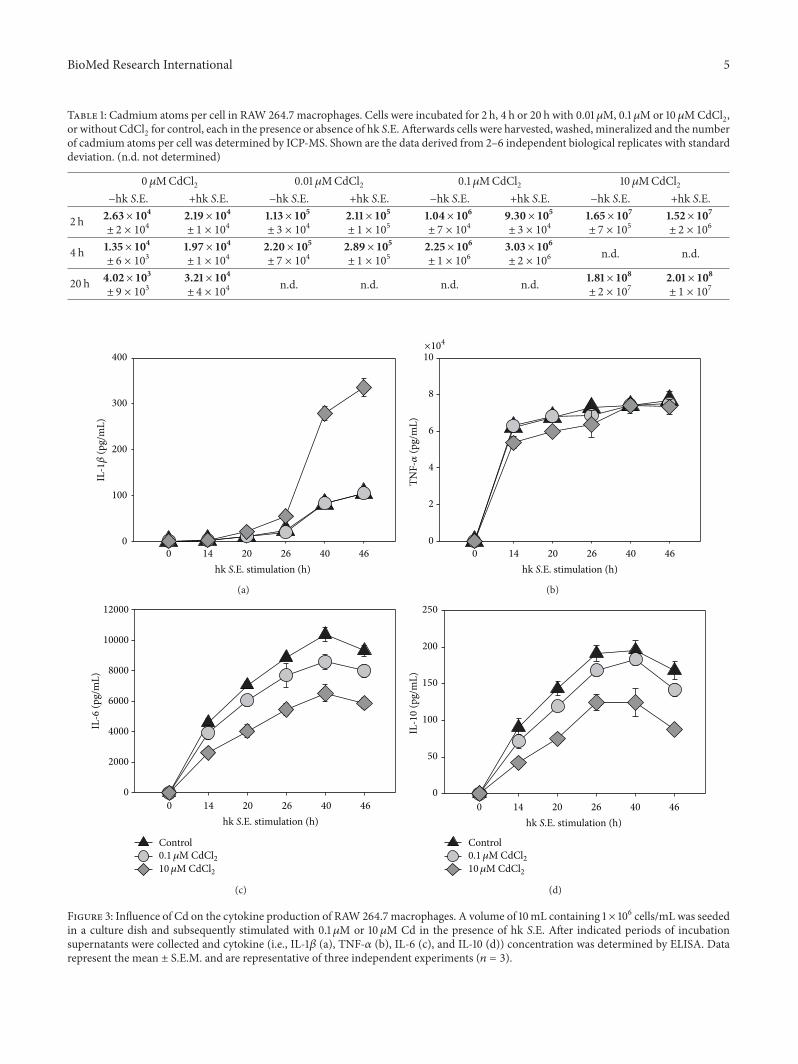
3.2. Cadmium Uptake of Macrophages. To get an impressionof the basal cadmium content in RAW 264.7 cells in cultureand the amount of Cd uptake and accumulation after incu-bation of the cells in the presence of CdCl
2over a period of
up to 20 h we determined the content of Cd as atoms per cellby means of ICP-MS. The result demonstrated a significantand continuous uptake and accumulation of Cd over timein the RAW 264.7 macrophages in dependence of the CdCl
2
concentration in the culture medium (Table 1). The resultsin the presence or absence of hk S.E. were found to be verysimilar.
3.3. Effects of Cadmium on Cytokine, Chemokine, and NOProduction. In order to identify immunoregulatory effects ofCd on macrophages the secretion of proinflammatory (i.e.,IL-1𝛽 and TNF-𝛼) and anti-inflammatory cytokines (IL-10and IL-6) in response to Cd exposure with or without hkS.E. stimulation were measured by ELISA. Thereby it was ofspecial interest to obtain data that may explain better survivalof antigen-stimulated cells in the presence of 10 𝜇M Cd.Therefore, we compared cytokine concentrations in super-natants of cells exposed to 0.1 𝜇Mor 10 𝜇MCd in comparisonto untreated controls (Figure 3). The results revealed that incase of IL-1𝛽 0.1 𝜇M Cd exhibited no effect compared tocontrol, whereas 10 𝜇M Cd led to a threefold increase of IL-1𝛽 concentration (Figure 3(a)). In terms of IL-10 and IL-60.1 𝜇MCd led to a slightly decreased cytokine secretion, while10 𝜇MCd reduced the concentration of both cytokines by half(Figures 3(b) and 3(c)).TheTNF-𝛼 secretion highly increasedafter hk S.E. stimulation, whereby the Cd exposition did notresult in significant alterations of the cytokine production(Figure 3(d)). The secretion of chemokine CXCL1 was noteffected by 0.1 𝜇M Cd compared to control, whereas 10 𝜇MCd caused twofold increase of CXCL1 concentration in RAW264.7 culture supernatants (Figure 4(a)). In contrast, NOproduction was found to be reduced by approximately 40%following exposure to 10 𝜇MCd; however, 0.1 𝜇MCd did notshow any effect on this functional parameter (Figure 4(b)).
4. Discussion
Proinflammatory effects of Cd in subtoxic dose ranges havebeen shown in diverse human and murine cell lines or
BioMed Research International 5
Table 1: Cadmium atoms per cell in RAW 264.7 macrophages. Cells were incubated for 2 h, 4 h or 20 h with 0.01𝜇M, 0.1 𝜇Mor 10𝜇MCdCl2,or without CdCl2 for control, each in the presence or absence of hk S.E. Afterwards cells were harvested, washed, mineralized and the numberof cadmium atoms per cell was determined by ICP-MS. Shown are the data derived from 2–6 independent biological replicates with standarddeviation. (n.d. not determined)
0 𝜇MCdCl2 0.01 𝜇MCdCl2 0.1 𝜇MCdCl2 10𝜇MCdCl2−hk S.E. +hk S.E. −hk S.E. +hk S.E. −hk S.E. +hk S.E. −hk S.E. +hk S.E.
2 h 2.63× 10⁴± 2 × 10⁴
2.19× 10⁴± 1 × 10⁴
1.13× 10⁵± 3 × 10⁴
2.11× 10⁵± 1 × 10⁵
1.04× 10⁶± 7 × 10⁴
9.30× 10⁵± 3 × 10⁴
1.65× 10⁷± 7 × 10⁵
1.52× 10⁷± 2 × 10⁶
4 h 1.35× 10⁴± 6 × 10³
1.97× 10⁴± 1 × 10⁴
2.20× 10⁵± 7 × 10⁴
2.89× 10⁵± 1 × 10⁵
2.25× 10⁶± 1 × 10⁶
3.03× 10⁶± 2 × 10⁶
n.d. n.d.
20 h 4.02× 10³± 9 × 10³
3.21× 10⁴± 4 × 10⁴
n.d. n.d. n.d. n.d. 1.81× 10⁸± 2 × 10⁷
2.01× 10⁸± 1 × 10⁷
400
300
200
100
00 14 20 26 40 46
IL-1𝛽
(pg/
mL)
hk S.E. stimulation (h)
(a)
TNF-𝛼
(pg/
mL)
0 14 20 26 40 46
hk S.E. stimulation (h)
×104
10
8
6
4
2
0
(b)
IL-6
(pg/
mL)
0 14 20 26 40 46
hk S.E. stimulation (h)
12000
10000
8000
6000
4000
2000
0
Control0.1 𝜇M CdCl210𝜇M CdCl2
(c)
IL-10
(pg/
mL)
0 14 20 26 40 46
hk S.E. stimulation (h)
250
200
150
100
50
0
Control0.1 𝜇M CdCl210𝜇M CdCl2
(d)
Figure 3: Influence of Cd on the cytokine production of RAW 264.7 macrophages. A volume of 10mL containing 1 × 10⁶ cells/mL was seededin a culture dish and subsequently stimulated with 0.1𝜇M or 10𝜇M Cd in the presence of hk S.E. After indicated periods of incubationsupernatants were collected and cytokine (i.e., IL-1𝛽 (a), TNF-𝛼 (b), IL-6 (c), and IL-10 (d)) concentration was determined by ELISA. Datarepresent the mean ± S.E.M. and are representative of three independent experiments (𝑛 = 3).

6 BioMed Research International
0 14 20 26 40 46
hk S.E. stimulation (h)
Control0.1 𝜇M CdCl210𝜇M CdCl2
35
30
25
20
15
10
5
0
CXCL
1(p
g/m
L)
(a)
0 14 20 26 40 46
hk S.E. stimulation (h)
Control0.1 𝜇M CdCl210𝜇M CdCl2
80
60
40
20
0
Nitr
ite/n
itrat
e (𝜇
M)
(b)
Figure 4: Influence of Cd on chemokine and NO production of RAW 264.7 macrophages. A volume of 10mL containing 1 × 10⁶ cells/mLwas seeded in a culture dish and subsequently stimulated with 0.1 𝜇M or 10𝜇M Cd in the presence of hk S.E. After indicated periods ofincubation supernatants were collected and chemokine (i.e., CXCL1 (a)) and nitrite/nitrate concentrations (b) were determined by ELISA orGriess reaction, respectively. Data represent the mean ± S.E.M. and are representative of three independent experiments (𝑛 = 3).
primary cells (reviewed in [28]). The upregulation of manycytokines such as IL-1𝛽, IL-6, IL-8, and TNF-𝛼 illustratesan immunomodulatory potential of Cd [29, 30]. Therefore,the aim of this study was to determine the effects of Cdin a relevant in vitro model of bacteria-driven ongoinginnate immune response. As macrophages play a key role inimmunity to bacterial infections as bactericidal effector cellsas well as APCs we started this complex of investigation withthe study of macrophages. For reasons of standardization andcomparability of immunotoxicological results we preferred touse the well-described and broadly accepted macrophage cellline RAW264.7 as a highly standardized in vitromodel ratherthan primary macrophages.
A basic requirement of an immunotoxicological in vitromodel is the exact knowledge of the toxic and subtoxicdose ranges of the compound to be tested. This requiresappropriate endpoints represented by classical cytotoxicityassays, such as MTT, XTT, WST-1, EZ4U, or LDH assay,in most cases. However, we have standardized a novelimpedance-based real-time cell analysis method (i.e., xCEL-Ligence RTCA and ACEA/Roche) for this purpose underGLP conditions. This methodology allows a very sensitivereal-time monitoring of toxic effects mediated by Cd andother xenobiotic compounds with high time resolution andoffers the opportunity of medium- and high-throughputtesting. Thus, in addition to the endpoint WST-1 assay theviability of nonactivated and activated macrophages exposedto different Cd concentrations was determined using thismethod. Using endpoint assays, dose-dependent toxicity ofCd on organisms and cells have been shown by several groups[24, 31, 32]. These data could be underlined and significantlycompleted in terms of the time course of toxic Cd effects
applying the impedance-based RTCA method in the presentstudy. Here, concentrations about 10 𝜇M Cd caused reducedadherence and cell viability in nonactivated and activatedmacrophages compared to respective controls. However,when the cells were simultaneously exposed to hk S.E. as abacterial antigenic stimulus, activating the macrophage viaseveral PRRs (i.e., Toll-like receptor (TLR)2, TLR4, TLR7,and TLR9 [33]) 10 𝜇MCd prevented the loss of adherence incomparison to control by 6 h as determined by xCELLigenceRTCA. Also the WST-1 assay revealed viability of stimulatedmacrophages exposed to 10𝜇M Cd after 48 h but not ofcontrol cells and cells exposed to Cd concentrations below10 𝜇M.These results demonstrate that 10 𝜇MCd is capable ofdelaying cell death caused by too strong activation through apotent bacterial antigenic stimulus, resulting in high produc-tion rates of cytotoxic proinflammatory cytokines, reactiveoxygen species (ROS), and nitric oxide (NO).
In order to exclude that different effects in nonactivatedand activated macrophages might be the result of differentCd-uptake rates, we determined Cd content in RAW 264.7cells. Using ICP-MS analysis we could show that Cd contentin RAW 264.7 macrophages is independent of cell activationwith hk S.E. Furthermore, we observed an increase of Cdconcentration per cell by exposure of increasing Cd concen-trations in the cell culture medium as expected.
Previous reports on nonactivated human cells (i.e.,human PBMCs or human monocytic cell line THP-1)revealed that lower concentrations of Cd showed stimulatingeffects on production of cytokines such as IL-1𝛽, IL-6, andTNF-𝛼 [29, 30]. However, in the present study using activatedmurine RAW 264.7 macrophages as a highly standardized invitro model, exposure to 10 𝜇M Cd did induce the secretion

BioMed Research International 7
of IL-1𝛽, too, whereas the production of IL-6 and IL-10 wassignificantly reduced. Lower expression of IL-10 might beexplained by decreased IL-6 production [34]. Interestingly,production of TNF-𝛼 was not effected by Cd in our in vitromodel. These results indicate that Cd effects on cytokineproduction depend on the cell type and also on the activationstate of immune cells. In terms of TNF-𝛼, the strong upregu-lation of this cytokine in activated RAW 264.7 macrophagesmight have overlain a possible TNF-𝛼-inducing effect of Cdobserved in nonactivated RAW 264.7 cells [31].
Additionally to IL-1𝛽, also the secretion of chemokineCXCL1 (former designation in mouse: KC) was upregulatedby exposure to 10 𝜇Mbut not to 0.1 𝜇MCd.This upregulationwas also reported by other groups for the homologous humanchemokine IL-8 in human monocytic cell line THP-1 andin bronchial epithelial cells after exposition with Cd [30,35]. Reduced IL-6 production is discussed as a possibleexplanation for upregulated secretion of CXCL1. Fieldinget al. and Hurst et al. showed that IL-6 regulates CXCL1expression via STAT3 resulting in recruitment of neutrophils[36, 37].
A decrease in NO production by 10 𝜇M Cd was alsoshown by other groups in LPS-stimulated RAW 264.7macrophages and in murine splenic macrophages stimulatedwith TNF-𝛼/IFN-𝛾 [31, 38].The reducedNO synthesis causedby 10 𝜇M Cd might be a significant reason for increased cellviability in activated macrophages, since it was previouslyshown that high cellular concentrations of NO may induceapoptosis in peritoneal macrophages [39]. But in conclusion,this result points out that macrophages exposed to such Cdconcentrationsmight show impaired killing of living bacteria.
5. Conclusion
The data presented in this work deliver clear evidence forthe capacity of Cd to modulate important cellular functionsof activated macrophages as key players in innate immunityto infection in a strongly limited subtoxic dose range ofthis toxic heavy metal. In particular, the results found inour hk S.E.-stimulated RAW 264.7 in vitro model indicatethat Cd-mediated immunomodulation increases cell survivaland inhibits the anti-inflammatory cytokines IL-6 and IL-10,while the secretion of proinflammatory cytokine IL-1𝛽 andthe neutrophil-recruiting chemokine CXCL1 are induced byCd.Although these findings alone are not sufficient to explainthe higher susceptibility to Salmonella infection under Cdexposure as previously observed in a mouse model in ourgroup but may contribute to explain the potential of long-term Cd exposure to elevate the risk of chronic inflammationfollowing bacterial infection as previously concluded fromepidemiologic studies.
Abbreviations
FBS: Fetal bovine serumCI: Cell indexHEPES: 4-(2-Hydroxyethyl)
piperazine-1-ethanesulfonic acidhk S.E.: Heat-killed salmonellae
NO: Nitric oxideICP-MS: Inductively coupled plasma-mass
spectrometryELISA: Enzyme-linked Immunosorbent AssayPRR: Pattern-recognition receptor.
Conflict of Interests
The authors declare that there is no conflict of interestsregarding the publication of this paper.
Acknowledgment
The authors are grateful to U. Scholz and M. Dahne forexcellent technical assistance.
References
[1] E. K. Leffel, C. Wolf, A. Poklis, and K. L. White Jr., “Drinkingwater exposure to cadmium, an environmental contaminant,results in the exacerbation of autoimmune disease in themurinemodel,” Toxicology, vol. 188, no. 2-3, pp. 233–250, 2003.
[2] S. Satarug and M. R. Moore, “Adverse health effects of chronicexposure to low-level cadmium in foodstuffs and cigarettesmoke,” Environmental Health Perspectives, vol. 112, no. 10, pp.1099–1103, 2004.
[3] S. Pillet, A. A. Rooney, J.-M. Bouquegneau, D. G. Cyr, andM. Fournier, “Sex-specific effects of neonatal exposures to lowlevels of cadmium through maternal milk on development andimmune functions of juvenile and adult rats,” Toxicology, vol.209, no. 3, pp. 289–301, 2005.
[4] K. Weggler, M. J. McLaughlin, and R. D. Graham, “Effect ofchloride in soil solution on the plant availability of biosolid-borne cadmium,” Journal of Environmental Quality, vol. 33, no.2, pp. 496–504, 2004.
[5] D.-M. Zhou, Y.-J. Wang, L. Cang, X.-Z. Hao, and X.-S. Luo,“Adsorption and cosorption of cadmium and glyphosate on twosoils with different characteristics,” Chemosphere, vol. 57, no. 10,pp. 1237–1244, 2004.
[6] E. Govasmark, J. Stab, B. Holen, D. Hoornstra, T. Nesbakk, andM. Salkinoja-Salonen, “Chemical and microbiological hazardsassociatedwith recycling of anaerobic digested residue intendedfor agricultural use,” Waste Management, vol. 31, no. 12, pp.2577–2583, 2011.
[7] A. E. Sahmoun, L. D. Case, S. A. Jackson, and G. G. Schwartz,“Cadmium and prostate cancer: a critical epidemiologic analy-sis,” Cancer Investigation, vol. 23, no. 3, pp. 256–263, 2005.
[8] K. V. Lopin, F.Thevenod, J. C. Page, and S.W. Jones, “Cd2+ blockand permeation of CaV3.1 (𝛼1G) T-type calcium channels: can-didate mechanism for Cd2+ influx,” Molecular Pharmacology,vol. 82, no. 6, pp. 1183–1193, 2012.
[9] Z. Liu, H. Li, M. Soleimani et al., “Cd2+ versus Zn2+ uptakeby the ZIP8 HCO
3−-dependent symporter: kinetics, electro-
genicity and trafficking,” Biochemical and Biophysical ResearchCommunications, vol. 365, no. 4, pp. 814–820, 2008.
[10] K. Girijashanker, L. He, M. Soleimani et al., “Slc39a14 geneencodes ZIP14, a metal/bicarbonate symporter: similarities tothe ZIP8 transporter,” Molecular Pharmacology, vol. 73, no. 5,pp. 1413–1423, 2008.
[11] J. P. Bressler, L. Olivi, J. H. Cheong, Y. Kim, and D. Bannon,“Divalent metal transporter 1 in lead and cadmium transport,”
8 BioMed Research International
Annals of the New York Academy of Sciences, vol. 1012, pp. 142–152, 2004.
[12] M. Okubo, K. Yamada, M. Hosoyamada, T. Shibasaki, and H.Endou, “Cadmium transport by human Nramp 2 expressed inXenopus laevis oocytes,” Toxicology and Applied Pharmacology,vol. 187, no. 3, pp. 162–167, 2003.
[13] D. Beyersmann and S. Hechtenberg, “Cadmium, gene regula-tion, and cellular signalling inmammalian cells,”Toxicology andApplied Pharmacology, vol. 144, no. 2, pp. 247–261, 1997.
[14] M. Satoh, T. Kaji, and C. Tohyama, “Low dose exposureto cadmium and its health effects. (3) toxicity in laboratoryanimals and cultured cells,” Nihon Eiseigaku Zasshi, vol. 57, no.4, pp. 615–623, 2003.
[15] D. Beyersmann and A. Hartwig, “Carcinogenic metal com-pounds: recent insight into molecular and cellular mecha-nisms,” Archives of Toxicology, vol. 82, no. 8, pp. 493–512, 2008.
[16] J. Thompson and J. Bannigan, “Cadmium: toxic effects on thereproductive system and the embryo,” Reproductive Toxicology,vol. 25, no. 3, pp. 304–315, 2008.
[17] H. C. Gonick, “Nephrotoxicity of cadmium & lead,”The IndianJournal of Medical Research, vol. 128, no. 4, pp. 335–352, 2008.
[18] D. Ibrahim, B. Froberg, A. Wolf, and D. E. Rusyniak, “Heavymetal poisoning: clinical presentations and pathophysiology,”Clinics in Laboratory Medicine, vol. 26, no. 1, pp. 67–97, 2006.
[19] C. Giaginis, E. Gatzidou, and S. Theocharis, “DNA repairsystems as targets of cadmium toxicity,” Toxicology and AppliedPharmacology, vol. 213, no. 3, pp. 282–290, 2006.
[20] P. Mendola, L. C. Messer, and K. Rappazzo, “Science link-ing environmental contaminant exposures with fertility andreproductive health impacts in the adult female,” Fertility andSterility, vol. 89, supplement 2, pp. e81–e94, 2008.
[21] T. Sanders, Y. Liu, V. Buchner, and P. B. Tchounwou, “Neuro-toxic effects and biomarkers of lead exposure: a review,”Reviewson Environmental Health, vol. 24, no. 1, pp. 15–45, 2009.
[22] F. O. Johnson and W. D. Atchison, “The role of environmentalmercury, lead and pesticide exposure in development of amy-otrophic lateral sclerosis,” NeuroToxicology, vol. 30, no. 5, pp.761–765, 2009.
[23] I. Lehmann, U. Sack, and J. Lehmann, “Metal ions affecting theimmune system,” Metal ions in life sciences, vol. 8, pp. 157–185,2011.
[24] N. Y. A. Hemdan, F. Emmrich, U. Sack et al., “The in vitroimmune modulation by cadmium depends on the way of cellactivation,” Toxicology, vol. 222, no. 1-2, pp. 37–45, 2006.
[25] W. C. Raschke, S. Baird, P. Ralph, and I. Nakoinz, “Functionalmacrophage cell lines transformed by Abelson leukemia virus,”Cell, vol. 15, no. 1, pp. 261–267, 1978.
[26] L. C. Green, D. A. Wagner, J. Glogowski, P. L. Skipper, J. S.Wishnok, and S. R. Tannenbaum, “Analysis of nitrate, nitrite,and [15N]nitrate in biological fluids,” Analytical Biochemistry,vol. 126, no. 1, pp. 131–138, 1982.
[27] M. Ishiyama, M. Suiga, K. Sasamoto, M. Mizoguchi, and P.-G.He, “A new sulfonated tetrazolium salt that produces a highlywater-soluble formazan dye,” Chemical and PharmaceuticalBulletin, vol. 41, no. 6, pp. 1118–1122, 1993.
[28] T. Olszowski, I. Baranowska-Bosiacka, I. Gutowska, and D.Chlubek, “Pro-inflammatory properties of cadmium,” ActaBiochimica Polonica, vol. 59, no. 4, pp. 475–482, 2012.
[29] E. Marth, S. Jelovcan, B. Kleinhappl, A. Gutschi, and S. Barth,“The effect of heavy metals on the immune system at lowconcentrations,” International Journal of Occupational Medicineand Environmental Health, vol. 14, no. 4, pp. 375–386, 2001.
[30] M. Freitas and E. Fernandes, “Zinc, cadmium and nickelincrease the activation of NF-𝜅B and the release of cytokinesfrom THP-1 monocytic cells,” Metallomics, vol. 3, no. 11, pp.1238–1243, 2011.
[31] P. L. Goering, R. K. Kuester, A. R. Neale et al., “Effects of partic-ulate and soluble cadmium species on biochemical and func-tional parameters in cultured murine macrophages,” In Vitro &Molecular Toxicology: Journal of Basic and Applied Research, vol.13, no. 2, pp. 125–136, 2000.
[32] Z. Krocova, A. MacEla, M. Kroca, and L. Hernychova, “Theimmunomodulatory effect(s) of lead and cadmium on the cellsof immune system in vitro,” Toxicology in Vitro, vol. 14, no. 1, pp.33–40, 2000.
[33] N. Arpaia, J. Godec, L. Lau et al., “TLR signaling is requiredfor Salmonella typhimurium virulence,” Cell, vol. 144, no. 5, pp.675–688, 2011.
[34] A. Steensberg, C. P. Fischer, C. Keller, K. Møller, and B. K.Pedersen, “IL-6 enhances plasma IL-1ra, IL-10, and cortisol inhumans,” The American Journal of Physiology— Endocrinologyand Metabolism, vol. 285, no. 2, pp. E433–E437, 2003.
[35] E. Cormet-Boyaka, K. Jolivette, A. Bonnegarde-bernard et al.,“An NF-𝜅b-independent and erk1/2-dependent mechanismcontrols CXCL8/IL-8 responses of airway epithelial cells tocadmium,” Toxicological Sciences, vol. 125, no. 2, pp. 418–429,2012.
[36] C. A. Fielding, R. M. McLoughlin, L. McLeod et al., “IL-6regulates neutrophil trafficking during acute inflammation viaSTAT3,” Journal of Immunology, vol. 181, no. 3, pp. 2189–2195,2008.
[37] S. M. Hurst, T. S. Wilkinson, R. M. McLoughlin et al., “IL-6 andits soluble receptor orchestrate a temporal switch in the patternof leukocyte recruitment seen during acute inflammation,”Immunity, vol. 14, no. 6, pp. 705–714, 2001.
[38] L. Tian and D. A. Lawrence, “Metal-induced modulation ofnitric oxide production in vitro by murine macrophages: lead,nickel, and cobalt utilize different mechanisms,” Toxicology andApplied Pharmacology, vol. 141, no. 2, pp. 540–547, 1996.
[39] J. E. Albina, S. Cui, R. B. Mateo, and J. S. Reichner, “Nitricoxide-mediated apoptosis in murine peritoneal macrophages,”The Journal of Immunology, vol. 150, no. 11, pp. 5080–5085, 1993.

Submit your manuscripts athttp://www.hindawi.com
Hindawi Publishing Corporationhttp://www.hindawi.com Volume 2014
Anatomy Research International
PeptidesInternational Journal of
Hindawi Publishing Corporationhttp://www.hindawi.com Volume 2014
Hindawi Publishing Corporation http://www.hindawi.com
International Journal of
Volume 2014
Zoology
Hindawi Publishing Corporationhttp://www.hindawi.com Volume 2014
Molecular Biology International
GenomicsInternational Journal of
Hindawi Publishing Corporationhttp://www.hindawi.com Volume 2014
The Scientific World JournalHindawi Publishing Corporation http://www.hindawi.com Volume 2014
Hindawi Publishing Corporationhttp://www.hindawi.com Volume 2014
BioinformaticsAdvances in
Marine BiologyJournal of
Hindawi Publishing Corporationhttp://www.hindawi.com Volume 2014
Hindawi Publishing Corporationhttp://www.hindawi.com Volume 2014
Signal TransductionJournal of
Hindawi Publishing Corporationhttp://www.hindawi.com Volume 2014
BioMed Research International
Evolutionary BiologyInternational Journal of
Hindawi Publishing Corporationhttp://www.hindawi.com Volume 2014
Hindawi Publishing Corporationhttp://www.hindawi.com Volume 2014
Biochemistry Research International
ArchaeaHindawi Publishing Corporationhttp://www.hindawi.com Volume 2014
Hindawi Publishing Corporationhttp://www.hindawi.com Volume 2014
Genetics Research International
Hindawi Publishing Corporationhttp://www.hindawi.com Volume 2014
Advances in
Virolog y
Hindawi Publishing Corporationhttp://www.hindawi.com
Nucleic AcidsJournal of
Volume 2014
Stem CellsInternational
Hindawi Publishing Corporationhttp://www.hindawi.com Volume 2014
Hindawi Publishing Corporationhttp://www.hindawi.com Volume 2014
Enzyme Research
Hindawi Publishing Corporationhttp://www.hindawi.com Volume 2014
International Journal of
Microbiology