respiratory: respiratory failure and ards marnie quick, rn, msn, cnrn
TRANSCRIPT
Respiratory: Respiratory Failure and ARDS
Marnie Quick, RN, MSN, CNRN
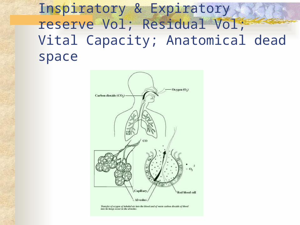
Normal Respirations: Tidal Vol; Inspiratory & Expiratory reserve Vol; Residual Vol; Vital Capacity; Anatomical dead space
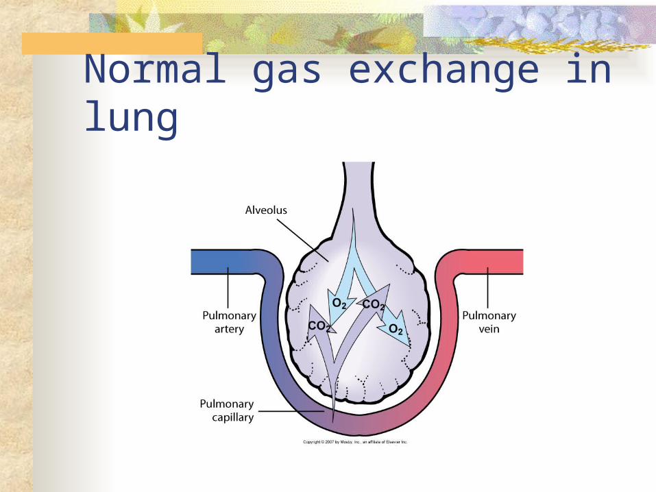
Normal gas exchange in lung
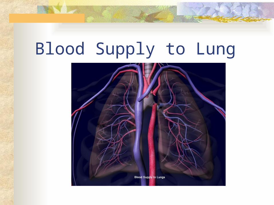
Blood Supply to Lung
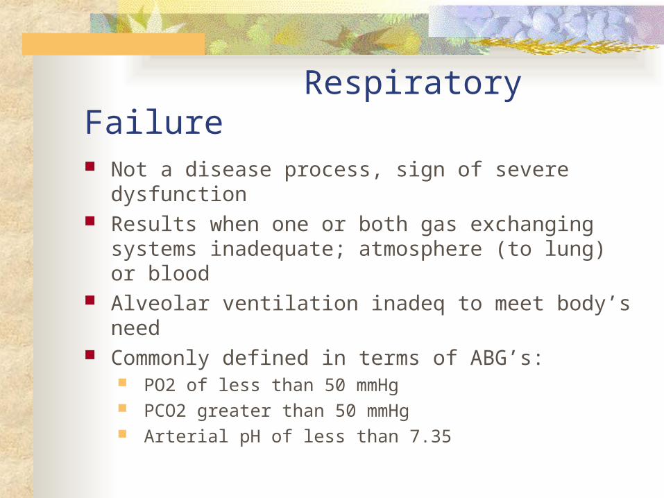
Respiratory Failure Not a disease process, sign of severe dysfunction Results when one or both gas exchanging systems
inadequate; atmosphere (to lung) or blood Alveolar ventilation inadeq to meet body’s need Commonly defined in terms of ABG’s:
PO2 of less than 50 mmHg PCO2 greater than 50 mmHg Arterial pH of less than 7.35
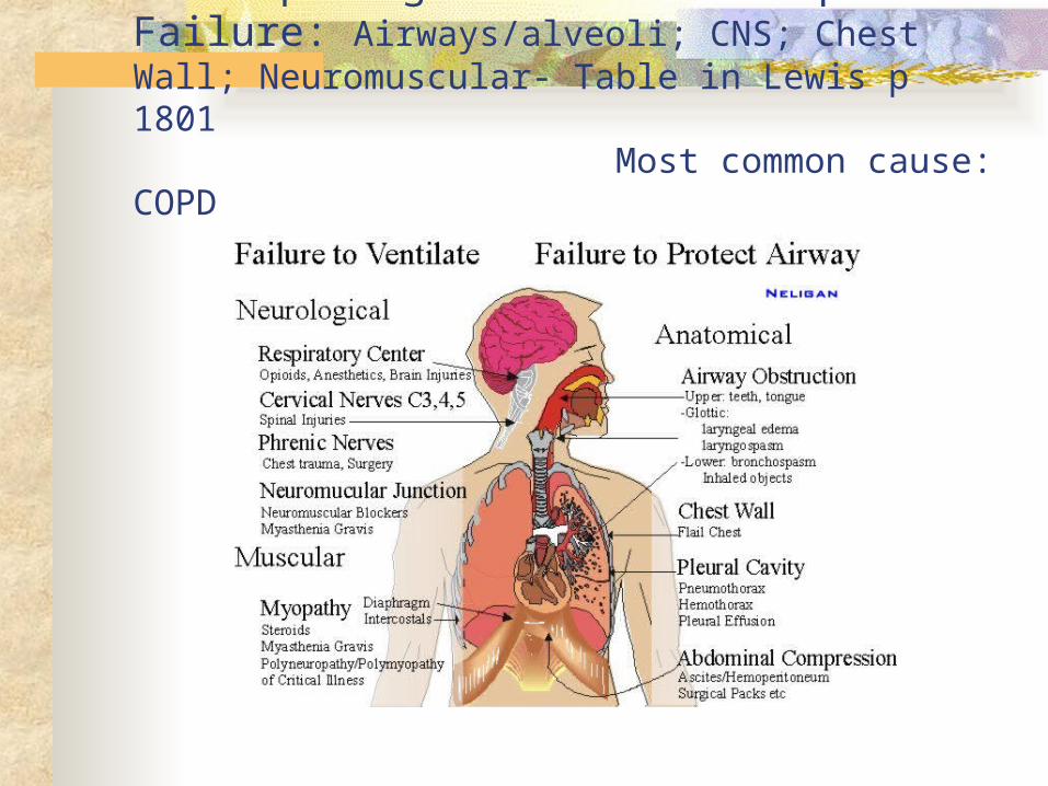
Predisposing factors for Resp Failure: Airways/alveoli; CNS; Chest Wall; Neuromuscular- Table in Lewis p 1801 Most common cause: COPD

Classification of Respiratory Failure
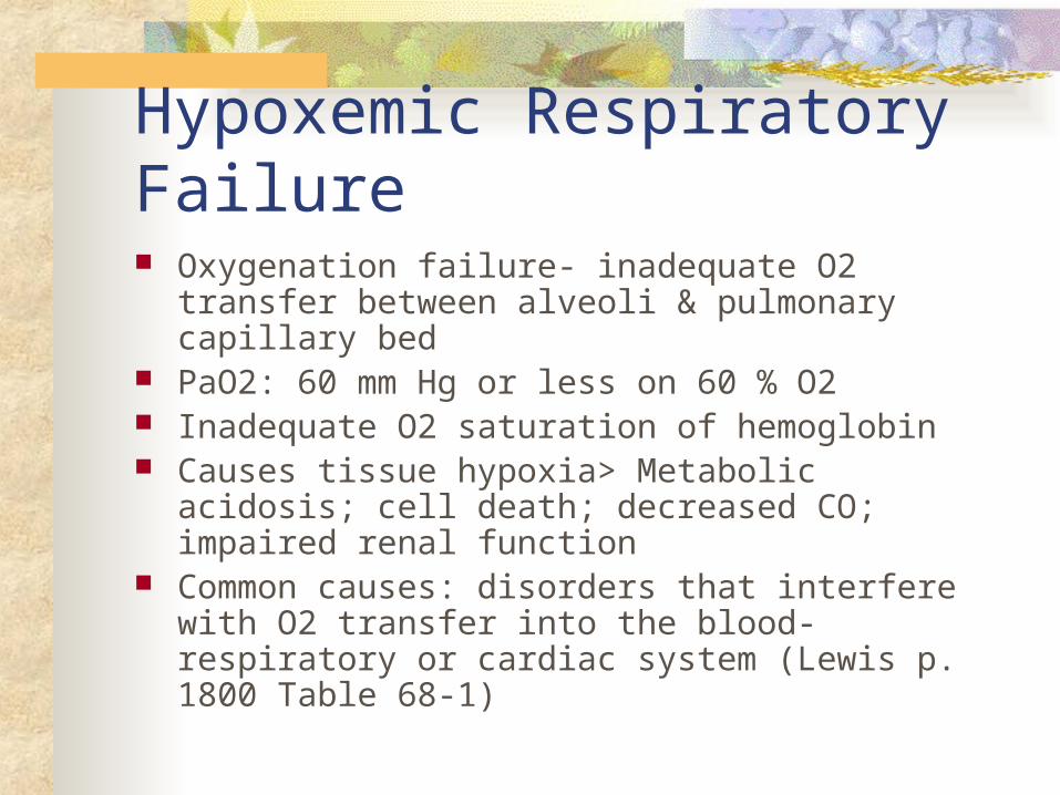
Hypoxemic Respiratory Failure Oxygenation failure- inadequate O2 transfer
between alveoli & pulmonary capillary bed PaO2: 60 mm Hg or less on 60 % O2 Inadequate O2 saturation of hemoglobin Causes tissue hypoxia> Metabolic acidosis; cell
death; decreased CO; impaired renal function Common causes: disorders that interfere with O2
transfer into the blood- respiratory or cardiac system (Lewis p. 1800 Table 68-1)
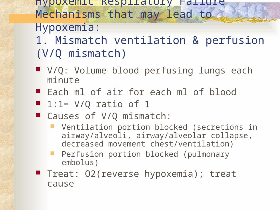
Hypoxemic Respiratory Failure Mechanisms that may lead to Hypoxemia: 1. Mismatch ventilation & perfusion (V/Q mismatch)
V/Q: Volume blood perfusing lungs each minute Each ml of air for each ml of blood 1:1= V/Q ratio of 1 Causes of V/Q mismatch:
Ventilation portion blocked (secretions in airway/alveoli, airway/alveolar collapse, decreased movement chest/ventilation)
Perfusion portion blocked (pulmonary embolus) Treat: O2(reverse hypoxemia); treat cause
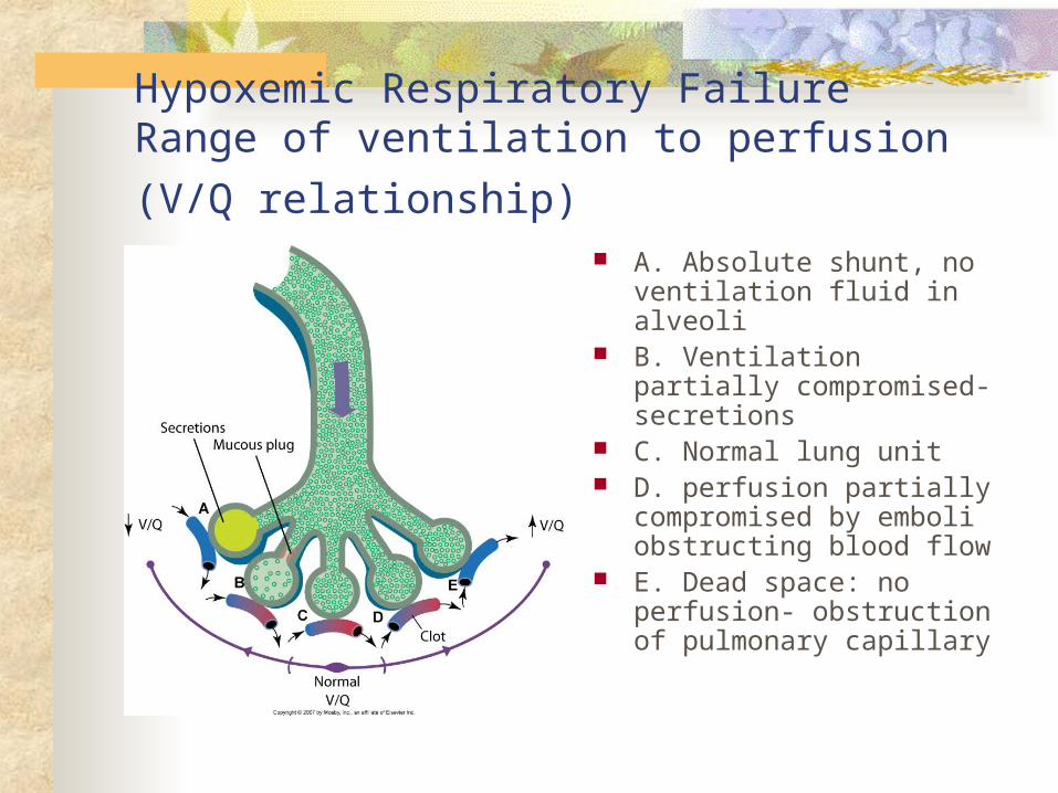
Hypoxemic Respiratory Failure
Range of ventilation to perfusion (V/Q relationship) A. Absolute shunt, no
ventilation fluid in alveoli B. Ventilation partially
compromised- secretions C. Normal lung unit D. perfusion partially
compromised by emboli obstructing blood flow
E. Dead space: no perfusion- obstruction of pulmonary capillary
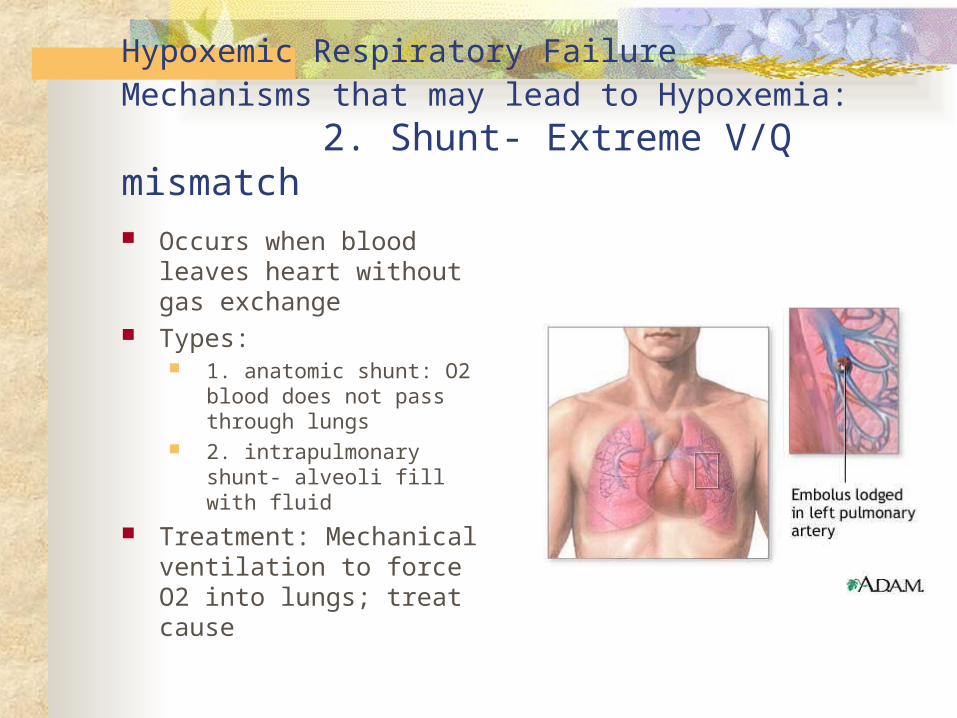
Hypoxemic Respiratory FailureMechanisms that may lead to Hypoxemia: 2. Shunt- Extreme V/Q mismatch
Occurs when blood leaves heart without gas exchange
Types: 1. anatomic shunt: O2
blood does not pass through lungs
2. intrapulmonary shunt- alveoli fill with fluid
Treatment: Mechanical ventilation to force O2 into lungs; treat cause
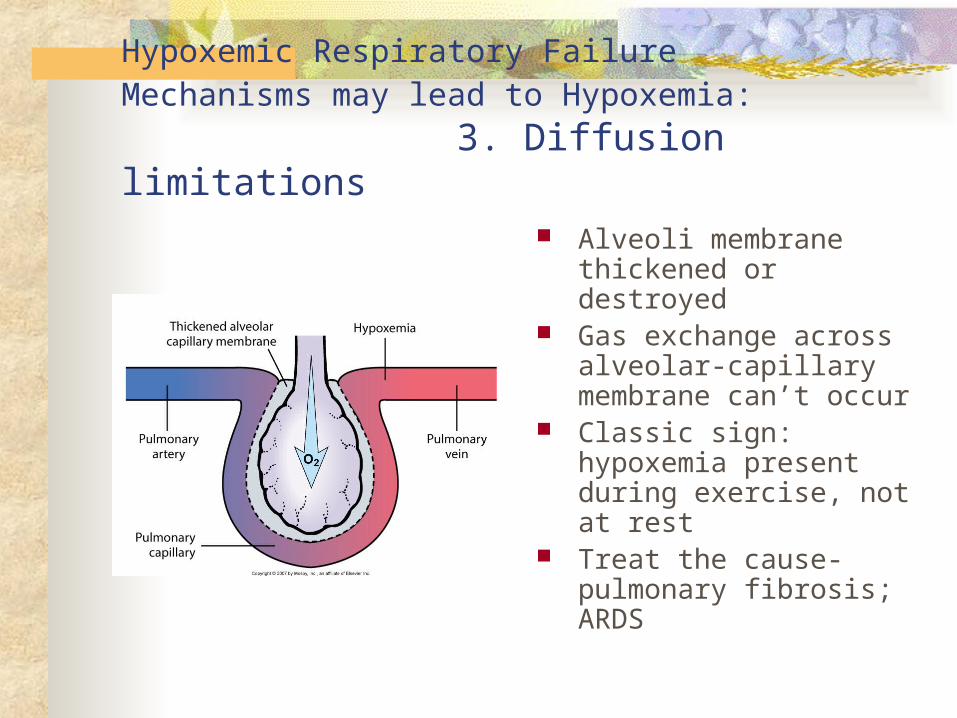
Hypoxemic Respiratory FailureMechanisms may lead to Hypoxemia: 3. Diffusion limitations
Alveoli membrane thickened or destroyed
Gas exchange across alveolar-capillary membrane can’t occur
Classic sign: hypoxemia present during exercise, not at rest
Treat the cause- pulmonary fibrosis; ARDS
Hypoxemic Respiratory FailureMechanisms may lead to Hypoxemia: Clinical Manifestations of Hypoxemia
Specific: Respiratory: Dyspnea; tachypnea; prolonged expiration;
intercostal muscle retraction; use of accessory muscles in resp;< 80% SpO2; paradoxic chest/abd wall movement with resp cycle (late); cyanosis (late)
Nonspecific: Cerebral: agitation, disorientation, delirium, restless,
combative, confusion, dec LOC, coma (late) Cardiac: tachycardia, hypertension, skin
cool/clammy, dysrhythmias (late), hypotension (late) Other: fatigue; need to pause to breath when
speaking

Hypercapnic Respiratory Failure Ventilatory failure with insufficient CO2 removal PaCO2 greater than 45 mm Hg Arterial pH less than 7.35 PCO2 rises rapidly and respiratory acidosis
develops: PO2 drops more slowly Common causes: disorders that compromise lung
ventilation and CO2 removal (Lewis Table 68-1)

Hypercapic Respiratory Failure
Ventatory failure: Inability of the respiratory system to ventilate out sufficient CO2 to maintain normal PaCO2
Specific Causes: Airways/alveoli: asthma, COPD, cystic fibrosis CNS: drug overdose- depressant, brainstem
dysfuction, metabolic causing decreased LOC; high SCI injuries- decrease/absent diaphragm/chest movement
Chest wall: pain, flail chest, rib fractures, mechanical restriction, kyphoscoliosis, obesity
Neuromuscular: resp muscles weak/paralysis- MS, MG, MD, Guilain-Barre Syndrome

Hypercapic Respiratory Failure Clinical Manifestations Specific:
Respiratory: Dyspnea; dec resp rate or rapid with shallow resp; dec tidal vol; dec min ventilation
Nonspecific: Cerebral: AM headache; disorientation, progressive
sommolence; coma (late) Cardiac: dyshythmias; hypertension; tachycardia;
bounding pulse Neuromuscular: muscle weakness; dec deep tendon
reflexes; Tremor/seizures (late)
Collaborative Care for Respiratory Failure: Diagnostic tests History/physical assessment Pulse oximetry ABG analysis Chest X-ray CBC, sputum/blood cultures, electrolytes EKG Urinalysis V/Q scan- if pulmonary embolism suspected Hemodynamic monitor/pulmonary function tests
Collaborative care for Respiratory Failure Respiratory Therapy Main treatment- correct underlying cause & restore adequate gas
exchange in lung Elevate HOB Oxygen Therapy
Maintain PaO2 at least 60 mm Hg SaO2 at least 90%
Mobilization of secretions Hydration & humidification Chest physical therapy Airway suctioning Effective coughing & positioning
Positive pressure ventilation Noninvasive positive pressure ventilation Intubation with mechanical ventilation
Collaborative Care for Respiratory Failure cont Drug Therapy
Relief bronchospasm; reduce airway inflam and pulmonary congestion; treat pulmonary infections; reduce anxiety, pain
Medical supportive therapy Treat underlying cause
Nutritional therapy Enteral; parenteral Protein and energy stores
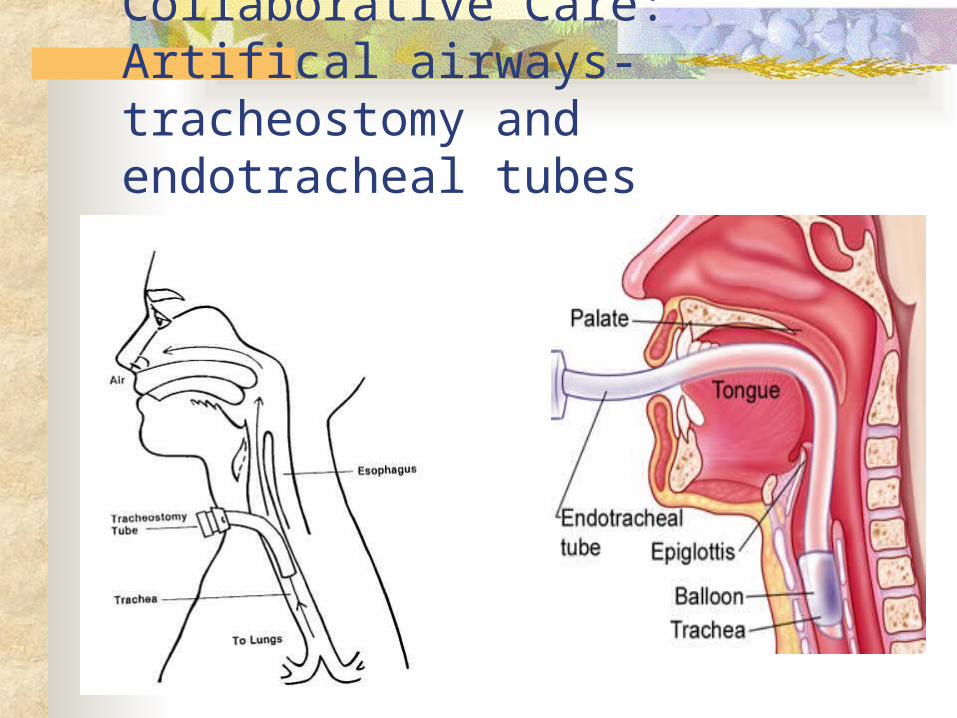
Collaborative Care: Artifical airways- tracheostomy and endotracheal tubes
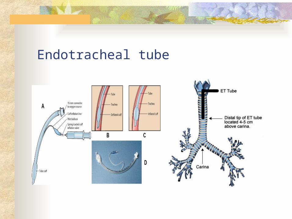
Endotracheal tube
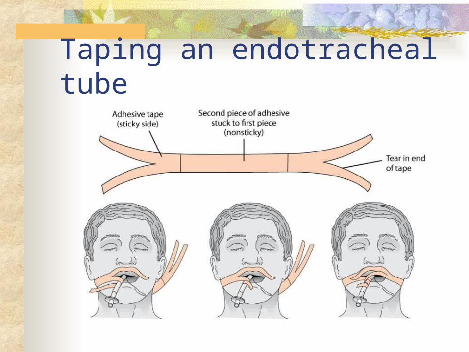
Taping an endotracheal tube
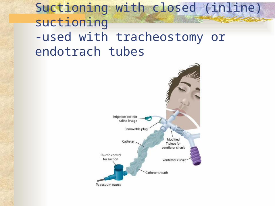
Suctioning with closed (inline) suctioning-used with tracheostomy or endotrach tubes
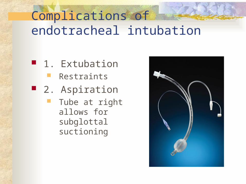
Complications of endotracheal intubation
1. Extubation Restraints
2. Aspiration Tube at right allows
for subglottal suctioning
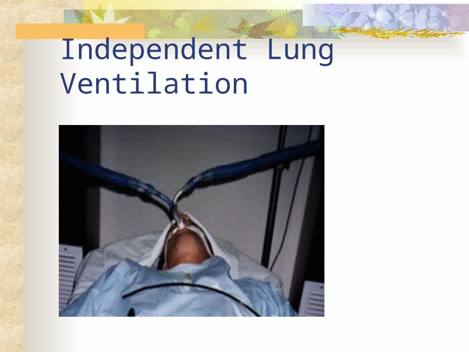
Independent Lung Ventilation
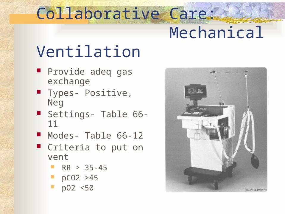
Collaborative Care: Mechanical Ventilation Provide adeq gas
exchange Types- Positive, Neg Settings- Table 66-11 Modes- Table 66-12 Criteria to put on vent
RR > 35-45 pCO2 >45 pO2 <50
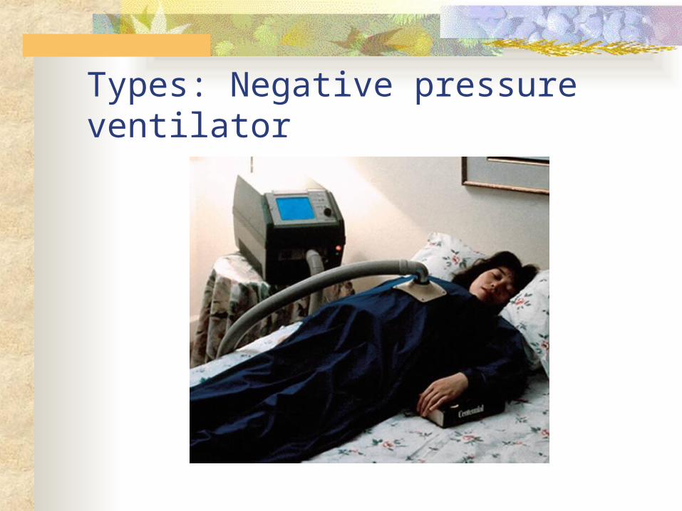
Types: Negative pressure ventilator
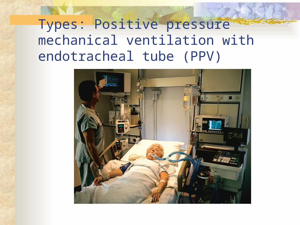
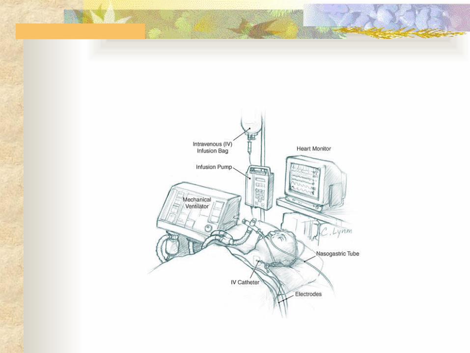
Types: Positive pressure mechanical ventilation with endotracheal tube (PPV)
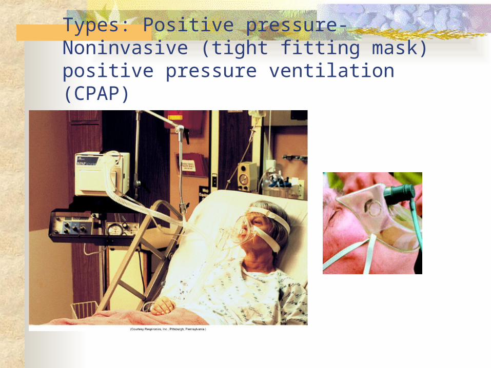
Types: Positive pressure- Noninvasive (tight fitting mask) positive pressure ventilation (CPAP)
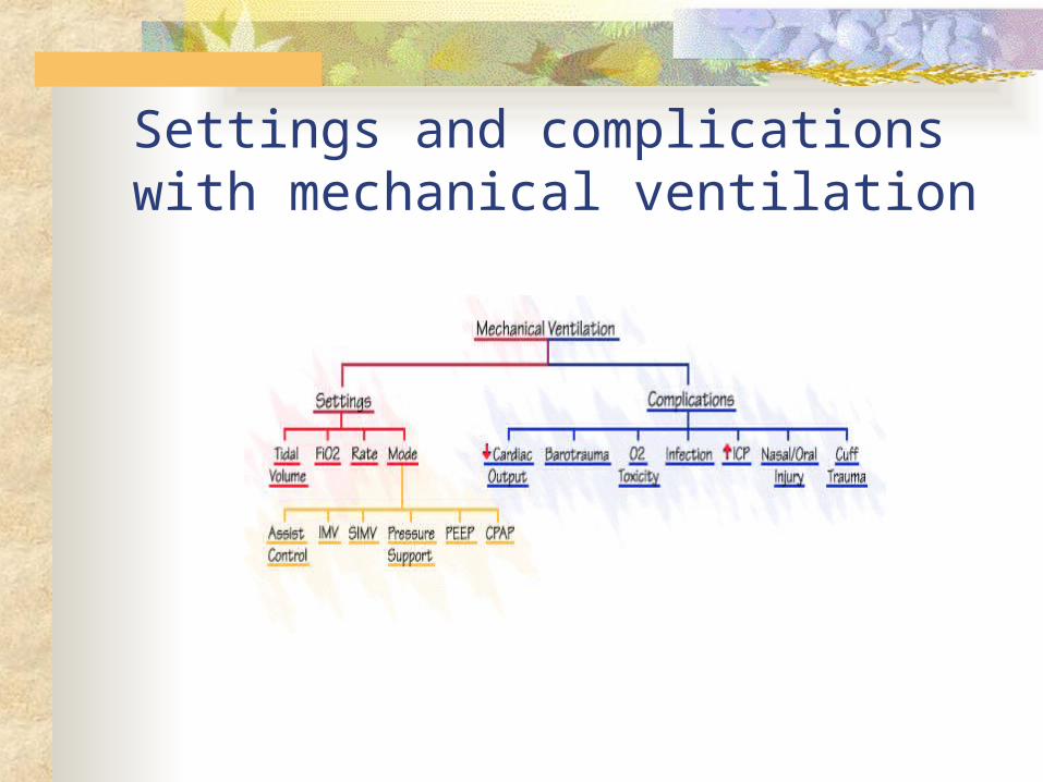
Settings and complications with mechanical ventilation
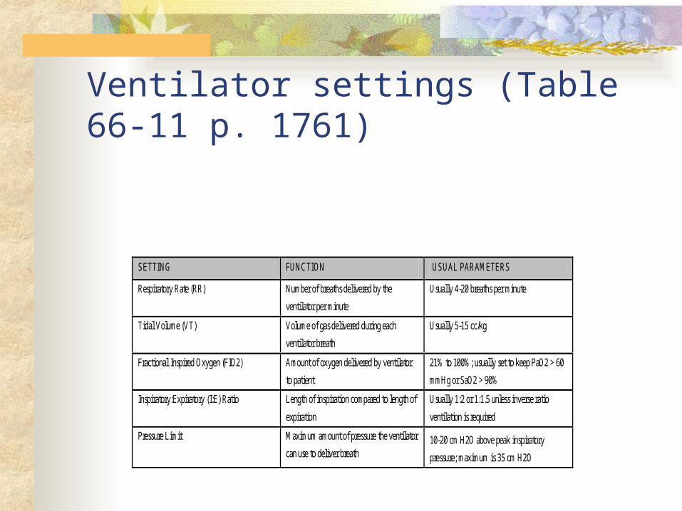
Ventilator settings (Table 66-11 p. 1761)
SETTING FUNCTION USUAL PARAMETERS
Respiratory Rate (RR) Number of breaths delivered by the
ventilator per minute
Usually 4-20 breaths per minute
Tidal Volume (VT) Volume of gas delivered during each
ventilator breath
Usually 5-15 cc/kg
Fractional Inspired Oxygen (FIO2) Amount of oxygen delivered by ventilator
to patient
21% to 100%; usually set to keep PaO2 > 60
mmHg or SaO2 > 90%
Inspiratory:Expiratory (I:E) Ratio Length of inspiration compared to length of
expiration
Usually 1:2 or 1:1.5 unless inverse ratio
ventilation is required
Pressure Limit Maximum amount of pressure the ventilator
can use to deliver breath 10-20 cm H2O above peak inspiratory
pressure; maximum is 35 cm H2O
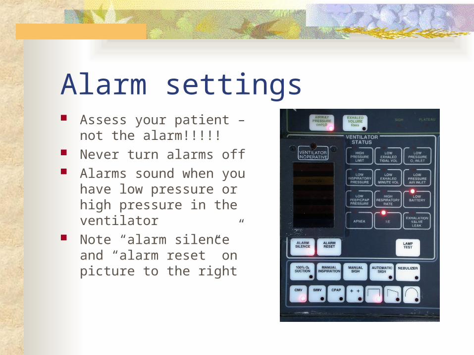
Alarm settings Assess your patient – not
the alarm!!!!! Never turn alarms off Alarms sound when you
have low pressure or high pressure in the ventilator
Note “alarm silence” and “alarm reset” on picture to the right
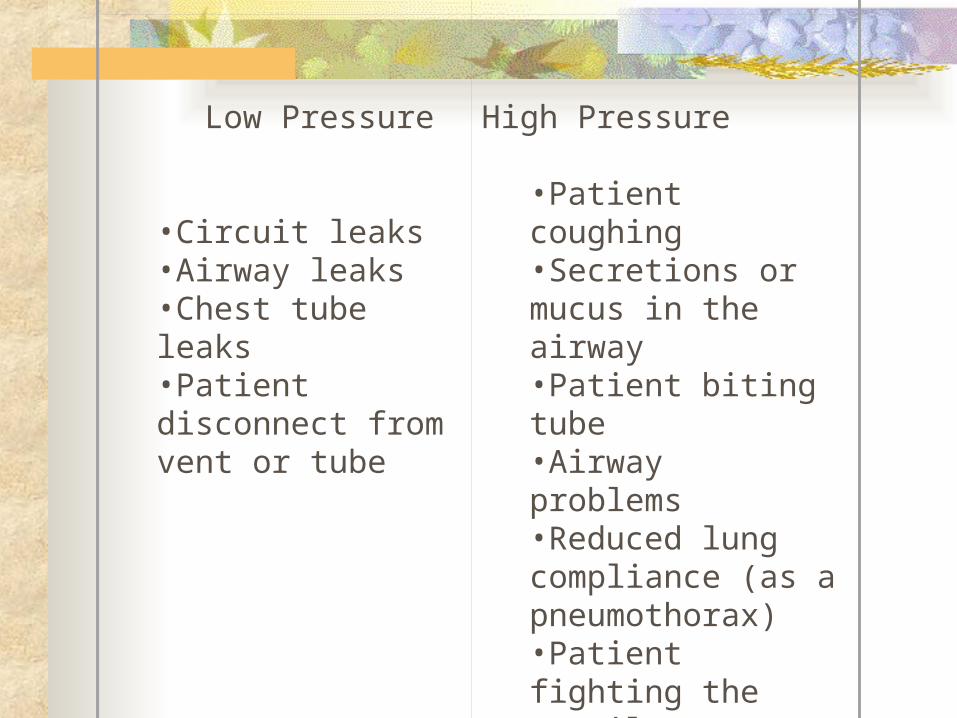
Low Pressure
•Circuit leaks •Airway leaks •Chest tube leaks •Patient disconnect from vent or tube
High Pressure
•Patient coughing •Secretions or mucus in the airway •Patient biting tube •Airway problems •Reduced lung compliance (as a pneumothorax) •Patient fighting the ventilator •Accumulation of water in the tube•Kinking of tube

Modes of PPV Volume Ventilation
Predetermined tidal volume (TV) is delivered with each inspiration
Tidal volume (TV) is consistent, airway pressures will vary
Pressure Ventilation Predetermined peak inspiratory pressure Tidal volume (TV) will vary, airway pressures
will be consistent

Ventilator settings of Modes (Table 66-12 p.1761) Volume Modes
CMV; AC; SIMV Predetermined tidal volume (TV) is delivered with each
inspiration Tidal volume (TV) is consistent, airway pressures will vary
Pressure Modes PSV; PC-IRV
Predetermined peak inspiratory pressure Tidal volume (TV) will vary, airway pressures will be
consistent Other Modes
PEEP and CPAP
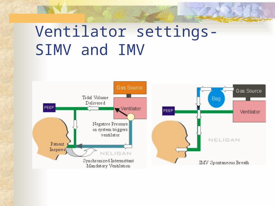
Ventilator settings- SIMV and IMV

Ventilator settings- Other modes Positive End-Expiratory Pressure
Positive pressure is maintained at the end of expiration
Pressure at end expiration keeps alveoli from collapsing, improving functional residual capacity (FRC)
Used with other modes on the ventilator Purpose is to improve oxygenation while limiting
risk of O2 toxicity Used to treat ARDS
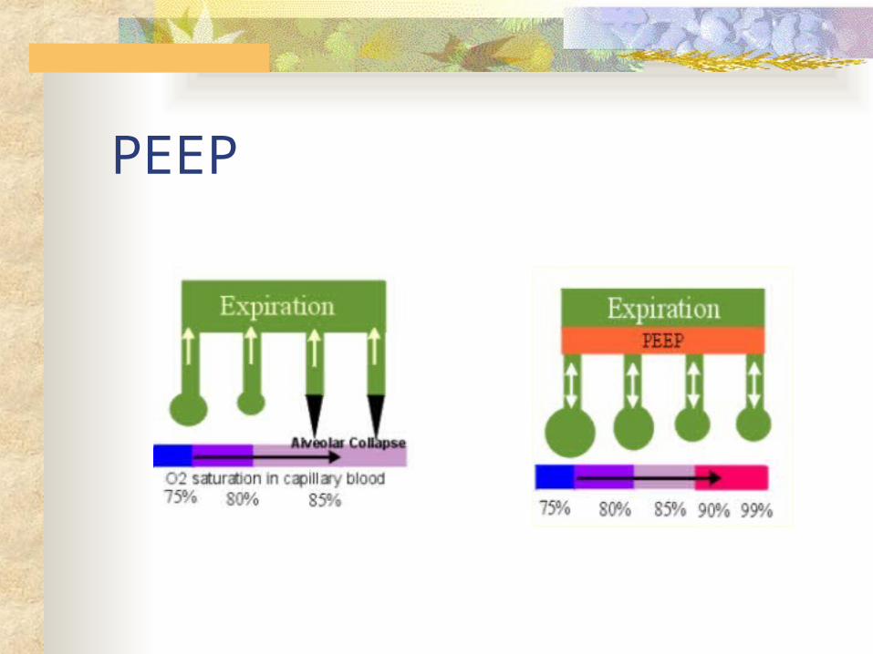
PEEP

Ventilator settings- other modes Continuous Positive Airway
Similar to PEEP However, pressure in CPAP is delivered
continuously Prevents airway pressure from falling to zero Measured in cm H20 Can be administered noninvasively (by mask) or
through ETube or TTube Used in treatment of obstructive sleep apnea
Complications of Positive Pressure Mechanical ventilation Cardiovascular: decreased CO; inc intrathoracic pressure Pulmonary: Barotrauma; Volutrauma; alveolar
hypoventilation/hyperventilation; ventilator-associated pneumonia
Sodium and water imbalance Neurological: impaired cerebral bl flow>IICP Gastrointestional: stress ulcer/GI bleed; gas; constipation Musculoskeletal: dec muscle tone; contractures; footdrop;
pressure ulcers from BR Psychosocial: physical & emotional stress; fight vent
Nursing Care for complications Neurological – elevate head of bed, keep body
in proper alignment Respiratory – monitor cuff inflation, vent settings,
ABG’s, for hyperventilation, hypoxemia Cardiovascular – monitor NIBP and arterial
pressures, CO, capillary refill, HR & rhythm Gastrointestinal – set up schedule for BM, admin
laxatives, PPI, admin tube feedings Musculoskeletal – passive & active ROM, turn
q2h, keep body in proper alignment
Psychological needs- Need for information; regain control; to hope; to trust Involve in discision making, medication for
sedation (proplfol), analgesia (fentanyl), neuromuscular blocking agents (Nimbex)
Other problems when on mechanical ventilation Machine disconnection or malfunction Nutrition needs Weaning from ventilator/ extubation
Spontanenous breathing trial (SBT) Hospital protocol
Document progress Table 66-13 p.1767- readiness/assessment
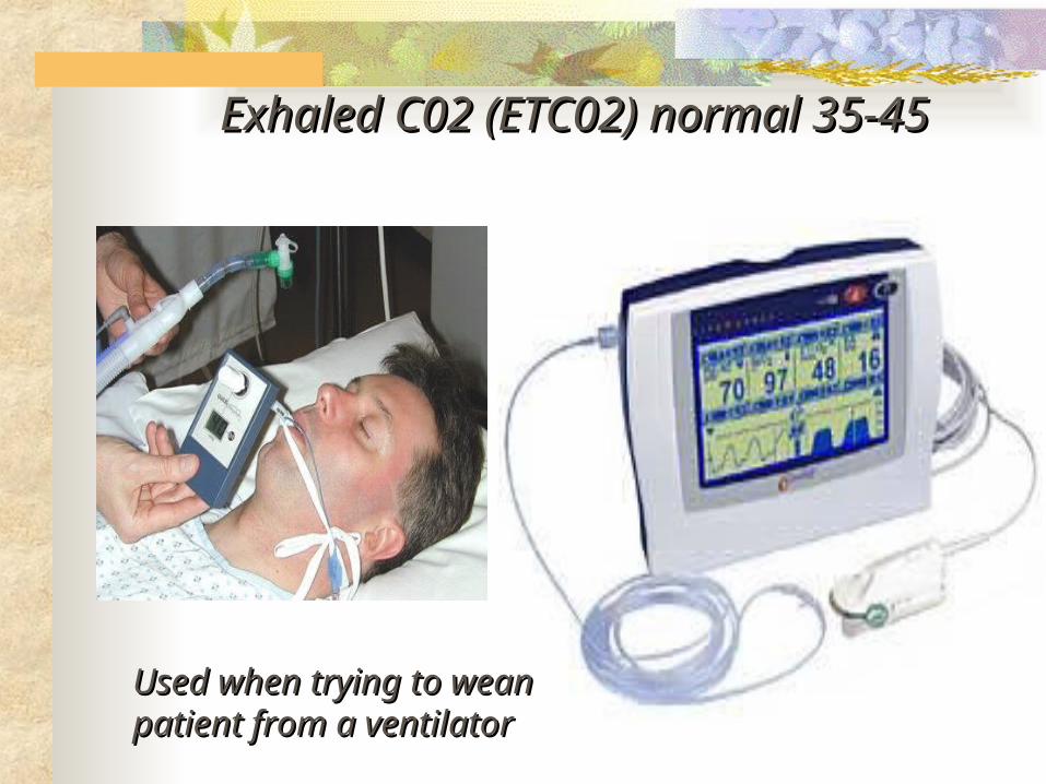
Exhaled C02 (ETC02) normal 35-45Exhaled C02 (ETC02) normal 35-45
Used when trying to wean Used when trying to wean patient from a ventilatorpatient from a ventilator
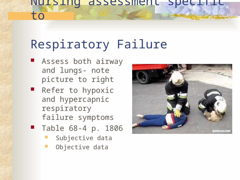
Nursing assessment specific to Respiratory Failure Assess both airway and
lungs- note picture to right
Refer to hypoxic and hypercapnic respiratory failure symptoms
Table 68-4 p. 1806 Subjective data Objective data
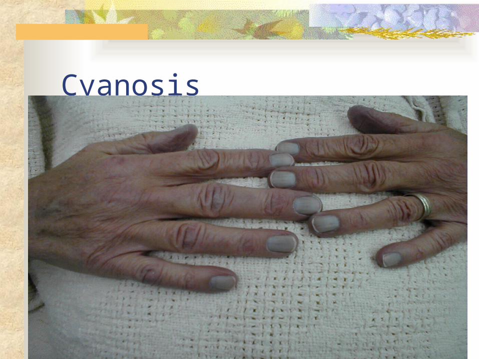
Cyanosis

Relevant Nursing Problems related to Respiratory Failure Prevention of acute respiratory failure Nursing Care Plans (p.1807-09) Gerontology considerations Nursing Care Plans Mechanical ventilation
(NCP 66-1 p.1754) Suctioning procedure and oral care
(p.1757-8)
Acute Respiratory Distress Syndrome ARDS Sudden progressive form of acute respiratory
failure Alveolar capillary membrane becomes damaged
and more permeable to intravascular fluid Results in noncardiac pulmonary edema and
progressive refractory hypoxemia ARDS is NOT primary! Follows various pulmonary or systemic
conditions Sepsis is the most common cause
Copyright © 2007, 2004, 2000, Mosby, Inc., an affiliate of Elsevier Inc. All Rights Reserved.
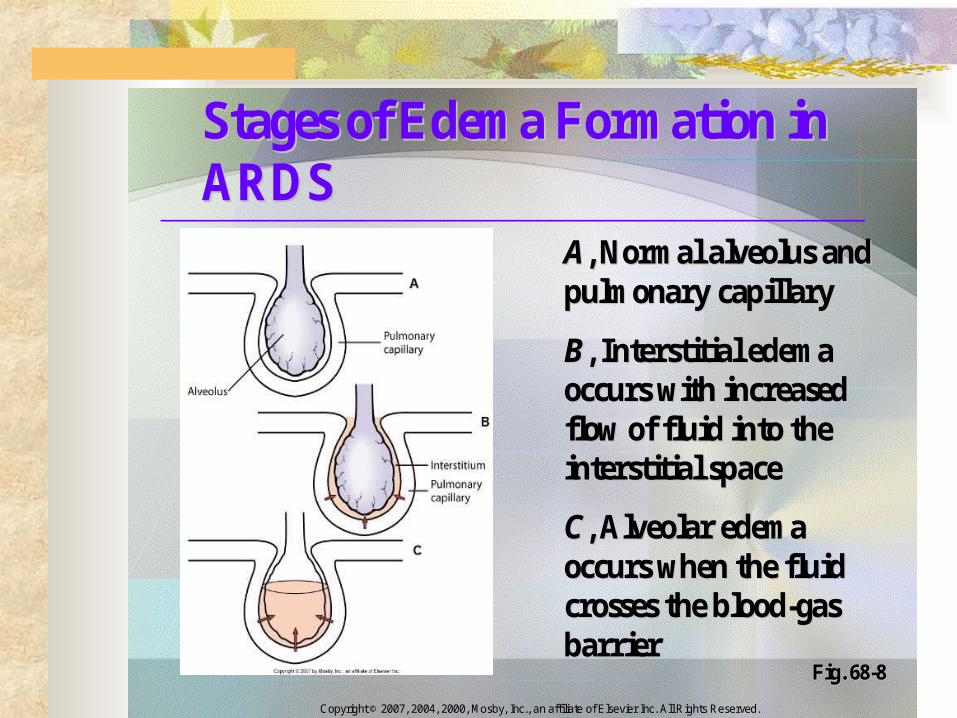
Stages of Edema Formation in Stages of Edema Formation in ARDSARDS
AA, Normal alveolus and , Normal alveolus and pulmonary capillary pulmonary capillary
BB, Interstitial edema , Interstitial edema occurs with increased occurs with increased flow of fluid into the flow of fluid into the interstitial space interstitial space
CC, Alveolar edema , Alveolar edema occurs when the fluid occurs when the fluid crosses the bloodcrosses the blood--gas gas barrierbarrier
Fig. 68Fig. 68--88
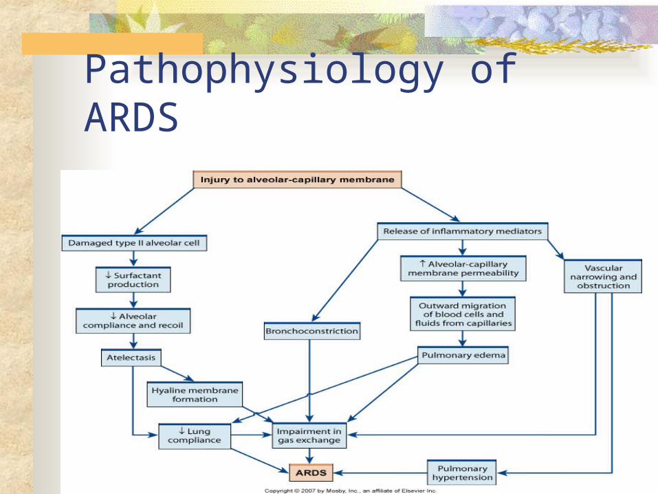
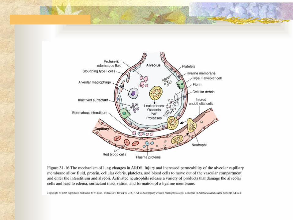
Pathophysiology of ARDS
Phases of ARDS Injury or Exudate Phase Occurs 24-48 hrs post direct or indirect inj Systemic inflammatory response and damage to
the alveolar-capillary membrane. Increased capillary permeability Fluid enters the alveoli Dilutes and deactivates surfactant Alveoli stiffen and collapse Interstitial, alveolar edema & atelectasis>
noncardiogenic pulmonary edema Hypoxemia becomes refractory
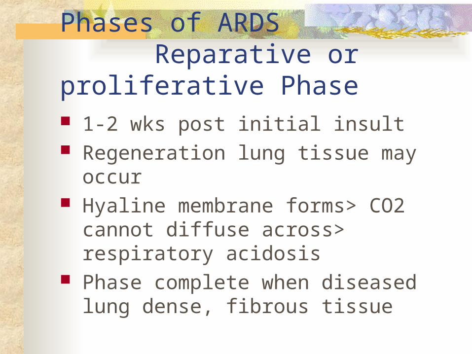
Phases of ARDS Reparative or proliferative Phase 1-2 wks post initial insult Regeneration lung tissue may occur Hyaline membrane forms> CO2 cannot
diffuse across> respiratory acidosis Phase complete when diseased lung dense,
fibrous tissue
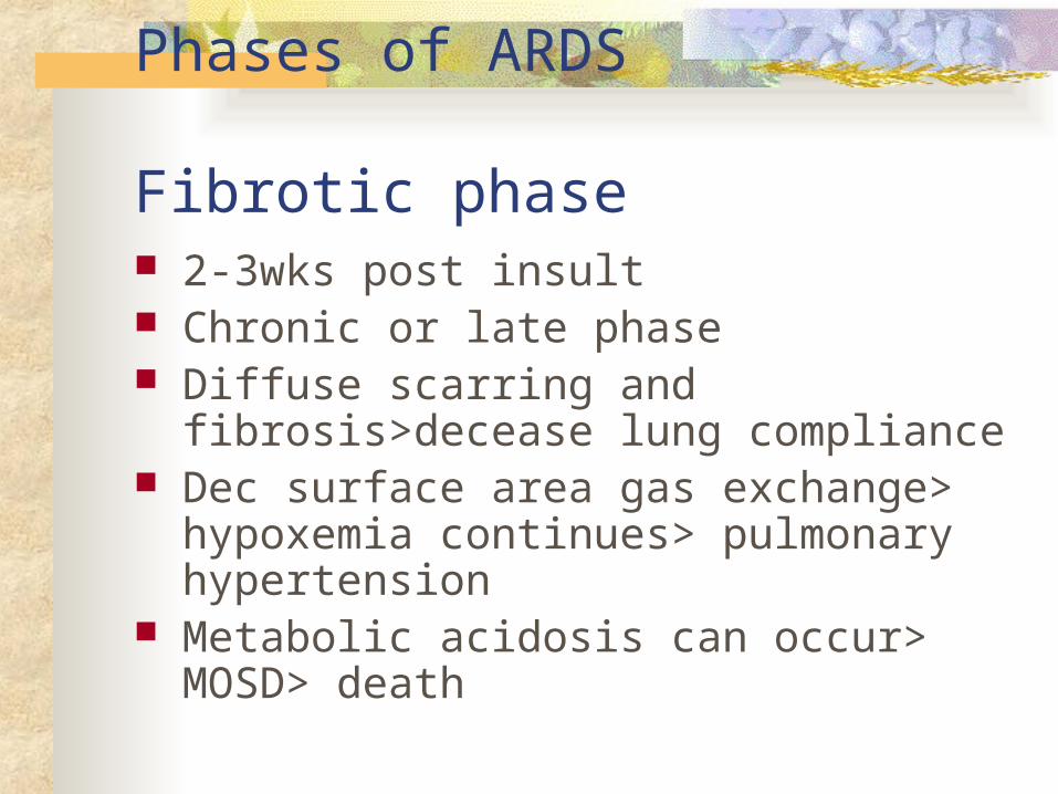
Phases of ARDS Fibrotic phase 2-3wks post insult Chronic or late phase Diffuse scarring and fibrosis>decease lung
compliance Dec surface area gas exchange> hypoxemia
continues> pulmonary hypertension Metabolic acidosis can occur> MOSD>
death
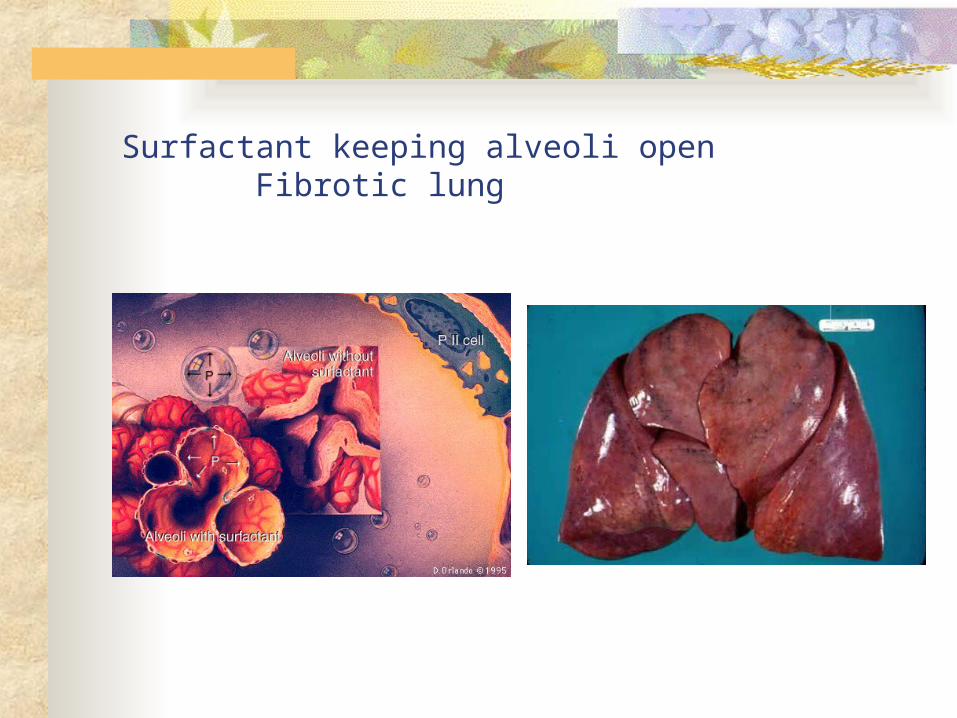
Surfactant keeping alveoli open Fibrotic lung
Clinical progression of ARDS Insidious onset- sym dev 24-48 hrs post
initial insult (direct or indirect lung injury) Course determined by nature of initial
injury, extent & severity of coexisting disease, and pulmonary complications
50% who develop ARDS die- even with aggressive treatment

Clinical manifestations of ARDS Progressive refractory hypoxemia> Hallmark sign Noncardiac pulmonary edema Early symptoms- labored R- dyspnea, tachypnea,
anxiety/restless, dry-nonproductive cough Later symptoms- cyanosis, adventitious breath
sounds, use of accessory muscles with retractions and decreased mental status
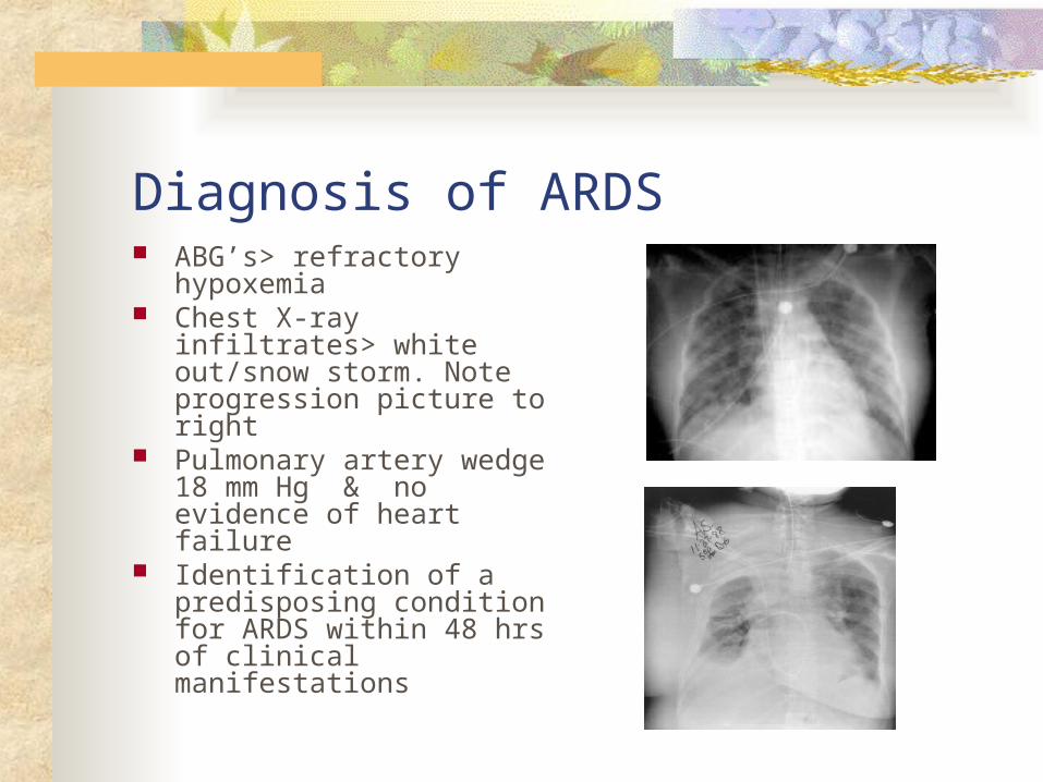
Diagnosis of ARDS ABG’s> refractory
hypoxemia Chest X-ray infiltrates>
white out/snow storm. Note progression picture to right
Pulmonary artery wedge 18 mm Hg & no evidence of heart failure
Identification of a predisposing condition for ARDS within 48 hrs of clinical manifestations
Complications of ARDS Hospital-acquired pneumonia Barotrauma Volu-pressure trauma Physiologic stress ulcer Renal failure
Collaborative Care for ARDS Respiratory therapy & medical support Oxygen Mechanical ventilation-
main treatment Positioning strategies
Proning CLRT-lateral rotation
bed Maintenance of CO &
tissue perfusion (fluids) Maintenance of nutrition
& fluid balance Treat underlying cause
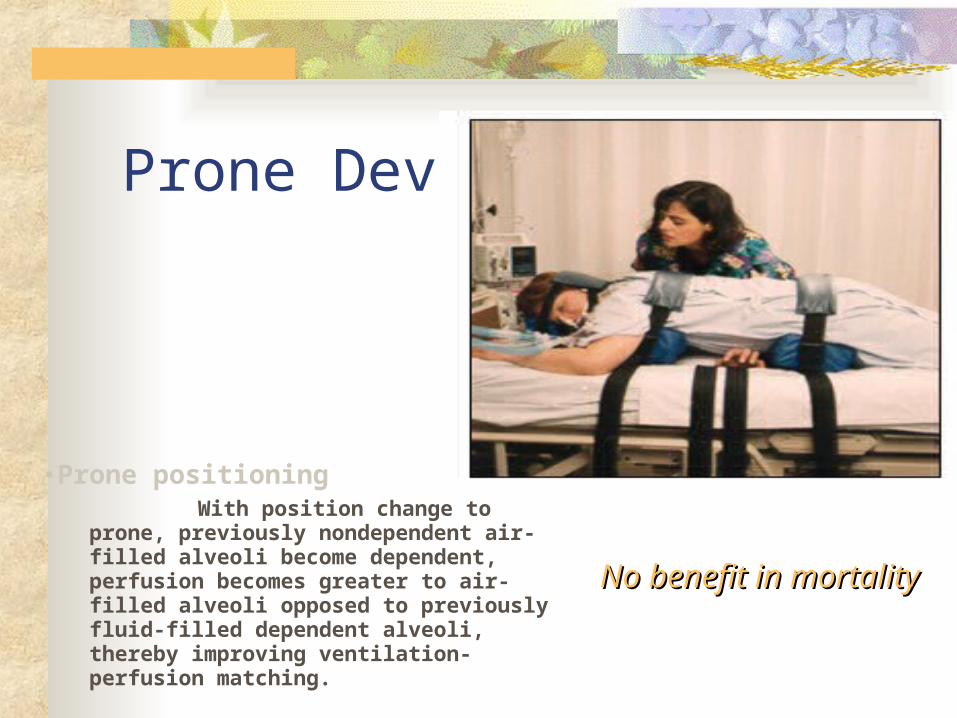
Prone Device
•Prone positioning With position change to
prone, previously nondependent air-filled alveoli become dependent, perfusion becomes greater to air-filled alveoli opposed to previously fluid-filled dependent alveoli, thereby improving ventilation-perfusion matching.
No benefit in mortalityNo benefit in mortality
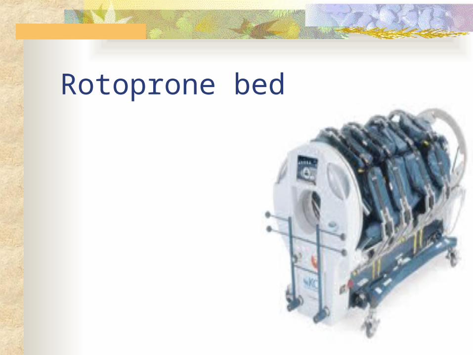
Rotoprone bed
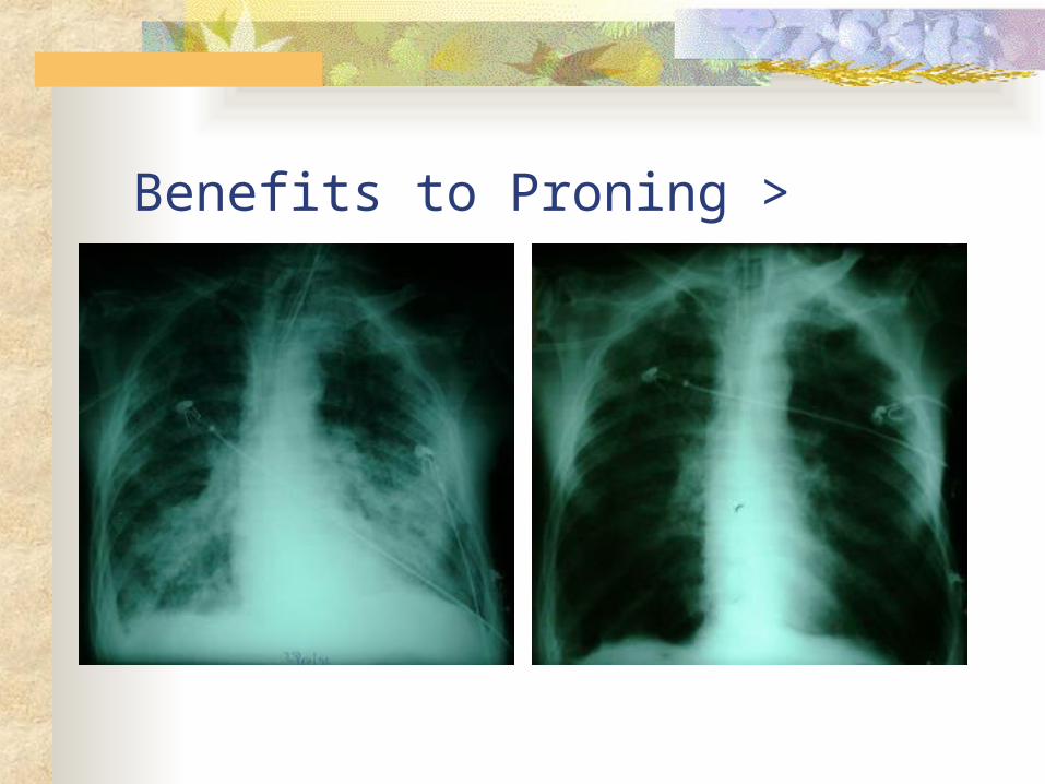
Benefits to Proning >
Nursing assessment specific to ARDS & Relevant nursing problems R/T ARDS Assessment
Refer to respiratory failure assessment Assess for clinical progression and clinical
manifestations as stated above Nursing care plans- refer to resp failure Goals for recovery from ARDS
PaO2 within normal limits on room air SaO2 greater 90% Patent airway Lungs clear on auscultation
