responsive pet nano/microfibers via surface-initiated ... · responsive pet nano/microfibers via...
TRANSCRIPT

Responsive PET Nano/Microfibers via Surface-InitiatedPolymerizationA. Evren Ozcam,† Kristen E. Roskov,† Jan Genzer,† and Richard J. Spontak*,†,‡
†Departments of Chemical & Biomolecular Engineering and ‡Materials Science & Engineering, North Carolina State University,Raleigh, North Carolina 27695, United States
*S Supporting Information
ABSTRACT: Poly(ethylene terephthalate) (PET) is one of the most importantthermoplastics in ubiquitous use today because of its mechanical properties, clarity,solvent resistance, and recyclability. In this work, we functionalize the surface ofelectrospun PET microfibers by growing poly(N-isopropylacrylamide) (PNIPAAm)brushes through a chemical sequence that avoids PET degradation to generatethermoresponsive microfibers that remain mechanically robust. Amidation of deposited3-aminopropyltriethoxysilane, followed by hydrolysis, yields silanol groups that permitsurface attachment of initiator molecules, which can be used to grow PNIPAAm via “grafting from” atom-transfer radicalpolymerization. Spectroscopic analyses performed after each step confirm the expected reaction and the ultimate growth ofPNIPAAm brushes. Water contact-angle measurements conducted at temperatures below and above the lower critical solutiontemperature of PNIPAAm, coupled with adsorption of Au nanoparticles from aqueous suspension, demonstrate that the brushesretain their reversible thermoresponsive nature, thereby making PNIPAAm-functionalized PET microfibers suitable for filtrationmedia, tissue scaffolds, delivery vehicles, and sensors requiring robust microfibers.
KEYWORDS: electrospinning, polymer brush, poly(ethylene terephthalate), responsive polymer, surface functionalization
Electrospinning is an emerging fabrication techniquecapable of generating solid polymer fibers that range
from tens of nanometers to several micrometers in diameter.Such nano/microfibers are of fundamental and technologicalinterest due to their high surface-to-volume ratio. During wetelectrospinning, a polymer solution of sufficiently high viscosityand conductivity is subjected to an electric field. When theelectrostatic forces overcome surface tension, a charged jetemitted from the tip of a Taylor cone1 undergoes a whippingaction2 (wherein the solvent evaporates) and is subsequentlycollected as a dry, randomly oriented fiber mat on a groundedcollector plate. This process strategy is appealing due to thesimple setup required and the ability to tailor fiber character-istics with relative ease.3 Although the morphology ofelectrospun nano/microfibers is desirable, they tend to lackthe functionality that is sought in contemporary applications.One way to overcome this deficiency is by developingmulticomponent nano/microfibers, in which the fiber-formingpolymer is modified with one or more species designed toenhance targeted properties.4−6 Surface-active compoundsadded to the polymer solution prior to electrospinning may,however, remain trapped within the resultant fiber uponsolidification and thus exhibit substantially reduced activity.7
While antibacterial biocides incorporated in this fashion losemuch of their efficacy,8 quaternary ammonium speciescovalently bonded to as-spun fibers can create a permanentantibacterial surface.9 Alternatively, polarizable antibacterialcopolymers codissolved with the fiber-forming polymer can bebrought to the fiber surface, where they remain anchored inplace, by the electric field during electrospinning.10,11 Recently,
Agarwal et al.12 have surveyed chemical routes by which tomodify and functionalize the surface of electrospun nanofibersfor diverse applications ranging from functional textiles, catalystsupports and ion-exchange membranes to drug delivery andtissue engineering.Polymers such as poly(ethylene terephthalate) (PET), which
is widely known for its mechanical strength, transparency andsolvent resistance, tend to possess a hydrophobic surface and alow surface energy,13 in which case electrospun nano/microfibers require post-treatment so that chemically activespecies are positioned on the fiber surface. Methods by whichto achieve such surface functionalization include plasmatreatment,8 mineralization,14 core−shell formation,15 chemicalvapor deposition16 or inclusion of reactive compounds.17,18
Once these chemically active groups are available, covalentbonding,19 immobilization20 or electrostatic interactions21 canbe used to introduce functional moieties to the fiber surfacewithout adversely affecting the bulk fiber properties. Whilesurface modification could permit the use of electrospun PET22
nano/microfibers in filtration media,23 protective textiles,24
tissue scaffolds,25 and drug-delivery vehicles,26 most of themodification approaches listed above purposefully or inadver-tently promote PET degradation. Thus, the conditions bywhich surface modification is conducted must be monitoredcarefully to avoid compromising the bulk properties of PET.
Received: November 8, 2011Accepted: December 27, 2011Published: January 10, 2012
Letter
www.acsami.org
© 2012 American Chemical Society 59 dx.doi.org/10.1021/am201559f | ACS Appl. Mater. Interfaces 2012, 4, 59−64

Grafting polymer brushes represents an alternative approachby which to modify and control the surface properties ofmaterials.27 Numerous studies have reported surface-initiatedgrafting on surfaces of various geometries with a plethora ofdifferent monomers by employing numerous polymerizationroutes. Poly(N-isopropylacrylamide) (PNIPAAm) is solelyconsidered here because of its thermoresponsive nature28 (itpossesses a lower critical solution temperature, LCST, in waterat ≈32 °C). Prior efforts to polymerize styrene29,30 andNIPAAm31,32 on flat PET surfaces have relied on differentmeans of activating the PET surface (e.g., saponification,plasma treatment and aminolysis) for the purpose of attachinginitiators. The major drawback of such treatments, however, isthat they may seriously deteriorate the mechanical properties ofPET and increase its surface roughness by chemicaldegradation, which would be catastrophic with regard toelectrospun PET nano/microfibers due to their fine dimen-sions. Independent studies33−35 have confirmed that 3-aminopropyltriethoxysilane (APTES) can be used to function-alize the surface of PET via amidation with negligibledegradation. Unlike short alkyl amines (which can diffuseinto and react throughout, and thus weaken, PET36,37), thebulky triethoxysilane group on APTES hinders diffusion,changes its chemical nature upon amidation and creates abarrier by restricting the diffusion of other APTES molecules.Moreover, since the ethoxysilane groups of APTES are exposedat the polymer/air interface after reaction, hydrolysis oftriethoxysilane yields silanol groups that facilitate initiatorattachment along the fiber surface.Thermoresponsive PNIPAAm brushes on electrospun fibers
have been recently reported. For instance, Brandl et al.38
describe the synthesis of a copolymer of 2-hydroxyethylmethacrylate (HEMA) and methyl methacrylate (MMA) andits postpolymerization modification with 2-bromoisobutyryl-bromide to prepare a macroinitiator. They claim thatelectrospinning of the macroinitiator and subsequent polymer-ization of NIPAAm results in thermoresponsive polymerbrushes. The disadvantage of this technique is that the locationof the “active” initiator group in the fiber depends on thedielectrophoretic forces, polarizability contrast and surfacetension of the comonomers, which invariably reduces theconcentration of “active” initiator centers on the fiber surface.The presence of ester groups between the butyrylbromidegroup of the initiator and hydroxyethyl group of HEMAlikewise yields hydrolytically unstable bonds at pH valuesgreater than 8 and lower than 5. Furthermore, the presence ofHEMA comonomer on the macroinitiator may result inswelling and absorption of NIPAAm monomer by theelectrospun fiber in the aqueous polymerization medium.Similarly, Fu et al.39 have synthesized and electrospun acopolymer of 4-vinylbenzylchloride and glycidyl methacrylate.Subsequent modification of the electrospun fibers with sodiumazide produces azide surface groups that can be coupled withalkyne-functionalized PNIPAAm chains via a click reaction togenerate PNIPAAm surface chains that affect the wettability ofthe fibers. Such grafting of PNIPAAm brushes on electrospunfibers would be necessarily low because of the sparsepopulation of active azide groups on the fiber surface and theaccompanying steric hindrance caused by “grafting to”polymerization.Despite the myriad of reports regarding fiber preparation via
electrospinning, the number of studies on electrospun PETfibers is rather limited.22,40−47 In this work, we aim to craft a
mild and universal way of modifying the surface of electrospunPET fibers to combine the mechanical robustness of PET andthe functionality of thermoresponsive PNIPAAm brushes thatare covalently attached to the fibers. First, electrospun PETmicrofibers are modified with APTES to create surface-boundhydroxyl groups for the attachment of [11-(2-bromo-2-methyl)propionyloxy] undecyltrichlorosilane (BMPUS),which serves as an ATRP initiator for the polymerization ofNIPAAm. Several analytical techniques are employed to (i)characterize the properties of as-spun and postmodified PETmicrofibers and (ii) follow the polymerization of NIPAAm viaATRP. In addition, we investigate the thermoresponsive natureof PNIPAAm-decorated PET microfibers by attaching Aunanoparticles at temperatures above and below the LCST ofPNIPAAm.
■ EXPERIMENTAL SECTION
Food-grade recycled PET flakes were kindly supplied by theUnited Resource Recovery Corp. (Spartanburg, SC). Hexa-fluoroisopropanol (HFIP) was obtained from OakwoodProducts Inc. (Estill, SC), and anhydrous toluene, 2-chlorophenol, APTES, NIPAAm, copper I bromide (CuBr),copper II bromide (CuBr2), and N,N,N′,N′,N″-pentamethyldie-thylenetriamine (PMDETA) were all purchased from Sigma-Aldrich and used as-received. The citrate-stabilized Aunanoparticles48 (diameter = 16.9 ± 1.8 nm) and BMPUSinitiator49 were prepared as described earlier. The PET flakeswere dissolved in HFIP at different concentrations andelectrospun at ambient temperature and 10 kV to generatemicrofibers varying in diameter. Thin films of PET measuring12 and 180 nm thick, as discerned by ellipsometry (v.i.), werespin-coated at 25 °C on silicon wafers from 0.5 and 3.0 wt %solutions, respectively, in 2-chlorophenol. Microfiber mats andthin films were stored under vacuum for at least 48 h prior touse to remove entrapped solvent.The APTES was deposited on the PET microfibers and thin
films by exposing the samples to 1% (v/v) APTES/anhydroustoluene solutions for 24 h at ambient temperature, followed bysonication in toluene for 10 min to remove loosely adsorbedAPTES molecules. The ethoxysilane groups of the surface-anchored APTES molecules were hydrolyzed in acidic water(pH ≈ 4.5−5.0) for 6 h at ambient temperature, and then thefiber mats were washed with copious amount of water. After thesamples were dried under reduced pressure, BMPUS wasdeposited on the PET−BMPUS surfaces by establishedprotocols.50,51 The PNIPAAm brushes were subsequentlygrown from PET−SiOH surfaces by ATRP of NIPAAm, asdescribed elsewhere.48 Briefly, 6.30 g of NIPAAm was dissolvedin a mixture of 4.86 g of methanol and 6.30 g of water in anargon-purged Schlenk flask, and oxygen was removed via threefreeze−thaw cycles. After removal of oxygen, PMDETA (0.56g), CuBr (0.16 g), and CuBr2 (0.016 g) were added to thesolution prior to an additional freeze−thaw cycle. The Schlenkflask was tightly sealed and transferred to an argon-purgedglovebox. Microfiber mats and thin films of PET weresubmersed in the solution for 8 h at ambient temperature,after which they were removed, promptly rinsed with methanoland deionized water, and then sonicated in deionized water for10 min. The length of the PNIPAAm brushes can besystematically changed by varying the polymerization time orthe rate of polymerization, which is governed by the CuBr/CuBr2 ratio. In the present work, we had no intention of
ACS Applied Materials & Interfaces Letter
dx.doi.org/10.1021/am201559f | ACS Appl. Mater. Interfaces 2012, 4, 59−6460
investigating different thicknesses of PNIPAAm brushes onelectrospun PET fibers.The thickness of the PET films was measured by variable-
angle spectroscopic ellipsometry (J.A. Woollam) at a 70°incidence angle before and after each modification step todiscern the PNIPAAm brush height. Surface chemicalcomposition was monitored at each reaction step by X-rayphotoelectron spectroscopy (XPS) performed on a KratosAnalytical AXIS ULTRA spectrometer at a single takeoff angleof 90°. Fourier-transform infrared (FTIR) spectroscopicanalysis of the PET microfibers was conducted in transmissionmode on a Nicolet 6700 spectrometer after embedding themicrofiber mats in potassium bromide pellets. For each sample,1024 scans were acquired after background correction at aresolution of 4 cm−1. Resultant XPS and FTIR spectra wereanalyzed using the Vision and Omnic Spectra software suites,respectively. The thermoresponsive behavior of PET and PET-PNIPAAm microfibers was interrogated by measuring the staticwater contact angle (WCA) at different temperatures via thesessile drop technique on a Rame-Hart Model 100−00instrument. The WCA measurements performed aboveambient temperature were recorded after thermal equilibrationin an environmental chamber manufactured by Rame-Hart. As-spun and modified PET microfibers were coated with ∼8 nm ofAu prior to analysis by field-emission scanning electronmicroscopy (SEM) performed on a JEOL 6400F electronmicroscope operated at 5 kV. The thickness of the Au layer wassufficiently thick to prevent specimen charging under theelectron beam, but sufficiently thin to avoid masking finefeatures on the surface of the microfibers. The averagemicrofiber diameter and corresponding standard deviationwere determined by measuring the diameters of at least 100microfibers and analyzing the results with the ImageJ softwarepackage.
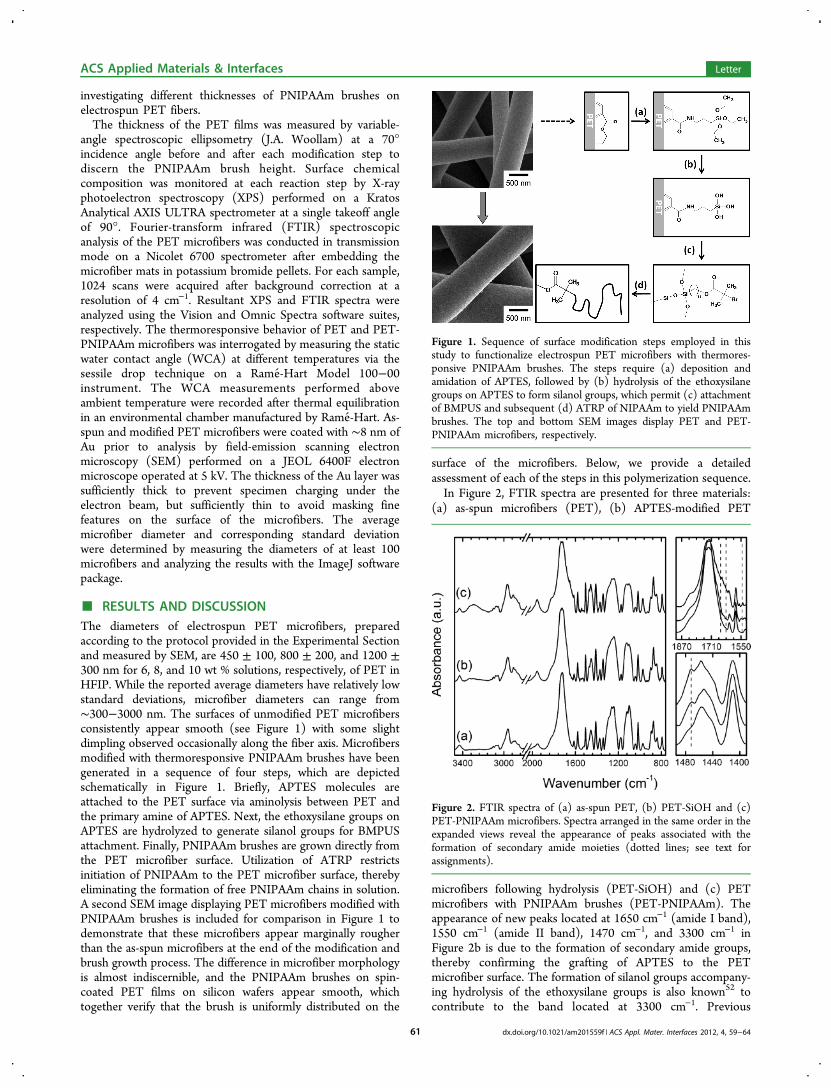
■ RESULTS AND DISCUSSIONThe diameters of electrospun PET microfibers, preparedaccording to the protocol provided in the Experimental Sectionand measured by SEM, are 450 ± 100, 800 ± 200, and 1200 ±300 nm for 6, 8, and 10 wt % solutions, respectively, of PET inHFIP. While the reported average diameters have relatively lowstandard deviations, microfiber diameters can range from∼300−3000 nm. The surfaces of unmodified PET microfibersconsistently appear smooth (see Figure 1) with some slightdimpling observed occasionally along the fiber axis. Microfibersmodified with thermoresponsive PNIPAAm brushes have beengenerated in a sequence of four steps, which are depictedschematically in Figure 1. Briefly, APTES molecules areattached to the PET surface via aminolysis between PET andthe primary amine of APTES. Next, the ethoxysilane groups onAPTES are hydrolyzed to generate silanol groups for BMPUSattachment. Finally, PNIPAAm brushes are grown directly fromthe PET microfiber surface. Utilization of ATRP restrictsinitiation of PNIPAAm to the PET microfiber surface, therebyeliminating the formation of free PNIPAAm chains in solution.A second SEM image displaying PET microfibers modified withPNIPAAm brushes is included for comparison in Figure 1 todemonstrate that these microfibers appear marginally rougherthan the as-spun microfibers at the end of the modification andbrush growth process. The difference in microfiber morphologyis almost indiscernible, and the PNIPAAm brushes on spin-coated PET films on silicon wafers appear smooth, whichtogether verify that the brush is uniformly distributed on the
surface of the microfibers. Below, we provide a detailedassessment of each of the steps in this polymerization sequence.In Figure 2, FTIR spectra are presented for three materials:
(a) as-spun microfibers (PET), (b) APTES-modified PET
microfibers following hydrolysis (PET-SiOH) and (c) PETmicrofibers with PNIPAAm brushes (PET-PNIPAAm). Theappearance of new peaks located at 1650 cm−1 (amide I band),1550 cm−1 (amide II band), 1470 cm−1, and 3300 cm−1 inFigure 2b is due to the formation of secondary amide groups,thereby confirming the grafting of APTES to the PETmicrofiber surface. The formation of silanol groups accompany-ing hydrolysis of the ethoxysilane groups is also known52 tocontribute to the band located at 3300 cm−1. Previous
Figure 1. Sequence of surface modification steps employed in thisstudy to functionalize electrospun PET microfibers with thermores-ponsive PNIPAAm brushes. The steps require (a) deposition andamidation of APTES, followed by (b) hydrolysis of the ethoxysilanegroups on APTES to form silanol groups, which permit (c) attachmentof BMPUS and subsequent (d) ATRP of NIPAAm to yield PNIPAAmbrushes. The top and bottom SEM images display PET and PET-PNIPAAm microfibers, respectively.
Figure 2. FTIR spectra of (a) as-spun PET, (b) PET-SiOH and (c)PET-PNIPAAm microfibers. Spectra arranged in the same order in theexpanded views reveal the appearance of peaks associated with theformation of secondary amide moieties (dotted lines; see text forassignments).
ACS Applied Materials & Interfaces Letter
dx.doi.org/10.1021/am201559f | ACS Appl. Mater. Interfaces 2012, 4, 59−6461
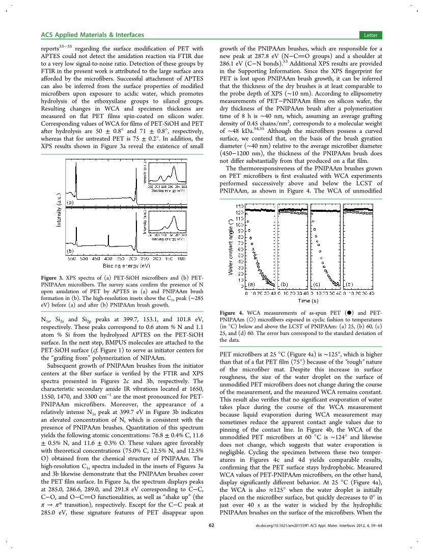
reports33−35 regarding the surface modification of PET withAPTES could not detect the amidation reaction via FTIR dueto a very low signal-to-noise ratio. Detection of these groups byFTIR in the present work is attributed to the large surface areaafforded by the microfibers. Successful attachment of APTEScan also be inferred from the surface properties of modifiedmicrofibers upon exposure to acidic water, which promoteshydrolysis of the ethoxysilane groups to silanol groups.Resulting changes in WCA and specimen thickness aremeasured on flat PET films spin-coated on silicon wafer.Corresponding values of WCA for films of PET-SiOH and PETafter hydrolysis are 50 ± 0.8° and 71 ± 0.8°, respectively,whereas that for untreated PET is 75 ± 0.2°. In addition, theXPS results shown in Figure 3a reveal the existence of small
N1s, Si2s and Si2p peaks at 399.7, 153.1, and 101.8 eV,respectively. These peaks correspond to 0.6 atom % N and 1.1atom % Si from the hydrolyzed APTES on the PET-SiOHsurface. In the next step, BMPUS molecules are attached to thePET-SiOH surface (cf. Figure 1) to serve as initiator centers forthe “grafting from” polymerization of NIPAAm.Subsequent growth of PNIPAAm brushes from the initiator
centers at the fiber surface is verified by the FTIR and XPSspectra presented in Figures 2c and 3b, respectively. Thecharacteristic secondary amide IR vibrations located at 1650,1550, 1470, and 3300 cm−1 are the most pronounced for PET-PNIPAAm microfibers. Moreover, the appearance of arelatively intense N1s peak at 399.7 eV in Figure 3b indicatesan elevated concentration of N, which is consistent with thepresence of PNIPAAm brushes. Quantitation of this spectrumyields the following atomic concentrations: 76.8 ± 0.4% C, 11.6± 0.5% N, and 11.6 ± 0.3% O. These values agree favorablywith theoretical concentrations (75.0% C, 12.5% N, and 12.5%O) obtained from the chemical structure of PNIPAAm. Thehigh-resolution C1s spectra included in the insets of Figures 3aand 3b likewise demonstrate that the PNIPAAm brushes coverthe PET film surface. In Figure 3a, the spectrum displays peaksat 285.0, 286.6, 289.0, and 291.8 eV corresponding to C−C,C−O, and O−CO functionalities, as well as “shake up” (theπ → π* transition), respectively. Except for the C−C peak at285.0 eV, these signature features of PET disappear upon
growth of the PNIPAAm brushes, which are responsible for anew peak at 287.8 eV (N−CO groups) and a shoulder at286.1 eV (C−N bonds).53 Additional XPS results are providedin the Supporting Information. Since the XPS fingerprint forPET is lost upon PNIPAAm brush growth, it can be inferredthat the thickness of the dry brushes is at least comparable tothe probe depth of XPS (∼10 nm). According to ellipsometrymeasurements of PET−PNIPAAm films on silicon wafer, thedry thickness of the PNIPAAm brush after a polymerizationtime of 8 h is ∼40 nm, which, assuming an average graftingdensity of 0.45 chains/nm2, corresponds to a molecular weightof ∼48 kDa.54,55 Although the microfibers possess a curvedsurface, we contend that, on the basis of the brush gyrationdiameter (∼40 nm) relative to the average microfiber diameter(450−1200 nm), the thickness of the PNIPAAm brush doesnot differ substantially from that produced on a flat film.The thermoresponsiveness of the PNIPAAm brushes grown
on PET microfibers is first evaluated with WCA experimentsperformed successively above and below the LCST ofPNIPAAm, as shown in Figure 4. The WCA of unmodified
PET microfibers at 25 °C (Figure 4a) is ∼125°, which is higherthan that of a flat PET film (75°) because of the ″rough″ natureof the microfiber mat. Despite this increase in surfaceroughness, the size of the water droplet on the surface ofunmodified PET microfibers does not change during the courseof the measurement, and the measured WCA remains constant.This result also verifies that no significant evaporation of watertakes place during the course of the WCA measurementbecause liquid evaporation during WCA measurement maysometimes reduce the apparent contact angle values due topinning of the contact line. In Figure 4b, the WCA of theunmodified PET microfibers at 60 °C is ∼124° and likewisedoes not change, which suggests that water evaporation isnegligible. Cycling the specimen between these two temper-atures in Figures 4c and 4d yields comparable results,confirming that the PET surface stays hydrophobic. MeasuredWCA values of PET-PNIPAAm microfibers, on the other hand,display significantly different behavior. At 25 °C (Figure 4a),the WCA is also ≈125° when the water droplet is initiallyplaced on the microfiber surface, but quickly decreases to 0° injust over 40 s as the water is wicked by the hydrophilicPNIPAAm brushes on the surface of the microfibers. When the
Figure 3. XPS spectra of (a) PET-SiOH microfibers and (b) PET-PNIPAAm microfibers. The survey scans confirm the presence of Nupon amidation of PET by APTES in (a) and PNIPAAm brushformation in (b). The high-resolution insets show the C1s peak (∼285eV) before (a) and after (b) PNIPAAm brush growth.
Figure 4. WCA measurements of as-spun PET (●) and PET-PNIPAAm (○) microfibers exposed in cyclic fashion to temperatures(in °C) below and above the LCST of PNIPAAm: (a) 25, (b) 60, (c)25, and (d) 60. The error bars correspond to the standard deviation ofthe data.
ACS Applied Materials & Interfaces Letter
dx.doi.org/10.1021/am201559f | ACS Appl. Mater. Interfaces 2012, 4, 59−6462
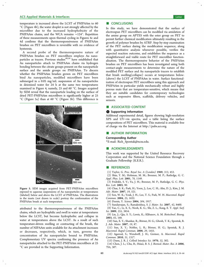
temperature is increased above the LCST of PNIPAAm to 60°C (Figure 4b), the water droplet is not strongly affected by themicrofiber due to the increased hydrophobicity of thePNIPAAm chains, and the WCA remains ∼124°. Repetitionof these measurements upon thermal cycling in Figures 4c and4d confirms that the thermoresponsiveness of PNIPAAmbrushes on PET microfibers is reversible with no evidence ofhysteresis.A second probe of the thermoresponsive nature of
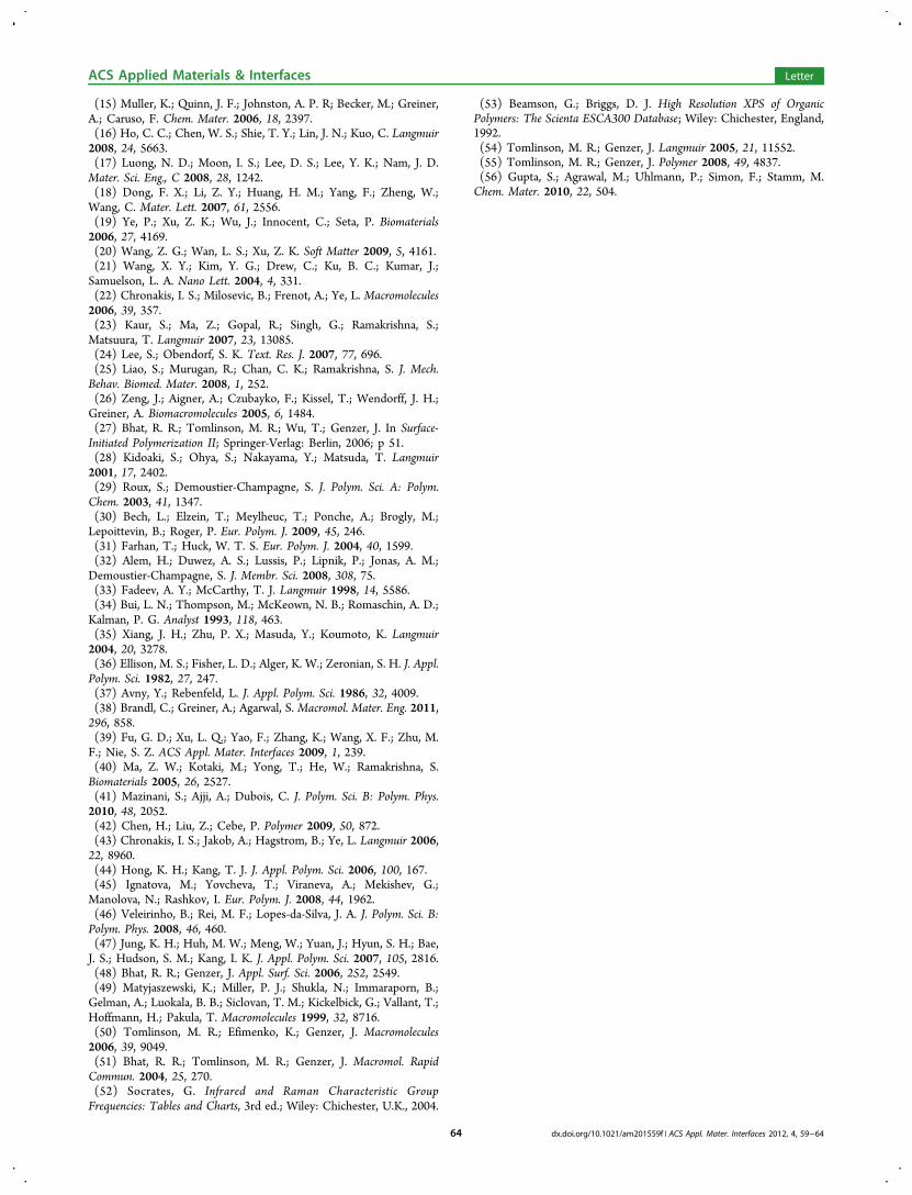
PNIPAAm brushes on PET microfibers employs Au nano-particles as tracers. Previous studies48,56 have established thatAu nanoparticles attach to PNIPAAm chains via hydrogenbonding between the citrate groups present on the nanoparticlesurface and the amide groups on PNIPAAm. To discernwhether the PNIPAAm brushes grown on PET microfibersbind Au nanoparticles, modified microfibers have beensubmerged in a 0.05 mg/mL suspension of Au nanoparticlesin deionized water for 24 h at the same two temperaturesexamined in Figure 4, namely, 25 and 60 °C. Images acquiredby SEM reveal that the nanoparticle loading on the surface ofdried PET-PNIPAAm microfibers is significantly higher at 25°C (Figure 5a) than at 60 °C (Figure 5b). This difference is
attributed to the thermoresponsiveness of the PNIPAAmchains, which are hydrophilic and swell in water at temperaturesbelow the LCST, but become hydrophobic and collapse inwater at temperatures above the LCST. As a result of suchtemperature-driven swelling or contracting of the brush, thenumber of NIPAAm units available for Au attachment increasesor decreases, respectively, which, in turn, governs theconcentration of Au nanoparticles bound to PNIPAAm.Complementary UV spectra confirming the presence of Aunanoparticles attached to the PET-PNIPAAm microfibers at 25°C are provided in the Supporting Information.
■ CONCLUSIONSIn this study, we have demonstrated that the surface ofelectrospun PET microfibers can be modified via amidation ofthe amine group on APTES with the ester group on PET topermit further chemical modification ultimately resulting in thegrowth of polymer brushes by ATRP. Step-by-step examinationof the PET surface during the modification sequence, alongwith quantitative analysis whenever possible, verifies theexpected reaction outcome, and establishes the sequence as astraightforward and viable route for PET microfiber function-alization. The thermoresponsive behavior of the PNIPAAmbrushes on PET microfibers has been investigated using bothcontact-angle measurements to determine the nature of themodified PET surface and Au nanoparticle tracers to establishthat brush swelling(collapse) occurs at temperatures below-(above) the LCST of PNIPAAm in water. Surface functional-ization of electrospun PET microfibers using this approach andPNIPAAm in particular yields mechanically robust and highlyporous mats that are temperature-sensitive, which means thatthey are suitable candidates for contemporary technologiessuch as responsive filters, scaffolds, delivery vehicles, andsensors.
■ ASSOCIATED CONTENT*S Supporting InformationAdditonal experimental detail, figures showing high-resolutionXPS and UV−vis spectra, and a table listing the surfacecompositions of PET microfibers. This material is available freeof charge via the Internet at http://pubs.acs.org.
■ AUTHOR INFORMATIONCorresponding Author*E-mail: [email protected].
■ ACKNOWLEDGMENTSThis work was supported by the United Resource RecoveryCorporation and the National Science Foundation through aGraduate Fellowship (K.E.R.).
■ REFERENCES(1) Taylor, G. Proc. Royal Soc. A (London) 1969, 313, 453.(2) Shin, Y. M.; Hohman, M. M.; Brenner, M. P.; Rutledge, G. C.Appl. Phys. Lett. 2001, 78, 1149.(3) Fridrikh, S. V.; Yu, J. H.; Brenner, M. P.; Rutledge, G. C. Phys.Rev. Lett. 2003, 90.(4) Chae, S. K.; Park, H.; Yoon, J.; Lee, C. H.; Ahn, D. J.; Kim, J. M.Adv. Mater. 2007, 19, 521.(5) Son, W. K.; Youk, J. H.; Lee, T. S.; Park, W. H. Macromol. RapidCommun. 2004, 25, 1632.(6) Dzenis, Y. Science 2004, 304, 1917.(7) Sundarrajan, S.; Ramakrishna, S. J. Mater. Sci. 2007, 42, 8400.(8) Yao, C.; Li, X. S.; Neoh, K. G.; Shi, Z. L.; Kang, E. T. Appl. Surf.Sci. 2009, 255, 3854.(9) Lin, J.; Qiu, S. Y.; Lewis, K.; Klibanov, A. M. Biotechnol. Bioeng.2003, 83, 168.(10) Sun, X. Y.; Shankar, R.; Borner, H. G.; Ghosh, T. K.; Spontak, R.J. Adv. Mater. 2007, 19, 87.(11) Sun, X. Y.; Nobles, L. R.; Borner, H. G.; Spontak, R. J.Macromol. Rapid Commun. 2008, 29, 1455.(12) Agarwal, S.; Wendorff, J. H.; Greiner, A. Macromol. RapidCommun. 2010, 31, 1317.(13) Dann, J. R. J. Colloid Interface Sci. 1970, 32, 302.(14) Chen, J. L.; Chu, B.; Hsiao, B. S. J. Biomed. Mater. Res. A 2006,79A, 307.
Figure 5. SEM images acquired from PET-PNIPAAm microfibersexposed to aqueous suspensions of Au nanoparticles at temperatures(labeled) below and above the LCST of PNIPAAm. The illustrationsin the insets (not drawn to scale) portray the conformation of thePNIPAAm brush at each temperature.
ACS Applied Materials & Interfaces Letter
dx.doi.org/10.1021/am201559f | ACS Appl. Mater. Interfaces 2012, 4, 59−6463
(15) Muller, K.; Quinn, J. F.; Johnston, A. P. R; Becker, M.; Greiner,A.; Caruso, F. Chem. Mater. 2006, 18, 2397.(16) Ho, C. C.; Chen, W. S.; Shie, T. Y.; Lin, J. N.; Kuo, C. Langmuir2008, 24, 5663.(17) Luong, N. D.; Moon, I. S.; Lee, D. S.; Lee, Y. K.; Nam, J. D.Mater. Sci. Eng., C 2008, 28, 1242.(18) Dong, F. X.; Li, Z. Y.; Huang, H. M.; Yang, F.; Zheng, W.;Wang, C. Mater. Lett. 2007, 61, 2556.(19) Ye, P.; Xu, Z. K.; Wu, J.; Innocent, C.; Seta, P. Biomaterials2006, 27, 4169.(20) Wang, Z. G.; Wan, L. S.; Xu, Z. K. Soft Matter 2009, 5, 4161.(21) Wang, X. Y.; Kim, Y. G.; Drew, C.; Ku, B. C.; Kumar, J.;Samuelson, L. A. Nano Lett. 2004, 4, 331.(22) Chronakis, I. S.; Milosevic, B.; Frenot, A.; Ye, L. Macromolecules2006, 39, 357.(23) Kaur, S.; Ma, Z.; Gopal, R.; Singh, G.; Ramakrishna, S.;Matsuura, T. Langmuir 2007, 23, 13085.(24) Lee, S.; Obendorf, S. K. Text. Res. J. 2007, 77, 696.(25) Liao, S.; Murugan, R.; Chan, C. K.; Ramakrishna, S. J. Mech.Behav. Biomed. Mater. 2008, 1, 252.(26) Zeng, J.; Aigner, A.; Czubayko, F.; Kissel, T.; Wendorff, J. H.;Greiner, A. Biomacromolecules 2005, 6, 1484.(27) Bhat, R. R.; Tomlinson, M. R.; Wu, T.; Genzer, J. In Surface-Initiated Polymerization II; Springer-Verlag: Berlin, 2006; p 51.(28) Kidoaki, S.; Ohya, S.; Nakayama, Y.; Matsuda, T. Langmuir2001, 17, 2402.(29) Roux, S.; Demoustier-Champagne, S. J. Polym. Sci. A: Polym.Chem. 2003, 41, 1347.(30) Bech, L.; Elzein, T.; Meylheuc, T.; Ponche, A.; Brogly, M.;Lepoittevin, B.; Roger, P. Eur. Polym. J. 2009, 45, 246.(31) Farhan, T.; Huck, W. T. S. Eur. Polym. J. 2004, 40, 1599.(32) Alem, H.; Duwez, A. S.; Lussis, P.; Lipnik, P.; Jonas, A. M.;Demoustier-Champagne, S. J. Membr. Sci. 2008, 308, 75.(33) Fadeev, A. Y.; McCarthy, T. J. Langmuir 1998, 14, 5586.(34) Bui, L. N.; Thompson, M.; McKeown, N. B.; Romaschin, A. D.;Kalman, P. G. Analyst 1993, 118, 463.(35) Xiang, J. H.; Zhu, P. X.; Masuda, Y.; Koumoto, K. Langmuir2004, 20, 3278.(36) Ellison, M. S.; Fisher, L. D.; Alger, K. W.; Zeronian, S. H. J. Appl.Polym. Sci. 1982, 27, 247.(37) Avny, Y.; Rebenfeld, L. J. Appl. Polym. Sci. 1986, 32, 4009.(38) Brandl, C.; Greiner, A.; Agarwal, S. Macromol. Mater. Eng. 2011,296, 858.(39) Fu, G. D.; Xu, L. Q.; Yao, F.; Zhang, K.; Wang, X. F.; Zhu, M.F.; Nie, S. Z. ACS Appl. Mater. Interfaces 2009, 1, 239.(40) Ma, Z. W.; Kotaki, M.; Yong, T.; He, W.; Ramakrishna, S.Biomaterials 2005, 26, 2527.(41) Mazinani, S.; Ajji, A.; Dubois, C. J. Polym. Sci. B: Polym. Phys.2010, 48, 2052.(42) Chen, H.; Liu, Z.; Cebe, P. Polymer 2009, 50, 872.(43) Chronakis, I. S.; Jakob, A.; Hagstrom, B.; Ye, L. Langmuir 2006,22, 8960.(44) Hong, K. H.; Kang, T. J. J. Appl. Polym. Sci. 2006, 100, 167.(45) Ignatova, M.; Yovcheva, T.; Viraneva, A.; Mekishev, G.;Manolova, N.; Rashkov, I. Eur. Polym. J. 2008, 44, 1962.(46) Veleirinho, B.; Rei, M. F.; Lopes-da-Silva, J. A. J. Polym. Sci. B:Polym. Phys. 2008, 46, 460.(47) Jung, K. H.; Huh, M. W.; Meng, W.; Yuan, J.; Hyun, S. H.; Bae,J. S.; Hudson, S. M.; Kang, I. K. J. Appl. Polym. Sci. 2007, 105, 2816.(48) Bhat, R. R.; Genzer, J. Appl. Surf. Sci. 2006, 252, 2549.(49) Matyjaszewski, K.; Miller, P. J.; Shukla, N.; Immaraporn, B.;Gelman, A.; Luokala, B. B.; Siclovan, T. M.; Kickelbick, G.; Vallant, T.;Hoffmann, H.; Pakula, T. Macromolecules 1999, 32, 8716.(50) Tomlinson, M. R.; Efimenko, K.; Genzer, J. Macromolecules2006, 39, 9049.(51) Bhat, R. R.; Tomlinson, M. R.; Genzer, J. Macromol. RapidCommun. 2004, 25, 270.(52) Socrates, G. Infrared and Raman Characteristic GroupFrequencies: Tables and Charts, 3rd ed.; Wiley: Chichester, U.K., 2004.
(53) Beamson, G.; Briggs, D. J. High Resolution XPS of OrganicPolymers: The Scienta ESCA300 Database; Wiley: Chichester, England,1992.(54) Tomlinson, M. R.; Genzer, J. Langmuir 2005, 21, 11552.(55) Tomlinson, M. R.; Genzer, J. Polymer 2008, 49, 4837.(56) Gupta, S.; Agrawal, M.; Uhlmann, P.; Simon, F.; Stamm, M.Chem. Mater. 2010, 22, 504.
ACS Applied Materials & Interfaces Letter
dx.doi.org/10.1021/am201559f | ACS Appl. Mater. Interfaces 2012, 4, 59−6464