results of the treatment of bone metastases with modular
TRANSCRIPT

Guzik Journal of Orthopaedic Surgery and Research (2016) 11:20 DOI 10.1186/s13018-016-0353-6
RESEARCH ARTICLE Open Access
Results of the treatment of bonemetastases with modular prostheticreplacement—analysis of 67 patients
Grzegorz GuzikAbstract
Background: Surgical treatment of long-bone metastases requires a comprehensive approach. The indicationsfor surgery are based on the patient’s general condition, type and stage of cancer, and survival time expectancy.Tumor modular endoprostheses have been increasingly used. Surgery should provide pain relief and improve thequality of life.
Methods: Between 2010 and 2013, 67 patients with malignant metastases were surgically treated with resectionprostheses. We performed a retrospective analysis of the indications for the surgery, its course, the type of theprostheses used, and the implantation techniques applied. We evaluated the most important clinical parametersinfluencing the postoperative quality of life of the patients.
Results: Breast, prostate, and lung cancers are the most common primary tumors that metastasize to bones. Themost common site of the lesions is the proximal femur; sporadically, they do occur in bones distal to the knee andelbow. After the surgery, all the patients could walk, most of them without crutches. The pain, rated on a VAS scale,decreased significantly, and the Karnofsky score improved. We observed that joint mobility and the strength of themuscles in the limbs allowed for normal functioning. Postoperative complications including infections and localtumor recurrences were rarely observed.
Conclusions: The use of modular prostheses is an adequate method of treatment in patients with bonemetastases. A radical resection of the tumor, which prevents local recurrences and loosening of implants, givesgood outcomes. Reduced joint mobility resulting from muscle attachment cutting is well tolerated and concernsmainly patients that underwent operations on the humerus.
Keywords: Bone metastases, Bone tumor resection, Postresectional bone alloplasty, Modular prostheses, Boneradiotherapy
BackgroundA significant progress in oncology has resulted in theprolonged survival of patients with myeloma as well asbreast, prostate, kidney, and thyroid cancer, but the inci-dence of bone metastasis has risen. Most patients withbone metastases need a combination of surgical andoncological treatment. The disease is associated withgeneral bad condition, pain, reduced mobility, troublewalking and working, and problems with independentfunctioning. Often, the patients have to give up their
Correspondence: [email protected] of Orthopaedic Oncology, Specialist Hospital in Brzozów,Podkarpacie Oncology Centre, Bielawskiego 18, 36-200 Brzozów, Poland
© 2016 Guzik. Open Access This article is distLicense (http://creativecommons.org/licenses/medium, provided you give appropriate crediCommons license, and indicate if changes wecreativecommons.org/publicdomain/zero/1.0/
independent living and move to care facilities. In thecase of large, lytic tumors posing a risk of pathologicalfractures and the already existing fractures, radiotherapyis ineffective [1–3].Bone metastases most commonly affect the axial skel-
eton involving the vertebral column, ribs, pelvis, and theproximal femur and humerus. In most cases of bonemetastases, bone resorption and formation processescoexist. The predominance of one process over the otherdetermines the type of metastasis. Sclerotic lesions, al-most exclusively arising from the prostatic carcinoma,are rarely an indication for surgical treatment. Lytic andmixed metastases occur most frequently and pose a risk
ributed under the terms of the Creative Commons Attribution 4.0 Internationalby/4.0/), which permits unrestricted use, distribution, and reproduction in anyt to the original author(s) and the source, provide a link to the Creativere made. The Creative Commons Public Domain Dedication waiver (http://) applies to the data made available in this article, unless otherwise stated.

Guzik Journal of Orthopaedic Surgery and Research (2016) 11:20 Page 2 of 9
of pathological fractures when involving load-bearingbones [1, 3, 4].It is very important to perform the surgery before frac-
ture occurs. Standard radiograms provide sufficient in-formation about the structure of the bone and the riskof a pathological fracture which is quantified in Mirel’sscoring system. In the case of the involvement of thevertebral column or the pelvis, CT scans and MRI priorto the surgery are recommended for the more preciseevaluation of the extent of the lesions and thus appropri-ate planning of the surgery [1, 5].The qualification for surgery should be multifaceted
with account being taken of the age of the patient, gen-eral condition and the type, staging, and grading of can-cer. Patients in poor general condition and with poorsurvival prognosis are referred for palliative care. In suchcases, it is usually agreed not to perform metastatictumor resection but to stabilize the fracture, which isfollowed by radiotherapy. Patients with better prognosesundergo resections of the metastatic tumor, which sig-nificantly decrease pain and reduce the risk of localrecurrences. The use of tumor endoprosthesis shouldprovide effective and fast pain relief, early mobilization,and longer implants survival. However, the use ofmegaprostheses has some drawbacks, such as a highrisk of infections arising in surgical wounds and re-duced functions of the limbs due to damaged muscleattachments [1, 6–8].
MethodsOver the period 2010–2013 at the Orthopaedic Depart-ment in Brzozów, 67 long-bone metastatic tumor resec-tions combined with modular prostheses implantationswere performed. The implants used at our departmentwere GMRS, Stryker (28) and MUTARS, Implantcast (39).We analyzed the medical records of the patients with
special attention being given to the type and the stage ofcancer, the duration of the disease, the type of treatment,and the prognosis. What was also assessed was the gen-eral condition of the patients, the location and the inten-sity of pain rated by VAS, Karnofsky performance statusscore in patients, their joint mobility and ability to move,and the provided orthopedic equipment. Before the sur-gery, radiographic examinations in two projections wereconducted. In the cases of particularly large or histo-pathologically unconfirmed tumors, we carried out CTand MRI scans of the involved regions to assess the sizeand location of the tumor, bone tissue, and cortical layercondition as well as the involvement of the medullarycavity. No vascular angiography of the tumor was per-formed. The analysis of preoperative imaging testsalways involved precise planning of the surgical ap-proach and the evaluation of the extent of bone and soft
tissue resection. Tumor resections were made with awide margin just like in the cases of primary bone tu-mors. After the surgery, we assessed the intensity of painwith VAS as well as the limb vasculature and innerv-ation. Rehabilitation records were reviewed consideringthe time when the walking and postoperative kinesither-apy was started and the provided orthopedic equipment.The passive and active range of joint motion was evalu-ated 14 days and 3 months after the surgery. We alsoconsidered the efficiency of different muscle groups,pain intensity, and the Karnofsky performance statusscore in patients. On the second, the 14th day, and3 months after the surgery, radiographic signs wereanalyzed in the context of the risk associated with re-currence or implant loosening. The patients are underthe medical care of orthopedists with follow-up visitsrepeated at 3-month intervals. The research has beenperformed in accordance with the declaration ofHelsinki. As this retrospective analysis consists ofanonymised clinical routine data, the Research EthicsCommittee (Okręgowa Izba Lekarska in Crakov, ulKrupnicza ) deems the application for and issue of anEthics approval not necessary. All the patients gave awritten consent to the use of data for research.
ResultsMost patients (41) were women, and 26 were men. Theaverage age of women was 67, and of men 69. The meanlength of the monitoring period was 2.3 years (range3.6–1.4 years). So far, 21 patients have died. Patho-logical fractures were diagnosed in 61 patients. In sixpatients, the size of the metastasis suggested a high riskof a fracture. In all the patients with no detected frac-tures, the lesions involved the proximal area of thefemur. Lytic lesions were noted in 66 cases (Fig 1), anda sclerotic lesion in one case. Large soft tissue tumorswere diagnosed in 52 patients.The incidence of primary cancers presented itself as
follows: breast cancer (32 patients), myeloma (12 patients),kidney cancer (11 patients), bowel cancer (2 patients), thy-roid cancer (2 patients), lung cancer (2 patients), prostatecancer (1 patient), and cancer of unknown primary site (5cases). The patients in whom the pathological fracture wasthe first symptom of neoplastic disease underwent biopsyand the surgery was postponed until its results were ob-tained (Fig 2). In all the five cases, the histopathologicalexamination confirmed the metastatic bone tumor. Withregard to the diagnosis, kidney cancer was detected in twocases, and lung cancer in one case. In the remainingtwo cases, it was not possible to precisely determine theprimary site of the neoplasm which revealed itself asadenocarcinoma in the histopathological analysis (Table 1).The implanted prostheses included: 2 total humerus
prostheses, 7 proximal humerus prostheses, 45 proximal

Fig. 1 Typical indications for modular endoprosthetic replacement inthe distal femur. Bone destruction by metastatic breast carcinoma (a, b)and radiograph of the modular endoprosthetic replacement (c)
Guzik Journal of Orthopaedic Surgery and Research (2016) 11:20 Page 3 of 9
femur prostheses, 6 distal femur prostheses, 2 total femurprostheses, and 5 proximal tibia prostheses (Table 2).The preoperative general condition of the majority of the
patients was relatively good. Naturally enough, the Karnofskyscores and the VAS scores varied from patient to pa-tient, depending on the site of the metastases (Table 3).
Patients with fractures of the lower extremity were notable to walk. The position of the limb was forced, andthe presence of various deformities was detected (shorten-ing, bent axis, thickened contour). Any attempts at movingresulted in a strong pain. No symptoms of ischemia ordamage to peripheral nerves were detected. six patientswith extensive lytic lesions in the lower limb were walkingwith the support of walking frames. The mobility of jointswas reduced due to pain. Attempts made in a lying positionat lifting the leg above the bed and extending the kneecaused pain or were impossible. Metastases in the upperextremity did not affect the patients’ ability to walk. The pa-tients were immobilized in Dessault vests or plaster splints.Also in this group, symptoms of ischemia or damage toperipheral nerves were not observed (Fig 3).Bone and soft tissue resections were wide margin. There
was no need to perform large vessel or nerve resections,with an exception of four patients with the axillary nervesituated in the tumor area. In any of the patients, the can-cer did not infiltrate the skin, so it was not necessary toperform excision followed by reconstruction with flaps.The extent of bone resections ranging from the minimumof 6 cm in the humerus and the tibia and the maximum of22 cm in the femur is shown below (Table 4).In the postoperative period, a significant improvement
in the quality of life was reported by all the patients as aresult of reduced pain or its complete regression. Themean pain intensity score by VAS in patients after lowerextremity surgeries was 3.8 and 3.1 in patients afterupper extremity surgeries. The mean Karnofsky perform-ance score was 65 in patients after lower extremity surger-ies and 75 in patients after upper extremity surgeries.In a group of 58 patients with metastases to lower
extremities, 12 have been walking normally without thesupport of crutches. Those are the patients with implantsin the following regions: the proximal area of the femur (6patients), the distal area of the femur (5 patients), and theproximal area of the tibia (1 patient). Thirty-nine patientshave been using only one crutch or stick when walkinglonger distances, while seven patients have been walkingwith both crutches.In each patient, a significant decrease in the strength
of muscles in the operated extremity was observed. Thepostoperative passive joint mobility was relatively good,but active joint mobility was significantly reduced in thepatients who underwent the surgery of the arm. Themean flexion of the arm was 70°, the mean extension47°, internal rotation achieved 30°, and external 15° ofmovement. Trendelenburg’s sign was clearly positive inpatients after femur surgeries which was indicative ofgluteal muscle dysfunction. The patients were able tomanage the stairs either alternating feet (47 patients) orreverting to each step (11 patients). We observed nocontractures of a knee joint which normally cause trouble
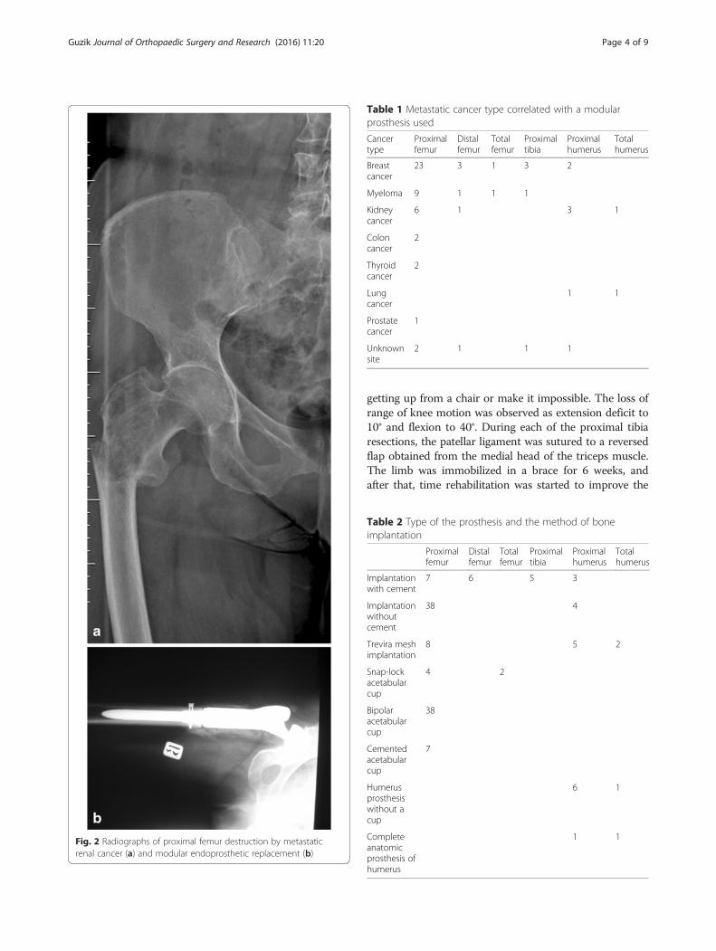
Table 1 Metastatic cancer type correlated with a modularprosthesis used
Cancertype
Proximalfemur
Distalfemur
Totalfemur
Proximaltibia
Proximalhumerus
Totalhumerus
Breastcancer
23 3 1 3 2
Myeloma 9 1 1 1
Kidneycancer
6 1 3 1
Coloncancer
2
Thyroidcancer
2
Lungcancer
1 1
Prostatecancer
1
Unknownsite
2 1 1 1
Fig. 2 Radiographs of proximal femur destruction by metastaticrenal cancer (a) and modular endoprosthetic replacement (b)
Guzik Journal of Orthopaedic Surgery and Research (2016) 11:20 Page 4 of 9
getting up from a chair or make it impossible. The loss ofrange of knee motion was observed as extension deficit to10° and flexion to 40°. During each of the proximal tibiaresections, the patellar ligament was sutured to a reversedflap obtained from the medial head of the triceps muscle.The limb was immobilized in a brace for 6 weeks, andafter that, time rehabilitation was started to improve the
Table 2 Type of the prosthesis and the method of boneimplantation
Proximalfemur
Distalfemur
Totalfemur
Proximaltibia
Proximalhumerus
Totalhumerus
Implantationwith cement
7 6 5 3
Implantationwithoutcement
38 4
Trevira meshimplantation
8 5 2
Snap-lockacetabularcup
4 2
Bipolaracetabularcup
38
Cementedacetabularcup
7
Humerusprosthesiswithout acup
6 1
Completeanatomicprosthesis ofhumerus
1 1
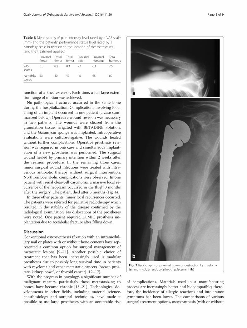
Table 3 Mean scores of pain intensity level rated by a VAS scale(mm) and the patients’ performance status level rated by aKarnofsky scale in relation to the location of the metastases(and the treatment applied)
Proximalfemur
Distalfemur
Totalfemur
Proximaltibia
Proximalhumerus
Totalhumerus
VASscores
6.8 8.2 8.3 7.1 6.1 7.5
Karnofskyscores
53 40 40 45 65 60
Fig. 3 Radiographs of proximal humerus destruction by myeloma(a) and modular endoprosthetic replacement (b)
Guzik Journal of Orthopaedic Surgery and Research (2016) 11:20 Page 5 of 9
function of a knee extensor. Each time, a full knee exten-sion range of motion was achieved.No pathological fractures occurred in the same bone
during the hospitalization. Complications involving loos-ening of an implant occurred in one patient (a case sum-marized below). Operative wound revision was necessaryin two patients. The wounds were cleared from thegranulation tissue, irrigated with BETADINE Solution,and the Garamycin sponge was implanted. Intraoperativeevaluations were culture-negative. The wounds healedwithout further complications. Operative prosthesis revi-sion was required in one case and simultaneous implant-ation of a new prosthesis was performed. The surgicalwound healed by primary intention within 2 weeks afterthe revision procedure. In the remaining three cases,minor surgical wound infections were treated with intra-venous antibiotic therapy without surgical intervention.No thromboembolic complications were observed. In onepatient with renal clear-cell carcinoma, a massive local re-currence of the neoplasm occurred in the thigh 3 monthsafter the surgery. The patient died after 5 months (Fig. 4).In three other patients, minor local recurrences occurred.
The patients were referred for palliative radiotherapy whichresulted in the stability of the disease confirmed by theradiological examination. No dislocations of the prostheseswere noted. One patient required LUMIC prosthesis im-plantation due to acetabular fracture after falling down.
DiscussionConventional osteosynthesis (fixation with an intramedul-lary nail or plates with or without bone cement) have rep-resented a common option for surgical management ofmetastatic lesions [9–11]. Another possible choice oftreatment that has been increasingly used is modularprostheses due to possibly long survival time in patientswith myeloma and other metastatic cancers (breast, pros-tate, kidney, bowel, or thyroid cancer) [12–17].With the progress in oncology, a significant number of
malignant cancers, particularly those metastasizing tobones, have become chronic [18–21]. Technological de-velopments in other fields, including material science,anesthesiology and surgical techniques, have made itpossible to use large prostheses with an acceptable risk
of complications. Materials used in a manufacturingprocess are increasingly better and biocompatible; there-fore, the incidence of allergic reactions and intolerancesymptoms has been lower. The comparisons of varioussurgical treatment options, osteosynthesis (with or without
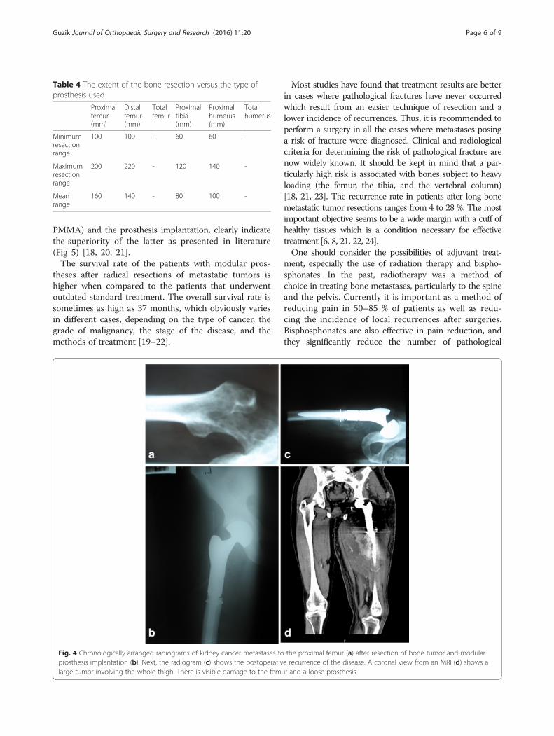
Table 4 The extent of the bone resection versus the type ofprosthesis used
Proximalfemur(mm)
Distalfemur(mm)
Totalfemur
Proximaltibia(mm)
Proximalhumerus(mm)
Totalhumerus
Minimumresectionrange
100 100 - 60 60 -
Maximumresectionrange
200 220 - 120 140 -
Meanrange
160 140 - 80 100 -
Guzik Journal of Orthopaedic Surgery and Research (2016) 11:20 Page 6 of 9
PMMA) and the prosthesis implantation, clearly indicatethe superiority of the latter as presented in literature(Fig 5) [18, 20, 21].The survival rate of the patients with modular pros-
theses after radical resections of metastatic tumors ishigher when compared to the patients that underwentoutdated standard treatment. The overall survival rate issometimes as high as 37 months, which obviously variesin different cases, depending on the type of cancer, thegrade of malignancy, the stage of the disease, and themethods of treatment [19–22].
Fig. 4 Chronologically arranged radiograms of kidney cancer metastases toprosthesis implantation (b). Next, the radiogram (c) shows the postoperativlarge tumor involving the whole thigh. There is visible damage to the femu
Most studies have found that treatment results are betterin cases where pathological fractures have never occurredwhich result from an easier technique of resection and alower incidence of recurrences. Thus, it is recommended toperform a surgery in all the cases where metastases posinga risk of fracture were diagnosed. Clinical and radiologicalcriteria for determining the risk of pathological fracture arenow widely known. It should be kept in mind that a par-ticularly high risk is associated with bones subject to heavyloading (the femur, the tibia, and the vertebral column)[18, 21, 23]. The recurrence rate in patients after long-bonemetastatic tumor resections ranges from 4 to 28 %. The mostimportant objective seems to be a wide margin with a cuff ofhealthy tissues which is a condition necessary for effectivetreatment [6, 8, 21, 22, 24].One should consider the possibilities of adjuvant treat-
ment, especially the use of radiation therapy and bispho-sphonates. In the past, radiotherapy was a method ofchoice in treating bone metastases, particularly to the spineand the pelvis. Currently it is important as a method ofreducing pain in 50–85 % of patients as well as redu-cing the incidence of local recurrences after surgeries.Bisphosphonates are also effective in pain reduction, andthey significantly reduce the number of pathological
the proximal femur (a) after resection of bone tumor and modulare recurrence of the disease. A coronal view from an MRI (d) shows ar and a loose prosthesis

Fig. 5 Failed fixation (gamma nail with PMMA) of a proximalfemoral fracture due to breast cancer metastasis (a) and radiographsafter modular endoprosthesis replacement (b)
Fig. 6 Radiographs of proximal humerus destruction by metastatic breast c
Guzik Journal of Orthopaedic Surgery and Research (2016) 11:20 Page 7 of 9
fractures by 25–50 %. They are particularly often used inthe treatment of metastatic breast cancer, prostate cancer,and multiple myeloma) [4, 6, 7, 25].In the case of the metastases of kidney cancer, thyroid
cancer, and myeloma, a trans-arterial embolization (TAE)can be performed. This procedure limits vascularization ofthe tumor, which results in a twofold or even threefoldreduction in bleeding during the surgery, and the oper-ation time is reduced by about 25 %. Many authors haveconfirmed the effectiveness of this treatment method inreducing pain and number of local recurrences after resec-tion of the tumor. Reduced intraoperative bleeding allowsfor more precise preparation of tissues and resection oflesions. According to some authors, embolization in-creases the sensitivity of tumor cells to chemotherapy andradiation therapy. We have not performed preoperativeembolization of tumors localized in the extremities at ourdepartment. This procedure has been performed exclu-sively when the tumor was localized in the pelvis and thespine [26–28].The indications for amputation due to cancer metastases
are extremely rare. It is performed in the case of largemetastatic tumors infiltrating the vascular and nervetrunks or skin when limb-salvage treatment is not pos-sible. An indication for amputation may be extensiveinflammation of bone and soft tissues that is localizedwithin the site of a former prosthetic implantation andwhich is impossible to treat [2, 3, 18].The present study clearly indicates that most metasta-
ses occur in the proximal area of the femur. Metastasesin other sites are less frequent; very rarely do they occurin the area below the elbow or knee joint. Breast canceris the most metastatic (Fig 6). Prostate cancer
ancer (a) and after modular endoprosthetic replacement (b)

Guzik Journal of Orthopaedic Surgery and Research (2016) 11:20 Page 8 of 9
metastases, frequent as they are, rarely necessitate surgi-cal treatment because of a relatively low risk of afracture.Most common complications that may follow modular
alloplasty are surgical wound infections [29, 30]. Patientsconstitute a group at the highest risk of infectious andthromboembolic complications because the surgeries aremost often urgent, therefore, MRSA screening or otherpathogen detection tests are rarely carried out. Thereare no clear recommendations as to the routine localantibiotic therapy in the case of primary resection allo-plasty. A rate of infectious complications ranges between1.2 and 19.5 %. Preoperative radiotherapy is seen as oneof the major risk factors for developing infections. Majorrisk factors include: decreased immunity as a result ofneoplastic disease and chemotherapy, wide surgicalapproach with significant blood loss, and the size ofmetal implants. Worse still, the patients are usually eld-erly and with various general health problems.Other potential complications include dislocation or
loosening of the implants and periprosthetic fractures.Revision procedures are required in 3–17 % of patients[31, 32].The procedures concerning preparation and implant-
ation of GMRS and MUTARS prostheses differ. TheMUTARS prostheses allow for a smooth rotation of theprosthesis stem. What is more, surgical management ofthe bone marrow canal is carried out not by reaming,but by rasping. The stems are bent, which minimizes therisk of damaging the cortical bone while driving them.The prostheses have a golden and silver coating, whichreduces the incidence of infectious complications andallergic reactions.Functional results of modular alloplasty are satisfac-
tory, especially in patients after proximal and distalfemur resections. Decreased gluteal muscles functionwas not a big problem to patients. Most of the patientswalk efficiently without crutches. A slight limb lengthdiscrepancy was not noticed by the patients and didnot affect their walking. The quality of life of the patientsafter metastasis resections and implantations of modu-lar prostheses improved significantly. The VAS andKarnofsky scales showed clearly reduced pain and im-proved functioning.
Conclusions
1. Modular tumor endoprosthesis may be an option insurgical treatment of long-bone metastases providingfast pain relief and early mobilization.
2. Radical metastatic tumor resection is a conditionnecessary for a good treatment outcome. It preventslocal recurrences and damage to implant or itsloosening.
3. Proximal humerus resections result in a reducedshoulder mobility and weakening of muscle strengthwhich impair normal limb functions.
4. The incidence rate of infections in patients aftermodular prostheses implantations varies. Effortsshould be directed at preventing infectiouscomplications because they are very difficult to treat.
AbbreviationsMRSA: methicillin-resistant Staphylococcus aureus; PMMA: polymethylmethacrylate; TAE: trans-arterial embolization.
Competing interestsThe author declares that he has no competing interest.
Author’s contributionsThe author read and approved the final manuscript.
Author’s informationI am the Head of the Orthopedic Oncology Department in Brzozów Hospital,Poland. I am an orthopedic surgeon and author of the book Spinemetastasis-diagnosis and treatment and about 30 publications from this field.
AcknowledgementsThis work is entirely my doing. I am the only author of this work.
Received: 4 September 2015 Accepted: 24 January 2016
References1. Hage WD, Aboulafia AJ, Aboulafia DM. Incidence, location and diagnostic
evaluation of metastatic bone disease. Orthop Clin North Am. 2000;31:515–28.2. Sherry HS, Levy RN, Siffert RS. Metastatic disease of bone in orthopedic
surgery. Clin Orthop. 1982;169:44–52.3. Riccio AI, Wodajo FM, Malawer M. Metastatic carcinoma of the long bones.
Am Fam Physician. 2007;76:1489–94.4. Coleman RE. Clinical features of metastatic bone disease and risk of skeletal
morbidity. Clin Cancer Res. 2006;12:6243–9.5. Utzschneider S, Weber P, Fottner A, Wegener B, Jansson V, Dürr HR.
Prognosis-adapted surgical management of bone metastases. Orthopade.2009;38(308):310–2. 314–305.
6. Hechmati G, Cure S, Gouépo A, Hoefeler H, Lorusso V, Lüftner D, et al. Costof skeletal-related events in European patients with solid tumours and bonemetastases: data from a prospective multinational observational study.J Med Econ. 2013;16(5):691–700.
7. Singh G, Lim CT, Jonathan TJ, Nathan SS. Evaluation of the role and cost-effectiveness of end-of-life orthopaedic interventions in cancer patients withskeletal metastases to the hip. J Palliat Care. 2013;29(2):83–90.
8. Ratasvuori M, Wedin R, Hansen BH, Keller J, Trovik C, Zaikova O, et al.Prognostic role of en-bloc resection and late onset of bone metastasis inpatients with bone-seeking carcinomas of the kidney, breast, lung, andprostate: SSG study on 672 operated skeletal metastases. J Surg Oncol.2014;110(4):360–5.
9. Sim FH, Daugherty TW, Ivins JC. The adjunctive use of methylmethacrylatein fixation of pathological fractures. J Bone Joint Surg Am. 1974;56-A:40–8.
10. Bauer HC, Wedin R. Survival after surgery for spinal and extremity metastases.Prognostication in 241 patients. Acta Orthop Scand 1995; 66:143–146.
11. Fottner A, Szalantzy M, Wirthmann L, Stähler M, Baur-Melnyk A, Jansson V,et al. Bone metastases from renal cell carcinoma: patient survival aftersurgical treatment. BMC Musculoskelet Disord 2010;11:145.
12. Murray JA, Parrish FF. Surgical management of secondary neoplasticfractures about the hip. Orthop Clin North Am 1974;5:887-901.
13. Capanna R, Morris H G, Campanacci D, Del Ben M,Campanacci M. Modularuncemented prosthetic reconstruction after resection of tumor of distalfemur. J Bone Joint Surg BR 1994;76:178-86.
14. Plotz W, Rechl H, Burgkart R, Messmer C, Schelter R, Hipp E, et al. Limb salvagewith tumor endoprostheses for malignant tumors of the knee. Clin OrthopRelat Res 2002;405:207–215.

Guzik Journal of Orthopaedic Surgery and Research (2016) 11:20 Page 9 of 9
15. Park DH, Jaiswal PK, Al-Hakim W, Aston WJ, Pollock RC, Skinner JA, et al. Theuse of massive endoprostheses for treatment of bone metastases. Sarcoma2007;62151.
16. Hwang N, Nandra R, Grimer RJ, Carter SR, Tillman RM, Abudu A, et al.Massive endoprosthetic replacement for bone metastases resulting fromrenal cell carcinoma: factors influencing patient survival. Eur J Surg Oncol2014;40(4):423-434.
17. Bickels J, Meller I, Henshaw RM, Malawer MM. Reconstruction of hip stability afterproximal and total femur resections. Clin Orthop Relat Res 2000;375:218–230.
18. Wedin R, Bauer HC, Wersall P. Failures after operation for skeletal metastaticlesions of long bones. Clin Orthop Relat Res 1999; 358:128–139.
19. Hansen BH, Keller J, Laitinen M, Berg P, Skjeldal S, Trovik C, et al. TheScandinavian sarcoma group skeletal metastasis register. Survival after surgeryfor bone metastases in the pelvis and extremities. Acta Orthop Scand Suppl2004;75:11–15.
20. Harvey N, Ahlmann ER, Allison DC, Wang L, Menendez LR. Endoprostheseslast longer than intramedullary devices in proximal femur metastases. ClinOrthop Relat Res 2012;470:684–691.
21. Wedin R, Bauer HC. Surgical treatment of skeletal metastatic lesions of theproximal femur: endoprosthesis or reconstruction nail? J Bone Joint Surg2005;87:1653–1657.
22. Menendez LR, Ahlmann ER, Kermani C, Gotha H. Endoprostheticreconstruction for neoplasms of the proximal femur. Clin Orthop Relat Res2006;450:46–51.
23. Pugh J, Sherry HS, Futterman B, Frankel VH. Biomechanics of pathologicfractures. Clin Orthop 1982;169:109-14.
24. Ashford RU, Hanna SA, Park DH, Pollock RC, Skinner JA, Briggs TW, et al.Proximal femoral replacements for metastatic bone disease: financialimplications for sarcoma units. Int Orthop 2010;34(5):709–713.
25. Pergolizzi S, Pontoriero A, Delia P, Santacaterina A. External beam irradiationin the palliation of bone metastases: a practice analysis among SicilianDepartments of Radiation Oncology Tumori. 2004;90(1):86-90.
26. Gupta G, Gamanagatti S. Preoperative transarterial embolisation in bonetumors. World J Radiol 2012;4(5):186-192.
27. Pazionis TJ, Papanastassiou ID, Maybody M, Healey JH. Embolistation ofhypervascular bone metastases reduces intraoperative blood loss: a casecontrol study. Clin Orthop Relat Res 2014;472(10):3179-3187.
28. Owen RJT. Embolisation of Musculoskeletal Bone Tumors. Semin Intervent Radiol2010;27(2):111-123.
29. Wang J, Temple HT, Pitcher JD, Mounasamy V, Malinin TI, Scully SP. Salvage offailed massive allograft reconstruction with endoprosthesis. Clin Orthop Relat Res2006;443:296-301.
30. Sarahrudi K, Hora K, Heinz T, Millington S, Vecsei V. Treatment results ofpathological fractures of the long bones: a retrospective analysis of 88patients. Int Orthop 2006;30:519–524.
31. Unwin PS, Cannon SR. Grimer RJ, Kemp HB, Sneath RS, Walker PS. Asepticloosening in cemented custom-made prosthetic replacements for bonetumours of the lower limb. JBJS 1996;78B:5-13.
32. Capanna R, Ruggieri P, Biagini R, Ferraro A, DeCristofaro R, McDonald D,et al. The effects of quadriceps excision on functional results after distalfemoral resection and prosthetic replacement of bone tumors. Clin Orthop1991;267:186-96.
• We accept pre-submission inquiries
• Our selector tool helps you to find the most relevant journal
• We provide round the clock customer support
• Convenient online submission
• Thorough peer review
• Inclusion in PubMed and all major indexing services
• Maximum visibility for your research
Submit your manuscript atwww.biomedcentral.com/submit
Submit your next manuscript to BioMed Central and we will help you at every step: