review article the role of nrf2-mediated pathway in...
TRANSCRIPT
Review ArticleThe Role of Nrf2-Mediated Pathway in CardiacRemodeling and Heart Failure
Shanshan Zhou, Wanqing Sun, Zhiguo Zhang, and Yang Zheng
The Cardiovascular Center, The First Hospital of Jilin University, 71 Xinmin Street, Changchun 130021, China
Correspondence should be addressed to Yang Zheng; [email protected]
Received 26 January 2014; Revised 12 April 2014; Accepted 30 April 2014; Published 1 July 2014
Academic Editor: MaheshThirunavukkarasu
Copyright © 2014 Shanshan Zhou et al.This is an open access article distributed under the Creative Commons Attribution License,which permits unrestricted use, distribution, and reproduction in any medium, provided the original work is properly cited.
Heart failure (HF) is frequently the consequence of sustained, abnormal neurohormonal, and mechanical stress and remains aleading cause of death worldwide. The key pathophysiological process leading to HF is cardiac remodeling, a term referring tomaladaptation to cardiac stress at the molecular, cellular, tissue, and organ levels. HF and many of the conditions that predisposeone to HF are associated with oxidative stress. Increased generation of reactive oxygen species (ROS) in the heart can directly leadto increased necrosis and apoptosis of cardiomyocytes which subsequently induce cardiac remodeling and dysfunction. Nuclearfactor-erythroid-2- (NF-E2-) related factor 2 (Nrf2) is a transcription factor that controls the basal and inducible expressionof a battery of antioxidant genes and other cytoprotective phase II detoxifying enzymes that are ubiquitously expressed in thecardiovascular system. Emerging evidence has revealed that Nrf2 and its target genes are critical regulators of cardiovascularhomeostasis via the suppression of oxidative stress, which is the key player in the development and progression of HF.The purposeof this review is to summarize evidence that activation ofNrf2 enhances endogenous antioxidant defenses and counteracts oxidativestress-associated cardiac remodeling and HF.
1. Introduction
Despite recent advances in treatment, the morbidity, mortal-ity, and economic burden of heart failure (HF) still remainvery high. Hypertension, ischemia, diabetes, and some anti-cancer drugs used in the clinic are common causes of cardiacremodeling andHF. Cardiac remodeling, a term that refers tocardiac maladaptation at the molecular, cellular, tissue, andorgan levels, is the key pathophysiological process leading toHF. It has been well established that oxidative stress is a majorcause of HF [1–8].
Free radicals and other reactive small molecules haveemerged as important regulators of many physiological andpathological processes [9]. Reactive oxygen species (ROS)and reactive nitrogen species (RNS) serve as signalingmessengers to mediate various biological responses [10,11], including numerous cardiovascular diseases, such asHF, coronary heart disease, and cardiac arrhythmias [12].Whether the effects of ROS/RNS are beneficial or harmfuldepends on the site, type, and amount of ROS/RNS produc-tion and the activity of the organism’s antioxidant defense
system [13]. As a rule, heart and cardiovascular diseases arecharacterized byROSoverproduction, whereas the formationofmajor RNS, nitric oxide (a free radical) and peroxynitrite (adiamagnetic molecule), can decrease or increase dependingon the nature of the heart injury [12]. ROS include superoxideanion (∙O2−), hydrogen peroxide (H
2O2), hydroxyl radical
(∙OH), and hypochlorite (OCl∙−), which impair cardiac func-tion [14] and increase susceptibility to cardiac arrhythmia[15] through direct toxic effect, resulting in increased necrosisand apoptosis of cardiomyocytes [16]. Critical componentsof the cellular antioxidative defense mechanisms are theROS scavengers, phase II detoxification enzymes, and otherdetoxification proteins which contain antioxidant responseelements (AREs) in their promoter regions. Amajor regulatorof the AREs is the highly conserved transcription factor,nuclear factor-erythroid-2- (NF-E2-) related factor 2 (Nrf2).Many of the Nrf2-regulated enzymes are essential in thepathogenesis of cardiovascular diseases [17] and are stronglyassociated with HF; in addition, they serve as sensitiveand specific markers to reflect the ventricular function inHF patients [18]. Nrf2 also can prolong graft survival and
Hindawi Publishing CorporationOxidative Medicine and Cellular LongevityVolume 2014, Article ID 260429, 16 pageshttp://dx.doi.org/10.1155/2014/260429
2 Oxidative Medicine and Cellular Longevity
modulate innate and adaptive immune responses after hearttransplantation [19]. Emerging evidence has revealed thatthe Nrf2/ARE signaling pathway plays an important role inpreventing oxidative cardiac cell injury in vitro [20, 21] aswell as protecting the heart frommaladaptive remodeling andcardiac dysfunction [7, 22–27].
The main purpose of this review is to discuss the currentevidence for the cardioprotective role of Nrf2 and its targetgenes in the development of cardiac remodeling and HFcaused by hypertension, ischemia, diabetes, and anticancerdrugs.
2. ROS and Cardiac Remodeling
ROS refer to a group of small reactive molecules thatinclude O2⋅−, H
2O2, OH⋅−, and OCl⋅−. These molecules
react avidly with other molecules such as cellular lipids,proteins, and nucleic acids [28]. The fine balance betweenROS generated during normal physiological processes andendogenous antioxidants presented in the body is essentialfor redox homeostasis. Increased production of ROS orimpaired endogenous antioxidant defense of the body thatleads to oxidative stress may cause adverse effects due toirreversible modification of macromolecules, membranes,proteins, and DNA [29]. Cardiac remodeling is an adaptive,regulatory process of cardiomyocytes that occurs over timein order to maintain homeostasis against external stress[30]. Progression of cardiac hypertrophy, cardiomyocyteapoptosis, and interstitial collagen deposition into systolicdysfunction has been reported in numerous clinical andanimal studies [31–35]. ROS can activate specific pathwaysleading to adaptive or maladaptive cardiac remodeling pro-cesses [28]. In experimental studies, oxidative stress has beenidentified as one of the molecular mechanisms and the keyplayers involved in the development of cardiac hypertrophy[36]. In addition, antioxidants have been found to preventthe development of cardiac hypertrophy [37]. Various studieshave suggested that oxidative stress is also an importantregulator of profibrotic processes in themyocardium [34, 35].NADPH oxidase-dependent ROS production contributes tothe development of left ventricular (LV) interstitial fibro-sis, reduction of ROS generation, and restoration of theredox balance; thus, it may be important in preventing ortreating myocardial fibrosis in HF [38]. Programmed celldeath (apoptosis) of cardiomyocytes has been identified asan essential process in the progression to HF [39]. ROSand the resulting oxidative stress also play a pivotal role inthis pathological process. Antioxidants and thiol reductants,such as N-acetylcysteine, and overexpression of manganesesuperoxide dismutase (MnSOD) can block or delay apoptosis[40]. Taken together, ROS play an important role in thedevelopment of cardiac remodeling and HF. The findings ofthe majority of the studies suggest that antioxidants may bepossible therapeutic candidates against cardiac remodeling.
3. The Nrf2-ARE Signaling Pathway
Nrf2 is the master regulator of oxidative stress signaling. It isa member of the cap-n-collar (CNC) family of transcription
factors, which include Nrf1-3 and Bach1-2 [41–43]. Thesegenes encode transcription factors that belong to the CNC-type of the basic region leucine zipper factor family (CNC-bZIP). CNC-bZIP factors are characterized by a highlyconserved 43-amino acid homology region that lies in theimmediate N-terminus of the basic DNA-binding domainand is referred to as the “CNC” domain after the Drosophilacap-n-collar protein. There is evidence indicating that CNC-bZIP factors function as obligate heterodimers by formingdimers with small Maf proteins (Maf G, Maf K, and Maf F)for DNA binding [44–46].
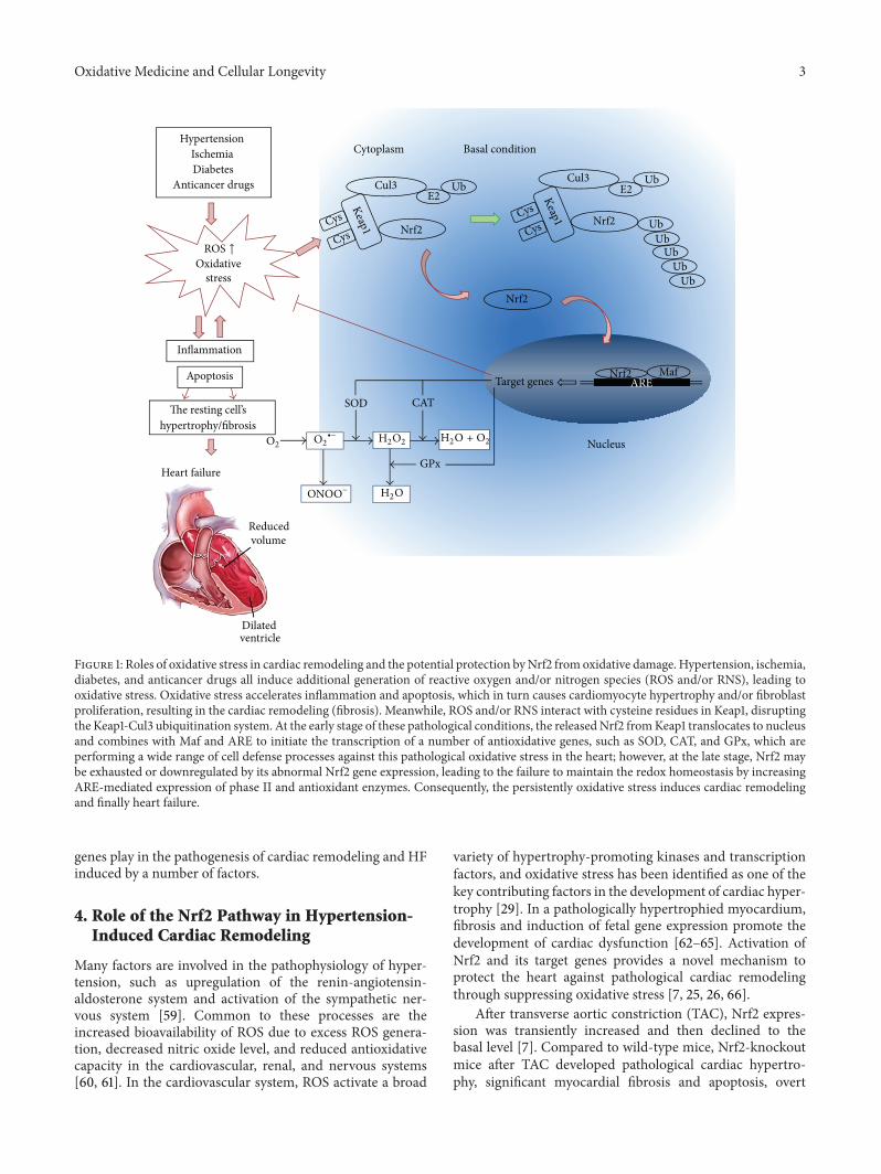
Under normal conditions, Nrf2 is kept in the cytoplasmby Kelch-like-ECH-associated protein 1 (Keap1) and Cullin3 [47]. Cullin 3 ubiquitinates its substrate, Nrf2; and Keap1serves as a substrate adaptor, which facilitates the ubiqui-tination of Nrf2 by Cullin 3. As a result, Nrf2 has a shorthalf-life that lasts only 20min under normal conditions[48]. As illustrated in Figure 1, oxidative stress destroyscritical cysteine residues in Keap1, disrupting the Keap1-Cul3ubiquitination system. If Nrf2 is not ubiquitinated, it buildsup in the cytoplasm [49] and is translocated into the nucleus.In the nucleus, Nrf2 combines with a small protein calledMafto form a heterodimer, and, by binding to the ARE in theupstream promoter region, it initiates the transcription of anumber of antioxidative genes, including heme oxygenase-1 (HO-1), NAD(P)H dehydrogenase (quinone 1) (NQO1),superoxide dismutases (SODs), catalase (CAT), glutathione-S-transferase (GST), 𝛾-glutamylcysteine synthase (𝛾-GCS),and glutathione peroxidase (GPx) [50, 51] (Figure 1). Theseare the first line of defensemechanism against ROS-mediatedcardiac injury. HO-1 is a rapidly inducible cytoprotective pro-tein that degrades heme to biliverdin, ferrous iron, and car-bon monoxide (CO) [52]. HO-1 mitigates cellular injury byexerting antioxidant, antiapoptotic, and anti-inflammatoryeffects [52, 53]. SOD catalyzes the dismutation of O2− intoH2O2and O
2. H2O2is a product of SOD activity and is
handled by GPx and CAT. Three SOD isozymes have beenidentified, including copper/zinc-containing SOD (CuZn-SOD, also SOD1), which is primarily cytosolic in location,Mn-SOD (also SOD2), and extracellular SOD (EC-SOD,also SOD3) [54]. The GPx/glutathione (GSH) system isimportant in low-level oxidative stress. It scavenges hydroxylradicals and singlet oxygen directly, detoxifying H
2O2and
lipid peroxides by the catalytic action of GPx [55]. GPxnot only scavenges H
2O2but also prevents the formation
of other more toxic radicals, such as ⋅OH [56]. CAT ismainly located in cellular peroxisomes and, to some extent,in the cytosol and catalyzes the reaction of H
2O2to water
and molecular oxygen [57]. Overexpression of mitochon-drial CAT has shown protection against cytotoxic drugs[58].
Through inducing the expression of this battery of genes,Nrf2 is able to augment a wide range of cell defense processes,thereby enhancing the overall capacity of cells to detoxifypotentially harmful substances. As such, the Nrf2-Keap1pathway is generally considered to be amajor cellular defensepathway. In addition, recent experimental and clinical studieshave shown the important roles that Nrf2 and its downstream
Oxidative Medicine and Cellular Longevity 3
Oxidative stress
Cul3
Nrf2
E2Ub
Cul3
Nrf2
E2Ub
UbUb
UbUb
UbNrf2
HypertensionIschemiaDiabetes
Anticancer drugs
Nucleus
Cytoplasm Basal condition
SOD CAT
GPx
Nrf2 MafTarget genes
The resting cell’s hypertrophy/fibrosis
Inflammation
Apoptosis
ROS ↑
Heart failure
Reducedvolume
Dilatedventricle
ARE
O2 O2∙− H2O2 H2O + O2
H2OONOO−
Cys
Cys
Keap1
Keap1Cys
Cys
Figure 1: Roles of oxidative stress in cardiac remodeling and the potential protection byNrf2 fromoxidative damage. Hypertension, ischemia,diabetes, and anticancer drugs all induce additional generation of reactive oxygen and/or nitrogen species (ROS and/or RNS), leading tooxidative stress. Oxidative stress accelerates inflammation and apoptosis, which in turn causes cardiomyocyte hypertrophy and/or fibroblastproliferation, resulting in the cardiac remodeling (fibrosis). Meanwhile, ROS and/or RNS interact with cysteine residues in Keap1, disruptingthe Keap1-Cul3 ubiquitination system. At the early stage of these pathological conditions, the releasedNrf2 fromKeap1 translocates to nucleusand combines with Maf and ARE to initiate the transcription of a number of antioxidative genes, such as SOD, CAT, and GPx, which areperforming a wide range of cell defense processes against this pathological oxidative stress in the heart; however, at the late stage, Nrf2 maybe exhausted or downregulated by its abnormal Nrf2 gene expression, leading to the failure to maintain the redox homeostasis by increasingARE-mediated expression of phase II and antioxidant enzymes. Consequently, the persistently oxidative stress induces cardiac remodelingand finally heart failure.
genes play in the pathogenesis of cardiac remodeling and HFinduced by a number of factors.
4. Role of the Nrf2 Pathway in Hypertension-Induced Cardiac Remodeling
Many factors are involved in the pathophysiology of hyper-tension, such as upregulation of the renin-angiotensin-aldosterone system and activation of the sympathetic ner-vous system [59]. Common to these processes are theincreased bioavailability of ROS due to excess ROS genera-tion, decreased nitric oxide level, and reduced antioxidativecapacity in the cardiovascular, renal, and nervous systems[60, 61]. In the cardiovascular system, ROS activate a broad
variety of hypertrophy-promoting kinases and transcriptionfactors, and oxidative stress has been identified as one of thekey contributing factors in the development of cardiac hyper-trophy [29]. In a pathologically hypertrophied myocardium,fibrosis and induction of fetal gene expression promote thedevelopment of cardiac dysfunction [62–65]. Activation ofNrf2 and its target genes provides a novel mechanism toprotect the heart against pathological cardiac remodelingthrough suppressing oxidative stress [7, 25, 26, 66].
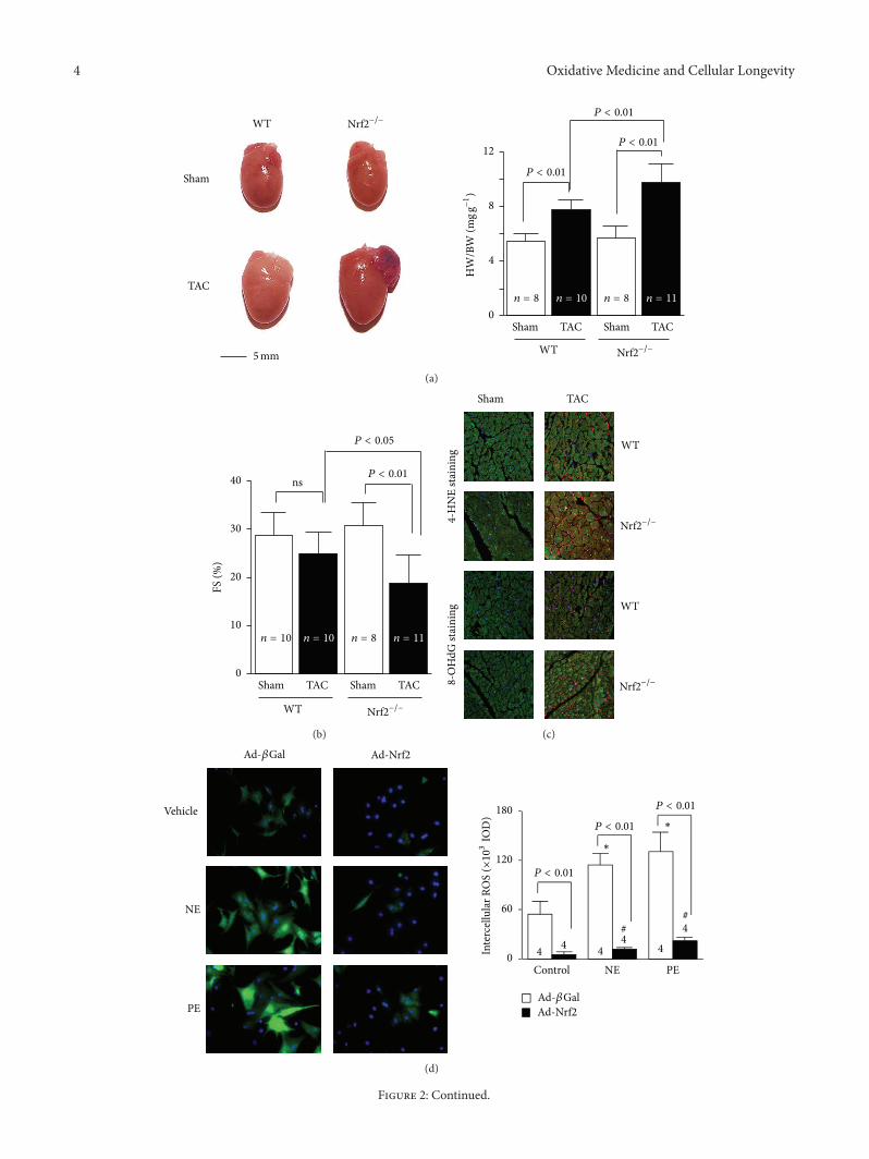
After transverse aortic constriction (TAC), Nrf2 expres-sion was transiently increased and then declined to thebasal level [7]. Compared to wild-type mice, Nrf2-knockoutmice after TAC developed pathological cardiac hypertro-phy, significant myocardial fibrosis and apoptosis, overt
4 Oxidative Medicine and Cellular Longevity
WT
Sham
TAC
0
4
8
12
Sham TAC Sham TAC
WT5mm
Nrf2−/−
Nrf2−/−
P < 0.01
P < 0.01
P < 0.01
n = 8n = 8 n = 10 n = 11
HW
/BW
(mg g
−1)
(a)
FS (%
)
0
10
20
30
40
Sham TAC Sham TAC
WT
ns
Nrf2−/−
n = 8n = 10n = 10 n = 11
P < 0.01
P < 0.05
(b)
4-H
NE
stai
ning
8-O
HdG
stai
ning
WT
WT
Sham TAC
Nrf2−/−
Nrf2−/−
(c)
Vehicle
NE
PE
0
60
120
180
Control NE PE4
444
4
4
##
Ad-Nrf2
P < 0.01
P < 0.01
P < 0.01
∗
∗
Ad-𝛽Gal
Ad-Nrf2Ad-𝛽Gal
Inte
rcel
lula
r RO
S(×10
3IO
D)
(d)
Figure 2: Continued.
Oxidative Medicine and Cellular Longevity 5
0
50
100
150
200
250
4 4 44 4 4
Ad-scramble shRNA
Ad-scramble shRNA
Ad-Nrf2 shRNA
Ad-Nrf2 shRNA
Control NE PE
Vehicle
NE
PE
P < 0.01P < 0.01P < 0.01
Inte
rcel
lula
r RO
S(×10
3IO
D)
∗∗
(e)
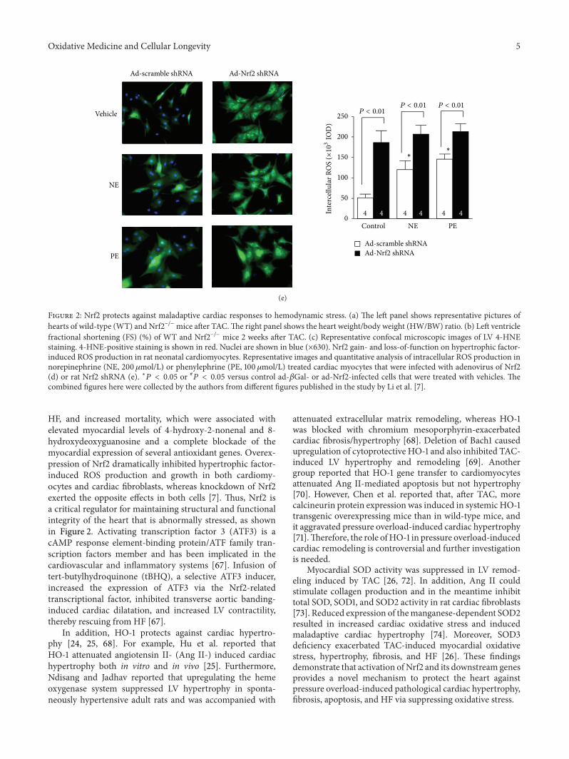
Figure 2: Nrf2 protects against maladaptive cardiac responses to hemodynamic stress. (a) The left panel shows representative pictures ofhearts of wild-type (WT) and Nrf2−/− mice after TAC.The right panel shows the heart weight/body weight (HW/BW) ratio. (b) Left ventriclefractional shortening (FS) (%) of WT and Nrf2−/− mice 2 weeks after TAC. (c) Representative confocal microscopic images of LV 4-HNEstaining. 4-HNE-positive staining is shown in red. Nuclei are shown in blue (×630). Nrf2 gain- and loss-of-function on hypertrophic factor-induced ROS production in rat neonatal cardiomyocytes. Representative images and quantitative analysis of intracellular ROS production innorepinephrine (NE, 200 𝜇mol/L) or phenylephrine (PE, 100𝜇mol/L) treated cardiac myocytes that were infected with adenovirus of Nrf2(d) or rat Nrf2 shRNA (e). ∗𝑃 < 0.05 or #
𝑃 < 0.05 versus control ad-𝛽Gal- or ad-Nrf2-infected cells that were treated with vehicles. Thecombined figures here were collected by the authors from different figures published in the study by Li et al. [7].
HF, and increased mortality, which were associated withelevated myocardial levels of 4-hydroxy-2-nonenal and 8-hydroxydeoxyguanosine and a complete blockade of themyocardial expression of several antioxidant genes. Overex-pression of Nrf2 dramatically inhibited hypertrophic factor-induced ROS production and growth in both cardiomy-ocytes and cardiac fibroblasts, whereas knockdown of Nrf2exerted the opposite effects in both cells [7]. Thus, Nrf2 isa critical regulator for maintaining structural and functionalintegrity of the heart that is abnormally stressed, as shownin Figure 2. Activating transcription factor 3 (ATF3) is acAMP response element-binding protein/ATF family tran-scription factors member and has been implicated in thecardiovascular and inflammatory systems [67]. Infusion oftert-butylhydroquinone (tBHQ), a selective ATF3 inducer,increased the expression of ATF3 via the Nrf2-relatedtranscriptional factor, inhibited transverse aortic banding-induced cardiac dilatation, and increased LV contractility,thereby rescuing from HF [67].
In addition, HO-1 protects against cardiac hypertro-phy [24, 25, 68]. For example, Hu et al. reported thatHO-1 attenuated angiotensin II- (Ang II-) induced cardiachypertrophy both in vitro and in vivo [25]. Furthermore,Ndisang and Jadhav reported that upregulating the hemeoxygenase system suppressed LV hypertrophy in sponta-neously hypertensive adult rats and was accompanied with
attenuated extracellular matrix remodeling, whereas HO-1was blocked with chromium mesoporphyrin-exacerbatedcardiac fibrosis/hypertrophy [68]. Deletion of Bach1 causedupregulation of cytoprotective HO-1 and also inhibited TAC-induced LV hypertrophy and remodeling [69]. Anothergroup reported that HO-1 gene transfer to cardiomyocytesattenuated Ang II-mediated apoptosis but not hypertrophy[70]. However, Chen et al. reported that, after TAC, morecalcineurin protein expression was induced in systemic HO-1transgenic overexpressing mice than in wild-type mice, andit aggravated pressure overload-induced cardiac hypertrophy[71].Therefore, the role ofHO-1 in pressure overload-inducedcardiac remodeling is controversial and further investigationis needed.
Myocardial SOD activity was suppressed in LV remod-eling induced by TAC [26, 72]. In addition, Ang II couldstimulate collagen production and in the meantime inhibittotal SOD, SOD1, and SOD2 activity in rat cardiac fibroblasts[73]. Reduced expression of themanganese-dependent SOD2resulted in increased cardiac oxidative stress and inducedmaladaptive cardiac hypertrophy [74]. Moreover, SOD3deficiency exacerbated TAC-induced myocardial oxidativestress, hypertrophy, fibrosis, and HF [26]. These findingsdemonstrate that activation ofNrf2 and its downstream genesprovides a novel mechanism to protect the heart againstpressure overload-induced pathological cardiac hypertrophy,fibrosis, apoptosis, and HF via suppressing oxidative stress.
6 Oxidative Medicine and Cellular Longevity
5. Role of the Nrf2 Pathway inIschemia-Induced Cardiac Remodeling
Coronary artery disease and ischemic heart disease are preva-lent worldwide. Cardiac hypertrophy, apoptosis, and fibrosisafter myocardial infarction (MI) have been identified as keydetrimental factors in the development of HF. The devel-opment of percutaneous coronary intervention and surgicalrevascularization has brought marked benefits to patientswith acute MI. However, ischemia/reperfusion (I/R) injuryduring revascularization can cause further cardiac injury [75,76]. Once blood flow is restored, ROS can be produced eitherby the myocardium itself or by infiltrating inflammatory cells[77]. ROS can subsequently lead to cellular damage througha number of pathways, including direct damage to mem-branes and proteins or indirect damage through activation ofproapoptotic pathways [77]. This damage can further causecardiac remodeling which leads to progressive heart chamberdilation, ventricular wall thinning, and eventually HF [78].Nrf2 and its target genes have been shown to play a protectiverole in cardiac ischemia-associated injury [79, 80].
Some antioxidants protect the heart from ischemia-induced cardiac injury via the Nrf2 pathway. For example, 𝛼-lipoic acid and prostaglandin D2 significantly increased Nrf2nuclear translocation and the expression of its downstreamgenes reduced lactate dehydrogenase (LDH) and creatinekinase (CK) release, attenuated myocardial infarct size,decreased cardiomyocyte apoptosis, and partially preservedheart function; and this effect was at least partially PI3K/Aktsignaling pathway dependent [79, 81]. The phenomenon ofischemic preconditioning (IPC) has been recognized as oneof the most potent mechanisms to protect against myocardialischemic injury, reducing infarct size, attenuating the inci-dence and severity of reperfusion-induced arrhythmias, andpreventing endothelial cell dysfunction [82]. In rat hearts, 30minutes of left anterior descending coronary artery occlusionresulted in a reduction in the Nrf2 protein level, whichwas prevented by IPC of the myocardium [83]. Recently,Zhang et al. [23] reported IPC-induced activation of proteinkinase C, which then promoted the translocation of Nrf2and the inductions of antioxidant genes HO-1 and MnSODand decreased tissue malondialdehyde content compared tocontrol hearts.
Prior studies have reported that HO-1 is upregulated inhuman failing hearts [84] and in animal models of rightventricular failure [85]. The HO-1 expression in the nonin-farct myocardium was increased four weeks after coronaryligation, and cardiomyocyte-specific HO-1 transgenic miceshowed improved postinfarction survival and attenuatedcardiac hypertrophy, interstitial fibrosis, oxidative stress, andapoptosis [86, 87]. Heterozygous HO-1+/− mice exhibitedexaggerated cardiac injury and dysfunction after I/R, patiallyrescued by antioxidants [88]. In contrast, mice with cardiac-restricted HO-1 overexpression were resistant to I/R injury,with improved contractile recovery and reduced infarctsize, inflammatory cell infiltration, oxidative damage, andapoptosis [89]. Similar results were obtained in rat heartssubjected to I/R 8 weeks after human HO-1 gene transfer
[90]; the improvement in LV function was maintained forup to 1 year after injury [91]. Kuzuya et al. [92, 93] haveshown that the infarct limitation observed in the caninemyocardium 24 hours after IPC was accompanied by asignificant increase in SOD2 activity. In addition, a recentstudy demonstrated that SOD3 expression was decreasedin MI-induced HF [54]. SOD3-knockout mice had greaterincreases of nitrotyrosine in the peri-infarct myocardium,and thiswas associatedwith a greater reduction of LV ejectionfraction, a greater decrease of sarcoplasmic or endoplasmicreticulum Calcium2+ ATPase, and a greater increase of atrialnatriuretic peptide in the peri-infarct zone compared to wild-type mice at 8 weeks after MI, which means that mice withSOD3 gene deletion developed more severe LV hypertrophy,more fibrosis, and worse cardiac function [54]. Moreover,it has been demonstrated that GPx-knockout mice weremore susceptible to myocardial I/R injury and transgenicmice were more resistant to myocardial I/R injury [94–96]. Overexpression of GPx also attenuated post-MI cardiacfailure and cardiac remodeling by decreasing myocyte hyper-trophy, apoptosis, and interstitial fibrosis in the noninfarctLV [56], which may be related to GPx preserving electrontransport chain complex activities [96]. Overexpression ofNrf2 and its downstream genes inhibited ischemia-inducedLV remodeling and HF; thus, therapies designed to interferewith oxidative stress might be beneficial to prevent ischemia-induced heart injury.
6. Role of the Nrf2 Pathway inDiabetes-Induced Cardiac Remodeling
Diabetic cardiomyopathy is a very common complicationof diabetes and also one of the major causes of mortalityin diabetic patients [97]. Evidence suggests that diabetes isassociated with a reduced overall antioxidant defense systemand increased oxidative stress [98]. Extra production of ROSand RNS causes the development of diabetic complications,including cardiomyopathy; on the other hand, antioxidantscan prevent or treat diabetic complications [97, 99, 100].Therefore, the activation of endogenous antioxidative compo-nents has been proposed as an appealing strategy to alleviatediabetic complications [101]. Nrf2 and its target genes mayprevent or inhibit this pathological process through theirantioxidative stress properties.
Emerging evidence has indicated that high glucose (HG)not only induces ROS and/or RNS production, but alsoenhances the expression and activation of Nrf2 and its down-streamgenes, both in vivo and in vitro [103, 115].However, Tanet al. have reported a different finding [116]. In tissue sectionsof left ventricles obtained from autopsied heart specimensof humans with or without diabetes, Nrf2 expression in thenuclei was significantly downregulated compared to controlhearts [116]. Tan et al. also demonstrated that Nrf2 proteinexpression was slightly increased in the hearts of mice withtwo-month hyperglycemia but significantly decreased in thehearts of mice with five-month hyperglycemia [116], whichsuggests that Nrf2 was adaptively overexpressed to combatdiabetic damages at the early stage of diabetes; at the late
Oxidative Medicine and Cellular Longevity 7
stage, however, cardiac antioxidant function was so severelyimpaired that it led to a decrease in cardiac Nrf2 expression[117]. Glucose at high concentrations induced ROS produc-tion in both primary neonatal and adult cardiomyocytes fromthe Nrf2 wild-type mouse heart, whereas, in Nrf2-knockoutcells, the amount of ROS was significantly greater underbasal conditions and high glucosemarkedly further increasedROS production in concentration- and time-dependentmanners. Concomitantly, high glucose induced significantlygreater levels of apoptosis at lower concentrations and in ashorter time in Nrf2-knockout cells than in wild-type cells[115].
HO-1 and its reaction products have been shown tohave both antioxidative and anti-inflammatory properties[89]. Association of a microsatellite polymorphism in thepromoter region of the human HO-1 gene with the riskof coronary artery disease in type 2 diabetic patients hasbeen reported [118]. Patients with type 2 diabetes withlonger repeats of the HO-1 gene promoter (with lower HO-1inducibility) were shown to have more oxidative stress andincreased susceptibility to coronary artery disease [118].In streptozotocin- (STZ-) induced diabetes rodent models,overexpression of HO-1 ameliorated LV dysfunction, myofib-ril structure disarray, oxidative stress, inflammation, apop-tosis, and autophagy [119]. Furthermore, deficiency of HO-1 significantly increased the infarct size in normoglycemicmice and exacerbated myocardial infarction in diabetic mice[120]. In addition to a larger infarct size, mortality wastwo-fold greater in diabetic HO-1-knockout mice than inwild-type mice after I/R injury, and 55% of the diabeticHO-1-knockout mice survived LV thrombi induced by I/R[120]. Myocardial SOD activity and the GSH level were alsosignificantly increased in the hearts of diabetic rats [114].Overexpression of SOD2 protected the cardiac morpholog-ical changes induced by diabetes and completely normalizedthe impaired contractility in diabetic cardiomyocytes [121].Moreover, the plasma type of GPx, GPx-3, was signifi-cantly upregulated in diabetic mice compared with controlmice [122]. Furthermore, GPx overexpression inhibited thedevelopment of LV remodeling and diastolic dysfunctionassociated with diabetes [27]. These beneficial effects ofGPx overexpression are thought to be associated with theattenuation of hypertrophy, apoptosis, and interstitial fibrosisof cardiomyocytes [27]. Thus, Nrf2 and its downstreamgenes play an essential protective role in the adaptation ofdiabetic cardiomyopathy. Taken together, these studies mayprovide a new target for developing therapeutic strategies totreat/prevent diabetic cardiomyopathy.
7. Role of the Nrf2 Pathway in AnticancerDrug-Induced Cardiac Remodeling
There are rising concerns among both cardiologists andoncologists about cardiotoxicity induced by cancer treat-ment, since it has a significant impact on themanagement andoutcomes of cancer patients. The most typical manifestationof cardiotoxicity is hypokinetic cardiomyopathy leading toHF [123]. Nrf2 and its target genes have been suggested to
be useful for protection against cardiotoxicity of anticancerdrugs [124, 125].
Anthracycline anticancer drugs (e.g., doxorubicin ordaunorubicin) can induce chronic cardiotoxicity and HF,both of which are believed to be caused by oxidative injuryand mitochondrial damage [126]. GPx activity was signif-icantly increased in both a daunorubicin-induced rabbitHF model and rat cardiomyocytes exposed to daunoru-bicin [127]. Incubation of H9c2 rat cardiomyoblasts withdoxorubicin resulted in a two-fold increase in Nrf2 proteincontent and enhanced transcription of several of the Nrf2-regulated downstream genes, including GSTp1 and NQO1[128]. Chronic arsenic exposure also has been linked toincreased risks of vascular diseases. Arsenic exposure affectsthe activity of Nrf2 [125], and HO-1 has been identifiedas a response biomarker [129]. H9c2 rat cardiomyocytesexposed to arsenic showed a modest activation of Nrf2 andlower GSH availability [130]. However, the results have beencontrastive. Recently, in a daunorubicin-induced rabbit HFmodel, Jirkovsky et al. showed that Nrf2 and its target genesdid not show upregulation and that global oxidative stressmay not be a factor for the development of anthracycline-induced HF [131]. Therefore, the role of Nrf2 and its targetgenes in anticancer drug-induced cardiotoxicity is still con-troversial and more investigations are needed.
8. Potential Clinical Interventions ofCardiac Remodeling by Targeting Nrf2 andIts Target Genes
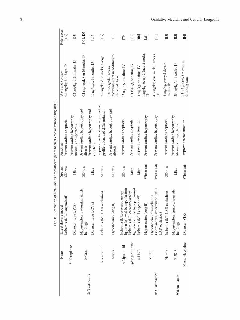
The exploration of mechanisms underlying the regulationof the Nrf2 pathway has led to the development of agentsmanipulating Nrf2 to treat HF. In fact, compounds thatincrease the activity of Nrf2 and its downstream genes arecurrently being tested for disease prevention [111, 132–134](Table 1).
8.1. Activation of Nrf2 to Treat Cardiac Remodeling andHF. Many Nrf2 activators are natural products and plant-derived phytochemicals. Some examples of natural Nrf2activators include sulforaphane, curcumin, resveratrol, andgarlic organosulfur compounds [135]. Several synthetic Nrf2activators have also been developed, such as carbobenzoxy-Leu-Leu-leucinal (MG132), 4-hydroxynonenal (4-HNE), 𝛼-lipoic acid, hydrogen sulfate, and 17𝛽-estradiol (E2) [79, 109,110]. These chemopreventive compounds exert their effectsby inducing an Nrf2-mediated defense response as well asactivation of phase II detoxification enzymes, antioxidants,and transporters that protect cells from oxidative stress.
8.1.1. Natural Nrf2 Activators. One of the most extensivelystudied natural products that target the Nrf2-Keap1 signalingpathway is sulforaphane, an isothiocyanate present in cru-ciferous vegetables such as broccoli [136]. Treatment withsulforaphane decreased infarct size, inhibited an increase inthe LV end-diastolic pressure, and improved coronary flowin mice after MI [102]. This protective effect may be partlymediated through HO-1, SOD, and CAT expression [102].
8 Oxidative Medicine and Cellular Longevity
Table1:Ac
tivationof
Nrf2
andits
downstre
amgenestotre
atcardiacr
emod
elingandHF.
Nam
eTargetdiseasem
odel
Species
Functio
nWaysa
ndvolume
References
Nrf2
activ
ators
Sulfo
raph
ane
Ischem
ia(I/R,L
angend
orff)
SDrats
Preventcardiac
apop
tosis
0.5m
g/kg/d,3
days,IP
[102]
Diabetes(type
1,ST
Z)Mice
Preventcardiac
hypertroph
y,fib
rosis,and
apop
tosis
0.5m
g/kg/d,3
mon
ths,IP
[103]
MG132
Hypertension(abd
ominalaortic
band
ing)
SDrats
Preventcardiac
hypertroph
yand
fibrosis
0.1m
g/kg/d,8
or16
weeks,IP
[104,105]
Diabetes(type
1,OVE)
Mice
Preventcardiac
hypertroph
yand
apop
tosis
10𝜇g/kg/d,3
mon
ths,IP
[106]
Resveratrol
Ischem
ia(M
I,LA
Docclu
sion)
SDrats
Improvec
ardiac
stem
cells’survival,
proliferatio
n,anddifferentiatio
n2.5m
g/kg/d,2
weeks,gavage
[107]
Allicin
Hypertension(A
ngII)
SDrats
Preventcardiac
hypertroph
yand
fibrosis
180m
g/kg/d,8
weeks,
receivingad
ietinadditio
nto
stand
ardchow
[108]
𝛼-Lipoica
cid
Ischem
ia(I/R,coron
aryartery
ligationfollo
wed
byreperfusion)
SDrats
Preventcardiac
apop
tosis
15mg/kg,one
time,IV
[79]
Hydrogensulfate
Ischem
ia(I/R,coron
aryartery
ligationfollo
wed
byreperfusion)
Mice
Preventcardiac
apop
tosis
0.1m
g/kg,one
time,IV
[109]
4-HNE
Ischem
ia(M
I,Langendo
rff)
Mice
Improvec
ardiac
functio
n4m
g/kg,one
time,IV
[110]
HO-1activ
ators
CoP
PHypertension(A
ngII)
Wistar
rats
Preventcardiac
hypertroph
y1m
g/kg,every
2days,2
weeks,
IP[25]
Hypertensionplus
ischemia
(spo
ntaneous
hypertensiv
erats+
LADocclu
sion)
Wistar
rats
Preventcardiac
hypertroph
y4.5m
g/kg,one/w
eek,6weeks,
IP[111]
Hem
inIschem
ia(M
I,LA
Docclu
sion)
SDrats
Preventcardiac
apop
tosis
4mg/kg,every
2days,4
weeks,IP
[112]
SODactiv
ators
EUK-
8Hypertension(tr
ansverse
aortic
band
ing)
Mice
Preventcardiac
hypertroph
y,fib
rosis,and
apop
tosis
25mg/kg/d,4
weeks,IP
[113]
N-Acetylcysteine
Diabetes(ST
Z)Wistar
rats
Improvec
ardiac
functio
n1.4
-1.5g/kg/d,8
weeks,in
drinking
water
[114]
Oxidative Medicine and Cellular Longevity 9
Curcumin is another well-studied chemopreventive natu-ral product that is capable of activating Nrf2 [132]. Curcuminhas been used to attenuate acute doxorubicin-induced car-diomyopathy in rats [133]. In this study, curcumin pretreat-ment reversed the increase in lipid peroxidation and CATcontent, as well as the decrease in GSH content and GPxactivity in rat cardiac tissues induced by doxorubicin [133].
By treating rat cardiac stem cells with resveratrol ina rat left anterior descending occlusion model, Gorbunovet al. found that resveratrol significantly improved cardiacfunction through enhancing Nrf2 expression, as well assignificantly increasing the survival and engraftment ofimplanted cardiac stem cells in the host [134]. Resveratrol canalso achieve the same effect by gavage in the same animalmodel [107].
In an Ang II-induced cardiac remodeling and HF ratmodel, allicin treatment could prevent the development ofcardiac remodeling and the progression of cardiac hypertro-phy to cardiac dysfunction, by enhancing the Nrf2 pathway[108]. In addition, dietary phytochemical intake could upreg-ulate the cardiac Nrf2 transcriptome and reduce oxidativedamage and HF in hypertensive rats [137]. The protectiveeffect of Nrf2 in myocardial remodeling and HF has beensuggested to be mediated through Nox4 [138], which isknown to be an important regulator of reduction-oxidationsignaling in many cell types including cardiomyocytes [139].
8.1.2. Synthetic Nrf2 Activators. Accumulating evidence hasdemonstrated thatMG132 can protect cells and tissues againstoxidative damage, since it can activate the Nrf2-ARE sig-naling pathway, leading to upregulation of detoxifying andantioxidant genes [140, 141]. Both sulforaphane and MG132could prevent diabetes-induced high blood pressure andcardiac dysfunction, as well as cardiac hypertrophy, fibrosis,oxidative damage, and inflammation [103, 106]. In pressure-overload-induced rodent HF models, treatment with MG132significantly attenuated cardiac hypertrophy and cardiacfibrosis as well as improving cardiac function [104, 105].Mechanistically, MG132 may enhance Nrf2-mediated antiox-idative function and inhibit NF-𝜅B-mediated inflammation[142].
4-HNE is an 𝛼, 𝛽-unsaturated hydroxyalkenal that isproduced by lipid peroxidation in cells. 𝛼-Lipoic acid isan organosulfur compound derived from octanoic acid. E2is a sex hormone. All three of these compounds couldprotect the heart from ischemia-induced cardiac remodelingand HF by activating Nrf2 and its target genes. Treatmentwith 4-HNE [110], 𝛼-lipoic acid [79], or hydrogen sulfate[109] could activate Nrf2 in the heart and increase theintramyocardial GSH content, consequently improving thefunctional recovery of the LV following I/R in Langendorff-perfused hearts [110] or reducing infarct size, decreasing car-diomyocyte apoptosis in vivo [79, 109]. The cardioprotectiveeffect of 4-HNE was not observed for Nrf2-knockout mice[110], indicating that this protection is Nrf2 dependent. Inan in vitro study, Yu et al. cultured primary cardiomyocytesand established a hypoxia/reoxygenation (H/R) model tosimulate myocardial I/R injury [143]. They found that E2
could upregulate Nrf2 expression in nuclear extracts and alsoincreased the expression ofHO-1, SOD1, GST, and glutamate-cysteine ligase (GCL) dramatically during H/R injury [143].These findings indicate that Nrf2 plays a pivotal role in pre-venting and alleviating cardiac I/R injury-induced oxidativestress.
8.2. Role of Nrf2 Target Genes Activation inCardiac Remodeling
8.2.1. Role of HO-1 Activation in Cardiac Remodeling. It hasbeen reported that elevation ofHO-1 by treatment with cobaltprotoporphyrin IX (CoPP IX) or a recombinant adenoviruscarrying the human HO-1 gene attenuated cardiac hypertro-phy and apoptosis, in both an Ang-II-induced HF modeland a spontaneously hypertensive rat model [25, 70], whilethis pathological process was exacerbated in the presence oftin protoporphyrin, an inhibitor of HO activity [70]. In aspontaneous hypertensive rat model, HO-1 upregulation byCoPP IX administration decreased blood pressure, inhibitedinflammation, and improved the ventricular remodelingprocess and postinfarct cardiac function [111]. In addition,cardiac stem cells (CSCs) were protected by pretreatmentwith CoPP against apoptosis through activation of theextracellular signal-regulated kinase (ERK)/Nrf2 signalingpathway and cytokine release [144].
Hemin could upregulate HO activity, reduce hyperten-sion, suppress oxidative stress, and attenuate cardiac fibrosis,apoptosis, and oxidative stress. This effect was modulatedthrough enhanced expression of the PI3K p85 regulatorysubunit [145]. In an acute I/R with STZ-induced hyper-glycemic rat model, intraperitoneal administration of hemin18 h before I/R increased the levels ofHO-1, providingmarkedprotection against myocardial injury [146]. Moreover, zincprotoporphyrin IX (an inhibitor ofHO activity) abolished theprotective effect by hemin [146]. Furthermore, chronic HO-1activation by prolonged administration of hemin improvedpostinfarction survival and exerted protective effects in a ratmodel of myocardial ischemia, through a potent antioxidantactivity [112].
Taurine treatment significantly improved LV systolic anddiastolic function in an STZ-induced diabetes rat model, andthere were persistent increases in activities of Akt/PKB andSOD, as well as the level of HO-1 protein [147].
Mesenchymal stem cells (MSCs) have been reported tohave the potential to release several kinds of cytokines,which could induce angiogenesis [148, 149]. However, almostall transplanted cells seemed to be lost by 6 weeks aftertransplantation, making it impossible for the limited numberof MSCs to achieve a maximum proangiogenesis effect [150].Due to its multiple catalytic byproducts, HO-1 has beenproposed to be involved in a number of cytoprotectiveeffects. HO-1 has been administered to improve the survivalenvironment of MSCs and to achieve maximum functionalbenefits ofMSCs [151]. HO-1 transduced byMSCs can induceangiogenic effects, reduce cardiac remodeling, and improveheart function after acute MI [152, 153]. Intramyocardialdelivery of the HO-1 gene using adeno-associated virus-2
10 Oxidative Medicine and Cellular Longevity
(AAV-2) before I/R also prevented cardiomyocyte apopto-sis and reduced infarct size and cardiac dysfunction aftermyocardial I/R in mice [90]. To evaluate the long-termeffects of HO-1 gene delivery, Liu et al. have showed thatdelivery of the HO-1 gene reduced mortality and preservedLV function and chamber dimensions 1 year after acute MI[91].These results suggest that preemptiveHO-1 gene deliverymay be useful as a therapeutic strategy to reduce post-MI LVremodeling and HF.
8.2.2. Role of SOD andGSHActivation in Cardiac Remodeling.EUK-8, as a SODmimetic, improved LV end-systolic dimen-sions and fractional shortening, prevented myocardial oxi-dant stress, attenuated necrotic and apoptotic cell death, andattenuated cardiac hypertrophy and fibrosis inmice subjectedto pressure overload [113]. In vitro, the SODmimetics tempoland EUK-8 could also reduce collagen production in Ang-II-treated fibroblasts [73]. Moreover, N-acetylcysteine, anantioxidant and GSH precursor, attenuated diabetic myocar-dial dysfunction via upregulatingmyocardial GSH and SOD2activity [114]. Treatment with polyethylene glycol-conjugatedSOD ameliorated doxorubicin-induced cardiac dysfunction,and this effect was mediated by inhibition of doxorubicin-induced upregulation of NF-𝜅B signaling, lowering the levelsof hexanoyl-lysine (a marker of free radical-induced lipidperoxidation) and suppressing the activation of Akt and Akt-regulated gene expression [124].
Recombinant SOD3 preserved cardiac function followingI/R in isolated rat hearts [154–156] and reduced the infarctsize when given just prior to coronary reperfusion in pigs[157]. Cardiac-selective expression of SOD3 from the cardiactroponin-T promoter after systemic administration of AAV-9 provided significant protection against both acute MI andLV remodeling [158]. Liu et al. also have demonstrated thatencapsulation of CSCs in SOD-loaded alginate hydrogelenhanced CSC survival in the presence of doxorubicin,indicating its potential application as a novel therapy forthe treatment of acute and early-onset doxorubicin-inducedcardiotoxicity [159].
9. Advantages of Nrf2 Activation in CardiacRemodeling and HF
Nrf2 dimerizes with members of the small Maf familyto bind to antioxidant or electrophile response elements(AREs/EpREs) located in the regulatory regions of cellulardefense enzyme genes [50]. The target genes of Nrf2 includemany antioxidant genes, such as HO-1, NQO1, SODs, CAT,GST, 𝛾-GCS, and GPx. By inducing the expression of thesegenes, Nrf2 activates a wide range of cell defense processes,thereby enhancing the overall capacity of cells to detoxify andeliminate harmful substances. As mentioned above, activatedNrf2 and its target genes, such as HO-1, SOD, and GPx,all play important roles in preventing cardiac remodeling.Although it cannot be excluded that some Nrf2 target genesmay protect the heart through anNrf2-independent pathway,Nrf2-induced cell defense processes remain to be the maindriving force. Because Nrf2 could induce transcriptional
activation of a number of ARE-bearing antioxidants, wespeculate that, acting as an upstream gene, activation of Nrf2may rescue from cardiac remodeling and HF induced bydeficiency of an individual downstream gene. So, activationof Nrf2 may have an advantage over its downstream targetgenes in protecting one fromoxidative stress-inducedHF, butfurther investigations are needed before a conclusion can bedrawn.
10. Conclusions
Increased generation of ROS in cardiomyocytes leads toincreased necrosis and apoptosis, which induce cardiacremodeling and dysfunction. Nrf2 and its target genes arekey components to maintain cellular redox homeostasisby attenuating oxidative stress-associated pathological pro-cesses. Patients with insufficient Nrf2 levels in their cardio-vascular system are more vulnerable to HF development. Itis conceivable that Nrf2 orchestrates a group of antioxidativeand other cytoprotective genes to provide a protectivemecha-nism against detrimental stress-induced cardiac dysfunction.However, further work is needed to understand the roleof Nrf2 in the pathogenesis of cardiac remodeling and HFin more detail before it can be seriously considered as atherapeutic target for HF.
Abbreviations
HF: Heart failureROS: Reactive oxygen speciesRNS: Reactive nitrogen speciesNrf2: Nuclear factor-erythroid-2- (NF-E2-)
related factor 2LV: Left ventricularAREs: Antioxidant response elementsCNC: Cap-n-collarCNC-bZIP: CNC type of basic region leucine zipperKeap1: Kelch-like-ECH-associated protein 1HO-1: Heme oxygenase-1NQO1: NAD(P)H dehydrogenase (quinone 1)SODs: Superoxide dismutasesCAT: CatalaseGST: Glutathione-S-transferases𝛾-GCS: 𝛾-Glutamylcysteine synthaseGPx: Glutathione peroxidasesTAC: Transverse aortic constrictionSTZ: StreptozotocinATF3: Activating transcription factor 3LDH: Lactate dehydrogenaseGCL: Glutamate-cysteine ligaseGSH: GlutathioneMG132: Carbobenzoxy-Leu-Leu-leucinal4-HNE: 4-HydroxynonenalE2: 17𝛽-EstradiolH/R: Hypoxia/reoxygenationCSCs: Cardiac stem cellsERK: Extracellular signal-regulated kinaseMSCs: Mesenchymal stem cellsAAV-2: Adeno-associated virus-2
Oxidative Medicine and Cellular Longevity 11
EpREs: Electrophile response elements∙O2−: Superoxide anion∙OH: Hydroxyl radicalH2O2: Hydrogen peroxide
OCl∙−: HypochloriteAng II: Angiotensin IIMI: Myocardial infarctionI/R: Ischemia/reperfusionHG: High glucosetBHQ: Tert-ButylhydroquinoneCK: Creatine kinase.
Conflict of Interests
The authors declare that there is no conflict of interestsregarding the publication of this paper.
Acknowledgment
This work was supported by the National Natural ScienceFoundation of China (no. 81370318, to Yang Zheng).
References
[1] J. Gutierrez, S.W. Ballinger, V.M.Darley-Usmar, andA. Landar,“Free radicals, mitochondria, and oxidized lipids: the emerg-ing role in signal transduction in vascular cells,” CirculationResearch, vol. 99, no. 9, pp. 924–932, 2006.
[2] T. J. Guzik and D. G. Harrison, “Vascular NADPH oxidases asdrug targets for novel antioxidant strategies,” Drug DiscoveryToday, vol. 11, no. 11-12, pp. 524–533, 2006.
[3] I. Jialal and S. Devaraj, “Antioxidants and atherosclerosis: don’tthrow out the baby with the bath water,”Circulation, vol. 107, no.7, pp. 926–928, 2003.
[4] N. R. Madamanchi, A. Vendrov, and M. S. Runge, “Oxidativestress and vascular disease,” Arteriosclerosis, Thrombosis, andVascular Biology, vol. 25, no. 1, pp. 29–38, 2005.
[5] C. E. Murdoch, M. Zhang, A. C. Cave, and A. M. Shah,“NADPH oxidase-dependent redox signalling in cardiac hyper-trophy, remodelling and failure,” Cardiovascular Research, vol.71, no. 2, pp. 208–215, 2006.
[6] E. Takimoto and D. A. Kass, “Role of oxidative stress in cardiachypertrophy and remodeling,” Hypertension, vol. 49, no. 2, pp.241–248, 2007.
[7] J. Li, T. Ichikawa, L. Villacorta et al., “Nrf2 protects againstmala-daptive cardiac responses to hemodynamic stress,” Arterioscle-rosis, Thrombosis, and Vascular Biology, vol. 29, no. 11, pp. 1843–1850, 2009.
[8] C. X. C. Santos, N. Anilkumar, M. Zhang, A. C. Brewer, and A.M. Shah, “Redox signaling in cardiac myocytes,” Free RadicalBiology and Medicine, vol. 50, no. 7, pp. 777–793, 2011.
[9] D. D.Thomas, L. A. Ridnour, J. S. Isenberg et al., “The chemicalbiology of nitric oxide: implications in cellular signaling,” FreeRadical Biology & Medicine, vol. 45, no. 1, pp. 18–31, 2008.
[10] D. K. Dowling and L. W. Simmons, “Reactive oxygen speciesas universal constraints in life-history evolution,” Proceedings ofthe Royal Society B: Biological Sciences, vol. 276, no. 1663, pp.1737–1745, 2009.
[11] R. Scherz-Shouval and Z. Elazar, “Regulation of autophagy byROS: physiology and pathology,”Trends in Biochemical Sciences,vol. 36, no. 1, pp. 30–38, 2011.
[12] I. Afanas’ev, “ROS and RNS signaling in heart disorders: couldantioxidant treatment be successful?” Oxidative Medicine andCellular Longevity, vol. 2011, Article ID 293769, 13 pages, 2011.
[13] M. L. Circu and T. Y. Aw, “Reactive oxygen species, cellularredox systems, and apoptosis,” Free Radical Biology and Medi-cine, vol. 48, no. 6, pp. 749–762, 2010.
[14] R. Bolli, W. X. Zhu, C. J. Hartley et al., “Attenuation ofdysfunction in the postischemic “stunned” myocardium bydimethylthiourea,” Circulation, vol. 76, no. 2, pp. 458–468, 1987.
[15] A. Beresewicz and M. Horackova, “Alterations in electrical andcontractile behavior of isolated cardiomyocytes by hydrogenperoxide: Possible ionic mechanisms,” Journal of Molecular andCellular Cardiology, vol. 23, no. 8, pp. 899–918, 1991.
[16] A. Chesley, M. S. Lundberg, T. Asai et al., “The 𝛽2-adrenergic
receptor delivers an antiapoptotic signal to cardiac myocytesthrough G
𝑖-dependent coupling to phosphatidylinositol 3’-
kinase,” Circulation Research, vol. 87, no. 12, pp. 1172–1179, 2000.[17] B. M. Hybertson, B. Gao, S. K. Bose, and J. M. McCord,
“Oxidative stress in health and disease: the therapeutic potentialof Nrf2 activation,”Molecular Aspects ofMedicine, vol. 32, no. 4–6, pp. 234–246, 2011.
[18] O. Andrukhova, M. Salama, R. Rosenhek et al., “Serum glu-tathione S-transferase P1 1 in prediction of cardiac function,”Journal of Cardiac Failure, vol. 18, no. 3, pp. 253–261, 2012.
[19] W. Wu, Q. Qiu, H. Wang et al., “Nrf2 is crucial to graft survivalin a rodent model of heart transplantation,”Oxidative Medicineand Cellular Longevity, vol. 2013, Article ID 919313, 9 pages,2013.
[20] H. Zhu, K. Itoh, M. Yamamoto, J. L. Zweier, and Y. Li, “Roleof Nrf2 signaling in regulation of antioxidants and phase2 enzymes in cardiac fibroblasts: protection against reactiveoxygen and nitrogen species-induced cell injury,” FEBS Letters,vol. 579, no. 14, pp. 3029–3036, 2005.
[21] H. Zhu, Z. Jia, B. R.Misra et al., “Nuclear factor E2-related factor2-dependent myocardiac cytoprotection against oxidative andelectrophilic stress,” Cardiovascular Toxicology, vol. 8, no. 2, pp.71–85, 2008.
[22] B. F. Peake, C. K. Nicholson, J. P. Lambert et al., “Hydrogensulfide preconditions the db/db diabetic mouse heart againstischemia-reperfusion injury by activating Nrf2 signaling in anErk-dependent manner,” American Journal of Physiology: Heartand Circulatory Physiology, vol. 304, no. 9, pp. H1215–H1224,2013.
[23] X. Zhang, Z. Xiao, J. Yao, G. Zhao, X. Fa, and J. Niu, “Partic-ipation of protein kinase C in the activation of Nrf2 signalingby ischemic preconditioning in the isolated rabbit heart,”Molecular and Cellular Biochemistry, vol. 372, no. 1-2, pp. 169–179, 2013.
[24] P. Wiesel, A. P. Patel, I. M. Carvajal et al., “Exacerbation ofchronic renovascular hypertension and acute renal failure inheme oxygenase-1-deficient mice,” Circulation Research, vol. 88,no. 10, pp. 1088–1094, 2001.
[25] C. M. Hu, Y. H. Chen, M. T. Chiang, and L. Y. Chau, “Hemeoxygenase-1 inhibits angiotensin II-induced cardiac hypertro-phy in vitro and in vivo,”Circulation, vol. 110, no. 3, pp. 309–316,2004.
12 Oxidative Medicine and Cellular Longevity
[26] Z. Lu, X. Xu, X. Hu et al., “Extracellular superoxide dismutasedeficiency exacerbates pressure overload-induced left ventricu-lar hypertrophy and dysfunction,” Hypertension, vol. 51, no. 1,pp. 19–25, 2008.
[27] S. Matsushima, S. Kinugawa, T. Ide et al., “Overexpression ofglutathione peroxidase attenuates myocardial remodeling andpreserves diastolic function in diabetic heart,”American Journalof Physiology—Heart and Circulatory Physiology, vol. 291, no. 5,pp. H2237–H2245, 2006.
[28] A. D. Hafstad, A. A. Nabeebaccus, and A. M. Shah, “Novelaspects of ROS signalling in heart failure,” Basic Research inCardiology, vol. 108, no. 4, article 359, 2013.
[29] S. K. Maulik and S. Kumar, “Oxidative stress and cardiachypertrophy: a review,” Toxicology Mechanisms and Methods,vol. 22, no. 5, pp. 359–366, 2012.
[30] S. Nattel, “Electrophysiologic remodeling: are ion channelsstatic players or dynamic movers?” Journal of CardiovascularElectrophysiology, vol. 10, no. 11, pp. 1553–1556, 1999.
[31] C. Yu, H. Lin, H. Yang, S. Kong, Q. Zhang, and S. W. Lee,“Progression of systolic abnormalities in patientswith “isolated”diastolic heart failure and diastolic dysfunction,” Circulation,vol. 105, no. 10, pp. 1195–1201, 2002.
[32] L. Cai, W. Li, G. Wang, L. Guo, Y. Jiang, and Y. James Kang,“Hyperglycemia-induced apoptosis in mouse myocardium:mitochondrial cytochrome c-mediated caspase-3 activationpathway,” Diabetes, vol. 51, no. 6, pp. 1938–1948, 2002.
[33] G. L. Chang, D. Y. Zhang, H. Yu et al., “Cardioprotectiveeffects of exenatide against oxidative stress-induced injury,”International Journal of Molecular Medicine, vol. 32, no. 5, pp.1011–1020, 2013.
[34] W. Y. Zhao, T. Q. Zhao, Y. J. Chen, R. A. Ahokas, and Y. Sun,“Oxidative stress mediates cardiac fibrosis by enhancing trans-forming growth factor-beta1 in hypertensive rats,” Molecularand Cellular Biochemistry, vol. 317, no. 1-2, pp. 43–50, 2008.
[35] S. Johar, A. C. Cave, A. Narayanapanicker, D. J. Grieve, andA. M. Shah, “Aldosterone mediates angiotensin II-inducedinterstitial cardiac fibrosis via a Nox2-containing NADPHoxidase,”The FASEB Journal, vol. 20, no. 9, pp. 1546–1548, 2006.
[36] S. Gupta, B.Das, and S. Sen, “Cardiac hypertrophy:mechanismsand therapeutic opportunities,” Antioxidants and Redox Signal-ing, vol. 9, no. 6, pp. 623–652, 2007.
[37] A. Sabri, H. H. Hughie, and P. A. Lucchesi, “Regulation ofhypertrophic and apoptotic signaling pathways by reactiveoxygen species in cardiac myocytes,” Antioxidants and RedoxSignaling, vol. 5, no. 6, pp. 731–740, 2003.
[38] K. Huynh, B. C. Bernardo, J. R. McMullen. et al., “Diabeticcardiomyopathy: mechanisms and new treatment strategiestargeting antioxidant signaling pathways,” Pharmacology &Therapeutics, vol. 142, no. 3, pp. 375–415, 2014.
[39] V. P. M. van Empel, A. T. A. Bertrand, L. Hofstra, H. J. Crijns, P.A. Doevendans, and L. J. deWindt, “Myocyte apoptosis in heartfailure,” Cardiovascular Research, vol. 67, no. 1, pp. 21–29, 2005.
[40] K. Kannan and S. K. Jain, “Oxidative stress and apoptosis,”Pathophysiology, vol. 7, no. 3, pp. 153–163, 2000.
[41] M. Kobayashi and M. Yamamoto, “Nrf2-Keap1 regulation ofcellular defense mechanisms against electrophiles and reactiveoxygen species,” Advances in Enzyme Regulation, vol. 46, no. 1,pp. 113–140, 2006.
[42] N. Li and A. E. Nel, “Role of the Nrf2-mediated signalingpathway as a negative regulator of inflammation: Implicationsfor the impact of particulate pollutants on asthma,”Antioxidantsand Redox Signaling, vol. 8, no. 1-2, pp. 88–98, 2006.
[43] T. W. Kensler, N. Wakabayashi, and S. Biswal, “Cell survivalresponses to environmental stresses via the Keap1-Nrf2-AREpathway,” Annual Review of Pharmacology and Toxicology, vol.47, pp. 89–116, 2007.
[44] A. Kobayashi, E. Ito, T. Toki et al., “Molecular cloning and func-tional characterization of a newCap’n’collar family transcriptionfactor Nrf3,”The Journal of Biological Chemistry, vol. 274, no. 10,pp. 6443–6452, 1999.
[45] Ø. Johnsen, P. Murphy, H. Prydz, and A. Kolstø, “Interaction ofthe CNC-bZIP factor TCF11/LCR-F1/Nrf1 with MafG: binding-site selection and regulation of transcription,” Nucleic AcidsResearch, vol. 26, no. 2, pp. 512–520, 1998.
[46] M. G.Marini, K. Chan, L. Casula, Y.W. Kan, A. Cao, and P.Moi,“hMAF, a small human transcription factor that heterodimer-izes specifically with Nrf1 and Nrf2,” The Journal of BiologicalChemistry, vol. 272, no. 26, pp. 16490–16497, 1997.
[47] K. Itoh, N. Wakabayashi, Y. Katoh et al., “Keap1 repressesnuclear activation of antioxidant responsive elements by Nrf2through binding to the amino-terminal Neh2 domain,” Genesand Development, vol. 13, no. 1, pp. 76–86, 1999.
[48] A. Kobayashi, M. Kang, H. Okawa et al., “Oxidative stresssensor Keap1 functions as an adaptor for Cul3-based E3 ligase toregulate proteasomal degradation ofNrf2,”Molecular andCellu-lar Biology, vol. 24, no. 16, pp. 7130–7139, 2004.
[49] T. Yamamoto, T. Suzuki, A. Kobayashi et al., “Physiological sig-nificance of reactive cysteine residues of Keap1 in determiningNrf2 activity,”Molecular and Cellular Biology, vol. 28, no. 8, pp.2758–2770, 2008.
[50] K. Itoh, T. Chiba, S. Takahashi et al., “An Nrf2/small Maf heter-odimer mediates the induction of phase II detoxifying enzymegenes through antioxidant response elements,” Biochemical andBiophysical Research Communications, vol. 236, no. 2, pp. 313–322, 1997.
[51] R. Howden, “Nrf2 and cardiovascular defense,”OxidativeMedi-cine and Cellular Longevity, vol. 2013, Article ID 104308, 10pages, 2013.
[52] L. E. Otterbein and A. M. K. Choi, “Heme oxygenase: colors ofdefense against cellular stress,” American Journal of Physiology:Lung Cellular and Molecular Physiology, vol. 279, no. 6, pp.L1029–L1037, 2000.
[53] N. G. Abraham andA. Kappas, “Heme oxygenase and the cardi-ovascular-renal system,” Free Radical Biology andMedicine, vol.39, no. 1, pp. 1–25, 2005.
[54] E. D. van Deel, Z. Lu, X. Xu et al., “Extracellular superoxidedismutase protects the heart against oxidative stress and hyper-trophy after myocardial infarction,” Free Radical Biology andMedicine, vol. 44, no. 7, pp. 1305–1313, 2008.
[55] H. J. Forman, H. Zhang, and A. Rinna, “Glutathione: overviewof its protective roles, measurement, and biosynthesis,”Molecu-lar Aspects of Medicine, vol. 30, no. 1-2, pp. 1–12, 2009.
[56] T. Shiomi, H. Tsutsui, H. Matsusaka et al., “Overexpression ofglutathione peroxidase prevents left ventricular remodeling andfailure aftermyocardial infarction inmice,”Circulation, vol. 109,no. 4, pp. 544–549, 2004.
[57] H. Cai, “Hydrogen peroxide regulation of endothelial func-tion: origins, mechanisms, and consequences,” CardiovascularResearch, vol. 68, no. 1, pp. 26–36, 2005.
[58] J. J. Kohler, I. Cucoranu, E. Fields et al., “Transgenic mito-chondrial superoxide dismutase and mitochondrially targetedcatalase prevent antiretroviral-induced oxidative stress andcardiomyopathy,” Laboratory Investigation, vol. 89, no. 7, pp.782–790, 2009.
Oxidative Medicine and Cellular Longevity 13
[59] C. Savoia, D. Burger, N. Nishigaki, A. Montezano, and R. M.Touyz, “Angiotensin II and the vascular phenotype in hyper-tension,” Expert Reviews in Molecular Medicine, vol. 13, articlee11, 2011.
[60] K. Sugamura and J. F. Keaney Jr., “Reactive oxygen species incardiovascular disease,” Free Radical Biology and Medicine, vol.51, no. 5, pp. 978–992, 2011.
[61] P. Puddu, G. M. Puddu, E. Cravero, M. Rosati, and A. Muscari,“The molecular sources of reactive oxygen species in hyperten-sion,” Blood Pressure, vol. 17, no. 2, pp. 70–77, 2008.
[62] J. C. Tardiff, T. E. Hewett, S. M. Factor, K. L. Vikstrom, J. Rob-bins, and L. A. Leinwand, “Expression of the 𝛽 (slow)-isoformof MHC in the adult mouse heart causes dominant-negativefunctional effects,” The American Journal of Physiology—HeartandCirculatory Physiology, vol. 278, no. 2, pp.H412–H419, 2000.
[63] H.Wakui, T.Dejima,K. Tamura et al., “Activation of angiotensinII type 1 receptor-associated protein exerts an inhibitory effecton vascular hypertrophy and oxidative stress in angiotensin II-mediated hypertension,” Cardiovascular Research, vol. 100, 3,pp. 511–519, 2013.
[64] H. X.Wang, H. Yang, Q. Y. Han et al., “Nadph oxidases mediatea cellular “memory” of angiotensin ii stress in hypertensivecardiac hypertrophy,” Free Radical Biology & Medicine, vol. 65,pp. 897–907, 2013.
[65] Y. Tsukamoto, T. Mano, Y. Sakata et al., “A novel heart failuremice model of hypertensive heart disease by angiotensin iiinfusion, nephrectomy, and salt loading,”The American Journalof Physiology—Heart and Circulatory Physiology, vol. 305, pp.H1658–H1667, 2013.
[66] Y. Xing, T. Niu,W.Wang et al., “Triterpenoid dihydro-cddo-tri-fluoroethyl amide protects against maladaptive cardiac remod-eling anddysfunction inmice: a critical role ofNrf2,”PLoSONE,vol. 7, no. 9, Article ID e44899, 2012.
[67] H. Lin, H. F. Li, H. H. Chen et al., “Activating transcriptionfactor 3 protects against pressure-overload heart failure via theautophagy molecule beclin-1 pathway,” Molecular Pharmacol-ogy, vol. 85, pp. 682–691, 2014.
[68] J. F. Ndisang and A. Jadhav, “Upregulating the heme oxygenasesystem suppresses left ventricular hypertrophy in adult spon-taneously hypertensive rats for 3 months,” Journal of CardiacFailure, vol. 15, no. 7, pp. 616–628, 2009.
[69] S. Mito, R. Ozono, T. Oshima et al., “Myocardial protectionagainst pressure overload in mice lacking bachl, a transcrip-tional repressor of heme oxygenase-1,”Hypertension, vol. 51, no.6, pp. 1570–1577, 2008.
[70] R. S. Y. Foo, R. C. M. Siow, M. J. Brown, and M. R. Ben-nett, “Heme oxygenase-1 gene transfer inhibits angiotensin II-mediated rat cardiac myocyte apoptosis but not hypertrophy,”Journal of Cellular Physiology, vol. 209, no. 1, pp. 1–7, 2006.
[71] C. Chen, R. Huo, Y. Tong et al., “Systemic heme oxygenase-1 transgenic overexpression aggravates pressure overload-induced cardiac hypertrophy in mice,” Cellular Physiology andBiochemistry, vol. 28, no. 1, pp. 25–32, 2011.
[72] S. Hikoso, O. Yamaguchi, Y. Nakano et al., “The I𝜅B Kinase𝛽/Nuclear factor 𝜅b signaling pathway protects the heart fromhemodynamic stress mediated by the regulation of manganesesuperoxide dismutase expression,” Circulation Research, vol.105, no. 1, pp. 70–79, 2009.
[73] P. Lijnen, V. Petrov, J. van Pelt, and R. Fagard, “Inhibition ofsuperoxide dismutase induces collagen production in cardiacfibroblasts,” American Journal of Hypertension, vol. 21, no. 10,pp. 1129–1136, 2008.
[74] L. Richters, N. Lange, R. Renner et al., “Exercise-induced adap-tations of cardiac redox homeostasis and remodeling in hetero-zygous SOD2-knockout mice,” Journal of Applied Physiology,vol. 111, no. 5, pp. 1431–1440, 2011.
[75] G. Takemura, M. Nakagawa, H. Kanamori, S. Minatoguchi,and H. Fujiwara, “Benefits of reperfusion beyond infarct sizelimitation,” Cardiovascular Research, vol. 83, no. 2, pp. 269–276,2009.
[76] F. Eefting, B. Rensing, J. Wigman et al., “Role of apoptosis inreperfusion injury,” Cardiovascular Research, vol. 61, no. 3, pp.414–426, 2004.
[77] V. Braunersreuther and V. Jaquet, “Reactive oxygen speciesin myocardial reperfusion injury: from physiopathology totherapeutic approaches,”Current Pharmaceutical Biotechnology,vol. 13, no. 1, pp. 97–114, 2012.
[78] Y. Sun, “Myocardial repair/remodelling following infarction:Roles of local factors,” Cardiovascular Research, vol. 81, no. 3,pp. 482–490, 2009.
[79] C. Deng, Z. Sun, G. Tong et al., “Alpha-lipoic acid reducesinfarct size and preserves cardiac function in rat myocardialischemia/reperfusion injury through activation of pi3k/akt/nrf2pathway,” PLoS ONE, vol. 8, no. 3, Article ID e58371, 2013.
[80] H. Ashrafian, G. Czibik, M. Bellahcene et al., “Fumarate iscardioprotective via activation of theNrf2 antioxidant pathway,”Cell Metabolism, vol. 15, no. 3, pp. 361–371, 2012.
[81] Y. Katsumata, K. Shinmura, Y. Sugiura et al., “Endogenousprostaglandin D
2and its metabolites protect the heart against
ischemia-reperfusion injury by activating Nrf2,” Hypertension,vol. 63, no. 1, pp. 80–87, 2014.
[82] E. K. Iliodromitis, A. Lazou, and D. T. Kremastinos, “Ischemicpreconditioning: protection against myocardial necrosis andapoptosis,” Vascular Health and Risk Management, vol. 3, no. 5,pp. 629–637, 2007.
[83] N. Gurusamy, G. Malik, N. V. Gorbunov, and D. K. Das, “Redoxactivation of Ref-1 potentiates cell survival followingmyocardialischemia reperfusion injury,” Free Radical Biology & Medicine,vol. 43, no. 3, pp. 397–407, 2007.
[84] F. Grabellus, C. Schmid, B. Levkau et al., “Reduction of hypoxia-inducible heme oxygenase-1 in the myocardium after leftventricular mechanical support,” Journal of Pathology, vol. 197,no. 2, pp. 230–237, 2002.
[85] V. S. Raju, N. Imai, and C. S. Liang, “Chamber-specific regu-lation of heme oxygenase-1 (heat shock protein 32) in right-sided congestive heart failure,” Journal ofMolecular and CellularCardiology, vol. 31, no. 8, pp. 1581–1589, 1999.
[86] G. Wang, T. Hamid, R. J. Keith et al., “Cardioprotective andantiapoptotic effects of heme oxygenase-1 in the failing heart,”Circulation, vol. 121, no. 17, pp. 1912–1925, 2010.
[87] S. R. Vulapalli, Z. Chen, B. H. L. Chua, T. Wang, and C.Liang, “Cardioselective overexpression of HO-1 prevents I/R-induced cardiac dysfunction and apoptosis,” American Journalof Physiology: Heart and Circulatory Physiology, vol. 283, no. 2,pp. H688–H694, 2002.
[88] T. Yoshida, N. Maulik, Y. S. Ho, J. Alam, and D. K. Das, “Hmox-1 constitutes an adaptive response to effect antioxidantcardioprotection: a studywith transgenicmice heterozygous fortargeted disruption of the heme oxygenase-1 gene,” Circulation,vol. 103, no. 12, pp. 1695–1701, 2001.
[89] S. Yet, R. Tian, M. D. Layne et al., “Cardiac-specific expressionof heme oxygenase-1 protects against ischemia and reperfusioninjury in transgenic mice,” Circulation Research, vol. 89, no. 2,pp. 168–173, 2001.
14 Oxidative Medicine and Cellular Longevity
[90] L. G. Melo, R. Agrawal, L. Zhang et al., “Gene therapy strategyfor long-term myocardial protection using adeno-associatedvirus-mediated delivery of heme oxygenase gene,” Circulation,vol. 105, no. 5, pp. 602–607, 2002.
[91] X. Liu, J. A. Simpson, K. R. Brunt et al., “Preemptive hemeoxygenase-1 gene delivery reveals reducedmortality and preser-vation of left ventricular function 1 yr after acute myocardialinfarction,” American Journal of Physiology: Heart and Circula-tory Physiology, vol. 293, no. 1, pp. H48–H59, 2007.
[92] T. Kuzuya, S. Hoshida, N. Yamashita et al., “Delayed effects ofsublethal ischemia on the acquisition of tolerance to ischemia,”Circulation Research, vol. 72, no. 6, pp. 1293–1299, 1993.
[93] A. Dana, G. F. Baxter, and D. M. Yellon, “Both protein kinase cand protein tyrosine kinase mediate adenosine induced delayedcardioprotection in rabbits,”Circulation, vol. 96, no. 6, pp. 1745–1749, 1997.
[94] T. Yoshida, M. Watanabe, D. T. Eagelman et al., “Transgenicmice overexpressing glutathione peroxidase are resistant tomyocardial ischemia reperfusion injury,” Journal of Molecularand Cellular Cardiology, vol. 28, no. 8, pp. 1759–1767, 1996.
[95] T. Yoshida, N. Maulik, R. M. Engelman et al., “Glutathione per-oxidase knockout mice are susceptible to myocardial ischemiareperfusion injury,” Circulation, vol. 96, no. 9, pp. II216–II220,1997.
[96] E. R. Dabkowski, C. L. Williamson, and J. M. Hollander,“Mitochondria-specific transgenic overexpression of phospho-lipid hydroperoxide glutathione peroxidase (GPx4) attenu-ates ischemia/reperfusion-associated cardiac dysfunction,” FreeRadical Biology and Medicine, vol. 45, no. 6, pp. 855–865, 2008.
[97] L. Cai and Y. J. Kang, “Oxidative stress and diabetic cardiomy-opathy: a brief review,” Cardiovascular Toxicology, vol. 1, no. 3,pp. 181–193, 2001.
[98] T. Ramprasath and G. S. Selvam, “Potential impact of geneticvariants in nrf2 regulated antioxidant genes and risk predictionof diabetes and associated cardiac complications,” CurrentMedicinal Chemistry, vol. 20, pp. 4680–4693, 2013.
[99] T. Nishikawa, D. Edelstein, X. L. Du et al., “Normalizingmitochondrial superoxide production blocks three pathways ofhyperglycaemic damage,” Nature, vol. 404, no. 6779, pp. 787–790, 2000.
[100] T. Inoguchi, P. Li, F. Umeda et al., “High glucose level and freefatty acid stimulate reactive oxygen species production throughprotein kinase C-dependent activation of NAD(P)H oxidase incultured vascular cells,” Diabetes, vol. 49, no. 11, pp. 1939–1945,2000.
[101] L. Cai, “Diabetic cardiomyopathy and its prevention by met-allothionein: experimental evidence, possible mechanisms andclinical implications,” Current Medicinal Chemistry, vol. 14, no.20, pp. 2193–2203, 2007.
[102] C. S. Piao, S. Gao, G. Lee et al., “Sulforaphane protects ischemicinjury of hearts through antioxidant pathway and mitochon-drial KATP channels,” Pharmacological Research, vol. 61, no. 4,pp. 342–348, 2010.
[103] Y. Bai, W. Cui, Y. Xin et al., “Prevention by sulforaphane of dia-betic cardiomyopathy is associated with up-regulation of Nrf2expression and transcription activation,” Journal of Molecularand Cellular Cardiology, vol. 57, no. 1, pp. 82–95, 2013.
[104] B. Chen, Y. Ma, R. Meng et al., “MG132, a proteasome inhibitor,attenuates pressure-overload-induced cardiac hypertrophy inrats bymodulation ofmitogen-activated protein kinase signals,”Acta Biochimica et Biophysica Sinica (Shanghai), vol. 42, no. 4,pp. 253–258, 2010.
[105] Y. Ma, Y. Chen, Y. Yang et al., “Proteasome inhibition attenuatesheart failure during the late stages of pressure overload throughalterations in collagen expression,” Biochemical Pharmacology,vol. 85, no. 2, pp. 223–233, 2013.
[106] Y. Wang, W. Sun, B. Du et al., “Therapeutic effect of MG-132on diabetic cardiomyopathy is associated with its suppressionof proteasomal activities: roles of Nrf2 and NF-𝜅B,” AmericanJournal of Physiology: Heart andCirculatory Physiology, vol. 304,no. 4, pp. H567–H578, 2013.
[107] N. Gurusamy, D. Ray, I. Lekli, and D. K. Das, “Red wineantioxidant resveratrol-modified cardiac stem cells regener-ate infarcted myocardium,” Journal of Cellular and MolecularMedicine, vol. 14, no. 9, pp. 2235–2239, 2010.
[108] X. Li, C. Li, Z. Xiang et al., “Allicin ameliorates cardiac hyper-trophy and fibrosis through enhancing of Nrf2 antioxidantsignaling pathways,” Cardiovascular Drugs andTherapy, vol. 26,no. 6, pp. 457–465, 2012.
[109] J. W. Calvert, S. Jha, S. Gundewar et al., “Hydrogen sulfidemediates cardioprotection through Nrf2 signaling,” CirculationResearch, vol. 105, no. 4, pp. 365–374, 2009.
[110] Y. Zhang, M. Sano, K. Shinmura et al., “4-Hydroxy-2-nonenalprotects against cardiac ischemia-reperfusion injury via theNrf2-dependent pathway,” Journal of Molecular and CellularCardiology, vol. 49, no. 4, pp. 576–586, 2010.
[111] T. M. Chen, J. Li, L. Liu et al., “Effects of heme oxygenase-1upregulation on blood pressure and cardiac function in an ani-malmodel of hypertensivemyocardial infarction,” InternationalJournal ofMolecular Sciences, vol. 14, no. 2, pp. 2684–2706, 2013.
[112] M. Collino, A. Pini, N. Mugelli et al., “Beneficial effect ofprolonged heme oxygenase 1 activation in a ratmodel of chronicheart failure,” Disease Models and Mechanisms, vol. 6, no. 4, pp.1012–1020, 2013.
[113] V. P. M. van Empel, A. T. Bertrand, R. J. van Oort et al., “EUK-8, a superoxide dismutase and catalase mimetic, reduces car-diac oxidative stress and ameliorates pressure overload-inducedheart failure in the harlequin mouse mutant,” Journal of theAmericanCollege of Cardiology, vol. 48, no. 4, pp. 824–832, 2006.
[114] Z. Xia, Z. Guo, P. R. Nagareddy, V. Yuen, E. Yeung, and J.H. McNeill, “Antioxidant N-acetylcysteine restores myocardialMn-SOD activity and attenuates myocardial dysfunction indiabetic rats,” European Journal of Pharmacology, vol. 544, no.1–3, pp. 118–125, 2006.
[115] X. He, H. Kan, L. Cai, and Q. Ma, “Nrf2 is critical in defenseagainst high glucose-induced oxidative damage in cardiomy-ocytes,” Journal of Molecular and Cellular Cardiology, vol. 46,no. 1, pp. 47–58, 2009.
[116] Y. Tan, T. Ichikawa, J. Li et al., “Diabetic downregulation of Nrf2activity via ERK contributes to oxidative stress-induced insulinresistance in cardiac cells in vitro and in vivo,”Diabetes, vol. 60,no. 2, pp. 625–633, 2011.
[117] B. Li, S. Liu, L.Miao, and L. Cai, “Prevention of diabetic compli-cations by activation of Nrf2: diabetic cardiomyopathy andnephropathy,” Experimental Diabetes Research, vol. 2012, ArticleID 216512, 7 pages, 2012.
[118] Y. Chen, S. Lin, M. Lin et al., “Microsatellite polymorphismin promoter of heme oxygenase-1 gene is associated withsusceptibility to coronary artery disease in type 2 diabeticpatients,” Human Genetics, vol. 111, no. 1, pp. 1–8, 2002.
[119] Y. Zhao, L. Zhang, Y. Qiao et al., “Heme oxygenase-1 preventscardiac dysfunction in streptozotocin-diabetic mice by reduc-ing inflammation, oxidative stress, apoptosis and enhancingautophagy,” PLoS ONE, vol. 8, no. 9, Article ID e75927, 2013.
Oxidative Medicine and Cellular Longevity 15
[120] X. Liu, J. Wei, D. H. Peng, M. D. Layne, and S. Yet, “Absenceof heme oxygenase-1 exacerbates myocardial ischemia/reper-fusion injury in diabetic mice,” Diabetes, vol. 54, no. 3, pp. 778–784, 2005.
[121] X. Shen, S. Zheng, N. S.Metreveli, and P.N. Epstein, “Protectionof cardiac mitochondria by overexpression of MnSOD reducesdiabetic cardiomyopathy,” Diabetes, vol. 55, no. 3, pp. 798–805,2006.
[122] K. Iwata, T. Nishinaka, K. Matsuno, and C. Yabe-Nishimura,“Increased gene expression of glutathione peroxidase-3 indiabetic mouse heart,” Biological and Pharmaceutical Bulletin,vol. 29, no. 5, pp. 1042–1045, 2006.
[123] D. Cardinale, G. Bacchiani, M. Beggiato, A. Colombo, and C.M. Cipolla, “Strategies to prevent and treat cardiovascular riskin cancer patients,” Seminars in Oncology, vol. 40, no. 2, pp. 186–198, 2013.
[124] S. Ichihara, Y. Yamada, Y. Kawai et al., “Roles of oxidative stressand Akt signaling in doxorubicin cardiotoxicity,” Biochemicaland Biophysical Research Communications, vol. 359, no. 1, pp.27–33, 2007.
[125] D. Sumi, T. Sasaki, H. Miyataka, and S. Himeno, “Rat H9c2cardiac myocytes are sensitive to arsenite due to a modestactivation of transcription factor Nrf2,” Archives of Toxicology,vol. 85, no. 12, pp. 1509–1516, 2011.
[126] T. D. Shenkenberg and D. D. VonHoff, “Mitoxantrone: a newanticancer drug with significant clinical activity,” Annals ofInternal Medicine, vol. 105, no. 1, pp. 67–81, 1986.
[127] A. Vavrova, O. Popelova, M. Sterba et al., “In vivo and invitro assessment of the role of glutathione antioxidant systemin anthracycline-induced cardiotoxicity,”Archives of Toxicology,vol. 85, no. 5, pp. 525–535, 2011.
[128] K. K. Nordgren and K. B. Wallace, “Keap1 redox-dependentregulation of doxorubicin-induced oxidative stress response incardiac myoblasts,” Toxicology and Applied Pharmacology, vol.274, pp. 107–116, 2013.
[129] D. B. Menzel, R. E. Rasmussen, E. Lee et al., “Human lympho-cyte home oxygenase 1 as a response biomarker to inorganicarsenic,” Biochemical and Biophysical Research Communica-tions, vol. 250, no. 3, pp. 653–656, 1998.
[130] Y. Kumagai and D. Sumi, “Arsenic: signal transduction, tran-scription factor, and biotransformation involved in cellularresponse and toxicity,” Annual Review of Pharmacology andToxicology, vol. 47, pp. 243–262, 2007.
[131] E. Jirkovsky,O. Popelova, P. Krivakova-Stankova et al., “Chronicanthracycline cardiotoxicity: molecular and functional analysiswith focus on nuclear factor erythroid 2-related factor 2 andmitochondrial biogenesis pathways,” Journal of Pharmacologyand Experimental Therapeutics, vol. 343, no. 2, pp. 468–478,2012.
[132] E. Balogun, M. Hoque, P. Gong et al., “Curcumin activatesthe haem oxygenase-1 gene via regulation of Nrf2 and theantioxidant-responsive element,” Biochemical Journal, vol. 371,no. 3, pp. 887–895, 2003.
[133] N. Venkatesan, “Curcumin attenuation of acute adriamycinmyocardial toxicity in rats,”British Journal of Pharmacology, vol.124, no. 3, pp. 425–427, 1998.
[134] N. Gorbunov, G. Petrovski, N. Gurusamy, D. Ray, D. H. Kim,and D. K. Das, “Regeneration of infarcted myocardium withresveratrol-modified cardiac stem cells,” Journal of Cellular andMolecular Medicine, vol. 16, no. 1, pp. 174–184, 2012.
[135] W. S. Jeong,M. Jun, andA.N. T. Kong, “Nrf2: a potential molec-ular target for cancer chemoprevention by natural compounds,”
Antioxidants and Redox Signaling, vol. 8, no. 1-2, pp. 99–106,2006.
[136] T. W. Kensler, T. J. Curphey, Y. Maxiutenko, and B. D. Roe-buck, “Chemoprotection by organosulfur inducers of phase 2enzymes: dithiolethiones and dithiins,” Drug Metabolism andDrug Interactions, vol. 17, no. 1–4, pp. 3–22, 2000.
[137] E. M. Seymour, M. R. Bennink, and S. F. Bolling, “Diet-relevant phytochemical intake affects the cardiac AhR and Nrf2transcriptome and reduces heart failure in hypertensive rats,”Journal of Nutritional Biochemistry, vol. 24, no. 9, pp. 1580–1586,2013.
[138] A. C. Brewer, T. V. A. Murray, M. Arno et al., “Nox4 regulatesNrf2 and glutathione redox in cardiomyocytes in vivo,” FreeRadical Biology and Medicine, vol. 51, no. 1, pp. 205–215, 2011.
[139] J. Kuroda, T. Ago, S. Matsushima, P. Zhai, M. D. Schneider, andJ. Sadoshima, “NADPH oxidase 4 (Nox4) is a major source ofoxidative stress in the failing heart,” Proceedings of the NationalAcademy of Sciences of the United States of America, vol. 107, no.35, pp. 15565–15570, 2010.
[140] H. Dreger, K. Westphal, A. Weller et al., “Nrf2-dependentupregulation of antioxidative enzymes: a novel pathway for pro-teasome inhibitor-mediated cardioprotection,” CardiovascularResearch, vol. 83, no. 2, pp. 354–361, 2009.
[141] S. K. Sahni, E. Rydkina, andA. Sahni, “Theproteasome inhibitorMG132 induces nuclear translocation of erythroid transcriptionfactorNrf2 and cyclooxygenase-2 expression in human vascularendothelial cells,”Thrombosis Research, vol. 122, no. 6, pp. 820–825, 2008.
[142] W. Cui, Y. Bai, P. Luo, L. Miao, and L. Cai, “Preventive andtherapeutic effects of mg132 by activating nrf2-are signalingpathway on oxidative stress-induced cardiovascular and renalinjury,” Oxidative Medicine and Cellular Longevity, vol. 2013,Article ID 306073, 10 pages, 2013.
[143] J. Yu, Y. Zhao, B. Li, L. Sun, and H. Huo, “17𝛽-Estradiol regu-lates the expression of antioxidant enzymes in myocardial cellsby increasing Nrf2 translocation,” Journal of Biochemical andMolecular Toxicology, vol. 26, no. 7, pp. 264–269, 2012.
[144] C. Cai, L. Teng, D. Vu et al., “The heme oxygenase 1 inducer(CoPP) protects human cardiac stem cells against apoptosisthrough activation of the extracellular signal-regulated kinase(ERK)/NRF2 signaling pathway and cytokine release,” Journalof Biological Chemistry, vol. 287, no. 40, pp. 33720–33732, 2012.
[145] M. E.Worou, K. Belmokhtar, P. Bonnet et al., “Hemin decreasescardiac oxidative stress and fibrosis in a rat model of sys-temic hypertension via PI3K/Akt signalling,” CardiovascularResearch, vol. 91, no. 2, pp. 320–329, 2011.
[146] C.Di Filippo, R.Marfella, S. Cuzzocrea et al., “Hyperglycemia instreptozotocin-induceddiabetic rat increases infarct size associ-ated with low levels of myocardial H0-1 during ischemia/reper-fusion,” Diabetes, vol. 54, no. 3, pp. 803–810, 2005.
[147] G. G. Wang, W. Li, X. H. Lu, X. Zhao, and L. Xu, “Taurineattenuates oxidative stress and alleviates cardiac failure in type1 diabetic rats,” Croatian Medical Journal, vol. 54, no. 2, pp. 171–179, 2013.
[148] M. Gnecchi, H. He, N. Noiseux et al., “Evidence supportingparacrine hypothesis for Akt-modified mesenchymal stem cell-mediated cardiac protection and functional improvement,”TheFASEB Journal, vol. 20, no. 6, pp. 661–669, 2006.
[149] B. Zeng, X. Ren, G. Lin et al., “Paracrine action of HO-1-modified mesenchymal stem cells mediates cardiac protectionand functional improvement,”Cell Biology International, vol. 32,no. 10, pp. 1256–1264, 2008.
16 Oxidative Medicine and Cellular Longevity
[150] J. Muller-Ehmsen, B. Krausgrill, V. Burst et al., “Effectiveengraftment but poor mid-term persistence of mononuclearand mesenchymal bone marrow cells in acute and chronicrat myocardial infarction,” Journal of Molecular and CellularCardiology, vol. 41, no. 5, pp. 876–884, 2006.
[151] Y. L. Tang, Y. C. Zhang, K. Qian, L. Shen, and M. I. Phillips,“Improved graft mesenchymal stem cell survival in ischemicheart with a hypoxia-regulated heme oxygenase-1 vector,” Jour-nal of the AmericanCollege of Cardiology, vol. 46, no. 7, pp. 1339–1350, 2005.
[152] T. Shu, B. Zeng, X. Ren, andY. Li, “HO-1modifiedmesenchymalstem cells modulate MMPs/TIMPs system and adverse remod-eling in infarcted myocardium,” Tissue and Cell, vol. 42, no. 4,pp. 217–222, 2010.
[153] B. Zeng, G. Lin, X. Ren, Y. Zhang, and H. Chen, “Over-expres-sion ofHO-1 onmesenchymal stem cells promotes angiogenesisand improves myocardial function in infarcted myocardium,”Journal of Biomedical Science, vol. 17, no. 1, article 80, 2010.
[154] N. Hatori, P.-. Sjoquist, S. L. Marklund, S. K. Pehrsson, and L.Ryden, “Effects of recombinant human extracellular-superoxidedismutase type C on myocardial reperfusion injury in isolatedcold-arrested rat hearts,” Free Radical Biology andMedicine, vol.13, no. 2, pp. 137–142, 1992.
[155] P. O. Sjoquist, L. Carlsson, G. Jonason, S. L. Marklund, and T.Abrahamsson, “Cardioprotective effects of recombinant humanextracellular-superoxide dismutase type C in rat isolated heartsubjected to ischemia and reperfusion,” Journal of Cardiovascu-lar Pharmacology, vol. 17, no. 4, pp. 678–683, 1991.
[156] P.-O. Sjoquist and S. L. Marklund, “Endothelium bound extra-cellular superoxide dismutase type C reduces damage in reper-fused ischaemic rat hearts,”Cardiovascular Research, vol. 26, no.4, pp. 347–350, 1992.
[157] N. Hatori, P.-O. Sjoquist, S. L. Marklund, and L. Ryden, “Effectsof recombinant human extracellular-superoxide dismutase typeC on myocardial infarct size in pigs,” Free Radical Biology andMedicine, vol. 13, no. 3, pp. 221–230, 1992.
[158] P. R. Konkalmatt, R. J. Beyers, D. M. O’Connor, Y. Xu, M.E. Seaman, and B. A. French, “Cardiac-selective expressionof extracellular superoxide dismutase after systemic injectionof adeno-associated virus 9 protects the heart against post-myocardial infarction left ventricular remodeling,” Circulation:Cardiovascular imaging, vol. 6, no. 3, pp. 478–486, 2013.
[159] T. C. K. Liu, S. Ismail, O. Brennan, C. Hastings, and G. P. Duffy,“Encapsulation of cardiac stem cells in superoxide dismutase-loaded alginate prevents doxorubicin-mediated toxicity,” Jour-nal of Tissue Engineering and Regenerative Medicine, vol. 7, no.4, pp. 302–311, 2013.

Submit your manuscripts athttp://www.hindawi.com
Stem CellsInternational
Hindawi Publishing Corporationhttp://www.hindawi.com Volume 2014
Hindawi Publishing Corporationhttp://www.hindawi.com Volume 2014
MEDIATORSINFLAMMATION
of
Hindawi Publishing Corporationhttp://www.hindawi.com Volume 2014
Behavioural Neurology
EndocrinologyInternational Journal of
Hindawi Publishing Corporationhttp://www.hindawi.com Volume 2014
Hindawi Publishing Corporationhttp://www.hindawi.com Volume 2014
Disease Markers
Hindawi Publishing Corporationhttp://www.hindawi.com Volume 2014
BioMed Research International
OncologyJournal of
Hindawi Publishing Corporationhttp://www.hindawi.com Volume 2014
Hindawi Publishing Corporationhttp://www.hindawi.com Volume 2014
Oxidative Medicine and Cellular Longevity
Hindawi Publishing Corporationhttp://www.hindawi.com Volume 2014
PPAR Research
The Scientific World JournalHindawi Publishing Corporation http://www.hindawi.com Volume 2014
Immunology ResearchHindawi Publishing Corporationhttp://www.hindawi.com Volume 2014
Journal of
ObesityJournal of
Hindawi Publishing Corporationhttp://www.hindawi.com Volume 2014
Hindawi Publishing Corporationhttp://www.hindawi.com Volume 2014
Computational and Mathematical Methods in Medicine
OphthalmologyJournal of
Hindawi Publishing Corporationhttp://www.hindawi.com Volume 2014
Diabetes ResearchJournal of
Hindawi Publishing Corporationhttp://www.hindawi.com Volume 2014
Hindawi Publishing Corporationhttp://www.hindawi.com Volume 2014
Research and TreatmentAIDS
Hindawi Publishing Corporationhttp://www.hindawi.com Volume 2014
Gastroenterology Research and Practice
Hindawi Publishing Corporationhttp://www.hindawi.com Volume 2014
Parkinson’s Disease
Evidence-Based Complementary and Alternative Medicine
Volume 2014Hindawi Publishing Corporationhttp://www.hindawi.com