review traduccion procariotas
DESCRIPTION
traduccionTRANSCRIPT

MICROBIOLOGY AND MOLECULAR BIOLOGY REVIEWS, Mar. 2005, p. 101–123 Vol. 69, No. 11092-2172/05/$08.00�0 doi:10.1128/MMBR.69.1.101–123.2005Copyright © 2005, American Society for Microbiology. All Rights Reserved.
Initiation of Protein Synthesis in BacteriaBrian Søgaard Laursen, Hans Peter Sørensen, Kim Kusk Mortensen,
and Hans Uffe Sperling-Petersen*Department of Molecular Biology, Aarhus University, Aarhus, Denmark
INTRODUCTION .......................................................................................................................................................101BACTERIAL TRANSLATION INITIATION ..........................................................................................................101COMPONENTS INVOLVED IN TRANSLATION INITIATION ........................................................................102
Ribosome..................................................................................................................................................................103Stabilization of the ribosomal structure..........................................................................................................103Small ribosomal subunit....................................................................................................................................103Large ribosomal subunit....................................................................................................................................104
mRNA .......................................................................................................................................................................105Initiator tRNA .........................................................................................................................................................107Translation Initiation Factors ..............................................................................................................................109
Initiation factor IF1............................................................................................................................................109Initiation factor IF2............................................................................................................................................110Initiation factor IF3............................................................................................................................................114
REGULATION OF TRANSLATION INITIATION ...............................................................................................115CONCLUDING REMARKS......................................................................................................................................118ACKNOWLEDGMENTS ...........................................................................................................................................118REFERENCES ............................................................................................................................................................118
INTRODUCTION
Protein biosynthesis occurs on large macromolecular ribo-nucleoprotein complexes named ribosomes in a processtermed translation. The ribosomes are enzymatic complexesthat catalyze peptide bond formation and synthesize polypep-tides based on the genetic code of the mRNA. Translation isconceptually divided into four phases: initiation, elongation,termination, and ribosome recycling.
The ribosome is composed of a large and a small subunit,which are assembled on the translation initiation region (TIR)of the mRNA during the initiation phase of translation. In thefollowing elongation phase, the mRNA is decoded as it slidesthrough the ribosome and a polypeptide chain is synthesized.Elongation continues until the ribosome encounters a stopcodon on the mRNA and the process enters the terminationphase of protein synthesis. Newly synthesized protein is re-leased from the ribosome. In the final ribosome recyclingphase, the ribosomal subunits dissociate and the mRNA isreleased. Each phase is regulated by a number of differentfactors. Reviews of the phases are available (52, 208).
Although the main events of the translation process areuniversally conserved, major differences in the detailed mech-anism of each phase exist. Bacterial translation involves rela-tively few factors, in contrast to the more complex process ineukaryotes (164). Here we focus on translation initiation inbacteria. Although parallels are drawn to the archaeal andeukaryotic systems where relevant, everything describedthroughout the rest of this review concerns the bacterial system
unless otherwise stated. Archaeal and eukaryotic processes oftranslation initiation are reviewed elsewhere (7, 44, 177).
BACTERIAL TRANSLATION INITIATION
Ribosomes initiate translation on mRNAs already duringtranscription. Hence, transcription and translation are tightlycoupled cellular processes. Translation initiation is the rate-limiting and most highly regulated phase of the four phases inprotein biosynthesis.
The rate at which ribosomes assemble on the mRNA is onthe order of seconds, although it is specific for each mRNA.The ribosomes subsequently translate the mRNA at a rate ofapproximately 12 amino acids per second (89). The ribosome,the aminoacylated and formylated initiator tRNA (fMet-tRNAf
Met), mRNA, and the three protein factors, initiationfactor IF1, initiation factor IF2, and initiation factor IF3, areinvolved in the translation initiation phase (Fig. 1).
The bacterial 70S ribosome is composed of a large 50S anda small 30S subunit. It has three tRNA binding sites designatedthe aminoacyl (A), peptidyl (P), and exit (E) sites. Binding ofIF3 to the 30S ribosomal subunit promotes dissociation of theribosome into subunits and thus couples ribosome recyclingand translation initiation (169). Initiation factor IF1 binds spe-cifically to the base of the A-site of the 30S ribosomal subunitand is thought to direct the initiator tRNA to the ribosomalP-site by blocking the A-site (26, 41). IF1 stimulates the activ-ities of IF3 and hence also the dissociation of the ribosomalsubunits (63).
Following subunit dissociation, IF2, mRNA, and fMet-tRNAf
Met associate with the 30S ribosomal subunit in an un-known and possibly random order. The Shine-Dalgamo (SD)sequence of canonical mRNAs interacts with the anti-SD se-quence of the 16S rRNA (258), and the initiation codon is
* Corresponding author. Mailing address: Department of MolecularBiology, Aarhus University, Gustav Wieds vej 10C, DK-8000 AarhusC, Denmark. Phone: 45 89425050. Fax: 45 86182812. E-mail: [email protected].
101

adjusted in the P-site of the ribosome. The initiation factors(especially IF3) seem to be responsible for this adjustment(101). The initiator tRNA is positioned in the P-site of the 30Sribosomal subunit in three steps that are designated codon-independent binding, codon-dependent binding, and fMet-tRNAf
Met adjustment (reference 231 and references citedtherein). All three steps are probably promoted by IF2, whichinteracts with fMet-tRNAf
Met on the ribosome. Furthermore,IF3 stabilizes the binding of fMet-tRNAf
Met to the ribosomalP-site and confers proofreading capability by destabilization ofa mismatched codon-anticodon interaction (60).
The 30S preinitiation complex consists of the 30S ribosomalsubunit, the three initiation factors, and mRNA in a standbyposition where fMet-tRNAf
Met is bound in a codon-indepen-dent manner. This relatively unstable complex undergoes arate-limiting conformational change that promotes the codon-anticodon interaction and forms the more stable 30S initiationcomplex (60, 174). Initiation factors IF1 and IF3 are ejected,
while IF2 stimulates association of the 50S ribosomal subunitto the complex. Initiator fMet-tRNAf
Met is adjusted to thecorrect position in the P-site, and IF2 is released from thecomplex. During this process, GTP bound to IF2 is hydrolyzedto GDP and Pi. The newly formed 70S initiation complexholding fMet-tRNAf
Met as a substrate for the peptidyltrans-ferase center of the 50S ribosomal subunit is ready to enter theelongation phase of translation.
Reviews of different aspects of bacterial translation initia-tion can be found in references 12, 61, 63, 208, and 210.
COMPONENTS INVOLVED INTRANSLATION INITIATION
The translation initiation event is a complex and highly reg-ulated process involving both RNA and protein components.Here we provide a detailed functional and structural descrip-tion of the individual components.
70S ribosome
50S subunit
70S initiation complex
IF1IF3
IF2
mRNA
fMet-tRNAfMet
IF1 IF3IF2
30S initiation complex
30S preinitiation complex
Codon-anticodon interactionconformational change
mRNA binding to stand-by position
codon independent fMet-tRNAfMet binding
Factor ejection
GDP + Pi
Subunit dissociation
Association of50S subunit
FIG. 1. Translation initiation pathway in bacteria. The 30S and 50S ribosomal subunits are shown in light and dark grey, respectively.Translation initiation factors IF1, IF2, and IF3, the mRNA, and the fMet-tRNAf
Met are shown in red, blue, green, yellow, and magenta, respectively.The components are placed on the ribosome according to current experimental knowledge. Details of the pathway are given in the text. Structuresare derived from PDB entries as follows: 30S ribosomal subunit, 1HR0; 50S ribosomal subunit, 1FFK; IF1, 1HR0; IF2, 1G7T; IF3N, 1TIF; IF3C,1TIG; mRNA, 1JGQ; fMet-tRNAf
Met, 1JGQ. Structural representations in this as well as other figures in this review were made using the programMolMol (93) and Pov-Ray unless otherwise stated.
102 LAURSEN ET AL. MICROBIOL. MOL. BIOL. REV.

Ribosome
The ribosome, which is composed of two subunits, is themacromolecular catalyst of protein synthesis. Bacterial ribo-somes have a relative sedimentation rate of 70S and a mass of2.4 MDa. The large subunit has a relative sedimentation rate of50S and a mass of 1.5 MDa, whereas the small subunit has arelative sedimentation rate of 30S and a mass of 0.8 MDa.Approximately two-thirds of the ribosome consists of RNAand one-third consists of proteins (219). The available struc-tures of ribosomes and ribosomal complexes are from differentsources. Escherichia coli residue numbering is used throughoutthis review.
The first visualizations of ribosomal structures were done byelectron microscopy and identified a particle subdivided intotwo subunits of unequal sizes (82). The first determination ofshapes came in the early 1970s (99). Today the resolution ofstructures of ribosomal particles made by cryoelectron micros-copy has increased to 7 A for the best reconstitutions (49, 247).Atomic resolution structures of ribosomes can, however, beobtained only by X-ray crystallography. High-resolution struc-tures of the intact 70S ribosome (239, 256), the 50S ribosomalsubunit (4, 72), and the 30S ribosomal subunit (195, 249) exist.Moreover, the structures of several ribosomal complexes, in-cluding different factors, RNAs, and antibiotics, have recentlybeen solved. Only those relevant for translation initiation arediscussed here. Reviews describing other complexes may befound elsewhere (5, 180, 219, 253, 259).
Stabilization of the ribosomal structure. Two-thirds of theribosome is composed of RNA, the ribosome is thus a largepolyanion. Three main types of interactions stabilize the ter-tiary structure of the rRNA: (i) Mg2� bridges, (ii) RNA-RNAinteractions, and (iii) RNA-protein interactions. The magne-sium ions form neutralizing bridges between two or more phos-phate groups from secondary-structure elements remote insequence. RNA-RNA interactions of different types exist: (i)base pairing between nucleotides associated with secondary-structure elements remote in sequence, and (ii) A-minor mo-tifs. The A-minor motif is an interaction between an adeninethat inserts its minor groove face into the minor groove of abase pair in a helix. This is most often a GC pair. The adenineforms hydrogen bonds with one or both of the backbone 2�hydroxyl groups of the RNA duplex (146). Different types ofhelix-helix packing interactions occur, involving the insertionof a ridge of phosphates into the minor groove of another helixor using an unpaired purine base to mediate the perpendicularpacking of one helix against the minor groove of another (249).
RNA-protein interactions occur mainly via the sugar-phos-phate backbone of the RNA. Thus, the ribosomal proteinsrecognize the unique shape of the rRNA rather than the bases,and the interactions are therefore sequence unspecific (146).Many of the ribosomal proteins have nonglobular extensionsthat are highly conserved in sequence. These tails penetrateinto the ribosome and fill the gaps between RNA helices. Inisolation, these protein tails, which contain approximately 26%arginine and lysine residues, look like random coils that prob-ably only assume the conformation they have on the ribosomewhen bound (4, 219).
Prior to peptide bond formation, an aminoacyl-tRNA isbound in the ribosomal A-site, a peptidyl-tRNA is bound in the
P-site, and a deacylated tRNA, which is ready for ejection fromthe ribosome, is bound to the E-site. Translation moves thetRNA from the A-site through the P- and E-sites before theyexit the ribosome again, with the exception of the initiatortRNA, which binds directly to the P-site. The small ribosomalsubunit contains the decoding center, where the triplet codonsof the mRNA are base-paired with the anticodons of the cog-nate tRNA, and hence determines the sequence of amino acidsto be incorporated in the synthesized protein. The large sub-unit contains the peptidyltransferase center and is thus thecatalytic unit.
Small ribosomal subunit The small ribosomal subunit iscomposed of 21 proteins and an RNA of approximately 1,500nucleotides sedimenting at 16S. Two individual researchgroups determined the structure of the Thermus thermophilussubunit at 3- and 3.3-A resolution (195, 249). The shape of thesubunit is determined largely by the RNA component, whichforms four secondary-structure domains (Fig. 2) (249). Tradi-tionally, the subunit has been divided into an upper third,called the head, connected by the neck to the body with ashoulder and platform. A protrusion in the lower part of thebody is called the spur (or toe). The side of the 30S subunitfacing the 50S subunit is called the front, whereas the solvent-exposed side is called the back. A complete description of thedomains and the location of proteins and their interactionswith RNA can be found in reference 18.
The small subunit binds mRNA and the anticodon loop andstem of tRNAs. Translational fidelity is controlled on the sub-unit by monitoring the base pairing between the codon andanticodon in the process known as decoding (56). The decod-ing center located at the upper part of the body and lower partof the head of the subunit is constructed entirely of RNA andcontains, among other elements, the upper part of helix 44 andthe 3� and 5� ends of the 16S rRNA (195). An interaction thatis important for translation initiation occurs at the 3� end of the16S rRNA (also known as the anti-SD [ASD] sequence) thatbase-pairs with the SD sequence of the mRNA.
E. coli has 41 different tRNA species with different antico-dons. The ribosome must select the tRNA with an anticodoncomplementary to the codon of the mRNA. This is termed thecognate tRNA. The error rate of tRNA selection in the de-coding process is 10�3 to 10�4. Pre-steady-state kineticsshowed that in addition to having lower dissociation rates fromthe ribosome, cognate tRNAs have higher rates of elongationfactor EF1A GTPase activation and accommodation (move-ment of the aminoacyl end of tRNA into the A-site of the 50Sribosomal subunit) than do near-cognate tRNAs. Based on thisresult, it was proposed that binding of cognate tRNA inducesa conformational change of the ribosome (162). Crystal struc-tures of the 30S subunit complexed with mRNA and cognatetRNA in the A-site reveal an induced fit mechanism. BasesA1492 and A1493 of the 16S rRNA flip out of helix 44 andinteract with the correctly base-paired codon-anticodon helixin an A-minor motif type interaction. A1492 and A1493 inter-act with the minor groove of a correctly paired codon-antico-don but not with incorrectly paired codons and anticodons.Binding of cognate tRNA also causes a flip of G530 of the 16SrRNA from syn to anti conformation (156). Bases A1492 andA1493 interact with the first and second base pairs of thecodon-anticodon helix, respectively. G530 interacts with the
VOL. 69, 2005 INITIATION OF PROTEIN SYNTHESIS IN BACTERIA 103

second position of the anticodon as well as the third position ofthe codon. The result is a strictly monitored codon-anticodoninteraction in the first two positions, whereas the ribosome isable to tolerate noncanonical base pairs at the third position(156). During decoding, the flipping of the 30S subunit basesA1492, A1493, and G530 translates to other parts of the sub-unit and leaves it in a closed conformation in which the shoul-der and head domains are rotated toward the subunit center,compared to the more open structure when the A-site is un-occupied. The transition to the more closed state is unfavor-able for near-cognate tRNAs (158). However, X-ray crystalstructures of the intact 70S E. coli ribosome reveal that theclosing of the head domain of the 30S subunit is connected toformation of the intact 70S ribosome and not to decoding.These structures do, however, indicate a movement of the
small-subunit body connected with decoding (239). Thus, ribo-somes play an active role in tRNA selection by direct recog-nition of the codon-anticodon base pairing. The extent towhich conformational changes occur needs further investiga-tion. An extensive review of decoding is available (157).
Large ribosomal subunit. The large ribosomal subunit iscomposed of 34 proteins and two RNAs sedimenting at 5S and23S, containing about 120 and 2,900 nucleotides, respectively.Six secondary-structure domains are defined by the 23S rRNA(149), whereas the 5S rRNA is regarded as the seventh domainof the subunit (219). A direct relationship between secondarystructure elements and morphological domains is not presentin the large subunit, which presents a more compact structurethan the small subunit (Fig. 2). The 50S subunit consists of arounded base with three protuberances called the L1 protu-
FIG. 2. Structures of the ribosomal subunits. (A) Overview of the 16S rRNA secondary structure. The domains are shown in colors accordingto the secondary structure: blue, 5� domain (bulk of body); magenta, central domain (platform); red, 3� major domain (head); yellow, 3� minordomain (helices 44 and 45 located at the subunit interface). (B) Overview of the 23S and 5S rRNA secondary structures. The RNA domains areshown in colors according to the secondary structure of the 23S rRNA: blue, domain I; cyan, domain II; green, domain III; yellow, domain IV;red, domain V; magenta, domain VI. The 5S rRNA is shown in orange. (C) Three-dimensional structure of the 30S ribosomal subunit from T.thermophilus at 3-A resolution (PDB entry 1J5E). RNA secondary-structure domains are colored as in panel A. Note that the secondary-structuredomains of the RNA correspond well to the tertiary domains. Proteins are omitted for clarity. The tRNA binding sites A, P, and E are indicated.(D) Three-dimensional structure of the 50S ribosomal subunit from H. marismortui at 2.4-A resolution (PDB entry 1FFK). Colors are the sameas in panel B. Note that the secondary-structure domains of the RNA do not correspond to the tertiary domains, unlike for the 30S subunit.Proteins are omitted for clarity. The L1 stalk, the central protuberance (CP), and the L7-L12 stalk are indicated.
104 LAURSEN ET AL. MICROBIOL. MOL. BIOL. REV.

berance, the central protuberance, and the L7/L12 stalk (Fig.2) (248). A tunnel starts at the peptidyltransferase center(PTC), where the formation of peptide bonds occurs. Thenascent polypeptide is thought to exit at the base of the cyto-plasmic side of the subunit through the approximately 100-A-long tunnel, which has an average diameter of 15 A (145).
During the peptidyl transfer reaction, the �-amino group ofA-site tRNA attacks the carbonyl group of the P-site peptidylgroup, which is linked to the tRNA via an ester bond. Thereaction proceeds via a tetrahedral intermediate to form apeptidyl bond. The reaction occurs in the PTC, where theamino acid of the A-site tRNA has been properly positionedrelative to the nascent peptide chain bound to the P-site tRNA.Peptide bond formation is then catalyzed. The PTC was iden-tified by soaking crystals of Haloarcula marismortui 50S sub-units with a transition state analogue, the so-called Yarus in-hibitor. Surprisingly, the subunit is completely devoid ofprotein within 18 A of the PTC, and the ribosome is thus aribozyme (145). It is beyond the scope of this review to go intodetail about the mechanism of the peptidyl transferase reac-tion. Reviews can be found elsewhere (55, 219, 248).
After the peptidyltransferase reaction has occurred, a deacy-lated tRNA is left in the P-site and the A-site tRNA is co-valently bound to a peptide chain extended by one residue. Forelongation to proceed, the P-site tRNA has to move into theE-site ready for ejection from the ribosome and the A-sitepeptidyl-tRNA has to move to the P-site. The E-site is specificfor deacylated tRNAs (196). The movement of tRNAs must beaccompanied by a precise movement of the mRNA to preservethe reading frame. A review of the translocation process can befound in reference 150.
In the following sections, the binding of mRNA, tRNA, andthe translation initiation factors to the ribosome is described,along with the properties of each individual component.
mRNA
mRNA interacts specifically with tRNA as well as the 30Sribosomal subunit during translation initiation. The mRNAcovered by the ribosome in the translation initiation phase iscalled the ribosomal binding site (RBS) and extends overabout 30 nucleotides (218). Bacterial mRNAs are normallypolycistronic and possess multiple signals for initiation andtermination of protein synthesis.
TIRs on mRNAs are not only characterized by the presenceof a putative initiation codon. Additional elements are neces-sary to promote correct initiation and avoid initiation from, forinstance, AUG codons encoding internal methionines of aprotein. Upstream from the initiation codon is the 5� untrans-lated region (5� UTR). This region contains the SD sequence,which can undergo base-pairing to the 3� of the 16S rRNA ofthe 30S ribosomal subunit (207). A direct consequence of theSD interaction is the adjustment of the initiation codon to theribosomal P-site, where it interacts with fMet-tRNAf
Met. E. colimRNAs typically have the SD sequence GGAGG located 7 �2 nucleotides upstream from the initiation codon, which can beAUG, GUG or UUG (123). The exceptional AUU initiationcodon has been observed in infC (encoding IF3) and pcnB[encoding E. coli poly(A) polymerase] (10). Initiation codons
in E. coli occur at a frequency of 90, 8, and 1% for AUG,GUG, and UUG, respectively (198).
Ribosomal protein S1 interacts with a pyrimidine-rich region5� to the SD region on mRNAs. This pyrimidine-rich regionacts as a ribosome recognition site (15). A direct interactionhas been confirmed by cryoelectron microscopy (EM) studiesof S1 on the 30S ribosomal subunit with a bound mRNA (200).
A region downstream from the initiation codon of severalmRNAs was found to show complementarity to bases 1469 to1483 within helix 44 of the 16S rRNA. This region was namedthe downstream box (DB), and there appeared to be a corre-lation between the degree of complementarity to the 16SrRNA and the translational efficiency of the mRNA. A mech-anism similar to the SD-ASD interaction was proposed (212).The presence of a DB-anti-DB (ADB) interaction has been amatter of debate, and a recent review concludes that there isno biochemical or genetic evidence in support of the proposedrole of the DB-ADB interaction in ribosomal recruitment ofmRNA (133, 134). This is supported by the crystal structure ofthe T. thermophilus 30S ribosomal subunit, which reveals thatthe shoulder of the subunit is located between the putative DBof the mRNA and the proposed anti-DB of the 16S rRNA(249).
Bacterial mRNAs are either canonical or leaderless, al-though the latter is rare, with no more than �40 identifiedcases in bacteria (133). Canonical mRNAs contain the 5� UTRelements described above, whereas leaderless mRNAs start at,or a few nucleotides 5� upstream of, the initiation codon. Aclear mechanism for binding of canonical mRNAs and theorder in which mRNA and fMet-tRNAf
Met enter the 30S ribo-somal subunit has not been established (Fig. 3). Initiationfactors do not affect the SD-ASD interaction or the associationbetween the 30S ribosomal subunit and canonical mRNA (61).However, site-directed cross-linking experiments have shownthat mRNA is partially relocated on the 30S ribosomal subunitfrom a “standby site” to a site closer to the P-site in a processinfluenced by IF1, IF2, and especially IF3 (101).
Binding of leaderless mRNAs to the ribosome involves amechanism that is somewhat different from binding of canon-ical mRNAs. The binding is dependent on the presence of theinitiator tRNA, whereas canonical mRNAs bind independentlyof the initiator tRNA, as observed for the archaea Sulfolobussolfataricus (9). Studies with E. coli revealed that the ratio ofIF2 to IF3 plays an important role in translation initiation ofleaderless mRNAs. It was suggested that leaderless mRNA isrecognized by a 30S–IF2–fMet-tRNAf
Met complex equivalentto that formed during translation initiation in eukaryotes (Fig.3) (57, 58, 227). This was based on the finding that an increasein the concentration of IF2 enhances the efficiency of leader-less mRNA translation, possibly by recruitment of fMet-tRNAf
Met to 30S ribosomal subunits, thus enabling codon-an-ticodon interaction. Recently, a cell-free translation systemwas used to show that leaderless mRNAs preferentially inter-act with 70S ribosomes and are able to proceed from theinitiation to the elongation phase even in the absence of initi-ation factors (233).
Biochemical experiments and immuno- as well as cryo-EMstudies establish a model in which the mRNA wraps aroundthe neck of the 30S ribosomal subunit, with its 5� end on theplatform side and its 3� end near the shoulder (50, 206). Struc-
VOL. 69, 2005 INITIATION OF PROTEIN SYNTHESIS IN BACTERIA 105

tural data for the interaction between mRNA and the ribo-some are now available from X-ray crystallographic studies(156, 258). The new data confirm the general features of theprevious models. Interaction between the ASD and SD se-
quences is located at a large cleft between the head and theback of the platform on the 30S ribosomal subunit (Fig. 4). ThemRNA wraps around the 30S ribosomal subunit while it passesthrough the up- and downstream tunnels (38). A latch-like
FIG. 3. Binding of mRNA to the 30S ribosomal subunit. (A) Binding of a canonical mRNA to the 30S ribosomal subunit. Two alternativepathways are shown where either the mRNA or the fMet-tRNAf
Met binds first, followed by the other component. The mRNA is bound via theSD-ASD interaction as well as the codon-anticodon interaction. (B) Binding of a leaderless mRNA to the 30S ribosomal subunit. The mRNA isbound to the ribosome mainly via the codon-anticodon interaction. IF2 stimulates the binding of leaderless mRNAs, presumably by recruitmentof fMet-tRNAf
Met to the subunit.
FIG. 4. mRNA bound to the 30S ribosomal subunit. (A) A 36-nucleotide mRNA is bound to the 30S ribosomal subunit. rRNA is shown in grey,mRNA is shown in yellow, and protein is shown in cyan. The ASD sequence of the 16S rRNA is shown in red to indicate the SD-ASD interaction.The P-site initiation codon is shown in green, and the A-site codon is shown in magenta. Note the kink in the mRNA between the two codons.(B) Close-up of the region indicated in panel A. The upstream and downstream tunnels are marked by arrows. Colors are the same as in panelA. The structure is derived from PDB entry 1JGQ, prepared using the program Ribbons (25), and rendered in Pov-Ray.
106 LAURSEN ET AL. MICROBIOL. MOL. BIOL. REV.

closure between the head and body on activation of the subunitforms the tunnels (195). Early studies indicated that binding ofmRNA to the ribosome through the SD interaction melts themRNA secondary structure in the TIR of the mRNA (161).The mRNA is probably unwound by mRNA helicases beforeentering the downstream tunnel, since an RNA helix is toolarge to pass.
Initiator tRNA
The first amino acid of a polypeptide chain is always amethionine that enters the ribosome bound to the initiatortRNAf
Met. Methionine incorporated internally in the polypep-tide is bound to elongator tRNAMet and carried to the ribo-some by elongation factor EF1A. The role of the initiatortRNA is to ensure correct initiation of translation at the TIRof the mRNA. In bacteria as well as chloroplasts and mito-chondria, the methionine bound to the tRNAf
Met is N- formy-lated, which selectively excludes the fMet-tRNAf
Met from theelongation phase of translation (95, 178, 179). As mentioned inthe previous section, alternative initiation codons related toAUG by a single base change are found in some genes. Thesecodons are all decoded by the initiator fMet-tRNAf
Met andtranslated as formylmethionine.
Two genes in the E. coli genome encode tRNAfMet (83). The
major fraction of cellular initiator tRNA (tRNAf1Met) is en-
coded by the metZ gene. Three identical copies of the geneoccur in tandem repeats within the operon known as the metZoperon (90). A relatively small fraction of tRNAf
Met
(tRNAf2Met) is encoded by the metY gene, located at the begin-
ning of the nusA/infB operon (83). The presence of adenosineinstead of 7-methylguanosine at position 46 is the only differ-ence between tRNAf2
Met and tRNAf1Met (84).
Initiator tRNAs bind directly to the P-site of the small sub-unit of the ribosome, whereas elongator tRNAs enter the ri-bosome at the A-site and subsequently translocate to the P-site. Binding of tRNAs to the P-site and the A-site is controlledby the initiation and elongation factors, respectively. There-fore, the initiator tRNAs have structural features that arerecognized by initiation factors and discriminated against byelongation factors. The initiator tRNA determinants are lo-cated in the anticodon stem, the acceptor stem, and the dihy-drouridine (D)-stem (Fig. 5). The significant features include(i) absence of a Watson-Crick base pair between positions 1and 72 in the acceptor stem, (ii) three conserved consecutiveGC base pairs in the anticodon stem, and (iii) the presence ofa purine-11–pyrimidine-24 in contrast to the pyrimidine-11–purine-24 base pair found in other tRNAs (235). The GC pairsmake the anticodon loop less flexible compared to the antico-don loop in elongator tRNAs and are important for targetingthe initiator tRNA to the ribosomal P-site (199, 201).
Methionine-isoaccepting initiator and elongator tRNAs areboth aminoacylated by methionyl tRNA synthetase. Two iden-tical subunits form the dimeric native enzyme, which binds twotRNAMet molecules in an anticooperative manner (125, 178).MetRS interacts with part of the acceptor stem and the anti-codon loop of the tRNA (Fig. 6). The major determinant forMetRS in tRNAMet binding is the anticodon. Aminoacylationwith methionine is not possible if this triplet is mutated,whereas other tRNAs provided with a CAU anticodon can be
methionylated by MetRS. Recognition of the anticodon byMetRS occurs through the helical C-terminal region of thesynthetase (125).
Aminoacylated initiator tRNA is formylated by methionyl-tRNA transformylase (MTF). The enzyme catalyzes the trans-fer of a formyl group from N10-formyltetrahydrofolate to the�-amino group of the methionine of Met-tRNAf
Met. The mostimportant determinant for formylation of the initiator tRNA isthe absence of a 1:72 base pair. This provides the tRNA witha 5-nucleotide single-stranded 3� end of the acceptor arm justlong enough to reach the active site of MTF (197). Both theinitiator tRNA and MTF undergo structural changes in aninduced-fit mechanism upon binding (122, 181). The regionson Met-tRNAf
Met that interact with MTF are shown in Fig. 6.Peptidyl-tRNA hydrolase (PTH) is a 21-kDa monomeric
enzyme that recycles all N-blocked aminoacyl-tRNA moleculesaccumulating from abortive translation. Initiator fMet-RNAf
Met is not a substrate for PTH. The presence of a 5�-terminal phosphate at the end of a fully base-paired acceptorstem is crucial for hydrolase activity (197). Therefore, the ab-sence of a base pair between nucleotides 1 and 72 protectsfMet-RNAf
Met against hydrolytic cleavage by PTH. The aminoacid attached to tRNAf
Met is also important since fMet-tRNAf
Met is completely resistant to hydrolysis by PTH whereasfGln-tRNAf
Met is not (228).In most cases, the formyl group and the methionine residue
are removed posttranslationally or even as the nascentpolypeptide chain emerges from the ribosomal exit tunnel (94).Formylation of Met-tRNAf
Met is important for protein synthe-sis in E. coli. Mutant initiator tRNAs defective in formylationare extremely poor in initiation of protein synthesis, and astrain of E. coli carrying disruptions in the fmt gene encodingMTF has severe growth defects (69, 126, 236). Formylation is,however, not necessary for translation initiation in all bacteria,as exemplified by Pseudosomonas aeruginosa (144).
FIG. 5. Initiator and elongator methionine-accepting tRNAs. Clo-verleaf representation of methionine-accepting tRNAs: (A) initiatortRNA and (B) elongator tRNA. The regions important for initiatortRNA identity are highlighted. Details are given in the text.
VOL. 69, 2005 INITIATION OF PROTEIN SYNTHESIS IN BACTERIA 107

Formylation favors selection of the fMet-tRNAfMet by IF2
(223), and blocks binding to the elongation factor EF1A andthus the function as elongator tRNA (71, 147, 202). The natureof the amino acid attached to the tRNA is less important forIF2 binding than is formylation. Hence, IF2 is able to bind tofVal-tRNA and fGln-tRNA but not to the unformylatedtRNAs (251). However, moderate overexpression of IF2 leadsto translation initiation without formylation of Met-tRNAf
Met,and IF2 stimulates the binding of unformylated Met-tRNAf
Met
to 30S ribosomal subunits in vitro (68, 250).IF2 protects the ester bond in fMet-tRNAf
Met against spon-taneous hydrolysis but does not protect the unformylated Met-tRNAf
Met (167). Discrimination by IF2 against the unformy-lated Met-tRNAf
Met has also been demonstrated byfootprinting experiments performed with Met-tRNAf
Met andfMet-tRNAf
Met in the presence of IF2. The experiments indi-cate binding of IF2 to the acceptor stem, position 12 to 13 inthe D-stem, two sites in the anticodon stem, and parts of theT-loop and the minor groove of the fMet-tRNAf
Met T-stem(Fig. 6) (166, 243). Based on a similar cleavage pattern ofRNase VI in the anticodon stem of Met-tRNAf
Met bound toMTF, it was proposed that the interaction between fMet-tRNAf
Met and IF2 induces a conformational change in theanticodon stem (122). However, site-directed mutagenesis and
nuclear magnetic resonance (NMR) spectroscopy studies re-veal that essentially all thermodynamic determinants govern-ing the stability and specificity of the interaction are locatedwithin the fMet-3� ACCAC part of fMet-tRNAf
Met (66).Original proposals suggested that IF2 carries fMet-tRNAf
Met
to the ribosome by analogy to EF1A for aminoacyl-tRNAs andeIF2 for Met-tRNAi
Met (reviewed in reference 94). A specificbinary complex can be formed between fMet-tRNAf
Met andIF2 in vitro, but the interaction is weak, with a Kd of 1.8 �M,and the complex dissociates readily in the presence of magne-sium ions (113, 118, 119, 167, 251). Most evidence suggests thatIF2 performs its interactions with fMet-tRNAf
Met on the 30Sribosomal subunit (251).
IF3 appears to inspect the anticodon stem of the P-sitebound tRNA (73). However, this inspection may occur throughindirect and not direct interactions, as discussed in the sectionon IF3 (see below).
Binding of fMet-tRNAfMet to the P-site of the 30S ribosomal
subunit is highly influenced by the conformation of the anti-codon stem. Site-directed mutagenesis has shown that thethree consecutive GC base pairs in the anticodon stem influ-ence the unique conformation of fMet-tRNAf
Met as well asP-site binding (202). The structure of the 70S ribosome incomplex with P-site-bound fMet-tRNAf
Met maps the interac-
FIG. 6. Interactions of the initiator tRNA. (A) Surface representations of the initiator tRNA. The regions that interact with the indicatedcomponent of the translational machinery are highlighted in red, and the nucleotide positions on the tRNA are indicated next to the structure.The structure of the initiator tRNA is derived from PDB entry 2FMT. (B) Detailed view of the interaction between the initiator tRNA and theribosome. On the left is a surface representation showing the important sites of interaction in red. On the right is the initiator tRNA on the 70Sribosome. The tRNA is shown in red, the mRNA is shown in yellow, and the 16S and 23S rRNA are shown in cyan and grey, respectively. Thecodon-anticodon interaction is shown (the structure is derived from PDB entries 1GIX and 1GIY, prepared using the program Ribbons [25], andrendered in Pov-Ray).
108 LAURSEN ET AL. MICROBIOL. MOL. BIOL. REV.
tions with 16S rRNA primarily to the anticodon stem and loop,whereas the 23S rRNA interacts with the CCA acceptor armand part of the D-stem of fMet-tRNAf
Met. The T-loop interactswith ribosomal protein L23 (256) (Fig. 6). A conformationalchange of the rRNA on P-site tRNA binding similar to thatobserved for A-site tRNA binding has not been found. How-ever, C1400 of the 16S rRNA appears to stabilize the wobblebase pair of the codon-anticodon interaction by stacking onbase 34 of the tRNA (256). Thus, the ribosome itself does notseem to perform as stringent a proofreading of the codon-anticodon interaction in the P-site as in the A-site. As de-scribed in the following sections, the initiation factors play akey role in the selection and adjustment of the initiator tRNAin the P-site.
Translation Initiation Factors
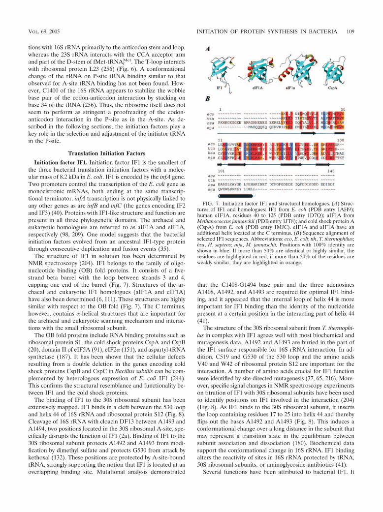
Initiation factor IF1. Initiation factor IF1 is the smallest ofthe three bacterial translation initiation factors with a molec-ular mass of 8.2 kDa in E. coli. IF1 is encoded by the infA gene.Two promoters control the transcription of the E. coli gene asmonocistronic mRNAs, both ending at the same transcrip-tional terminator. infA transcription is not physically linked toany other genes as are infB and infC (the genes encoding IF2and IF3) (40). Proteins with IF1-like structure and function arepresent in all three phylogenetic domains. The archaeal andeukaryotic homologues are referred to as aIF1A and eIF1A,respectively (98, 209). One model suggests that the bacterialinitiation factors evolved from an ancestral IF1-type proteinthrough consecutive duplication and fusion events (35).
The structure of IF1 in solution has been determined byNMR spectroscopy (204). IF1 belongs to the family of oligo-nucleotide binding (OB) fold proteins. It consists of a five-strand beta barrel with the loop between strands 3 and 4,capping one end of the barrel (Fig. 7). Structures of the ar-chaeal and eukaryotic IF1 homologues (aIF1A and eIF1A)have also been determined (6, 111). These structures are highlysimilar with respect to the OB fold (Fig. 7). The C terminus,however, contains �-helical structures that are important forthe archaeal and eukaryotic scanning mechanism and interac-tions with the small ribosomal subunit.
The OB fold proteins include RNA binding proteins such asribosomal protein S1, the cold shock proteins CspA and CspB(20), domain II of eIF5A (91), eIF2� (151), and aspartyl-tRNAsynthetase (187). It has been shown that the cellular defectsresulting from a double deletion in the genes encoding coldshock proteins CspB and CspC in Bacillus subtilis can be com-plemented by heterologous expression of E. coli IF1 (244).This confirms the structural resemblance and functionality be-tween IF1 and the cold shock proteins.
The binding of IF1 to the 30S ribosomal subunit has beenextensively mapped. IF1 binds in a cleft between the 530 loopand helix 44 of 16S rRNA and ribosomal protein S12 (Fig. 8).Cleavage of 16S rRNA with cloacin DF13 between A1493 andA1494, two positions located in the 30S ribosomal A-site, spe-cifically disrupts the function of IF1 (2a). Binding of IF1 to the30S ribosomal subunit protects A1492 and A1493 from modi-fication by dimethyl sulfate and protects G530 from attack bykethoxal (132). These positions are protected by A-site-boundtRNA, strongly supporting the notion that IF1 is located at anoverlapping binding site. Mutational analysis demonstrated
that the C1408-G1494 base pair and the three adenosinesA1408, A1492, and A1493 are required for optimal IF1 bind-ing, and it appeared that the internal loop of helix 44 is moreimportant for IF1 binding than the identity of the nucleotidepresent at a certain position in the interacting part of helix 44(41).
The structure of the 30S ribosomal subunit from T. thermophi-lus in complex with IF1 agrees well with most biochemical andmutagenesis data. A1492 and A1493 are buried in the part ofthe IF1 surface responsible for 16S rRNA interaction. In ad-dition, C519 and G530 of the 530 loop and the amino acidsV40 and W42 of ribosomal protein S12 are important for theinteraction. A number of amino acids crucial for IF1 functionwere identified by site-directed mutagenesis (37, 65, 216). More-over, specific signal changes in NMR spectroscopy experimentson titration of IF1 with 30S ribosomal subunits have been usedto identify positions on IF1 involved in the interaction (204)(Fig. 8). As IF1 binds to the 30S ribosomal subunit, it insertsthe loop containing residues 17 to 25 into helix 44 and therebyflips out the bases A1492 and A1493 (Fig. 8). This induces aconformational change over a long distance in the subunit thatmay represent a transition state in the equilibrium betweensubunit association and dissociation (180). Biochemical datasupport the conformational change in 16S rRNA. IF1 bindingalters the reactivity of sites in 16S rRNA protected by tRNA,50S ribosomal subunits, or aminoglycoside antibiotics (41).
Several functions have been attributed to bacterial IF1. It
FIG. 7. Initiation factor IF1 and structural homologues. (A) Struc-tures of IF1 and homologues: IF1 from E. coli (PDB entry 1AH9);human eIF1A, residues 40 to 125 (PDB entry 1D7Q); aIF1A fromMethanococcus jannaschii (PDB entry 1JT8); and cold shock protein A(CspA) from E. coli (PDB entry 1MJC). eIF1A and aIF1A have anadditional helix located at the C terminus. (B) Sequence alignment ofselected IF1 sequences. Abbreviations: eco, E. coli; tth, T. thermophilus;hsa, H. sapiens; mja, M. jannaschii. Positions with 100% identity areshown in blue. If more than 50% are identical or highly similar, theresidues are highlighted in red; if more than 50% of the residues areweakly similar, they are highlighted in orange.
VOL. 69, 2005 INITIATION OF PROTEIN SYNTHESIS IN BACTERIA 109

enhances the dissociation and association rate for 70S ribo-somes (46, 59), primarily through the stimulating effect on theactivity of IF2 and IF3 (174). Interaction between IF2 and the30S ribosomal subunit is favored when IF1 is bound, and therelease of IF2 is indirectly promoted when IF1 is ejected (27,136, 222). IF1 cooperates with IF2 to ensure that only theinitiator tRNA binds to the P-site and that it interacts with theinitiation codon of the mRNA (24, 73, 127, 251). IF1 occludetRNAs from the A-site until the 70S initiation complex hasformed. Ejection of IF1 consequently opens the A-site forincoming aminoacyl-tRNAs. In vivo studies have shown thatIF1-depleted cells have low growth rates and short polysomes(39). These data demonstrate that IF1 is essential for cellviability and suggest that one or more of its functions arecrucial. However, no clear function has been assigned to theinitiation factor yet (37).
Initiation factor IF2. IF2 is the largest of the initiation fac-tors. It is encoded by the infB gene. The infB gene is part of thepolycistronic nusA operon containing metY (minor form of theinitiator tRNA), ylxC (protein of unknown function), nusA (atranscriptional termination factor), infB (translation initiationfactor IF2), rbfA (ribosome binding factor A), truB (tRNApseudouridine 5S synthase), rpsO (ribosomal protein S15), andpnp (polynucleotide phosphorylase). All these genes are tran-scribed from metY toward pnp on a part of the DNA thatcontains several transcriptional promoters (172, 173, 189, 193).Transcription of the nusA/infB operon occurs primarily from apromoter separated by three genes upstream from infB. Thispromoter is autogenously controlled by the translation productof nusA (143, 171). The organization of nusA and infB inbacteria reveals that they are simultaneously present in oper-ons. The rather conserved distribution of the genes within theoperon leads to the proposal that this organization may beimportant for regulation (241).
Three isoforms of the initiation factor, named IF2-1 (97.3
kDa), IF2-2 (79.7 kDa), and IF2-3 (78.8 kDa), exist in E. coliand other members of the family Enterobacteriaceae (107, 154).Bacillus subtilis is the only organism that does not belong to theEnterobacteriaceae where more than one isoform of IF2 hasbeen experimentally demonstrated (81). IF2-1, IF2-2, andIF2-3 are translated from three independent but in-frametranslational start sites of the infB mRNA. This feature hasbeen referred to as tandem translation. Hence, IF2-2 and IF2-3differ from IF2-1 only by the absence of the first 157 and 164amino acid residues, respectively (Fig. 9) (139). The presenceof both the large and smaller isoforms is required for optimalgrowth of E. coli. The cellular content of IF2-2 and IF2-3 isclose to the level of IF2-1 at optimal growth conditions (79,191), but the ratio of IF2-2 and IF2-3 to IF2-1 increases as aresponse to cold shock (53). An open single-stranded structureis present in the intracistronic TIR of the infB mRNA. Wesuggest that this structure is required for the translation ofIF2-2 and IF2-3 (107).
IF2 can be divided into domains based on interspecies ho-mology. The domain nomenclature differs somewhat amongspecies. Throughout this review, the nomenclature for E. colisuggested by Mortensen et al. is used (Fig. 9) (140). DomainVI was subsequently divided into two subdomains, designatedVI-1 and VI-2.
The factor can be divided into a conserved C-terminal re-gion consisting of domains IV to VI and a less highly conservedN-terminal region corresponding to domains I to III (209,217). Homologues of IF2 have been found in archaeabacteriaand eukaryotes, where the factor is referred to as aIF5B andeIF5B, respectively (98). Remarkable interspecies homology inthe C-terminal region is present among the homologues (209).The homologues have functions similar to those of bacterialIF2, including GTPase activity, promotion of ribosomal sub-unit association, and probably interaction with the initiatortRNA (30, 165).
FIG. 8. IF1 bound to the 30S ribosomal subunit. (A) Structure of IF1 on the 30S ribosomal subunit. IF1 is shown in blue, helix 44 is shown inmagenta, the 530 loop is shown in yellow, and protein S12 is shown in green. The structure is derived from PDB entry 1HR0. (B) Close-up of theinteraction of IF1 with the 30S subunit. IF1 is shown in a surface representation colored according to the electrostatic potential (positive charges,blue; negative charges, red). Helix 44 and the 530 loop of 16S rRNA are shown in magenta and yellow, respectively. Protein S12 is shown in a greenribbon representation. Bases A1492 and A1493 of the 16S rRNA are indicated in red. Note that they have flipped out of helix 44 and are buriedin a pocket in IF1 and a pocket between IF1 and S12, respectively.
110 LAURSEN ET AL. MICROBIOL. MOL. BIOL. REV.

FIG. 9. IF2 and structural homologues. (A) Schematic representation of the E. coli IF2 primary structure. The domain boundaries and the lengthsof the three IF2 isoforms are indicated. Ribbon diagrams of the structures of the IF2N domain from E. coli (PDB entry 1ND9) and the IF2homologue aIF5B from M. thermoautotrophicum (PDB entry 1G7T) are shown. The domains are indicated in different colors, and the E. coli domainnomenclature is used. (B) Sequence alignment of selected bacterial IF2 and archaeal and eukaryotic homologues. Only a small part of theN-terminal nonconserved region is shown. Abbreviations: tth; T. thermophilus, bst, B. stearothermophilus; eco, E. coli; mth, M. thermoautotrophicum; hsa,H. sapiens. Secondary-structure elements defined from the structure of aIF5B from M. thermoautotrophicum are indicated by cylinders for helicalsegments and arrows for segments in �-strand conformation. The domain boundaries are indicated by yellow arrows. Color codes are as in Fig. 7.
111
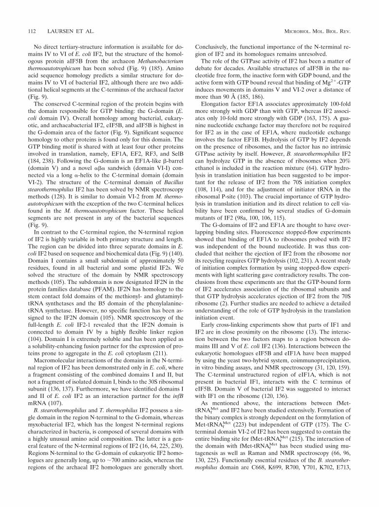
No direct tertiary-structure information is available for do-mains IV to VI of E. coli IF2, but the structure of the homol-ogous protein aIF5B from the archaeon Methanobacteriumthermoautotrophicum has been solved (Fig. 9) (185). Aminoacid sequence homology predicts a similar structure for do-mains IV to VI of bacterial IF2, although there are two addi-tional helical segments at the C-terminus of the archaeal factor(Fig. 9).
The conserved C-terminal region of the protein begins withthe domain responsible for GTP binding: the G-domain (E.coli domain IV). Overall homology among bacterial, eukary-otic, and archaeabacterial IF2, eIF5B, and aIF5B is highest inthe G-domain area of the factor (Fig. 9). Significant sequencehomology to other proteins is found only for this domain. TheGTP binding motif is shared with at least four other proteinsinvolved in translation, namely, EF1A, EF2, RF3, and SelB(184, 238). Following the G-domain is an EF1A-like �-barrel(domain V) and a novel ��� sandwich (domain VI-I) con-nected via a long �-helix to the C-terminal domain (domainVI-2). The structure of the C-terminal domain of Bacillusstearothermophilus IF2 has been solved by NMR spectroscopymethods (128). It is similar to domain VI-2 from M. thermo-autotrophicum with the exception of the two C-terminal helicesfound in the M. thermoautotrophicum factor. These helicalsegments are not present in any of the bacterial sequences(Fig. 9).
In contrast to the C-terminal region, the N-terminal regionof IF2 is highly variable in both primary structure and length.The region can be divided into three separate domains in E.coli IF2 based on sequence and biochemical data (Fig. 9) (140).Domain I contains a small subdomain of approximately 50residues, found in all bacterial and some plastid IF2s. Wesolved the structure of the domain by NMR spectroscopymethods (105). The subdomain is now designated IF2N in theprotein families database (PFAM). IF2N has homology to thestem contact fold domains of the methionyl- and glutaminyl-tRNA synthetases and the B5 domain of the phenylalanine-tRNA synthetase. However, no specific function has been as-signed to the IF2N domain (105). NMR spectroscopy of thefull-length E. coli IF2-1 revealed that the IF2N domain isconnected to domain IV by a highly flexible linker region(104). Domain I is extremely soluble and has been applied asa solubility-enhancing fusion partner for the expression of pro-teins prone to aggregate in the E. coli cytoplasm (211).
Macromolecular interactions of the domains in the N-termi-nal region of IF2 has been demonstrated only in E. coli, wherea fragment consisting of the combined domains I and II, butnot a fragment of isolated domain I, binds to the 30S ribosomalsubunit (136, 137). Furthermore, we have identified domains Iand II of E. coli IF2 as an interaction partner for the infBmRNA (107).
B. stearothermophilus and T. thermophilus IF2 possess a sin-gle domain in the region N-terminal to the G-domain, whereasmyxobacterial IF2, which has the longest N-terminal regionscharacterized in bacteria, is composed of several domains witha highly unusual amino acid composition. The latter is a gen-eral feature of the N-terminal regions of IF2 (16, 64, 225, 230).Regions N-terminal to the G-domain of eukaryotic IF2 homo-logues are generally long, up to �700 amino acids, whereas theregions of the archaeal IF2 homologues are generally short.
Conclusively, the functional importance of the N-terminal re-gion of IF2 and its homologues remains unresolved.
The role of the GTPase activity of IF2 has been a matter ofdebate for decades. Available structures of aIF5B in the nu-cleotide free form, the inactive form with GDP bound, and theactive form with GTP bound reveal that binding of Mg2�-GTPinduces movements in domains V and VI-2 over a distance ofmore than 90 A (185, 186).
Elongation factor EF1A associates approximately 100-foldmore strongly with GDP than with GTP, whereas IF2 associ-ates only 10-fold more strongly with GDP (163, 175). A gua-nine nucleotide exchange factor may therefore not be requiredfor IF2 as in the case of EF1A, where nucleotide exchangeinvolves the factor EF1B. Hydrolysis of GTP by IF2 dependson the presence of ribosomes, and the factor has no intrinsicGTPase activity by itself. However, B. stearothermophilus IF2can hydrolyze GTP in the absence of ribosomes when 20%ethanol is included in the reaction mixture (64). GTP hydro-lysis in translation initiation has been suggested to be impor-tant for the release of IF2 from the 70S initiation complex(108, 114), and for the adjustment of initiator tRNA in theribosomal P-site (103). The crucial importance of GTP hydro-lysis in translation initiation and its direct relation to cell via-bility have been confirmed by several studies of G-domainmutants of IF2 (98a, 100, 106, 115).
The G-domains of IF2 and EF1A are thought to have over-lapping binding sites. Fluorescence stopped-flow experimentsshowed that binding of EF1A to ribosomes probed with IF2was independent of the bound nucleotide. It was thus con-cluded that neither the ejection of IF2 from the ribosome norits recycling requires GTP hydrolysis (102, 231). A recent studyof initiation complex formation by using stopped-flow experi-ments with light scattering gave contradictory results. The con-clusions from these experiments are that the GTP-bound formof IF2 accelerates association of the ribosomal subunits andthat GTP hydrolysis accelerates ejection of IF2 from the 70Sribosome (2). Further studies are needed to achieve a detailedunderstanding of the role of GTP hydrolysis in the translationinitiation event.
Early cross-linking experiments show that parts of IF1 andIF2 are in close proximity on the ribosome (13). The interac-tion between the two factors maps to a region between do-mains III and V of E. coli IF2 (136). Interactions between theeukaryotic homologues eIF5B and eIF1A have been mappedby using the yeast two-hybrid system, coimmunoprecipitation,in vitro binding assays, and NMR spectroscopy (31, 120, 159).The C-terminal unstructured region of eIF1A, which is notpresent in bacterial IF1, interacts with the C terminus ofeIF5B. Domain V of bacterial IF2 was suggested to interactwith IF1 on the ribosome (120, 136).
As mentioned above, the interactions between fMet-tRNAf
Met and IF2 have been studied extensively. Formation ofthe binary complex is strongly dependent on the formylation ofMet-tRNAf
Met (223) but independent of GTP (175). The C-terminal domain VI-2 of IF2 has been suggested to contain theentire binding site for fMet-tRNAf
Met (215). The interaction ofthe domain with fMet-tRNAf
Met has been studied using mu-tagenesis as well as Raman and NMR spectroscopy (66, 96,130, 225). Functionally essential residues of the B. stearother-mophilus domain are C668, K699, R700, Y701, K702, E713,
112 LAURSEN ET AL. MICROBIOL. MOL. BIOL. REV.
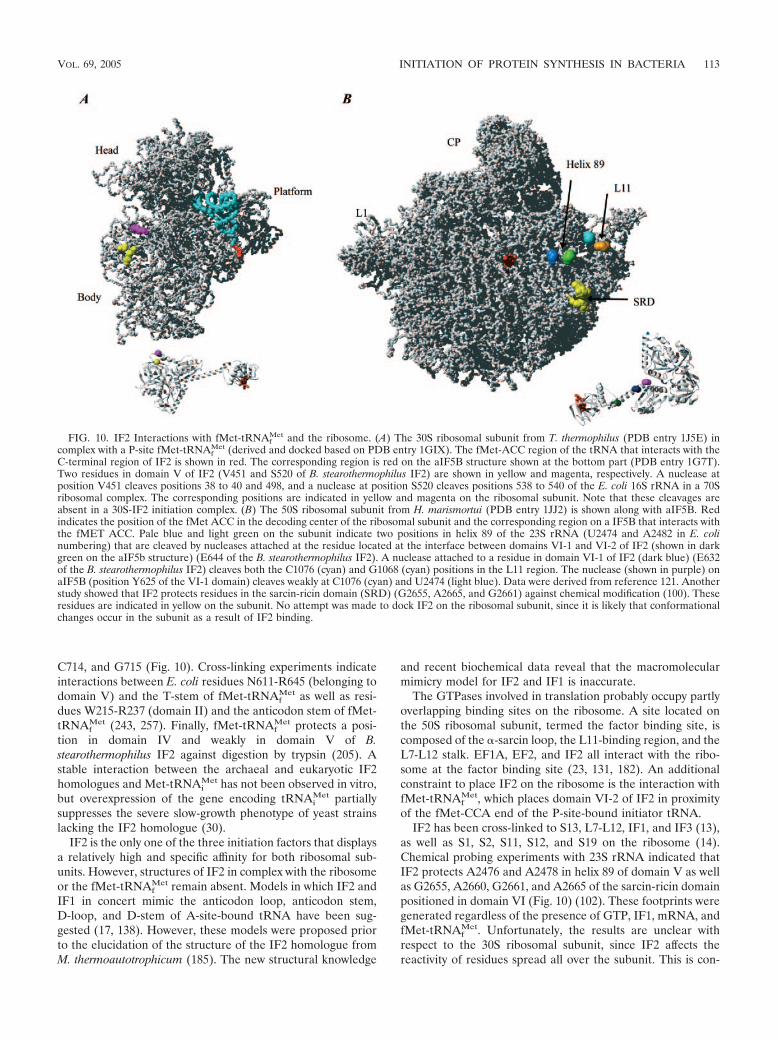
C714, and G715 (Fig. 10). Cross-linking experiments indicateinteractions between E. coli residues N611-R645 (belonging todomain V) and the T-stem of fMet-tRNAf
Met as well as resi-dues W215-R237 (domain II) and the anticodon stem of fMet-tRNAf
Met (243, 257). Finally, fMet-tRNAfMet protects a posi-
tion in domain IV and weakly in domain V of B.stearothermophilus IF2 against digestion by trypsin (205). Astable interaction between the archaeal and eukaryotic IF2homologues and Met-tRNAi
Met has not been observed in vitro,but overexpression of the gene encoding tRNAi
Met partiallysuppresses the severe slow-growth phenotype of yeast strainslacking the IF2 homologue (30).
IF2 is the only one of the three initiation factors that displaysa relatively high and specific affinity for both ribosomal sub-units. However, structures of IF2 in complex with the ribosomeor the fMet-tRNAf
Met remain absent. Models in which IF2 andIF1 in concert mimic the anticodon loop, anticodon stem,D-loop, and D-stem of A-site-bound tRNA have been sug-gested (17, 138). However, these models were proposed priorto the elucidation of the structure of the IF2 homologue fromM. thermoautotrophicum (185). The new structural knowledge
and recent biochemical data reveal that the macromolecularmimicry model for IF2 and IF1 is inaccurate.
The GTPases involved in translation probably occupy partlyoverlapping binding sites on the ribosome. A site located onthe 50S ribosomal subunit, termed the factor binding site, iscomposed of the �-sarcin loop, the L11-binding region, and theL7-L12 stalk. EF1A, EF2, and IF2 all interact with the ribo-some at the factor binding site (23, 131, 182). An additionalconstraint to place IF2 on the ribosome is the interaction withfMet-tRNAf
Met, which places domain VI-2 of IF2 in proximityof the fMet-CCA end of the P-site-bound initiator tRNA.
IF2 has been cross-linked to S13, L7-L12, IF1, and IF3 (13),as well as S1, S2, S11, S12, and S19 on the ribosome (14).Chemical probing experiments with 23S rRNA indicated thatIF2 protects A2476 and A2478 in helix 89 of domain V as wellas G2655, A2660, G2661, and A2665 of the sarcin-ricin domainpositioned in domain VI (Fig. 10) (102). These footprints weregenerated regardless of the presence of GTP, IF1, mRNA, andfMet-tRNAf
Met. Unfortunately, the results are unclear withrespect to the 30S ribosomal subunit, since IF2 affects thereactivity of residues spread all over the subunit. This is con-
FIG. 10. IF2 Interactions with fMet-tRNAfMet and the ribosome. (A) The 30S ribosomal subunit from T. thermophilus (PDB entry 1J5E) in
complex with a P-site fMet-tRNAfMet (derived and docked based on PDB entry 1GIX). The fMet-ACC region of the tRNA that interacts with the
C-terminal region of IF2 is shown in red. The corresponding region is red on the aIF5B structure shown at the bottom part (PDB entry 1G7T).Two residues in domain V of IF2 (V451 and S520 of B. stearothermophilus IF2) are shown in yellow and magenta, respectively. A nuclease atposition V451 cleaves positions 38 to 40 and 498, and a nuclease at position S520 cleaves positions 538 to 540 of the E. coli 16S rRNA in a 70Sribosomal complex. The corresponding positions are indicated in yellow and magenta on the ribosomal subunit. Note that these cleavages areabsent in a 30S-IF2 initiation complex. (B) The 50S ribosomal subunit from H. marismortui (PDB entry 1JJ2) is shown along with aIF5B. Redindicates the position of the fMet ACC in the decoding center of the ribosomal subunit and the corresponding region on a IF5B that interacts withthe fMET ACC. Pale blue and light green on the subunit indicate two positions in helix 89 of the 23S rRNA (U2474 and A2482 in E. colinumbering) that are cleaved by nucleases attached at the residue located at the interface between domains VI-1 and VI-2 of IF2 (shown in darkgreen on the aIF5b structure) (E644 of the B. stearothermophilus IF2). A nuclease attached to a residue in domain VI-1 of IF2 (dark blue) (E632of the B. stearothermophilus IF2) cleaves both the C1076 (cyan) and G1068 (cyan) positions in the L11 region. The nuclease (shown in purple) onaIF5B (position Y625 of the VI-1 domain) cleaves weakly at C1076 (cyan) and U2474 (light blue). Data were derived from reference 121. Anotherstudy showed that IF2 protects residues in the sarcin-ricin domain (SRD) (G2655, A2665, and G2661) against chemical modification (100). Theseresidues are indicated in yellow on the subunit. No attempt was made to dock IF2 on the ribosomal subunit, since it is likely that conformationalchanges occur in the subunit as a result of IF2 binding.
VOL. 69, 2005 INITIATION OF PROTEIN SYNTHESIS IN BACTERIA 113

sistent with an observed rearrangement of the subunit inducedby IF2 (242). Recently, a model of IF2 binding to the ribosomewas presented, based on cleavage of the rRNA by chemicalnucleases tethered to cysteine residues introduced at specificsites of IF2 (121). No cleavage of the 16S rRNA was observedwhen IF2 was bound to 30S ribosomal subunits or to thecomplete 30S initiation complex. However, cleavage of the 16SrRNA was observed when IF2 was bound to the 70S initiationcomplex (Fig. 10). These data indicate that domain V of IF2 islocalized toward the 30S subunit in the 70S initiation complex.As described above, cross-linking data of the 30S complex andfootprinting data on the binary fMet-tRNAf
Met–IF2 complexplace domain V of IF2 in proximity to the elbow of the P-site-bound fMet-tRNAf
Met. The distance between the 16S rRNAand the elbow of the fMet-tRNAf
Met appears to be too far fordomain V of IF2 to establish contact with both simultaneously.Conclusively, IF2 changes localization during the transitionfrom the 30S to the 70S initiation complex (121). The cleavageexperiments were performed in the presence of excess GTP.Large domain movements take place in IF2 during GTP hy-drolysis (185), and the cleavage patterns in the rRNA might bedependent on whether IF2 is in the GTP- or GDP-bound form.To fully understand the function of IF2 during translationinitiation, detailed atomic resolution structures of both the 30Sand 70S initiation complexes as well as a better understandingof the timing and not least the consequences of GTP hydrolysisare needed.
Besides the function as a translation factor, IF2 has theproperties of a chaperone. It promotes functional folding ofproteins and forms stable complexes with unfolded proteins(22). Furthermore, the expression of IF2 is upregulated duringthe cold shock response (3), and the factor is important for thetranslation of leaderless transcripts (57). Finally, we have in-troduced the use of IF2 sequence data for the classification oforganisms of closely related organisms (75–77, 152, 209, 226).
Initiation factor IF3. E. coli IF3 is a 20.4-kDa protein com-posed of 180 amino acids encoded by the essential infC gene(160, 190). The infC-rpmI-rplT operon contains the genes en-coding IF3 and the two ribosomal proteins L35 and L20 (28,110). These genes are transcribed from four promoters andterminated by two transcriptional terminators (110, 245). Atthe translational level, the expression from the operon is reg-ulated by two different control circuits, discussed further in thelast section of this review. Whereas IF1 and IF2 are universallypresent and important for the function of all living cells, IF3 islimited to a number of bacterial species and has been found insome plastids (112, 254, 255). The human mitochondrial IF3mt
has short extensions in the N and C termini surrounding aregion homologous to bacterial IF3. It promotes initiationcomplex formation on mitochondrial ribosomes (92).
IF3 is composed of two structural domains of approximatelyequal size (Fig. 11) (48, 97). The two domains, called the IF3Nand IF3C, are separated by a � 45-A lysine-rich flexible linker(80, 135). The IF3N domain consists of a globular �/�-fold,with helix �1 packed against a mixed five-strand �-sheet (Fig.11). This fold is followed by helix �2, which connects IF3N toIF3C. The length of �2 was found to be different in the struc-tures derived from NMR spectroscopy (51) and X-ray diffrac-tion (11) experiments and has been the subject of debate (re-viewed in reference 12). The linker is essential for IF3
function, but variation of its length and composition does notconsiderably change the activity (43).
The structure of IF3C has been solved by NMR spectros-copy and X-ray diffraction (11, 51). It consists of a two-layer�/� sandwich fold composed of a four-strand mixed �-sheetpacked against two parallel �-helices (�3 and �4), leading to a������ topology (Fig. 11). The structure is similar to U1A(RNA binding protein involved in RNA splicing) (51) andYppH (a protein involved in cell division) (88).
IF3 perform several different functions. (i) It prevents asso-ciation of the ribosomal subunits by binding to the 30S subunit,thereby blocking binding of the 50S subunit (59, 188). (ii) Itmonitors the codon-anticodon interaction by promoting thedissociation of fMet-tRNAf
Met from initiation complexes
FIG. 11. IF3 structure and alignment. (A) Structures of the IF3Ndomain from B. stearothermophilus (PDB entry 1TIF) and the IF3Cdomain from E. coli (PDB entry 2IFE). The side chains of the arginineresidues in the IF3C domain are shown and labeled with the residuenumber. Mutations in the arginine residues that affect binding to the30S ribosomal subunit are residue numbers 99, 112, 116, 147, andpossibly 168. These roughly define the surface that binds to the 30Sribosomal subunit. Mutations of arginine residues reducing IF3 activityinvolved in mRNA-related functions define a surface comprising res-idues 129, 131, and 133. (B) Sequence alignment of selected sequencesof IF3. Abbreviations, color codes, and secondary-structure nomencla-ture are as in Fig. 7. Secondary-structure elements are as defined inreference 203. Black vertical arrows indicate residues that have beenidentified as interacting with the 30S ribosomal subunit by mutagenesisand/or chemical modification. Grey triangles indicate residues whoseintensity was most strongly affected by titration with 30S ribosomalsubunits in NMR spectroscopy studies (reference 203 and referencescited therein). Yellow triangles indicate approximate domain bound-aries.
114 LAURSEN ET AL. MICROBIOL. MOL. BIOL. REV.

formed at the 5� initiation codon of leaderless mRNAs (227).Likewise, initiation complexes with an incorrectly bound ami-noacyl-tRNA (noninitiator tRNA) (73, 74) and complexes withtriplets other than AUG, GUG, and UUG in the P-site aredissociated by IF3 (70, 127, 224). (iii) It stimulates the rapidformation of codon-anticodon interaction at the ribosomal P-site (60, 250). (iv) It is involved in the adjustment of the mRNAfrom the standby site to the decoding P-site of the 30S ribo-somal subunit (101). Finally, a role for IF3 in recycling ofsubunits has been proposed. It was observed to enhance thedissociation of deacylated tRNAs from posttermination com-plexes and to dissociate 70S ribosomes into subunits (78, 87).
All functions of the native IF3 can be accomplished by theisolated IF3C domain in vitro, while the IF3N domain proba-bly serves the purpose of modulating the thermodynamic sta-bility of the IF3-30S complexes (169). Site-directed mutagen-esis of the eight arginine residues in the IF3C domain has beenused to map the active sites (168). The arginines at positions99, 112, 116, 147, and 168 are important for the binding to the30S ribosomal subunit (Fig. 11). The ability of IF3 to dissociatethe ribosome into subunits was affected mainly by mutations ofR112 and R147 (and less extensively by mutations of R99 andR116). The stimulation of the pseudoinitiation complex disso-ciation (with a noninitiator tRNA bound) was affected by mu-tations of R99 and R112 (and less extensively of the arginineresidues at positions 116, 129, 133, and 147). Dissociation ofnoncanonical initiation complexes (initiation codons otherthan AUG, GUG, and UUG) was not affected in any of themutants. Stimulation of translation was affected by mutationsof R116 and R129 (and less extensively of the arginine residuesat positions 99, 112, and 131), whereas inhibition of nonca-nonical mRNA translation was affected by mutations of R99,R112, and R168 (and less extensively of the arginine residuesat positions 116, 129, and 131). Finally, the repositioning of themRNA from the standby site to the P-decoding site was weaklyaffected by mutations of the arginine residues at positions 129,131, 133, 147, and 168. The data indicate that IF3C contains atleast two active surfaces, one embedded in the 30S subunit andthe other facing the mRNA (Fig. 11) (168).
Both IF3 domains are RNA binding and interact indepen-dently with the 30S ribosomal subunit. IF3C interacts with thehighest affinity through a large surface of symmetrically dis-tributed residues in loops and �-helices, whereas IF3N inter-acts mainly via a small number of asymmetrically distributedresidues (203). Results of mapping of IF3 residues implicatedin binding to the 30S ribosomal subunit by NMR spectroscopy,site-directed mutagenesis, and other chemical methods are inexcellent agreement (203) (Fig. 11).
The localization of IF3 on the 30S ribosomal subunit hasbeen studied by various methods, with conflicting results. Im-muno-EM located the factor at the cleft of the 30S ribosomalsubunit (220). The ribosomal proteins S7, S11, S12, S13, S18,S19, and S21 have been cross-linked to IF3 but are spread overa wide area of the 30S subunit (13, 33, 34, 117). Helices 26(central domain) and 45 (3� minor domain) of the 16S rRNAhave been cross-linked to IF3 (47). Chemical probing revealedIF3 contacts to helices 23 and 24 in the central domain of the16S rRNA (132, 141), and NMR spectroscopy indicated thatIF3 interacts with the 3� end of the 16S rRNA (246). Cryo-EMlocated the IF3C domain to the interface side of the small
ribosomal subunit (124). However, X-ray diffraction of 30Sribosomal subunit crystals soaked with IF3C places the domainat the solvent side of the platform (170).
A model based on hydroxyl radical footprinting and directedprobing from Fe(II)-derivatized IF3 has been presented (42).This model is in agreement with the cryo-EM data, and theresults are summarized in Fig. 12. It was suggested that theobservations in the crystallographic studies represent bindingto a secondary site in the crystal-soaking experiments as aresult of blockage of the primary binding site by crystal con-tacts. IF3C is located in the same area as helix 69 of the 23SrRNA in the 70S ribosome, which explains why IF3 blockssubunit association.
The two domains of IF3 were shown to be on opposite sidesof the fMet-tRNAf
Met (42). IF3 has been thought to interactwith the anticodon stem and loop of fMet-tRNAf
Met (73). How-ever, IF3 is unable to reach the three conserved discriminatorGC base pairs in the anticodon stem of fMet-tRNAf
Met in thecurrent model. Hence, discrimination against elongator tRNAspromoted by IF3 is probably indirect (42).
REGULATION OF TRANSLATION INITIATION
Bacteria must be able to adjust to environmental changes intemperature, the availability of nutrients and water, presenceof toxic molecules, etc. A prerequisite for induction of anappropriate stress response is precise monitoring of internaland external parameters. Although transcriptional regulationis the primary mechanism in stress responses, regulation oftranslation is faster and consequently important. Post-tran-scriptional regulation occurs at different stages includingmRNA stability and translation initiation. Here we focus onresponses involving the translation initiation phase.
Regulation occurs by a variety of events that control theformation of elongation-competent translation initiation com-plexes. The only variable component in translation initiation isthe mRNA. The sequence and structure of the mRNA deter-mine its interaction with the translational machinery and hencethe efficiency and frequency of translation. A highly expressedmRNA contains some or all of the following elements at theRBS: (i) a cognate initiation codon for interaction with thefMet-tRNAf
Met (183, 237); (ii) an SD sequence complementaryto the ASD sequence of the 16S rRNA (207); (iii) a pyrimidinetract for interaction with ribosomal protein S1 (15, 232, 260);and (iv) base-specific enhancer elements upstream (155) ordownstream (213, 214) of the initiation codon. The interde-pendence and relative importance of these mRNA elementsare poorly understood (234). Translational regulation can in-volve cis-acting elements of the mRNA that form secondary ortertiary structures which sequester the ribosomal binding site.trans-Acting elements include protein, antisense RNA, andother factors that control the alternative structures of the RBSand thus affect the efficiency of initiation complex formation. Arecent review describes the translational repression mecha-nisms (194).
Initial binding of the mRNA RBS to the ribosome occursprimarily through interactions with the ribosomal protein S1and the ASD sequence of the 16S rRNA. Both interactionsrequire local single-stranded mRNA (reference 194 and refer-ences cited therein). Secondary structures in the RBS can
VOL. 69, 2005 INITIATION OF PROTEIN SYNTHESIS IN BACTERIA 115

lower the translational efficiency of an mRNA. Figure 13 sum-marizes some mechanisms for translational repression and ac-tivation caused by changes in the secondary structure of themRNA. The thermodynamic stability of secondary structuresin the RBS plays a crucial role, but the kinetics of the mRNAfolding is also an important factor (reference 194 and refer-ences cited therein). This was demonstrated for the phage MS2maturation protein. The mRNA contains a leader sequenceforming a cloverleaf structure, which inhibits translation initi-ation. The effects of mRNA renaturation time on translationinitiation were studied, and it was observed that the formationof the cloverleaf structure is slower than the formation ofinitiation complexes on the mRNA (176).
The interactions involved in forming the secondary and ter-tiary structures of mRNA are sensitive to temperature. Struc-tural changes of the mRNA as a consequence of temperaturechanges may regulate the translational activity of mRNAs. Thisis especially seen for the mRNAs involved in the expression ofheat shock genes. The E. coli heat shock factor 32 is thebest-characterized example (reference 194 and referencescited therein). The structure of the RBS of this mRNA isextremely sensitive to changes in temperature near 42°C.
Regulation is also mediated by trans-acting factors that sta-bilize or destabilize mRNA structures. A typical example is theautoregulation of the S10 operon mediated by the ribosomalprotein L4. L4 is encoded by the S10 operon and stabilizes ahairpin in the mRNA, which represses translation (45). Aprotein is usually responsible for the regulation, but recentexamples show that other molecules can control the expressionof an mRNA. For example, thiamine (also known as vitamin
B1) controls the expression of the genes involved in thiaminebiosynthesis via the thi box of the mRNA (129). Similar modelshave been proposed for cobalamin and vitamin B12, indicatingthat regulation can be conferred not exclusively by proteins butalso by small molecules (142, 221). The mRNA elements thatdirectly monitor environmental conditions have been termedriboswitches and are involved in several metabolic pathways(for example, biosynthesis of vitamins and metabolism of me-thionine, lysine, and purines) (240). Intermolecular RNA in-teractions also play an important regulatory role. For example,the translation initiation site of the mRNA of outer membraneprotein F (OmpF) in E. coli can be blocked by a naturalantisense RNA that is transcribed from the micF gene in re-sponse to changes in osmolarity (142, 221).
Protein synthesis needs to be tightly coupled to the nutri-tional conditions met by the cell. The cellular content of GTPis an indicator of the overall nutritional conditions. GTP levelshave been proposed to be directly coupled to the activity ofIF2, which is active only in the GTP-bound form (12). mRNA-mediated detection of environmental conditions has been re-viewed previously (32, 153, 240).
Regulation by competition is also a common method ofregulation. For example, the threonyl-tRNA synthetase re-presses its own expression by binding to the RBS of the mRNA(214). The homodimeric tRNA synthetase recognizes two do-mains in the mRNA that structurally mimic the anticodon armof tRNAThr. Thus, if excess threonyl-tRNA synthetase ispresent it binds to its own mRNA and represses expression,whereas if excess tRNAThr is present the synthetase binds tothe tRNAThr instead of the mRNA and translation will be
FIG. 12. Interaction of IF3 with the 30S ribosomal subunit. The interactions between IF3 and the 30S ribosomal subunit identified by hydroxylradical footprinting and directed probing are shown (data from reference 42). (A) 30S ribosomal subunit from T. thermophilus with P-site-boundtRNA (derived from PDB entries 1J5E and 1GIX). The tRNA is shown in yellow. (B) Close-up of the ribosomal subunit. Sites in the 16S rRNAand on the tRNA that are cleaved by nucleases attached to IF3 are indicated. The IF3N and IF3C domains (PDB entries 1TIF and 2IFE) are shownin a ribbon representation, with the modified cysteines indicated by spheres. Orange spheres on the IF3 domains indicate residues where anattached nuclease does not cleave the 16S rRNA or tRNA. The magenta spheres in the IF3C domain indicate residues K97 and MI35 from wherenucleases cleave in the 790 loop of the 16S rRNA (magenta) seen below the acceptor arm of the tRNA. Nucleases at these positions also cleavethe side of the tRNA marked in blue (residues 3 to 5 and 12 to 24). The cyan sphere in the IF3C domain indicates E104 from which nucleasescleave in the 790 loop (magenta) and at residues 1482 to 1487, indicated in cyan on the 30S subunit. Green spheres in the linker region on theIF3N domain indicate residues (E76 and S80) on which nucleases cleave on the other side of the tRNA, marked in green (residues 26 to 29 and35 to 37), the 790 loop region (magenta), and the 690 loop region (red) of the 16S rRNA. A nuclease attached to position R11 in the IF3N domain(red) cleaves at positions in the 690 loop (red). No attempt was made to dock IF3 on the ribosomal subunit, since conformational changes mostprobably take place in the subunit as a result of IF3 binding.
116 LAURSEN ET AL. MICROBIOL. MOL. BIOL. REV.

promoted. The mechanism of the repression caused by pro-teins that bind to their own mRNA is in some cases morecomplicated. Ribosomal protein S15 selectively binds to itsown mRNA when it is in a pseudoknot conformation; an in-active ternary complex of S15, the mRNA, and the 30S ribo-somal subunit is then formed. This prevents the formation ofactive translation initiation complexes (8).
Bacterial translation initiation can occur at multiple sites ona polycistronic mRNA. Translation of the individual cistrons isoften coupled by base pairing between the TIR of a down-stream cistron and part of the preceding coding sequence. Thebase pairing is disrupted when the ribosome advances towardthe TIR of the downstream cistron that, as a consequence,becomes activated (109). Translational coupling also occurs bya reinitiation mechanism in which a ribosome translating acistron reinitiates at the next cistron when a stop codon isreached (54, 192). Overlapping between the end of the firstcistron and the beginning of the second cistron is often in-volved in reinitiation. When the terminating ribosome loosens,
it may slip forward or backward to locate a reinitiation site ina process with potential modulating roles for the terminationfactors (1, 36, 85).
Other features including the initiation and even terminationcodons are involved in translational control. Expression fromthe infC-rpmI-rplT operon, which contains the genes encodingIF3, L35, and L20, is regulated at the translational level by twodifferent control circuits. The initiation codon on the infCmRNA is the unusual AUU codon. As discussed above, IF3discriminates against non-cognate initiation codons and hencerepresses the expression of its own gene (19). The secondcontrol circuit in the infC-rpmI-rplT operon concerns L20. L20directly represses the translation of rpmI and indirectly re-presses that of its own gene via translational coupling (109).The L20-mediated repression requires a base-pairing interac-tion between nucleotides within infC and nucleotides in theTIR of rpmI (28, 29). The result is a pseudoknot recognized byL20. An irregular stem located upstream from rpmI is alsorecognized by L20, and it was suggested that L20 binds to its
FIG. 13. Examples of translational regulation mechanisms. (A) Repression of translation by binding of a metabolite that stabilizes an alternativemRNA secondary structure and leaves the SD sequence and initiation codon (AUG) in a base-paired region. (B) Activation of translation bybinding of a metabolite that stabilizes an alternative mRNA secondary structure and leaves the SD sequence and initiation codon (AUG) in anunpaired region, thus providing ribosomal access. (C) Repression of translation by the formation of an alternative mRNA secondary structure asa result of a change in temperature. (D) Activation of translation by an increase in temperature, causing a local melting of the mRNA secondarystructure covering the SD and AUG region.
VOL. 69, 2005 INITIATION OF PROTEIN SYNTHESIS IN BACTERIA 117

mRNA and rRNA partners in a similar manner (67). Recentlyit was suggested that the stop codon could influence the initi-ation process (86). An inefficient termination codon was sug-gested to cause ribosomal pausing and queuing along the up-stream mRNA region, thus blocking the translation initiationof short genes.
The examples mentioned above are concerned mostly withregulation of translation from specific mRNAs. However,more nonspecific control mechanisms also exist. These primar-ily involve regulation of a whole class of genes in response to,for example, environmental changes. A temperature downshiftcauses a transient inhibition of protein synthesis, which resultsin a growth lag called the acclimation phase, where proteinsynthesis is arrested. However, a certain class of cold shockproteins is synthesized during this phase. The most extensivelyinduced genes have an unusually long 5� UTR in common(229). This 5� UTR contains a conserved cold-box sequencethat stabilizes the mRNA at low temperature. Moreover, the 5�UTR has a remarkably high affinity for ribosomes at low tem-perature (252). After the acclimation phase, ribosomes be-come cold adapted, presumably by the binding of RbfA, a 30Sribosomal subunit-associated protein, and CsdA, a member ofthe DEAD box family of helicases. Subsequently, synthesis ofnon-cold-shock proteins is induced (229). This induction iscaused in part by the cold shock proteins of the CspA family,which are able to destabilize mRNA secondary structures atlow temperature (reference 148 and references cited therein).
The relative levels of the initiation factors may also play arole in regulation. Expression from leaderless mRNAs is sup-pressed by IF3 and stimulated by IF2 (58). This led to theproposal that the relative IF3 deficiency present when an in-crease in growth rate alters the ribosome/IF3 ratio could favoran increased translational rate of leaderless mRNAs. More-over, the stoichiometry of the three translation initiation fac-tors relative to ribosomes is more than doubled during theacclimation period of the cold shock response. These factorsselectively stimulate translation of cold shock mRNAs at lowtemperature (62).
CONCLUDING REMARKS
During the last few years, a vast amount of structural andfunctional knowledge about the components involved in initi-ation of protein synthesis have accumulated. The interactionsbetween some of the components have been mapped at atomicresolution, whereas other interactions have been mapped atlow resolution. Binding of tRNA, mRNA, and initiation fac-tors to the small ribosomal subunit as well as subunit associa-tion are associated with conformational changes of the ribo-some. To fully understand the mechanism of translationinitiation and its regulation, atomic-resolution structures offunctional states of the initiation complexes, along with sup-porting biochemical data, are necessary. This task is the nextchallenge in the elucidation of the exact mechanism of trans-lation initiation.
ACKNOWLEDGMENTS
This work was funded by grants from the Familien Hede NielsensFund (grant 9901722), The Carlsberg Foundation (grants 0987/40 and1649/40), and the Danish Natural Science Research Council (grants
21-03-0592 and 21-03-0465) to H. U. Sperling-Petersen and K. K.Mortensen.
REFERENCES
1. Adhin, M. R., and J. van Duin. 1990. Scanning model for translationalreinitiation in eubacteria. J. Mol. Biol. 213:811–818.
2. Antoun, A., M. Y. Pavlov, K. Andersson, T. Tenson, and M. Ehrenberg.2003. The roles of initiation factor 2 and guanosine triphosphate in initia-tion of protein synthesis. EMBO J. 22:5593–5601.
2a.Baan, R. A., J. J. Duijfjes, E. van Leerdam, P. H. van Knippenberg, and L.Bosch. 1976. Specific in situ cleavage of 16S ribosomal RNA of Escherichiacoli interferes with the function of initiation factor IF-1. Proc. Natl. Acad.Sci. USA 73:702–706.
3. Bae, W., B. Xia, M. Inouye, and K. Severinov. 2000. Escherichia coli CspA-family RNA chaperones are transcription antiterminators. Proc. Natl.Acad. Sci. USA 97:7784–7789.
4. Ban, N., P. Nissen, J. Hansen, P. B. Moore, and T. A. Steitz. 2000. Thecomplete atomic structure of the large ribosomal subunit at 2.4 A resolu-tion. Science 289:905–920.
5. Bashan, A., R. Zarivach, F. Schluenzen, I. Agmon, J. Harms, T. Auerbach,D. Baram, R. Berisio, H. Bartels, H. A. Hansen, P. Fucini, D. Wilson, M.Peretz, M. Kessler, and A. Yonath. 2003. Ribosomal crystallography: pep-tide bond formation and its inhibition. Biopolymers 70:19–41.
6. Battiste, J. L., T. V. Pestova, C. U. Hellen, and G. Wagner. 2000. The eIF1Asolution structure reveals a large RNA-binding surface important for scan-ning function. Mol. Cell 5:109–119.
7. Bell, S. D., and S. P. Jackson. 1998. Transcription and translation in Ar-chaea: a mosaic of eukaryal and bacterial features. Trends Microbiol.6:222–228.
8. Benard, L., C. Philippe, B. Ehresmann, C. Ehresmann, and C. Portier.1996. Pseudoknot and translational control in the expression of the S15ribosomal protein. Biochimie 78:568–576.
9. Benelli, D., E. Maone, and P. Londei. 2003. Two different mechanisms forribosome/mRNA interaction in archaeal translation initiation. Mol. Micro-biol. 50:635–643.
10. Binns, N., and M. Masters. 2002. Expression of the Escherichia coli pcnBgene is translationally limited using an inefficient start codon: a secondchromosomal example of translation initiated at AUU. Mol. Microbiol.44:1287–1298.
11. Biou, V., F. Shu, and V. Ramakrishnan. 1995. X-ray crystallography showsthat translational initiation factor IF3 consists of two compact alpha/betadomains linked by an alpha-helix. EMBO J. 14:4056–4064.
12. Boelens, R., and C. O. Gualerzi. 2002. Structure and function of bacterialinitiation factors. Curr. Protein Pept. Sci. 3:107–119.
13. Boileau, G., P. Butler, J. W. Hershey, and R. R. Traut. 1983. Direct cross-links between initiation factors 1, 2, and 3 and ribosomal proteins promotedby 2-iminothiolane. Biochemistry 22:3162–3170.
14. Bollen, A., R. L. Heimark, A. Cozzone, R. R. Traut, and J. W. Hershey.1975. Cross-linking of initiation factor IF-2 to Escherichia coli 30 S ribo-somal proteins with dimethylsuberimidate. J. Biol. Chem. 250:4310–4314.
15. Boni, I. V., D. M. Isaeva, M. L. Musychenko, and N. V. Tzareva. 1991.Ribosome-messenger recognition: mRNA target sites for ribosomal proteinS1. Nucleic Acids Res. 19:155–162.
16. Bremaud, L., S. Laalami, B. Derijard, and Y. Cenatiempo. 1997. Transla-tion initiation factor IF2 of the myxobacterium Stigmatella aurantiaca: pres-ence of a single species with an unusual N-terminal sequence. J. Bacteriol.179:2348–2355.
17. Brock, S., K. Szkaradkiewicz, and M. Sprinzl. 1998. Initiation factors ofprotein biosynthesis in bacteria and their structural relationship to elonga-tion and termination factors. Mol. Microbiol. 29:409–417.
18. Brodersen, D. E., W. M. Clemons, Jr., A. P. Carter, B. T. Wimberly, and V.Ramakrishnan. 2002. Crystal structure of the 30 S ribosomal subunit fromThermus thermophilus: structure of the proteins and their interactions with16 S RNA. J. Mol. Biol. 316:725–768.
19. Butler, J. S., M. Springer, J. Dondon, M. Graffe, and M. Grunberg-Manago. 1986. Escherichia coli protein synthesis initiation factor IF3 con-trols its own gene expression at the translational level in vivo. J. Mol. Biol.192:767–780.
20. Bycroft, M., T. J. Hubbard, M. Proctor, S. M. Freund, and A. G. Murzin.1997. The solution structure of the S1 RNA binding domain: a member ofan ancient nucleic acid-binding fold. Cell 88:235–242.
21. Reference deleted.22. Caldas, T., S. Laalami, and G. Richarme. 2000. Chaperone properties of
bacterial elongation factor EF-G and initiation factor IF2. J. Biol. Chem.275:855–860.
23. Cameron, D. M., J. Thompson, P. E. March, and A. E. Dahlberg. 2002.Initiation factor IF2, thiostrepton and micrococcin prevent the binding ofelongation factor G to the Escherichia coli ribosome. J. Mol. Biol. 319:27–35.
24. Canonaco, M. A., R. A. Calogero, and C. O. Gualerzi. 1986. Mechanism oftranslational initiation in prokaryotes. Evidence for a direct effect of IF2 onthe activity of the 30 S ribosomal subunit. FEBS Lett. 207:198–204.
118 LAURSEN ET AL. MICROBIOL. MOL. BIOL. REV.

25. Carson, M. 1997. Ribbons. Methods Enzymol. 277:493–505.26. Carter, A. P., W. M. Clemons, Jr., D. E. Brodersen, R. J. Morgan-Warren,
T. Hartsch, B. T. Wimberly, and V. Ramakrishnan. 2001. Crystal structureof an initiation factor bound to the 30S ribosomal subunit. Science 291:498–501.
27. Celano, B., R. T. Pawlik, and C. O. Gualerzi. 1988. Interaction of Esche-richia coli translation-initiation factor IF-1 with ribosomes. Eur. J. Biochem.178:351–355.
28. Chiaruttini, C., M. Milet, and M. Springer. 1996. A long-range RNA-RNAinteraction forms a pseudoknot required for translational control of theIF3-L35-L20 ribosomal protein operon in Escherichia coli. EMBO J. 15:4402–4413.
29. Chiaruttini, C., M. Milet, and M. Springer. 1997. Translational coupling bymodulation of feedback repression in the IF3 operon of Escherichia coli.Proc. Natl. Acad. Sci. USA 94:9208–9213.
30. Choi, S. K., J. H. Lee, W. L. Zoll, W. C. Merrick, and T. E. Dever. 1998.Promotion of met-tRNAiMet binding to ribosomes by yIF2, a bacterial IF2homolog in yeast. Science 280:1757–1760.
31. Choi, S. K., D. S. Olsen, A. Roll-Mecak, A. Martung, K. L. Remo, S. K.Burley, A. G. Hinnebusch, and T. E. Dever. 2000. Physical and functionalinteraction between the eukaryotic orthologs of prokaryotic translationinitiation factors IF1 and IF2. Mol. Cell. Biol. 20:7183–7191.
32. Condon, C. 2003. RNA processing and degradation in Bacillus subtilis.Microbiol. Mol. Biol. Rev. 67:157–174.
33. Cooperman, B. S., J. Dondon, J. Finelli, M. Grunberg-Manago, and A. M.Michelson. 1977. Photosensitized cross-linking of IF-3 to Escherichia coli 30S subunits. FEBS Lett. 76:59–63.
34. Cooperman, B. S., A. Expert-Bezancon, L. Kahan, J. Dondon, and M.Grunberg-Manago. 1981. IF-3 crosslinking to Escherichia coli ribosomal 30S subunits by three different light-dependent procedures: identification of30 S proteins crosslinked to IF-3-utilization of a new two-stage crosslinkingreagent, p-nitrobenzylmaleimide. Arch. Biochem. Biophys. 208:554–562.
35. Cousineau, B., F. Leclerc, and R. Cedergren. 1997. On the origin of proteinsynthesis factors: a gene duplication/fusion model. J. Mol. Evol. 45:661–670.
36. Crawford, D. J., K. Ito, Y. Nakamura, and W. P. Tate. 1999. Indirectregulation of translational termination efficiency at highly expressed genesand recoding sites by the factor recycling function of Escherichia coli releasefactor RF3. EMBO J. 18:727–732.
37. Croitoru, V., M. Bucheli-Witschel, P. Hagg, F. Abdulkarim, and L. A.Isaksson. 2004. Generation and characterization of functional mutants inthe translation initiation factor IF1 of Escherichia coli. Eur. J. Biochem.271:534–544.
38. Culver, G. M. 2001. Meanderings of the mRNA through the ribosome.Structure (Cambridge) 9:751–758.
39. Cummings, H. S., and J. W. Hershey. 1994. Translation initiation factor IF1is essential for cell viability in Escherichia coli. J. Bacteriol. 176:198–205.
40. Cummings, H. S., J. F. Sands, P. C. Foreman, J. Fraser, and J. W. Hershey.1991. Structure and expression of the infA operon encoding translationalinitiation factor IF1. Transcriptional control by growth rate. J. Biol. Chem.266:16491–16498.
41. Dahlquist, K. D., and J. D. Puglisi. 2000. Interaction of translation initia-tion factor IF1 with the E. coli ribosomal A site. J. Mol. Biol. 299:1–15.
42. Dallas, A., and H. F. Noller. 2001. Interaction of translation initiation factor3 with the 30S ribosomal subunit. Mol. Cell 8:855–864.
43. de Cock, E., M. Springer, and F. Dardel. 1999. The interdomain linker ofEscherichia coli initiation factor IF3: a possible trigger of translation initi-ation specificity. Mol. Microbiol. 32:193–202.
44. Dennis, P. P. 1997. Ancient ciphers: translation in Archaea. Cell 89:1007–1010.
45. de Smit, M. H., and J. van Duin. 1990. Control of prokaryotic translationalinitiation by mRNA secondary structure. Prog. Nucleic Acid Res. Mol. Biol.38:1–35.
46. Dottavio-Martin, D., D. P. Suttle, and J. M. Ravel. 1979. The effects ofinitiation factors IF-1 and IF-3 on the dissociation of Escherichia coli 70 Sribosomes. FEBS Lett. 97:105–110.
47. Ehresmann, C., F. Baudin, M. Mougel, P. Romby, J. P. Ebel, and B.Ehresmann. 1987. Probing the structure of RNAs in solution. Nucleic AcidsRes. 15:9109–9128.
48. Fortier, P. L., J. M. Schmitter, C. Garcia, and F. Dardel. 1994. The N-terminal half of initiation factor IF3 is folded as a stable independentdomain. Biochimie 76:376–383.
49. Frank, J. 2003. Electron microscopy of functional ribosome complexes.Biopolymers 68:223–233.
50. Frank, J., J. Zhu, P. Penczek, Y. Li, S. Srivastava, A. Verschoor, M.Radermacher, R. Grassucci, R. K. Lata, and R. K. Agrawal. 1995. A modelof protein synthesis based on cryo-electron microscopy of the E. coli ribo-some. Nature 376:441–444.
51. Garcia, C., P. L. Fortier, S. Blanquet, J. Y. Lallemand, and F. Dardel. 1995.Solution structure of the ribosome-binding domain of E. coli translationinitiation factor IF3. Homology with the U1A protein of the eukaryoticspliceosome. J. Mol. Biol. 254:247–259.
52. Garret, R. A., S. R. Douthwaite, A. Liljas, A. T. Matheson, P. B. Moore, and
H. F. Noller. 2000. The ribosome: structure, function, antibiotics, and cel-lular interactions. ASM Press, Washington, D.C.
53. Giuliodori, A. M., A. Brandi, C. O. Gualerzi, and C. L. Pon. 2004. Prefer-ential translation of cold-shock mRNAs during cold adaptation. RNA 10:265–276.
54. Govantes, F., E. Andujar, and E. Santero. 1998. Mechanism of translationalcoupling in the nifLA operon of Klebsiella pneumoniae. EMBO J. 17:2368–2377.
55. Green, R., and J. R. Lorsch. 2002. The path to perdition is paved withprotons. Cell 110:665–668.
56. Green, R., and H. F. Noller. 1997. Ribosomes and translation. Annu. Rev.Biochem. 66:679–716.
57. Grill, S., C. O. Gualerzi, P. Londei, and U. Blasi. 2000. Selective stimulationof translation of leaderless mRNA by initiation factor 2: evolutionary im-plications for translation. EMBO J. 19:4101–4110.
58. Grill, S., I. Moll, D. Hasenohrl, C. O. Gualerzi, and U. Blasi. 2001. Mod-ulation of ribosomal recruitment to 5�-terminal start codons by translationinitiation factors IF2 and IF3. FEBS Lett. 495:167–171.
59. Grunberg-Manago, M., P. Dessen, D. Pantaloni, T. Godefroy-Colburn,A. D. Wolfe, and J. Dondon. 1975. Light-scattering studies showing theeffect of initiation factors on the reversible dissociation of Escherichia coliribosomes. J. Mol. Biol. 94:461–478.
60. Gualerzi, C., G. Risuleo, and C. L. Pon. 1977. Initial rate kinetic analysis ofthe mechanism of initiation complex formation and the role of initiationfactor IF-3. Biochemistry 16:1684–1689.
61. Gualerzi, C. O., L. Brandi, E. Caserta, A. L. Teana, R. Spurio, J. Tomsic,and C. L. Pon. 2000. Translation initiation in bacteria, p. 477–494. In R. A.Garret, S. R. Douthwaite, A. Liljas, A. T. Matheson, P. B. Moore, and H. F.Noller (ed.), The ribosome: structure, function, antibiotics, and cellularinteractions. ASM Press, Washington, D.C.
62. Gualerzi, C. O., A. M. Giuliodori, and C. L. Pon. 2003. Transcriptional andpost-transcriptional control of cold-shock genes. J. Mol. Biol. 331:527–539.
63. Gualerzi, C. O., and C. L. Pon. 1990. Initiation of mRNA translation inprokaryotes. Biochemistry 29:5881–5889.
64. Gualerzi, C. O., M. Severini, R. Spurio, A. La Teana, and C. L. Pon. 1991.Molecular dissection of translation initiation factor IF2. Evidence for twostructural and functional domains. J. Biol. Chem. 266:16356–16362.
65. Gualerzi, C. O., R. Spurio, A. La Teana, R. Calogero, B. Celano, and C. L.Pon. 1989. Site-directed mutagenesis of Escherichia coli translation initia-tion factor IF1. Identification of the amino acid involved in its ribosomalbinding and recycling. Protein Eng. 3:133–138.
66. Guenneugues, M., E. Caserta, L. Brandi, R. Spurio, S. Meunier, C. L. Pon,R. Boelens, and C. O. Gualerzi. 2000. Mapping the fMet-tRNA(f)(Met)binding site of initiation factor IF2. EMBO J. 19:5233–5240.
67. Guillier, M., F. Allemand, S. Raibaud, F. Dardel, M. Springer, and C.Chiaruttini. 2002. Translational feedback regulation of the gene for L35 inEscherichia coli requires binding of ribosomal protein L20 to two sites in itsleader mRNA: a possible case of ribosomal RNA-messenger RNA molec-ular mimicry. RNA 8:878–889.
68. Guillon, J. M., S. Heiss, J. Soutourina, Y. Mechulam, S. Laalami, M.Grunberg-Manago, and S. Blanquet. 1996. Interplay of methionine tRNAswith translation elongation factor Tu and translation initiation factor 2 inEscherichia coli. J. Biol. Chem. 271:22321–22325.
69. Guillon, J. M., Y. Mechulam, J. M. Schmitter, S. Blanquet, and G. Fayat.1992. Disruption of the gene for Met-tRNA(fMet) formyltransferase se-verely impairs growth of Escherichia coli. J. Bacteriol. 174:4294–4301.
70. Haggerty, T. J., and S. T. Lovett. 1997. IF3-mediated suppression of a GUAinitiation codon mutation in the recJ gene of Escherichia coli. J. Bacteriol.179:6705–6713.
71. Hansen, P. K., F. Wikman, B. F. Clark, J. W. Hershey, and H. Uffe Petersen.1986. Interaction between initiator Met-tRNAf
Met and elongation factorEF-Tu from E. coli. Biochimie 68:697–703.
72. Harms, J., F. Schluenzen, R. Zarivach, A. Bashan, S. Gat, I. Agmon, H.Bartels, F. Franceschi, and A. Yonath. 2001. High resolution structure ofthe large ribosomal subunit from a mesophilic eubacterium. Cell 107:679–688.
73. Hartz, D., J. Binkley, T. Hollingsworth, and L. Gold. 1990. Domains ofinitiator tRNA and initiation codon crucial for initiator tRNA selection byEscherichia coli IF3. Genes Dev. 4:1790–1800.
74. Hartz, D., D. S. McPheeters, and L. Gold. 1989. Selection of the initiatortRNA by Escherichia coli initiation factors. Genes Dev. 3:1899–1912.
75. Hedegaard, J., M. Hauge, J. Fage-Larsen, K. K. Mortensen, M. Kilian,H. U. Sperling-Petersen, and K. Poulsen. 2000. Investigation of the trans-lation-initiation factor IF2 gene, infB, as a tool to study the populationstructure of Streptococcus agalactiae. Microbiology 146:1661–1670.
76. Hedegaard, J., H. Okkels, B. Bruun, M. Kilian, K. K. Mortensen, and N.Norskov-Lauritsen. 2001. Phylogeny of the genus Haemophilus as deter-mined by comparison of partial infB sequences. Microbiology 147:2599–2609.
77. Hedegaard, J., S. A. Steffensen, N. Norskov-Lauritsen, K. K. Mortensen,and H. U. Sperling-Petersen. 1999. Identification of Enterobacteriaceae by
VOL. 69, 2005 INITIATION OF PROTEIN SYNTHESIS IN BACTERIA 119

partial sequencing of the gene encoding translation initiation factor 2. Int.J. Syst. Bacteriol. 49:1531–1538.
78. Hirokawa, G., M. C. Kiel, A. Muto, M. Selmer, V. S. Raj, A. Liljas, K.Igarashi, H. Kaji, and A. Kaji. 2002. Post-termination complex disassemblyby ribosome recycling factor, a functional tRNA mimic. EMBO J. 21:2272–2281.
79. Howe, J. G., and J. W. Hershey. 1983. Initiation factor and ribosome levelsare coordinately controlled in Escherichia coli growing at different rates.J. Biol. Chem. 258:1954–1959.
80. Hua, Y., and D. P. Raleigh. 1998. On the global architecture of initiationfactor IF3: a comparative study of the linker regions from the Escherichiacoli protein and the Bacillus stearothermophilus protein. J. Mol. Biol. 278:871–878.
81. Hubert, M., N. R. Nyengaard, K. Shazand, K. K. Mortensen, S. F. Lassen,M. Grunberg-Manago, and H. U. Sperling-Petersen. 1992. Tandem trans-lation of Bacillus subtilis initiation factor IF2 in E. coli. Over-expression ofinfBB.su in E. coli and purification of alpha- and beta-forms of IF2B.su. FEBSLett. 312:132–138.
82. Huxley, H. E., and G. Zubay. 1960. Electron microscope observations onthe structure of microsomal particles from Escherichia coli. J. Mol. Biol.2:10–18.
83. Ikemura, T., and H. Ozeki. 1977. Gross map location of Escherichia colitransfer RNA genes. J. Mol. Biol. 117:419–446.
84. Ishii, S., K. Kuroki, and F. Imamoto. 1984. tRNAMetf2 gene in the leaderregion of the nusA operon in Escherichia coli. Proc. Natl. Acad. Sci. USA81:409–413.
85. Janosi, L., S. Mottagui-Tabar, L. A. Isaksson, Y. Sekine, E. Ohtsubo, S.Zhang, S. Goon, S. Nelken, M. Shuda, and A. Kaji. 1998. Evidence for invivo ribosome recycling, the fourth step in protein biosynthesis. EMBO J.17:1141–1151.
86. Jin, H., A. Bjornsson, and L. A. Isaksson. 2002. Cis control of gene expres-sion in E. coli by ribosome queuing at an inefficient translational stop signal.EMBO J. 21:4357–4367.
87. Karimi, R., M. Y. Pavlov, R. H. Buckingham, and M. Ehrenberg. 1999.Novel roles for classical factors at the interface between translation termi-nation and initiation. Mol. Cell 3:601–609.
88. Katoh, E., T. Hatta, H. Shindo, Y. Ishii, H. Yamada, T. Mizuno, and T.Yamazaki. 2000. High precision NMR structure of YhhP, a novel Esche-richia coli protein implicated in cell division. J. Mol. Biol. 304:219–229.
89. Kennell, D., and H. Riezman. 1977. Transcription and translation initiationfrequencies of the Escherichia coli lac operon. J. Mol. Biol. 114:1–21.
90. Kenri, T., F. Imamoto, and Y. Kano. 1994. Three tandemly repeated struc-tural genes encoding tRNA(f1Met) in the metZ operon of Escherichia coliK-12. Gene 138:261–262.
91. Kim, K. K., L. W. Hung, H. Yokota, R. Kim, and S. H. Kim. 1998. Crystalstructures of eukaryotic translation initiation factor 5A from Methanococ-cus jannaschii at 1.8 A resolution. Proc. Natl. Acad. Sci. USA 95:10419–10424.
92. Koc, E. C., and L. L. Spremulli. 2002. Identification of mammalian mito-chondrial translational initiation factor 3 and examination of its role ininitiation complex formation with natural mRNAs. J. Biol. Chem. 277:35541–35549.
93. Koradi, R., M. Billeter, and K. Wuthrich. 1996. MOLMOL: a program fordisplay and analysis of macromolecular structures. J. Mol. Graph. 14:29–32,51–55.
94. Kozak, M. 1983. Comparison of initiation of protein synthesis in pro-caryotes, eucaryotes, and organelles. Microbiol. Rev. 47:1–45.
95. Kozak, M. 1999. Initiation of translation in prokaryotes and eukaryotes.Gene 234:187–208.
96. Krafft, C., A. Diehl, S. Laettig, J. Behlke, U. Heinemann, C. L. Pon, C. O.Gualerzi, and H. Welfle. 2000. Interaction of fMet-tRNA(fMet) with theC-terminal domain of translational initiation factor IF2 from Bacillusstearothermophilus. FEBS Lett. 471:128–132.
97. Kycia, J. H., V. Biou, F. Shu, S. E. Gerchman, V. Graziano, and V. Ra-makrishnan. 1995. Prokaryotic translation initiation factor IF3 is an elon-gated protein consisting of two crystallizable domains. Biochemistry 34:6183–6187.
98. Kyrpides, N. C., and C. R. Woese. 1998. Universally conserved translationinitiation factors. Proc. Natl. Acad. Sci. USA 95:224–228.
98a.Laalami, S., A. V. Timofeev, H. Putzer, J. Leautey, and M. Grunberg-Manago. 1994. In vivo study of engineered G-domain mutants of Esche-richia coli translation initiation factor IF2. Mol. Microbiol. 11:293–302.
99. Lake, J. A. 1976. Ribosome structure determined by electron microscopy ofEscherichia coli small subunits, large subunits and monomeric ribosomes. J.Mol. Biol. 105:131–139.
100. Larigauderie, G., S. Laalami, N. R. Nyengaard, M. Grunberg-Manago, Y.Cenatiempo, K. K. Mortensen, and H. U. Sperling-Petersen. 2000. Muta-tion of Thr445 and Ile500 of initiation factor 2 G-domain affects Escherichiacoli growth rate at low temperature. Biochimie 82:1091–1098.
101. La Teana, A., C. O. Gualerzi, and R. Brimacombe. 1995. From stand-by todecoding site. Adjustment of the mRNA on the 30S ribosomal subunitunder the influence of the initiation factors. RNA 1:772–782.
102. La Teana, A., C. O. Gualerzi, and A. E. Dahlberg. 2001. Initiation factor IF2 binds to the alpha-sarcin loop and helix 89 of Escherichia coli 23S ribo-somal RNA. RNA 7:1173–1179.
103. La Teana, A., C. L. Pon, and C. O. Gualerzi. 1996. Late events in translationinitiation. Adjustment of fMet-tRNA in the ribosomal P-site. J. Mol. Biol.256:667–675.
104. Laursen, B. S., A. C. Kjærgaard, K. K. Mortensen, D. W. Hoffman, andH. U. Sperling-Petersen. 2004. The N-terminal domain (IF2N) of bacterialtranslation initiation factor IF2 is connected to the conserved C-terminaldomains by a flexible linker. Protein Sci. 13:230–239.
105. Laursen, B. S., K. K. Mortensen, H. U. Sperling-Petersen, and D. W.Hoffman. 2003. A conserved structural motif at the N terminus of bacterialtranslation initiation factor IF2. J. Biol. Chem. 278:16320–16328.
106. Laursen, B. S., I. Siwanowicz, G. Larigauderie, J. Hedegaard, K. Ito, Y.Nakamura, K. K. Mortensen, and H. U. Sperling-Petersen. 2002. Charac-terization of mutations in the GTP-binding domain of IF2 resulting incold-sensitive growth of Escherichia coli. J. Mol. Biol. 326:543–551.
107. Laursen, B. S., S. A. Steffensen, J. Hedegaard, J. M. Moreno, K. K.Mortensen, and H. U. Sperling-Petersen. 2002. Structural requirements ofthe mRNA for intracistronic translation initiation of the enterobacterialinfB gene. Genes Cells 7:901–910.
108. Lelong, J. C., M. Grunberg-Manago, J. Dondon, D. Gros, and F. Gros.1970. Interaction between guanosine derivatives and factors involved in theinitiation of protein synthesis. Nature 226:505–510.
109. Lesage, P., C. Chiaruttini, M. Graffe, J. Dondon, M. Milet, and M.Springer. 1992. Messenger RNA secondary structure and translational cou-pling in the Escherichia coli operon encoding translation initiation factorIF3 and the ribosomal proteins, L35 and L20. J. Mol. Biol. 228:366–386.
110. Lesage, P., H. N. Truong, M. Graffe, J. Dondon, and M. Springer. 1990.Translated translational operator in Escherichia coli. Auto-regulation in theinfC-rpmI-rplT operon. J. Mol. Biol. 213:465–475.
111. Li, W., and D. W. Hoffman. 2001. Structure and dynamics of translationinitiation factor aIF-1A from the archaeon Methanococcus jannaschii de-termined by NMR spectroscopy. Protein Sci. 10:2426–2438.
112. Lin, Q., N. J. Yu, and L. L. Spremulli. 1996. Expression and functionalanalysis of Euglena gracilis chloroplast initiation factor 3. Plant Mol. Biol.32:937–945.
113. Lockwood, A. H., P. R. Chakraborty, and U. Maitra. 1971. A complexbetween initiation factor IF2, guanosine triphosphate, and fMet-tRNA: anintermediate in initiation complex formation. Proc. Natl. Acad. Sci. USA68:3122–3126.
114. Lockwood, A. H., P. Sarkar, and U. Maitra. 1972. Release of polypeptidechain initiation factor IF-2 during initiation complex formation. Proc. Natl.Acad. Sci. USA 69:3602–3605.
115. Luchin, S., H. Putzer, J. W. Hershey, Y. Cenatiempo, M. Grunberg-Manago, and S. Laalami. 1999. In vitro study of two dominant inhibitoryGTPase mutants of Escherichia coli translation initiation factor IF2. Directevidence that GTP hydrolysis is necessary for factor recycling. J. Biol.Chem. 274:6074–6079.
116. Reference deleted.117. MacKeen, L. A., L. Kahan, A. J. Wahba, and I. Schwartz. 1980. Photo-
chemical cross-linking of initiation factor-3 to Escherichia coli 30 S ribo-somal subunits. J. Biol. Chem. 255:10526–10531.
118. Majumdar, A., K. K. Bose, and N. K. Gupta. 1976. Specific binding ofEscherichia coli chain initiation factor 2 to fMet-tRNAf
Met. J. Biol. Chem.251:137–140.
119. Mangroo, D., X. Q. Wu, and U. L. RajBhandary. 1995. Escherichia coliinitiator tRNA: structure-function relationships and interactions with thetranslational machinery. Biochem. Cell Biol. 73:1023–1031.
120. Marintchev, A., V. G. Kolupaeva, T. V. Pestova, and G. Wagner. 2003.Mapping the binding interface between human eukaryotic initiation factorsIA and 5B: a new interaction between old partners. Proc. Natl. Acad. Sci.USA 100:1535–1540.
121. Marzi, S., W. Knight, L. Brandi, E. Caserta, N. Soboleva, W. E. Hill, C. O.Gualerzi, and J. S. Lodmell. 2003. Ribosomal localization of translationinitiation factor IF2. RNA 9:958–969.
122. Mayer, C., C. Kohrer, E. Kenny, C. Prusko, and U. L. RajBhandary. 2003.Anticodon sequence mutants of Escherichia coli initiator tRNA: effects ofoverproduction of aminoacyl-tRNA synthetases, methionyl-tRNA formyl-transferase, and initiation factor 2 on activity in initiation. Biochemistry42:4787–4799.
123. McCarthy, J. E., and R. Brimacombe. 1994. Prokaryotic translation: theinteractive pathway leading to initiation. Trends Genet. 10:402–407.
124. McCutcheon, J. P., R. K. Agrawal, S. M. Philips, R. A. Grassucci, S. E.Gerchman, W. M. Clemons, Jr., V. Ramakrishnan, and J. Frank. 1999.Location of translational initiation factor IF3 on the small ribosomal sub-unit. Proc. Natl. Acad. Sci. USA 96:4301–4306.
125. Mechulam, Y., E. Schmitt, L. Maveyraud, C. Zelwer, O. Nureki, S.Yokoyama, M. Konno, and S. Blanquet. 1999. Crystal structure of Esche-richia coli methionyl-tRNA synthetase highlights species-specific features.J. Mol. Biol. 294:1287–1297.
126. Meinnel, T., J. M. Guillon, Y. Mechulam, and S. Blanquet. 1993. The
120 LAURSEN ET AL. MICROBIOL. MOL. BIOL. REV.

Escherichia coli fmt gene, encoding methionyl-tRNA(fMet) formyltrans-ferase, escapes metabolic control. J. Bacteriol. 175:993–1000.
127. Meinnel, T., C. Sacerdot, M. Graffe, S. Blanquet, and M. Springer. 1999.Discrimination by Escherichia coli initiation factor IF3 against initiation onnon-canonical codons relies on complementarity rules. J. Mol. Biol. 290:825–837.
128. Meunier, S., R. Spurio, M. Czisch, R. Wechselberger, M. Guenneugues,C. O. Gualerzi, and R. Boelens. 2000. Structure of the fMet-tRNA(fMet)-binding domain of B. stearothermophilus initiation factor IF2. EMBO J.19:1918–1926.
129. Miranda-Rios, J., M. Navarro, and M. Soberon. 2001. A conserved RNAstructure (thi box) is involved in regulation of thiamin biosynthetic geneexpression in bacteria. Proc. Natl. Acad. Sci. USA 98:9736–9741.
130. Misselwitz, R., K. Welfle, C. Krafft, H. Welfle, L. Brandi, E. Caserta, andC. O. Gualerzi. 1999. The fMet-tRNA binding domain of translationalinitiation factor IF2: role and environment of its two Cys residues. FEBSLett. 459:332–336.
131. Moazed, D., J. M. Robertson, and H. F. Noller. 1988. Interaction of elon-gation factors EF-G and EF-Tu with a conserved loop in 23S RNA. Nature334:362–364.
132. Moazed, D., R. R. Samaha, C. Gualerzi, and H. F. Noller. 1995. Specificprotection of 16 S rRNA by translational initiation factors. J. Mol. Biol.248:207–210.
133. Moll, I., S. Grill, C. O. Gualerzi, and U. Blasi. 2002. Leaderless mRNAs inbacteria: surprises in ribosomal recruitment and translational control. Mol.Microbiol. 43:239–246.
134. Moll, I., M. Huber, S. Grill, P. Sairafi, F. Mueller, R. Brimacombe, P.Londei, and U. Blasi. 2001. Evidence against an interaction between themRNA downstream box and 16S rRNA in translation initiation. J. Bacte-riol. 183:3499–3505.
135. Moreau, M., E. de Cock, P. L. Fortier, C. Garcia, C. Albaret, S. Blanquet,J. Y. Lallemand, and F. Dardel. 1997. Heteronuclear NMR studies of E.coli translation initiation factor IF3. Evidence that the inter-domain regionis disordered in solution. J. Mol. Biol. 266:15–22.
136. Moreno, J. M., L. Drskjotersen, J. E. Kristensen, K. K. Mortensen, andH. U. Sperling-Petersen. 1999. Characterization of the domains of E. coliinitiation factor IF2 responsible for recognition of the ribosome. FEBSLett. 455:130–134.
137. Moreno, J. M., J. Kildsgaard, I. Siwanowicz, K. K. Mortensen, and H. U.Sperling-Petersen. 1998. Binding of Escherichia coli initiation factor IF2 to30S ribosomal subunits: a functional role for the N-terminus of the factor.Biochem. Biophys. Res. Commun. 252:465–471.
138. Moreno, J. M., H. P. Sørensen, K. K. Mortensen, and H. U. Sperling-Petersen. 2000. Macromolecular mimicry in translation initiation: a modelfor the initiation factor IF2 on the ribosome. IUBMB Life 50:347–354.
139. Mortensen, K. K., E. Hajnsdorf, P. Regnier, and H. U. Sperling-Petersen.1995. Improved recombinant tandem expression of translation initiationfactor IF2 in RNASE E deficient E. coli cells. Biochem. Biophys. Res.Commun. 214:1254–1259.
140. Mortensen, K. K., J. Kildsgaard, J. M. Moreno, S. A. Steffensen, J. Egeb-jerg, and H. U. Sperling-Petersen. 1998. A six-domain structural model forEscherichia coli translation initiation factor IF2. Characterisation of twelvesurface epitopes. Biochem. Mol. Biol. Int. 46:1027–1041.
141. Muralikrishna, P., and E. Wickstrom. 1989. Escherichia coli initiation fac-tor 3 protein binding to 30S ribosomal subunits alters the accessibility ofnucleotides within the conserved central region of 16S rRNA. Biochemistry28:7505–7510.
142. Nahvi, A., N. Sudarsan, M. S. Ebert, X. Zou, K. L. Brown, and R. R.Breaker. 2002. Genetic control by a metabolite binding mRNA. Chem.Biol. 9:1043.
143. Nakamura, Y., J. Plumbridge, J. Dondon, and M. Grunberg-Manago. 1985.Evidence for autoregulation of the nusA-infB operon of Escherichia coli.Gene 36:189–193.
144. Newton, D. T., C. Creuzenet, and D. Mangroo. 1999. Formylation is notessential for initiation of protein synthesis in all eubacteria. J. Biol. Chem.274:2214–22146.
145. Nissen, P., J. Hansen, N. Ban, P. B. Moore, and T. A. Steitz. 2000. Thestructural basis of ribosome activity in peptide bond synthesis. Science289:920–930.
146. Nissen, P., J. A. Ippolito, N. Ban, P. B. Moore, and T. A. Steitz. 2001. RNAtertiary interactions in the large ribosomal subunit: the A-minor motif.Proc. Natl. Acad. Sci. USA 98:4899–4903.
147. Nissen, P., M. Kjeldgaard, S. Thirup, G. Polekhina, L. Reshetnikova, B. F.Clark, and J. Nyborg. 1995. Crystal structure of the ternary complex ofPhe-tRNAPhe, EF-Tu, and a GTP analog. Science 270:1464–1472.
148. Nogueira, T., and M. Springer. 2000. Post-transcriptional control by globalregulators of gene expression in bacteria. Curr. Opin. Microbiol 3:154–158.
149. Noller, H. F., J. Kop, V. Wheaton, J. Brosius, R. R. Gutell, A. M. Kopylov,F. Dohme, W. Herr, D. A. Stahl, R. Gupta, and C. R. Waese. 1981. Sec-ondary structure model for 23S ribosomal RNA. Nucleic Acids Res.9:6167–6189.
150. Noller, H. F., M. M. Yusupov, G. Z. Yusupova, A. Baucom, and J. H. Cate.
2002. Translocation of tRNA during protein synthesis. FEBS Lett. 514:11–16.
151. Nonato, M. C., J. Widom, and J. Clardy. 2002. Crystal structure of theN-terminal segment of human eukaryotic translation initiation factor 2al-pha. J. Biol. Chem. 277:17057–17061.
152. Norskov-Lauritsen, N., D. Sandvang, J. Hedegaard, V. Fussing, K. K.Mortensen, H. U. Sperling-Petersen, and H. C. Schonheyder. 2001. Clonalorigin of aminoglycoside-resistant Citrobacter freundii isolates in a Danishcounty. J. Med. Microbiol. 50:636–641.
153. Nudler, E., and A. S. Mironov. 2004. The riboswitch control of bacterialmetabolism. Trends Biochem. Sci. 29:11–17.
154. Nyengaard, N. R., K. K. Mortensen, S. F. Lassen, J. W. Hershey, and H. U.Sperling-Petersen. 1991. Tandem translation of E. coli initiation factor IF2beta: purification and characterization in vitro of two active forms. Biochem.Biophys. Res. Commun. 181:1572–1579.
155. O’Connor, M., T. Asai, C. L. Squires, and A. E. Dahlberg. 1999. Enhance-ment of translation by the downstream box does not involve base pairing ofmRNA with the penultimate stem sequence of 16S rRNA. Proc. Natl. Acad.Sci. USA 96:8973–8978.
156. Ogle, J. M., D. E. Brodersen, W. M. Clemons, Jr., M. J. Tarry, A. P. Carter,and V. Ramakrishnan. 2001. Recognition of cognate transfer RNA by the30S ribosomal subunit. Science 292:897–902.
157. Ogle, J. M., A. P. Carter, and V. Ramakrishnan. 2003. Insights into thedecoding mechanism from recent ribosome structures. Trends Biochem.Sci. 28:259–266.
158. Ogle, J. M., F. V. Murphy, M. J. Tarry, and V. Ramakrishnan. 2002.Selection of tRNA by the ribosome requires a transition from an open to aclosed form. Cell 111:721–732.
159. Olsen, D. S., E. M. Savner, A. Mathew, F. Zhang, T. Krishnamoorthy, L.Phan, and A. G. Hinnebusch. 2003. Domains of eIF1A that mediate bindingto eIF2, eIF3 and eIF5B and promote ternary complex recruitment in vivo.EMBO J. 22:193–204.
160. Olsson, C. L., M. Graffe, M. Springer, and J. W. Hershey. 1996. Physio-logical effects of translation initiation factor IF3 and ribosomal protein L20limitation in Escherichia coli. Mol. Gen. Genet. 250:705–714.
161. Olsthoorn, R. C., S. Zoog, and J. van Duin. 1995. Coevolution of RNA helixstability and Shine-Dalgarno complementarity in a translational start re-gion. Mol. Microbiol. 15:333–339.
162. Pape, T., W. Wintermeyer, and M. Rodnina. 1999. Induced fit in initialselection and proofreading of aminoacyl-tRNA on the ribosome. EMBO J.18:3800–3807.
163. Parmeggiani, A., G. W. Swart, K. K. Mortensen, M. Jensen, B. F. Clark, L.Dente, and R. Cortese. 1987. Properties of a genetically engineered Gdomain of elongation factor Tu. Proc. Natl. Acad. Sci. USA 84:3141–3145.
164. Pestova, T. V., and C. U. Hellen. 2000. The structure and function ofinitiation factors in eukaryotic protein synthesis. Cell Mol. Life Sci 57:651–674.
165. Pestova, T. V., I. B. Lomakin, J. H. Lee, S. K. Choi, T. E. Dever, and C. U.Hellen. 2000. The joining of ribosomal subunits in eukaryotes requireseIF5B. Nature 403:332–335.
166. Petersen, H. U., T. A. Kruse, H. Worm-Leonhard, G. E. Siboska, B. F.Clark, A. S. Boutorin, P. Remy, J. P. Ebel, J. Dondon, and M. Grunberg-Manago. 1981. Study of the interaction of Escherichia coli initiation factorIF2 with formylmethionyl-tRNAMetf by partial digestion with cobra venomribonuclease. FEBS Lett. 128:161–165.
167. Petersen, H. U., T. Roll, M. Grunberg-Manago, and B. F. Clark. 1979.Specific interaction of initiation factor IF2 of E. coli with formylmethionyl-tRNAf
Met Biochem. Biophys. Res Commun. 91:1068–1074.168. Petrelli, D., C. Garofalo, M. Lammi, R. Spurio, C. L. Pon, C. O. Gualerzi,
and A. La Teana. 2003. Mapping the active sites of bacterial translationinitiation factor IF3. J. Mol. Biol. 331:541–556.
169. Petrelli, D., A. LaTeana, C. Garofalo, R. Spurio, C. L. Pon, and C. O.Gualerzi. 2001. Translation initiation factor IF3: two domains, five func-tions, one mechanism? EMBO J. 20:4560–4569.
170. Pioletti, M., F. Schlunzen, J. Harms, R. Zarivach, M. Gluhmann, H. Avila,A. Bashan, H. Bartels, T. Auerbach, C. Jacobi, T. Hartsch, A. Yonath, andF. Franceschi. 2001. Crystal structures of complexes of the small ribosomalsubunit with tetracycline, edeine and IF3. EMBO J. 20:1829–1839.
171. Plumbridge, J. A., J. Dondon, Y. Nakamura, and M. Grunberg-Manago.1985. Effect of NusA protein on expression of the nusA, infB operon in E.coli. Nucleic Acids Res. 13:3371–3388.
172. Plumbridge, J. A., J. G. Howe, M. Springer, D. Touati-Schwartz, J. W.Hershey, and M. Grunberg-Manago. 1982. Cloning and mapping of a genefor translational initiation factor IF2 in Escherichia coli. Proc. Natl. Acad.Sci. USA 79:5033–5037.
173. Plumbridge, J. A., and M. Springer. 1983. Organization of the Escherichiacoli chromosome around the genes for translation initiation factor IF2(infB) and a transcription termination factor (nusA). J. Mol. Biol. 167:227–243.
174. Pon, C. L., and C. O. Gualerzi. 1984. Mechanism of protein biosynthesis inprokaryotic cells. Effect of initiation factor IF1 on the initial rate of 30 Sinitiation complex formation. FEBS Lett. 175:203–207.
VOL. 69, 2005 INITIATION OF PROTEIN SYNTHESIS IN BACTERIA 121

175. Pon, C. L., M. Paci, R. T. Pawlik, and C. O. Gualerzi. 1985. Structure-function relationship in Escherichia coli initiation factors. Biochemical andbiophysical characterization of the interaction between IF-2 and guanosinenucleotides. J. Biol. Chem. 260:8918–8924.
176. Poot, R. A., N. V. Tsareva, I. V. Boni, and J. van Duin. 1997. RNA foldingkinetics regulates translation of phage MS2 maturation gene. Proc. Natl.Acad. Sci. USA 94:10110–10115.
177. Preiss, T., and M. W. Hentze. 2003. Starting the protein synthesis machine:eukaryotic translation initiation. Bioessays 25:1201–1211.
178. RajBhandary, U. L. 1994. Initiator transfer RNAs. J. Bacteriol. 176:547–552.
179. Rajbhandary, U. L., and C. Ming Chow. 1995. Initiator tRNAs and initia-tion of protein synthesis, p. 511–528. In D. Soll and U. L. Rajbhandary(ed.), tRNA: structure, biosynthesis, and function. American Soceity forMicrobiology, Washington, D.C.
180. Ramakrishnan, V. 2002. Ribosome structure and the mechanism of trans-lation. Cell 108:557–572.
181. Ramesh, V., C. Mayer, M. R. Dyson, S. Gite, and U. L. RajBhandary. 1999.Induced fit of a peptide loop of methionyl-tRNA formyltransferase trig-gered by the initiator tRNA substrate. Proc. Natl. Acad. Sci. USA 96:875–880.
182. Richman, N., and J. W. Bodley. 1972. Ribosomes cannot interact simulta-neously with elongation factors EF Tu and EF G. Proc. Natl. Acad. Sci.USA 69:686–689.
183. Ringquist, S., S. Shinedling, D. Barrick, L. Green, J. Binkley, G. D. Stormo,and L. Gold. 1992. Translation initiation in Escherichia coli: sequenceswithin the ribosome-binding site. Mol. Microbiol. 6:1219–1229.
184. Rodnina, M. V., H. Stark, A. Savelsbergh, H. J. Wieden, D. Mohr, N. B.Matassova, F. Peske, T. Daviter, C. O. Gualerzi, and W. Wintermeyer.2000. GTPases mechanisms and functions of translation factors on theribosome. Biol. Chem. 381:377–387.
185. Roll-Mecak, A., C. Cao, T. E. Dever, and S. K. Burley. 2000. X-ray struc-tures of the universal translation initiation factor IF2/eIF5B: conforma-tional changes on GDP and GTP binding. Cell 103:781–792.
186. Roll-Mecak, A., B. S. Shin, T. E. Dever, and S. K. Burley. 2001. Engagingthe ribosome: universal IFs of translation. Trends Biochem. Sci. 26:705–709.
187. Ruff, M., S. Krishnaswamy, M. Boeglin, A. Poterszman, A. Mitschler, A.Podjarny, B. Rees, J. C. Thierry, and D. Moras. 1991. Class II aminoacyltransfer RNA synthetases: crystal structure of yeast aspartyl-tRNA syn-thetase complexed with tRNA(Asp). Science 252:1682–1689.
188. Sacerdot, C., C. Chiaruttini, K. Engst, M. Graffe, M. Milet, N. Mathy, J.Dondon, and M. Springer. 1996. The role of the AUU initiation codon inthe negative feedback regulation of the gene for translation initiation factorIF3 in Escherichia coli. Mol. Microbiol. 21:331–346.
189. Sacerdot, C., P. Dessen, J. W. Hershey, J. A. Plumbridge, and M. Grun-berg-Manago. 1984. Sequence of the initiation factor IF2 gene: unusualprotein features and homologies with elongation factors. Proc. Natl. Acad.Sci. USA 81:7787–7791.
190. Sacerdot, C., G. Fayat, P. Dessen, M. Springer, J. A. Plumbridge, M.Grunberg-Manago, and S. Blanquet. 1982. Sequence of a 1:26-kb DNAfragment containing the structural gene for E. coli initiation factor IF3:presence of an AUU initiator codon. EMBO J. 1:311–315.
191. Sacerdot, C., G. Vachon, S. Laalami, F. Morel-Deville, Y. Cenatiempo, andM. Grunberg-Manago. 1992. Both forms of translational initiation factorIF2 (alpha and beta) are required for maximal growth of Escherichia coli.Evidence for two translational initiation codons for IF2 beta. J. Mol. Biol.225:67–80.
192. Saito, K., L. C. Mattheakis, and M. Nomura. 1994. Post-transcriptionalregulation of the str operon in Escherichia coli. Ribosomal protein S7inhibits coupled translation of S7 but not its independent translation. J.Mol. Biol. 235:111–124.
193. Sands, J. F., P. Regnier, H. S. Cummings, M. Grunberg-Manago, and J. W.Hershey. 1988. The existence of two genes between infB and rpsO in theEscherichia coli genome: DNA sequencing and S1 nuclease mapping. Nu-cleic Acids Res. 16:10803–10816.
194. Schlax, P. J., and D. J. Worhunsky. 2003. Translational repression mech-anisms in prokaryotes. Mol. Microbiol. 48:1157–1169.
195. Schluenzen, F., A. Tocilj, R. Zarivach, J. Harms, M. Gluehmann, D. Janell,A. Bashan, H. Bartels, I. Agmon, F. Franceschi, and A. Yonath. 2000.Structure of functionally activated small ribosomal subunit at 3.3 angstromsresolution. Cell 102:615–623.
196. Schmeing, T. M., P. B. Moore, and T. A. Steitz. 2003. Structures of deacy-lated tRNA mimics bound to the E site of the large ribosomal subunit.RNA 9:1345–1352.
197. Schmitt, E., M. Panvert, S. Blanquet, and Y. Mechulam. 1998. Crystalstructure of methionyl-tRNAf
Met transformylase complexed with the initia-tor formyl-methionyl-tRNAf
Met. EMBO J. 17:6819–6826.198. Schneider, T. D., G. D. Stormo, L. Gold; and A. Ehrenfeucht. 1986. Infor-
mation content of binding sites on nucleotide sequences. J. Mol. Biol.188:415–431.
199. Schweisguth, D. C., and P. B. Moore. 1997. On the conformation of the
anticodon loops of initiator and elongator methionine tRNAs. J. Mol. Biol.267:505–519.
200. Sengupta, J., R. K. Agrawal, and J. Frank. 2001. Visualization of protein S1within the 30S ribosomal subunit and its interaction with messenger RNA.Proc. Natl. Acad. Sci. USA 98:11991–11996.
201. Seong, B. L., and U. L. RajBhandary. 1987. Escherichia coli formylmethi-onine tRNA: mutations in GGGCCC sequence conserved in anticodonstem of initiator tRNAs affect initiation of protein synthesis and conforma-tion of anticodon loop. Proc. Natl. Acad. Sci. USA 84:334–338.
202. Seong, B. L., and U. L. RajBhandary. 1987. Mutants of Escherichia coliformylmethionine tRNA: a single base change enables initiator tRNA to actas an elongator in vitro. Proc. Natl. Acad. Sci. USA 84:8859–8863.
203. Sette, M., R. Spurio, P. van Tilborg, C. O. Gualerzi, and R. Boelens. 1999.Identification of the ribosome binding sites of translation initiation factorIF3 by multidimensional heteronuclear NMR spectroscopy. RNA 5:82–92.
204. Sette, M., P. van Tilborg, R. Spurio, R. Kaptein, M. Paci, C. O. Gualerzi,and R. Boelens. 1997. The structure of the translational initiation factor IF1from E. coli contains an oligomer-binding motif. EMBO J. 16:1436–1443.
205. Severini, M., T. Choli, A. La Teana, and C. O. Gualerzi. 1992. Proteolysisof Bacillus stearothermophilus IF2 and specific protection by fMet-tRNA.FEBS Lett. 297:226–228.
206. Shatsky, I. N., A. V. Bakin, A. A. Bogdanov, and V. D. Vasiliev. 1991. Howdoes the mRNA pass through the ribosome? Biochimie. 73:937–945.
207. Shine, J., and L. Dalgarno. 1974. The 3�-terminal sequence of Escherichiacoli 16S ribosomal RNA: complementarity to nonsense triplets and ribo-some binding sites. Proc. Natl. Acad. Sci. USA 71:1342–1346.
208. Sonenberg, N., J. W. Hershey, and M. B. Mathews. 2000. Translationalcontrol of gene expression. Cold Spring Harbor Laboratory Press, ColdSpring Harbor, N.Y.
209. Sørensen, H. P., J. Hedegaard, H. U. Sperling-Petersen, and K. K.Mortensen. 2001. Remarkable conservation of translation initiation factors:IF1/eIF1A and IF2/eIF5B are universally distributed phylogenetic markers.IUBMB Life 51:321–327.
210. Sørensen, H. P., B. S. Laursen, K. K. Mortensen, and H. U. Sperling-Petersen. 2002. Bacterial translation initiation—mechanism and regulation.Recent Res. Dev. Biophys. Biochem. 2:243–270.
211. Sørensen, H. P., H. Sperling-Petersen, and K. K. Mortensen. 2003. Afavorable solubility partner for the recombinant expression of streptavidin.Protein Exp. Purif. 32:252–259.
212. Sprengart, M. L., H. P. Fatscher, and E. Fuchs. 1990. The initiation oftranslation in E. coli: apparent base pairing between the 16s rRNA anddownstream sequences of the mRNA. Nucleic Acids Res. 18:1719–1723.
213. Sprengart, M. L., E. Fuchs, and A. G. Porter. 1996. The downstream box:an efficient and independent translation initiation signal in Escherichia coli.EMBO J. 15:665–674.
214. Springer, M., C. Portier, and M. Grunberg-Manago. 1998. RNA mimicry inthe translational apparatus, p. 377–413. In R. W. Simons and M. Grunberg-Manago (ed.), RNA structure and function. Cold Spring Harbor Labora-tory Press, Cold Spring Harbor, N.Y.
215. Spurio, R., L. Brandi, E. Caserta, C. L. Pon, C. O. Gualerzi, R. Misselwitz,C. Krafft, K. Welfle, and H. Welfle. 2000. The C-terminal subdomain (IF2C-2) contains the entire fMet-tRNA binding site of initiation factor IF2.J. Biol. Chem. 275:2447–2454.
216. Spurio, R., M. Paci, R. T. Pawlik, A. La Teana, B. V. DiGiacco, C. L. Pon,and C. O. Gualerzi. 1991. Site-directed mutagenesis and NMR spectro-scopic approaches to the elucidation of the structure-function relationshipsin translation initiation factors IF1 and IF3. Biochimie 73:1001–1006.
217. Steffensen, S. A., A. B. Poulsen, K. K. Mortensen, and H. U. Sperling-Petersen. 1997. E. coli translation initiation factor IF2—an extremely con-served protein. Comparative sequence analysis of the infB gene in clinicalisolates of E. coli. FEBS Lett. 419:281–284.
218. Steitz, J. A. 1969. Polypeptide chain initiation: nucleotide sequences of thethree ribosomal binding sites in bacteriophage R17 RNA. Nature 224:957–964.
219. Steitz, T. A., and P. B. Moore. 2003. RNA, the first macromolecular cata-lyst: the ribosome is a ribozyme. Trends Biochem. Sci. 28:411–418.
220. Stoffler, G., and M. Stoffler-Meilicke. 1984. Immunoelectron microscopy ofribosomes. Annu. Rev. Biophys. Bioeng. 13:303–330.
221. Stormo, G. D., and Y. Ji. 2001. Do mRNAs act as direct sensors of smallmolecules to control their expression? Proc. Natl. Acad. Sci. USA 98:9465–9467.
222. Stringer, E. A., P. Sarkar, and U. Maitra. 1977. Function of initiation factor1 in the binding and release of initiation factor 2 from ribosomal initiationcomplexes in Escherichia coli. J. Biol. Chem. 252:1739–1744.
223. Sundari, R. M., E. A. Stringer, L. H. Schulman, and U. Maitra. 1976.Interaction of bacterial initiation factor 2 with initiator tRNA. J. Biol.Chem. 251:3338–3345.
224. Sussman, J. K., E. L. Simons, and R. W. Simons. 1996. Escherichia colitranslation initiation factor 3 discriminates the initiation codon in vivo. Mol.Microbiol. 21:347–360.
225. Szkaradkiewicz, K., T. Zuleeg, S. Limmer, and M. Sprinzl. 2000. Interac-tion of fMet-tRNAf
Met and fMet-AMP with the C-terminal domain of
122 LAURSEN ET AL. MICROBIOL. MOL. BIOL. REV.

Thermus thermophilus translation initiation factor 2. Eur. J. Biochem. 267:4290–4299.
226. Takahashi, S., S. Detrick, A. A. Whiting, A. J. Blaschke-Bonkowksy, Y.Aoyagi, E. E. Adderson, and J. F. Bohnsack. 2002. Correlation of phyloge-netic lineages of group B Streptococci, identified by analysis of restriction-digestion patterns of genomic DNA, with infB alleles and mobile geneticelements. J. Infect. Dis. 186:1034–1038.
227. Tedin, K., I. Moll, S. Grill, A. Resch, A. Graschopf, C. O. Gualerzi, and U.Blasi. 1999. Translation initiation factor 3 antagonizes authentic start codonselection on leaderless mRNAs. Mol. Microbiol. 31:67–77.
228. Thanedar, S., N. V. Kumar, and U. Varshney. 2000. The fate of the initiatortRNAs is sensitive to the critical balance between interacting proteins.J. Biol. Chem. 275:20361–20367.
229. Thieringer, H. A., P. G. Jones, and M. Inouye. 1998. Cold shock andadaptation. Bioessays 20:49–57.
230. Tiennault-Desbordes, E., Y. Cenatiempo, and S. Laalami. 2001. Initiationfactor 2 of Myxococcus xanthus, a large version of prokaryotic translationinitiation factor 2. J. Bacteriol. 183:207–213.
231. Tomsic, J., L. A. Vitali, T. Daviter, A. Savelsbergh, R. Spurio, P. Striebeck,W. Wintermeyer, M. V. Rodnina, and C. O. Gualerzi. 2000. Late events oftranslation initiation in bacteria: a kinetic analysis. EMBO J. 19:2127–2136.
232. Tzareva, N. V., V. I. Makhno, and I. V. Boni. 1994. Ribosome-messengerrecognition in the absence of the Shine-Dalgarno interactions. FEBS Lett.337:189–194.
233. Udagawa, T., Y. Shimizu, and T. Ueda. 2004. Evidence for the translationinitiation of leaderless mRNAs by the intact 70 S ribosome without itsdissociation into subunits in eubacteria. J. Biol. Chem. 279:8539–8546.
234. Van Etten, W. J., and G. R. Janssen. 1998. An AUG initiation codon, notcodon-anticodon complementarity, is required for the translation of unlea-dered mRNA in Escherichia coli. Mol. Microbiol. 27:987–1001.
235. Varshney, U., C. P. Lee, and U. L. RajBhandary. 1993. From elongatortRNA to initiator tRNA. Proc. Natl. Acad. Sci. USA 90:2305–2309.
236. Varshney, U., and U. L. RajBhandary. 1992. Role of methionine and formy-lation of initiator tRNA in initiation of protein synthesis in Escherichia coli.J. Bacteriol. 174:7819–7826.
237. Vellanoweth, R. L., and J. C. Rabinowitz. 1992. The influence of ribosome-binding-site elements on translational efficiency in Bacillus subtilis andEscherichia coli in vivo. Mol. Microbiol. 6:1105–1114.
238. Vetter, I. R., and A. Wittinghofer. 2001. The guanine nucleotide-bindingswitch in three dimensions. Science 294:1299–1304.
239. Vila-Sanjurjo, A., W. K. Ridgeway, V. Seymaner, W. Zhang, S. Santoso, K.Yu, and J. H. Cate. 2003. X-ray crystal structures of the WT and a hyper-accurate ribosome from Escherichia coli. Proc. Natl. Acad. Sci. USA 100:8682–8687.
240. Vitreschak, A. G., D. A. Rodionov, A. A. Mironov, and M. S. Gelfand. 2004.Riboswitches: the oldest mechanism for the regulation of gene expression?Trends Genet. 20:44–50.
241. Vornlocher, H. P., R. Kreutzer, and M. Sprinzl. 1997. Organization of theThermus thermophilus nusA/infB operon and overexpression of the infBgene in Escherichia coli. Biochimie 79:195–203.
242. Wakao, H., P. Romby, J. P. Ebel, M. Grunberg-Manago, C. Ehresmann,and B. Ehresmann. 1991. Topography of the Escherichia coli ribosomal 30Ssubunit-initiation factor 2 complex. Biochimie 73:991–1000.
243. Wakao, H., P. Romby, E. Westhof, S. Laalami, M. Grunberg-Manago, J. P.Ebel, C. Ehresmann, and B. Ehresmann. 1989. The solution structure of
the Escherichia coli initiator tRNA and its interactions with initiation factor2 and the ribosomal 30 S subunit. J. Biol. Chem. 264:20363–20371.
244. Weber, M. H., C. L. Beckering, and M. A. Marahiel. 2001. Complementa-tion of cold shock proteins by translation initiation factor IF1 in vivo. J.Bacteriol. 183:7381–7386.
245. Wertheimer, S. J., R. A. Klotsky, and I. Schwartz. 1988. Transcriptionalpatterns for the thrS-infC-rplT operon of Escherichia coli. Gene 63:309–320.
246. Wickstrom, E., H. A. Heus, C. A. Haasnoot, and P. H. van Knippenberg.1986. Circular dichroism and 500-MHz proton magnetic resonance studiesof the interaction of Escherichia coli translational initiation factor 3 proteinwith the 16S ribosomal RNA 3� cloacin fragment. Biochemistry 25:2770–2777.
247. Wilson, D. N., G. Blaha, S. R. Connell, P. V. Ivanov, H. Jenke, U. Stelzl, Y.Teraoka, and K. H. Nierhaus. 2002. Protein synthesis at atomic resolution:mechanistics of translation in the light of highly resolved structures for theribosome. Curr. Protein Pept. Sci. 3:1–53.
248. Wilson, D. N., and K. H. Nierhaus. 2003. The ribosome through the lookingglass. Angew. Chem. Int. Ed. Engl. 42:3464–3486.
249. Wimberly, B. T., D. E. Brodersen, W. M. Clemons, Jr., R. J. Morgan-Warren, A. P. Carter, C. Vonrhein, T. Hartsch, and V. Ramakrishnan.2000. Structure of the 30S ribosomal subunit. Nature 407:327–339.
250. Wintermeyer, W., and C. Gualerzi. 1983. Effect of Escherichia coli initiationfactors on the kinetics of N-Acphe-tRNAPhe binding to 30S ribosomalsubunits. A fluorescence stopped-flow study. Biochemistry 22:690–694.
251. Wu, X. Q., and U. L. RajBhandary. 1997. Effect of the amino acid attachedto Escherichia coli initiator tRNA on its affinity for the initiation factor IF2and on the IF2 dependence of its binding to the ribosome. J. Biol. Chem.272:1891–1895.
252. Xia, B., H. Ke, W. Jiang, and M. Inouye. 2002. The Cold Box stem-loopproximal to the 5�-end of the Escherichia coli cspA gene stabilizes its mRNAat low temperature. J. Biol. Chem. 277:6005–6011.
253. Yonath, A. 2002. The search and its outcome: high-resolution structures ofribosomal particles from mesophilic, thermophilic, and halophilic bacteriaat various functional states. Annu. Rev. Biophys. Biomol. Struct. 31:257–273.
254. Yu, N. J., and L. L. Spremulli. 1998. Regulation of the activity of chloro-plast translational initiation factor 3 by NH2- and COOH-terminal exten-sions. J. Biol. Chem. 273:3871–3877.
255. Yu, N. J., and L. L. Spremulli. 1997. Structural and mechanistic studies onchloroplast translational initiation factor 3 from Euglena gracilis. Biochem-istry 36:14827–14835.
256. Yusupov, M. M., G. Z. Yusupova, A. Baucom, K. Lieberman, T. N. Earnest,J. H. Cate, and H. F. Noller. 2001. Crystal structure of the ribosome at 5.5A resolution. Science 292:883–896.
257. Yusupova, G., J. Reinbolt, H. Wakao, S. Laalami, M. Grunberg-Manago, P.Romby, B. Ehresmann, and C. Ehresmann. 1996. Topography of the Esch-erichia coli initiation factor 2/fMet-tRNA(f)(Met) complex as studied bycross-linking. Biochemistry 35:2978–2984.
258. Yusupova, G. Z., M. M. Yusupov, J. H. Cate, and H. F. Noller. 2001. Thepath of messenger RNA through the ribosome. Cell 106:233–241.
259. Zarivach, R., A. Bashan, F. Schluenzen, J. Harms, M. Pioletti, F. France-schi, and A. Yonath. 2002. Initiation and inhibition of protein biosynthesisstudies at high resolution. Curr. Protein Pept. Sci. 3:55–65.
260. Zhang, J., and M. P. Deutscher. 1992. A uridine-rich sequence required fortranslation of prokaryotic mRNA. Proc. Natl. Acad. Sci. USA 89:2605–2609.
VOL. 69, 2005 INITIATION OF PROTEIN SYNTHESIS IN BACTERIA 123