safer radiotherapy - triannual rte analysis and learning
TRANSCRIPT

1
Safer radiotherapy
Radiotherapy error and near miss reporting: the unseen pathway

Safer radiotherapy
2
Contents The unseen pathway .................................................................................................................... 3
Methodology ............................................................................................................................. 3
Number of RTE reports ............................................................................................................ 5
Classification (level) of RTE ..................................................................................................... 6
Breakdown of process codes ................................................................................................... 8
Reportable radiation incident (Level 1) RTE ............................................................................. 9
Non-reportable radiation incident (Level 2) RTE .................................................................... 10
Minor radiation incident (Level 3) RTE ................................................................................... 11
Near miss (Level 4) RTE ........................................................................................................ 12
Other non-conformance (Level 5) RTE ................................................................................... 13
Causative factors .................................................................................................................... 14
Conclusion ................................................................................................................................. 16
Case study A. Management of variations, unexpected events or errors ................................ 17
Case study B. Communication between treatment unit and V&R ........................................... 19
Case study C. Production of other accessories or personalised beam shaping device .......... 22
Case study D. Commissioning ............................................................................................... 25
Case study E. Production of images demonstrating correct detail.......................................... 27
Acknowledgements .................................................................................................................... 29
References ................................................................................................................................. 30
Appendix 1 ............................................................................................................................. 31
Appendix 2 ............................................................................................................................. 34
Safer radiotherapy
3
The unseen pathway The radiotherapy (RT) pathway includes a number of safety critical activities usually unseen by the patient. These relate to the safety of the infrastructure, room design, equipment, machine QA, mould room and workshop activities. Much of the focus on the radiotherapy error and near miss (RTE) analysis over the last 10 years has been on the most frequently reported RTE which have been associated with referral, treatment planning and treatment activities. Following a discussion with members of the Institute of Physics and Engineering in Medicine, Radiotherapy Special Interest Group (IPEM RT SIG) a review and analysis of the unseen pathway was undertaken. The results are included in this report with the aim of disseminating learning to the wider RT community. Previous notable events associated with the unseen pathway are well documented. One clinically significant event was the unintended overexposure of a patient in Glasgow in 2006, resulting in an overexposure of 58% of the prescribed dose (1). This was a calculation error in part due to a change in operational procedures in the use of the treatment planning system after upgrading the oncology management system. Another 2 notable clinically significant events affected multiple patients. One occurred in 1988, following a calibration error after changing a 60Co teletherapy source. No independent calibration was undertaken on the beam. Some 207 patients received a 25% overdose over a 5-month period (2). The second event resulted from the reapplication of a correction factor within a new planning system and a lack of procedures for treatment planning system acceptance. This event affected 1,045 patients between 1982 and 1991. These patients received an underdose in the range 5% to 30% (3). Much has changed in radiotherapy practice since these events were identified to mitigate these types of errors. However, these events highlight that errors in the unseen pathway can have significant consequences for the patient. Also, there is a greater opportunity for systematic failures that affect multiple patients in this part of the pathway. This review aims to focus on RTE reported on the unseen pathway over the last ten years to identify trends and learning opportunites. If individual providers would like to comment on the analysis or share experience of learning from RTE please email the RT team at [email protected].
Methodology This RTE analysis has been undertaken by Public Health England (PHE) and includes anonymised voluntary reports from NHS RT providers and UK inspectorates. RTE reports are submitted from England and Wales to the National Reporting and Learning System (NRLS) at NHS England and NHS Improvement using the TSRT9 trigger code

Safer radiotherapy
4
(4), and directly to PHE from providers in Northern Ireland and Scotland. There is a requirement on RT providers to notify the Ionising Radiation (Medical Exposure) Regulations (IR(ME)R) (5 to 7) inspectorates of all significant accidental or unintended exposures (SAUE) (or ‘reportable radiation incidents’ (level 1) as defined in ‘Towards Safer Radiotherapy’ (8)). The UK inspectorates for IR(ME)R; Care Quality Commission, Healthcare Inspectorate Wales, Healthcare Improvement Scotland and the Regulation and Quality Improvement Authority, shared anonymised closed synopses of reported significant accidental or unintended exposures (SAUE) for analysis. A 2018 survey (9) of RT providers, showed providers were less likely to submit level 5 RTE nationally where dual local reporting and learning systems were in operation as to do so would require additional resource. As with any voluntary reporting system, the data will only reflect those incidents that are reported and may not necessarily be representative of the actual level of occurrence. As such, this data needs interpreting with care. The classification from ‘Towards Safer Radiotherapy’ (8) and specific pathway coding, and causative factor (CF) taxonomy from the ‘Development of learning from radiotherapy errors’ (10) were employed for the analysis. ‘Towards Safer Radiotherapy’ (8) provides definitions for the terminology to be used in defining RT errors that include near misses (RTE) and proposed the ‘Classification of radiotherapy errors grid’ which describes the severity or level of the error. ‘Development of learning from radiotherapy errors’ (10) provides the ‘radiotherapy pathway coding’ which describes where on the pathway the error occurred. This document introduces ‘safety barriers’ (SB) which are activities included in the pathway specifically to identify and stop errors propagating across the pathway. It also proposes a ‘causative factor (CF) taxonomy’ to describe the root cause of the RTE (10). A search of the RTE database was undertaken on 30 June 2020 with an incident report date range of 14 August 2009 until 30 June 2020. Using the definitions from ‘Development of learning from radiotherapy errors’ (10) the search was focused on all activity sub-codes related to Infrastructure, Equipment-specific activities (Room Design, New equipment, Routine machine QA) and Mould room or workshop activities. Specific Treatment Unit Process sub-codes were also included: On-set imaging: production process, Management of variations or unexpected events or errors, Communication between treatment unit, Verify and Record (V&R), Recording of delivered treatment data and Other. Therefore, the search was limited to 47 out of 206 pathway activity sub-codes for the reporting period. An additional search of causative factor codes including Technical and Environmental was also completed, covering a total of 5 out of 24 causative factor sub-codes. The activity pathway and causative factor sub-codes included are listed in Appendix 1.

Safer radiotherapy
5
Duplicate reports were identified through use of incident unique identifier and PHE identifier and removed. This revealed a total of 9,680 out of a possible 63,078 RTE reports for inclusion in the analysis. This anonymised data was shared by a total of 65 providers including the IR(ME)R inspectorates. Frequency trend analysis was completed on the data and is presented below. Five activities were highlighted through this analysis as most frequently reported or potentially significant for a study of the risk of these activities and is presented as 5 case studies in this report. The analysis has been reviewed and added to by the Patient Safety in Radiotherapy Steering Group (PSRT) and the IPEM RT SIG.
Number of RTE reports A total of 63,078 RTE were reported between 14 August 2009 and 30 June 2020. Of these the unseen pathway was indicated in 9,680 RTE reports. Figure 1. Reported unseen pathway RTE per year Aug 2009 to June 2020 (n = 9,078/9,680 subset of RTE)

Safer radiotherapy
6
The number of RTE associated with the unseen pathway has increased since 2009 (Figure 1). This increase mirrors the growth of mature reporting cultures seen across providers and the RT pathway for the overall period. Furthermore, the complexity of techniques and technologies used in radiotherapy has increased since 2009 with additional QA and imaging undertaken which may have had an impact on reporting levels. While there has been an increase in number of reports the proportion of level 1 reports has reduced. The proportion of level 1 RTE reports peaked in 2011 and 2012 at 2.7% and was at 0.9% in 2018 and 2019. No level 1 RTE reports were received in the first 6 months of 2020. RTE from 2009 (n = 3) and 2020 (n = 599) were excluded from Figure 1 as they do not include a full year of data. However, in January 2021 the average number of monthly reports of RTE associated with the unseen pathway was calculated for the entire year of 2020 at 92 reports. This was 99.8 for the first 6 months of 2020, which includes the data in this analysis. This is considerably less than the 177.4 RTE month average of the year 2019. A decrease in reporting levels has been seen in the wider RT pathway in 2020. It is clear there are new pressures on RT providers during the response to the coronavirus (COVID-19) pandemic. This may have led to a decrease in RTE reporting. Lower referral rates due to delays in presentation or diagnosis, shielding or screening due to COVID-19 will have been a factor in lower patient attendance. In addition, a move to more hypofractionated prescriptions for breast and prostate patients has led to a lower number of patient attendances which also reduces the opportunity for error. Lower activity overall means more “time” for each patient, also enforced by PPE changes and cleaning. This leaves more time for ‘thought and reflection’ and less opportunity for automaticity to have an effect.
Classification (level) of RTE Each of the 9,680 RTE reports was classified by severity of the event as ‘other non-conformance (Level 5)’, ‘near miss (Level 4)’, ‘minor radiation incident (Level 3)’, ‘non-reportable radiation incident (Level 2)’ or ‘reportable radiation incident (Level 1)’. Figure 2 includes data for the unseen pathway associated RTE presented against all RTE reports for the same time period. Of the RTE reports, 97.7% (n = 9,455) were minor radiation, near miss or other non-conformities with little or no impact on patient outcome. Of the remaining 2.3% (n = 225) RTE reports, 1.1% (n = 110) were reportable under IR(ME)R to the appropriate authority. There are differences in the spread of reported classification levels of unseen pathway RTE reports and all RTE as seen in Figure 2.

Safer radiotherapy
7
Figure 2. Classification (level) as percentage of RTE reports. Unseen pathway RTE compared with all RTE data and unseen pathway with 13z ‘on-set imaging production process’ removed
A higher percentage of RTE were classified as minor radiation incidents (Level 3) across RTE associated with the unseen pathway (71.6%, n = 6,929) than all Level 3 RTE reports (31.9%, n = 20,102). This is due to the inclusion of the pathway code 13z ‘on-set imaging production process’. These events were identified as part of ‘equipment failure’. They accounted for 6,350 of all RTE associated with the unseen pathway, the majority of which were coded as level 3 RTE, which skewed the results. An example of this type of RTE includes CBCT faults during acquisition leading to an additional verification exposure for a patient. During a previous review of all ‘on-set imaging – production process’ RTE it was found that 53.2% were attributed to equipment malfunction. If all 13z reports were removed from the analysis the unseen pathway data would be more comparable with the entire dataset. Further guidance on mitigating and reporting 13z on-set imaging production process RTE can be seen in the PHE good practice guidance series (11) and Safer RT Triannual RTE analysis (12). For this reason, the RTE pathway code (13z) ‘on-set imaging production process’, irrespective of classification level, has been removed from further analysis within this report. Of the RTE reports (with (13z) reports removed) 97.1% (n = 3,232) were minor radiation (Level 3), near miss (Level 4) or other non-conformities (Level 5) with little or no impact on patient outcome. 1.0% (n=34) of the total were ‘reportable radiation incident (Level 1)’ (Figure 3).

Safer radiotherapy
8
Figure 3. Classification (level) of RTE reports. Unseen pathway RTE with 13z ‘on-set imaging production process’ removed (n = 3,330)
A high proportion of ‘reportable radiation incident (Level 1)’ and ‘non-reportable radiation incident (Level 2) RTE in the total dataset usually affect a single patient or a single fraction of treatment (13). This may differ in the unseen pathway where it has been seen in notable events that these types of RTE have the potential to affect multiple patients. Where these are reported as single events, the number of patients affected may be underrepresented in figure 3. Level 3 events typically affect a single exposure and as such will be represented proportionally in the analysis.
Breakdown of process codes The most frequently reported process subcodes in the RT pathway associated with the unseen pathway are presented in Figure 4. This subset of data was also broken down by level with the greatest proportion of RTE falling in level 3 (minor radiation incidents). As discussed above this does not include (13z) ‘on-set imaging production process’. The reports were spread across a total of 132 pathway activities or subcodes. The most frequently occurring RTE reported was ‘management of variations, unexpected events or errors’ at 15.0% (n = 499). This is discussed further in Case Study A, within this report. The second most frequently occurring RTE was ‘communication between treatment unit and V&R’ at 8.3% (n = 275), discussed further in Case Study B.

Safer radiotherapy
9
Figure 4. Breakdown of most frequently reported RTE process subcodes by level (n = 1,682/3,330 subset of RTE)
Reportable radiation incident (Level 1) RTE Reportable radiation incidents (Level 1), as defined in TSRT (8) fall into the category of notifiable accidental and unintended exposure under IR(ME)R (5 to 7). These incidents have greater potential to be clinically significant, although they may be correctable within the course of treatment. The majority of these reportable radiation incident reports affected a single exposure. This meant that corrective action could often be taken over the remaining treatment fractions, so the incident did not have a significant impact on the patient or the outcome of their treatment. There were 64 reportable radiation incidents included for this reporting period, comprising 1.9% of the RTE reviewed. Further analysis of the reports indicates the points in the pathway at which the reportable incidents occurred (Figure 5).

Safer radiotherapy
10
Figure 5. Breakdown of most frequently reported level 1 RTE by process subcode (n = 43/64 subset of RTE)
The reports were spread across 30 different pathway activities or subcodes. ‘Production of images demonstrating correct details’, comprised 9.4% (n = 6) and was the most frequently occurring event within the reportable radiation incidents. An example of this type of RTE includes the CT planning scan terminating part way through a 4DCT scan. Mitigations for this type of RTE include reviewing equipment and QA and maintenance programme and training staff to understand frequent equipment malfunctions. Further mitigations include reviewing this type of malfunction and reporting to both manufacturers and the MHRA.
Non-reportable radiation incident (Level 2) RTE A non-reportable radiation incident (Level 2) is defined as a radiation incident which is not reportable, but of potential clinical significance (8). Non-reportable radiation incidents (Level 2) comprised 0.7 % (n = 23) of the RTE reported for this time period. Further analysis indicates the points in the pathway at which non-reportable radiation incidents occurred (Figure 6).

Safer radiotherapy
11
Figure 6. Breakdown of most frequently reported level 2 RTE by process subcode (n = 23/34 subset of RTE)
The reports were spread across just 19 different subcodes. ‘Production of immobilisation devices’ comprised 14.7% (n = 5) and was the most frequently occurring event within the non-reportable radiation incidents. An example of this type of RTE includes the incorrect production of a mouth bite leading to addition CT planning scans or the deflating of a vacuum fixed bag which led to multiple verification images or the re-planning of treatment. Mitigation for this type of RTE include ensuring standard mouth bite designs are used where possible and appropriate instructions are available for the manufacturing of immobilisations devices.
Minor radiation incident (Level 3) RTE A minor radiation incident is defined as a radiation incident in the technical sense, but of no potential or actual clinical significance (8). Minor radiation incidents comprised 39.8% (n = 1,327) of the RTE reported for this reporting period. A breakdown of most frequently reported level 3 RTE by process subcode can be seen in Figure 7. These reports were spread across 75 different pathway activities or subcodes.

Safer radiotherapy
12
Figure 7. Breakdown of most frequently reported level 3 RTE by process subcode (n = 938/1,327 subset of RTE)
‘Management of variations, unexpected events or errors’ was the most frequently occurring event (27.9%, n = 370) within this subset. Case study A contains further information on this type of RTE.
Near miss (Level 4) RTE A near miss is defined as a potential radiation incident that was detected and prevented before treatment delivery (8). Near misses comprised 22.6% (n = 752) of the RTE reported. Figure 8 shows the most frequently occurring process subcodes for level 4 RTE. Reports were spread across 88 different pathway codes. ‘Production of other accessories or personalised beam shaping devices’ comprised of 13.0% (n = 98) making is the most frequently reported level 4 RTE. Further information on this type of RTE can be seen in Case Study C.

Safer radiotherapy
13
Figure 8. Breakdown of most frequently reported level 4 RTE by process subcode (n = 456/752 subset of RTE)
Other non-conformance (Level 5) RTE Other non-conformance is defined as a non-compliance with some other aspect of a documented procedure, but not directly affecting RT delivery (8). Level 5 RTE comprised 34.6% (n = 1,153) of all RTE reported for this period. Reports were spread across 109 pathway activities or process subcodes. The most frequently reported level 5 process subcode was ‘communication between treatment unit and V&R’ (8.3%, n = 96), (Figure 9). This is discussed further in Case Study B. This was followed by ‘management of variations, unexpected events or errors’ (discussed as part of Case Study A) and ‘labelling of mould room or workshop outputs. Accurate and clear labelling of mould room outputs is key to mitigating these events. Also ensuring the labelling is permanent is important so that stickers do not fall off or ink marks accidentally smudged or rendered illegible over time.

Safer radiotherapy
14
Figure 9. Breakdown of most frequently reported level 5 RTE by process subcode (n = 611/1,153 subset of RTE)
Causative factors The use of a causative factor (CF) taxonomy enables identification of system problems or root causes that could precipitate a range of different incidents (14). The first CF code was reported in January 2017; therefore, not all RTE included a CF. Of the 3,330 RTE reported 2,210 contained a CF. The reports were spread across all 24 causative factor sub-codes. Figure 10 shows the most frequently reported primary causative factors which are the root cause (RC) of an incident. The most frequently reported RC was individual ‘equipment failure’ (64.4%, n = 1,424), followed by ‘slips and lapses’ (8.3%, n = 184). When compared to data across the entire pathway code the most frequently assigned RC is ‘slips and lapses’. This difference in RC is reflective of the pathway activities providing the focus for this analysis. Typical examples of ‘equipment failure’ RTE reports included OMS loss of connection or communication with treatment machine or linac, server failure, DICOM faults, OMS crash or freeze, MLC fault and treatment machine fault.

Safer radiotherapy
15
Figure 10. Breakdown of most frequently reported RC by level (n = 2,164/2,210 subset of data)

Safer radiotherapy
16
Conclusion The number of RTE reported related to the unseen pathway has increased since 2009. The growth of reporting culture and the infrastructure to support the use of incident learning systems coupled with the rise in complexity of techniques and technologies employed in radiotherapy will have contributed to the increase in RTE reports. However, as this data is collected as part of a voluntary reporting system it may be that not all RTE are submitted. As such, this data needs interpreting with care. Careful interpretation of the breakdown of RTE by classification is also required. The Level 1 and 2 RTE reports included pathway subcodes such as commissioning which have been reported as single RTE reports but have the potential to affect multiple patients. Therefore, these additional patient incidents may be underrepresented in this analysis. There is a high incidence of on-set imaging production process RTE in the data. The likelihood of image associated RTE may be in part due to the large number of imaging exposures undertaken as part of radiotherapy verification. This may be due to the dynamic nature of online review and the rapid pace of development of new technology. However, the benefit that image guided radiotherapy brings to the patient is clear. Across the 5 subcodes that included a study of risk there were several mitigations repeated. These mitigations may provide a useful focus for reducing the risk of RTE on the unseen pathway. The ranked most frequently recommended mitigations listed within this report include: 1. Review equipment malfunctions and report to MHRA. 2. Review equipment malfunctions and report to manufacturers. 3. Train staff to understand equipment malfunction procedure. 4. Have in place contingency plans in case of equipment failure, practice and rehearse these
contingency plans where practicable. 5. Review equipment QA and maintenance programme. RTE across the unseen pathway should continue to be reported. RTE should include sufficient information within the RTE report to aid analysis and enhance learning. RTE should be reviewed at a local, network and national level to minimise the risk of re-occurrence. This review should include the adoption of corrective and preventative actions and encourage a culture of safety. Ongoing review of the unseen pathway is required to identify emerging trends in RTE and to mitigate against significant events.

Safer radiotherapy
17
Case study A. Management of variations, unexpected events or errors ‘Management of variation, unexpected events or errors’ is the most frequently reported RTE process subcode within this analysis. This type of RTE occurs at the treatment stage of the patient pathway. The following table is a breakdown of this RTE by classification. Level 1 Level 2 Level 3 Level 4 Level 5 Total
5 3 370 42 79 499 Synopsis In the case of a patient receiving treatment to the prostate, daily verification CBCT imaging required. Patient set up in treatment room and imaging acquired. Match completed and patient in correct treatment position. Arc treatment began but treatment did not start due to linac fault. Three further attempts to repeat the CBCT were made resulting in 3 incomplete images. Fault logged in on-treatment fault log and technician called to set. The patient was removed from the bed and the fault was cleared. The patient was set up to the correct position and a new CBCT taken. Patient in correct position so treatment commenced. The same fault occurred again, fault logged, and technicians called. Patient removed from bed whilst fault investigated. Patient waited back in waiting room and then taken to another linac for treatment, treatment given correctly on other treatment linac following another CBCT. Investigation indicated that original linac fault was not cleared and second CBCT and restart of treatment should not have occurred. Patient received additional 3 CBCT images for this one fraction. Coding: TSRT9/ Level 1/13cc/ 13hh/ CF1a/ CF1b/ MD13hh Root causes and contributing factors The root cause for this case study was identified as failure to recognise hazard due to not recognising that initial fault was not cleared. Contributory factors included ‘decision making process’. The engineer decided that fault was clear when there should have been a test with the patient off the bed to ensure this was the case.
Following a simple risk matrix (see Appendix 2) a study of risk was produced for this pathway code.

Safer radiotherapy
18
(13cc) Management of
variations/ unexpected events/
errors
Initial Risk Following mitigation
Consequence Likelihood Risk score Consequence Likelihood Risk
score Area of Risk
Machine breakdown (no further details) 2 3 6 2 2 4
Unspecified error message leading to
stop treatment 2 2 4 2 1 2
Machine breakdown - gantry 2 2 4 2 1 2
Machine breakdown - collimator 2 2 4 2 1 2
Machine breakdown - imaging 2 3 6 2 2 4
Beam generation & monitoring (BGM)
fault 2 2 4 2 1 2
Mitigations identified in these RTE reports 1. Train staff to understand equipment malfunction procedure. 2. Train staff to confirm equipment is performing correctly prior to returning to clinical use. 3. Ensure adequate procedures are in place and are followed. 4. Have in place contingency plans in case of equipment failure. 5. Practice and rehearse contingency plans where practicable. 6. Review equipment malfunctions and report to MHRA. 7. Review equipment malfunctions and report to manufacturers. Learning from excellence 1. Investigate repeat incidents, consider removal of equipment or technique from practice (11). 2. Consider keeping an electronic record of faults to easily search and collate incidents specific
to each piece of equipment and how these were resolved (IPEM RT-SIG). 3. Review equipment testing and QA processes to ensure imaging is included (11). 4. Review equipment QA and maintenance programme (11). 5. Monitor for re-occurrence (15). 6. Staff are now alerted to this problem from both planned maintenance and treatment aspects
(15). 7. Raise awareness at staff meetings (15).

Safer radiotherapy
19
Case study B. Communication between treatment unit and V&R ‘Communication between treatment unit and V&R’ is the second most frequently reported RTE process subcode within this analysis. This type of RTE occurs at the treatment stage of the patient pathway. The following table is a breakdown of this RTE by classification. Level 1 Level 2 Level 3 Level 4 Level 5 Total
4 0 132 43 96 275
Synopsis In the case of a patient receiving treatment to the breast, patient was set up and treated correctly. At the end of treatment radiographer A went into the treatment room to remove the patient from the bed, whilst radiographer B saved the patient’s treatment record. At this point there was a power cut leading to a network error. This meant that the treatment could not be saved in the patient’s notes. When the network was back up and running the treatment was retrospectively recorded in the patient’s notes. If this retrospective addition to the patient’s record was not completed there was a potential for an additional treatment exposure. Coding: TSRT9/ Level 4/13dd/ CF6a/ CF3a/ MD13hh Root causes and contributing factors The root cause for this case study was identified as physical as there was a power cut leading to not being able to record the patient’s treatment. Contributory factors included ‘equipment or IT network failure’. Due to the power cut there was a local network failure which led to not being able to record the treatment.
Following a simple risk matrix (see Appendix B) a study of risk was produced for this pathway code:
(13dd) Communication
between treatment unit and V&R
Initial Risk Following mitigation
Consequence Likelihood Risk score Consequence Likelihood Risk
score Area of Risk
V&R did not record treatment (exposure not
specified) 3 2 6 3 1 3

Safer radiotherapy
20
(13dd) Communication
between treatment unit and V&R
Initial Risk Following mitigation
Consequence Likelihood Risk score Consequence Likelihood Risk
score Area of Risk
V&R did not record imaging exposure 2 2 4 2 1 2
V&R did not record treatment of single field,
leading to re-treat of single field
3 2 6 3 1 3
V&R did not record MU 3 2 6 3 1 3
Network error leading to treatment or imaging exposure not being
recorded
3 2 6 3 1 3
Treat in DICOM mode leading to exposure not
recorded 3 2 6 3 1 3
Mitigations identified in these RTE reports 1. Review equipment QA and maintenance programme to include recording of data. 2. Review infrastructure, for example, UPS or back-up generators may have prevented data
loss. 3. Train staff to understand equipment malfunction procedure. 4. Ensure adequate procedures are in place and followed by staff. 5. Record all equipment errors in the fault log. 6. Escalate all equipment errors according to local procedure. 7. Review equipment malfunctions and report to MHRA. 8. Review equipment malfunctions and report to manufacturers. 9. Review equipment testing and QA processes to ensure V&R is included. 10. Review the storage space on hard drive to ensure the daily workload can be appropriately
stored. 11. Ensure equipment is in treatment mode not QA mode (will not record treatment in QA
mode). Learning from excellence 1. Have in place contingency plans in case of viral attack, practice and rehearse these
contingency plans where practicable (11). 2. Plan a procedure for recovering the data from an interrupted session before restarting the
medical device concerned (16). 3. The device must include a double verification of any manual re-entry of parameters (16).

Safer radiotherapy
21
4. Draw up an operating procedure to validate resumption of an interrupted treatment session in the information system and make it available at treatment stations (16).
5. Ensure the traceability of these events in the patient’s medical files (16). 6. Meet IEC standards (15). 7. Establish stable electrical supply for equipment (15). 8. Develop procedures to respond to loss of power and verify equipment output (15). 9. Provide training to hospital maintenance staff and manufacturers’ engineers (15). 10. Communication of any planned outages to mitigate patient disruption (IPEM RT-SIG). 11. Consider contingency plans for connections to satellite centres (IPEM RT-SIG).

Safer radiotherapy
22
Case study C. Production of other accessories or personalised beam shaping device ‘Production of other accessories or personalised beam shaping device’ is one of the most frequently reported RTE process subcode within this analysis. This type of RTE occurs at the mould room or workshop stage of the patient pathway, this area has different management structures across providers. The following table is a breakdown of this RTE by classification. Level 1 Level 2 Level 3 Level 4 Level 5 Total
5 3 56 98 60 222
Synopsis A patient was undergoing electron boost treatment. The treatment was planned at the same time as the photon treatment. A message was communicated to the workshop area that a custom electron cut-out was required for a patient. The electron cut-out was made and sent for checking during data input. During first day of treatment the patient was in position as per the treatment set up. The electron cut-out was not recognised as correct on the linac. The orientation of the cut-out was incorrect and did not follow the set-up information. The patient treatment was delayed a day whilst the correct cut-out and data entry was completed. Coding: TSRT9/ Level 4/ 9e/ 9k/ 12g/ CF1c/ CF2c/ MD13s Root causes and contributing factors The root cause for this case study was identified as individual ‘slips and lapses’ as the cut-out was incorrectly made with the incorrect orientation. Contributory factors included ‘adherence to procedures or protocols’. The checks conducted during data entry did not identify that the cut-out was made incorrectly.
Following a simple risk matrix (see Appendix 2) a study of risk was produced for this pathway code:
(9e) Production of other accessories or personalised beam
shaping device
Initial Risk Following mitigation
Consequence Likelihood Risk score Consequence Likelihood Risk
score Area of Risk
Bolus made too small 3 2 6 3 1 3

Safer radiotherapy
23
(9e) Production of other accessories or personalised beam
shaping device
Initial Risk Following mitigation
Consequence Likelihood Risk score Consequence Likelihood Risk
score Area of Risk
Bolus thickness made too thick; treatment
continued with incorrect bolus for entire treatment
4 1 4 4 1 4
Bolus thickness made too large, treatment for a
single # only 2 2 4 2 1 2
Lead cut out for superficial treatment
wrong thickness 3 2 6 3 1 3
Lead blocks made with incorrect collimator angle 2 2 4 2 1 2
Custom electron cut-out incorrect, treatment
could not start 2 3 6 2 1 2
Custom electron cut-out incorrect, treatment
incorrect 3 2 6 3 1 3
Mitigations identified in these RTE reports 1. Ensure appropriate instructions are available for manufacture of accessories or personalised
beam shaping device. 2. Ensure mould room tasks are competency based. 3. Ensure local procedures are sufficiently detailed and clear for infrequently performed tasks. 4. Ensure standard nomenclature is used across the department. 5. Utilise standard design wherever possible to reduce production of incorrect accessory or
personalised beam shaping device. 6. Ensure independent end of process checks are conducted before accessory or personalised
beam shaping leaves the production area. 7. Check all accessories and personalised beam shaping devices before treatment
commences.
Learning from excellence 1. Ensure the same parameters are recorded in all documents (15). 2. Introduce check list to be checked prior to patient starting. This might include confirming
details against primary patient referral, review of patient record (15). 3. Consider barcoding of accessories (15). 4. Check if appropriate training has been given (15).

Safer radiotherapy
24
5. Ensure communication is clear for complex techniques (15). 6. Ensure teamwork is effective including cross checking of shielding templates are used
correctly (15). 7. Consider the use of labels, with patient name, hospital number and DOB. Two labels could
be attached to each patient device and transferred with patient data along the pathway (IPEM RT-SIG).
8. Ensure protocol includes the need to document bolus thickness. This should be documented on the bolus and signed and dated (15).
9. Ensure excellent communication between referral, pre-treatment imaging area, mould room and treatment unit (IPEM RT-SIG).

Safer radiotherapy
25
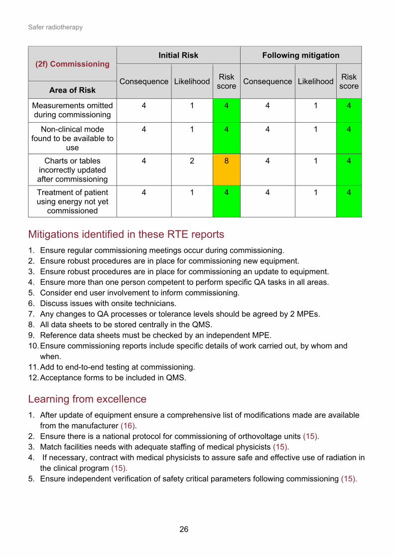
Case study D. Commissioning By the nature of the tasks involved, ‘Commissioning’ associated RTE have a greater opportunity to lead systematic failures that affect multiple patients. Therefore, a study of risk based on the submitted RTE was undertaken. This type of RTE occurs at the new equipment stage. The following table is a breakdown of this RTE by classification. Level 1 Level 2 Level 3 Level 4 Level 5 Total
5 3 11 7 6 32 Synopsis Via routine quality assurance checks a commissioning error of the radiotherapy planning system was identified. The error was made when entering some CT number to electron density data for cortical bone. This had the potential to have affected the dosimetric accuracy of radiotherapy treatments subsequently delivered. The incorrect Hounsfield number error in the treatment planning system was only for one data point which was not the most important part of the attenuation curve for most RT. The dosimetry error in treatment was estimated at 0.5% resulting in a small under-dose in radiotherapy treatments delivered. In the majority of cases the error was calculated to be less than 0.5% and, in all cases, less than 1%. The likely effect upon patients was considered at an expert panel meeting which concluded that there would be no significant effect upon patient outcomes. The department used the identification of the error as an opportunity to review the methodology and independent check at commissioning of radiotherapy equipment. Coding: TSRT9/ Level 1/ 2f/ 2h/ 0c/ CF3b/ CF5d/ MD3f Root causes and contributing factors The root cause for this case study was identified as technical ‘commissioning, calibration or maintenance’. This was due to incorrect entry of Hounsfield units to inform electron density for one structure in a new treatment planning system. Contributory factors included teamwork, management and organisation ‘inadequate staffing’, as a single individual had completed the competency for commissioning processes.
Following a simple risk matrix (see Appendix 2) a study of risk was produced for this pathway code.
Safer radiotherapy
26
(2f) Commissioning Initial Risk Following mitigation
Consequence Likelihood Risk score Consequence Likelihood Risk
score Area of Risk
Measurements omitted during commissioning
4 1 4 4 1 4
Non-clinical mode found to be available to
use
4 1 4 4 1 4
Charts or tables incorrectly updated after commissioning
4 2 8 4 1 4
Treatment of patient using energy not yet
commissioned
4 1 4 4 1 4
Mitigations identified in these RTE reports 1. Ensure regular commissioning meetings occur during commissioning. 2. Ensure robust procedures are in place for commissioning new equipment. 3. Ensure robust procedures are in place for commissioning an update to equipment. 4. Ensure more than one person competent to perform specific QA tasks in all areas. 5. Consider end user involvement to inform commissioning. 6. Discuss issues with onsite technicians. 7. Any changes to QA processes or tolerance levels should be agreed by 2 MPEs. 8. All data sheets to be stored centrally in the QMS. 9. Reference data sheets must be checked by an independent MPE. 10. Ensure commissioning reports include specific details of work carried out, by whom and
when. 11. Add to end-to-end testing at commissioning. 12. Acceptance forms to be included in QMS. Learning from excellence 1. After update of equipment ensure a comprehensive list of modifications made are available
from the manufacturer (16). 2. Ensure there is a national protocol for commissioning of orthovoltage units (15). 3. Match facilities needs with adequate staffing of medical physicists (15). 4. If necessary, contract with medical physicists to assure safe and effective use of radiation in
the clinical program (15). 5. Ensure independent verification of safety critical parameters following commissioning (15).

Safer radiotherapy
27
Case study E. Production of images demonstrating correct detail ‘Production of images demonstrating correct detail’ is the most frequently reported level 1 RTE process subcode within this analysis. This type of RTE occurs at the pretreatment activity stage of the patient pathway. The following table is a breakdown of this RTE by classification. Level 1 Level 2 Level 3 Level 4 Level 5 Total
6 2 34 3 9 54
Synopsis Patient being treated for lung cancer, due to have 4DCT scan. The Patient was coached in use of breathing apparatus and positioned correctly in CT bore. CT scan initiated for free-breathing scan and scan data saved, free-breathing scan initiated. During scan CT scanner terminated part way through. CT re-initiated and part way through second scan CT terminated. Physics called to advise on immediate action. Patient removed from CT couch and fault with CT scanner fixed, along with testing. CT scanner handed back for clinical use to continue planning scan. Patient reset up in CT room and 4DCT achieved. Post investigation identified that the second CT scan taken after the scan terminated should not have been initiated and was against procedure. Coding: TSRT9/ Level 1/ 10f/ 10l/ 10q/ CF3a/ CF2c/ MD10l Root causes and contributing factors The root cause for this case study was identified as technical ‘equipment or IT network failure’ as the scan terminated part way through. Contributory factors included ‘adherence to procedures or protocols. The individuals should not have conducted the second scan as this was out of protocol.
Following a simple risk matrix (see Appendix 2) a study of risk was produced for this pathway code.

Safer radiotherapy
28
(10f) Production of images
demonstrating correct detail
Initial Risk Following mitigation
Consequence Likelihood Risk score Consequence Likelihood Risk
score Area of Risk
Scan terminated part way through 3DCT
scan 2 3 6 2 2 4
Scan terminated part way through 4DCT
scan 2 4 8 2 2 4
Scan not recorded 2 2 4 2 1 2
Scan not recorded for 4DCT scan 2 4 8 2 2 4
Artefacts leading to need to rescan 2 2 4 2 1 2
Breathing not recorded due to
machine malfunction 2 2 4 2 1 2
Mitigations identified in these RTE reports 1. Review equipment QA and maintenance programme. 2. Ensure there is a regular verification on the transfer of data from CT to the planning system. 3. Train staff on correct use of 4DCT scanning. 4. Have in place contingency plans in case of equipment failure, practice and rehearse these
contingency plans where practicable. 5. Train staff to understand equipment malfunction procedures. 6. Ensure adequate procedures are in place and are followed. 7. Review equipment malfunctions and report to MHRA. 8. Review equipment malfunctions and report to manufacturers. 9. Record all equipment errors in the fault log. 10. Escalate all equipment errors according to local procedure. Learning from excellence 1. Review scout scan to ensure appropriate detail is captured (12).

Safer radiotherapy
29
Acknowledgements Thank you to all stakeholders for their ongoing commitment to improving safer RT. In particular, we would like to thank: NHS RT Providers and RTE reporters across the UK National Reporting and Learning System (NRLS) at NHS England and Improvement UK IR(ME)R inspectorates Care Quality Commission (CQC) in England Regulation and Quality Improvement Authority (RQIA) in Northern Ireland Healthcare Improvement Scotland (HIS) in Scotland Healthcare Inspectorate Wales (HIW) in Wales Patient Safety in Radiotherapy Steering Group Peter Colley (IPEM RT-SIG (2016 to 2020). Consultant Physicist Lead for Radiotherapy Physics, Hull and East Yorkshire Teaching Hospitals NHS Trust) IPEM Radiotherapy-Special Interest Group

Safer radiotherapy
30
References 1. Scottish Executive. Unintended overexposure of patient Lisa Norris during radiotherapy
treatment at the Beatson Oncology Centre, Glasgow in January 2006. Report of an investigation by the Inspector appointed by the Scottish Ministers for The Ionising Radiation (Medical Exposures) Regulations 2000. 2006.
2. Tobias J. What went wrong at Exeter? BMJ. 1988; 297 3. Ash D & Bates T. Report on the clinical effects of inadvertent radiation underdosage in
1,045 patients. Clinical Oncology. 1994;6(4):214-6. 4. National Patient Safety Agency. Implementing Towards Safer Radiotherapy: guidance on
reporting radiotherapy errors and near misses effectively. 5. The Ionising Radiation (Medical Exposure) (Amendment) Regulations 2018. The Stationery
Office, London, SI 2018/121. 6. The Ionising Radiation (Medical Exposure) Regulations (Northern Ireland) 2018. The
Stationery Office, London, SR 2018/17. 7. The Ionising Radiation (Medical Exposure) Regulations 2017. The Stationery Office,
London, SI 2017/1322. 8. Royal College of Radiologists, Society and College of Radiographers, Institute of Physics
and Engineering in Medicine, National Patient Safety Agency, British Institute of Radiology. Towards Safer Radiotherapy. London: Royal College of Radiologists, 2008.
9. Public Health England. Safer Radiotherapy: Newsletter Jan 2019. 10. Public Health England. Radiotherapy: learning from errors. 11. Public Health England. Radiotherapy: good practice. 12. Public Health England. Safer Radiotherapy: Triannual analysis. 13. Public Health England. Radiotherapy error and near misses: biennial report. 14. Clark B et al. The management of radiation treatment error through incident learning.
Radiotherapy and Oncology, 2010, Vol 95, pp344-349. 15. International Atomic Energy Agency. Safety in Radiation Oncology (SAFRON). 16. ASN. Newsletters for Medical Professionals. Record and verify: recording errors. 17. The Radiotherapy Board made up of the Society and College of Radiographers; Institute of
Physics and Engineering in Medicine and the Royal College of Radiologists. Ionising Radiation (Medical Exposure) Regulations: Implications for clinical practice in radiotherapy. London: The Royal College of Radiologists,2020 Ref RTBoard 2020.

Safer radiotherapy
31
Appendix 1 Taxonomies utilised for unseen pathway search. A list of all taxonomies can be found in the Development of Learning from RTE (10). Pathway Subcode
Pathway Description
0 Infrastructure
0a (0a) Implementation of national and international codes of practice for radiation dosimetry
0b (0b) Development of dosimetry algorithms for local application
0c (0c) Development of treatment planning algorithms for local application
0d (0d) Other
0e (0e) IT Infrastructure
1 Room design
1a (1a) Patient safety
1b (1b) Staff and public safety
1c (1c) Environmental controls
1d (1d) Access control
1e (1e) Other
2 New equipment
2a (2a) Installation
2b (2b) Manufacturer's tests
2c (2c) Acceptance tests
2d (2d) Critical examination under IRR99 (now 2017)
2e (2e) Customisation and configuration of equipment
2f (2f) Commissioning
2g (2g) Data recording
2h (2h) Preparation of data files for planning systems
2i (2i) Other
3 Routine machine QA
3a (3a) Daily consistency checks – geometric parameters
3b (3b) Daily consistency checks – dosimetric parameters

Safer radiotherapy
32
Pathway Subcode
Pathway Description
3c (3c) Daily consistency checks – safety (IRR compliance)
3d (3d) Daily verification of accuracy of data transfer between TPS, R&V system and treatment equipment
3e (3e) Planned QA programme checks – geometric parameters
3f (3f) Planned QA programme checks – dosimetric calibration
3g (3g) Planned QA programme checks – safety (IRR compliance)
3h (3h) Planned QA programme checks – image quality parameters
3i (3i) Regular preventative maintenance and repair programme
3j (3j) Handover of radiotherapy equipment after planned QA & maintenance
3k (3k) Routine radiation safety checks
3l (3l) Other
9 Mould room or workshop activities
9a (9a) Confirmation of ID
9b (9b) Pre mould room diagnostics/interventions
9c (9c) Production of immobilisation devices
9d (9d) Checking/fitting of immobilisation devices
9e (9e) Production of other accessories/personalised beam shaping device
9f (9f) Checking of other accessories/personalised beam shaping device
9g (9g) Labelling of mould room/workshop outputs
9h (9h) Recording of information in patient record
9i (9i) Instructions to patient
9k (9k) End of process checks
9l (9l) Other
13 Treatment unit process (including EXBRT, Protons and Superficial)
13z (13z) On-set imaging: production process
13cc (13cc) Management of variations, unexpected events or errors
13dd (13dd) Communication between treatment unit and V&R
13ff (13ff) Recording of delivered treatment data
13jj (13jj) Other

Safer radiotherapy
33
Causative Factor Subcode Causative Factor Description
CF 3 Technical
CF 3a Equipment or IT network failure
CF 3b Commissioning, calibration or maintenance
CF 3c Device or product design
CF 6 Environmental
CF 6a Physical (power cut, control area excessively noisy, distractions and so on)
CF 6b Natural factors (fire, flood ad so on)
Safer radiotherapy
34
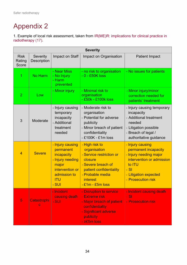
Appendix 2 1. Example of local risk assessment, taken from IR(ME)R: implications for clinical practice in radiotherapy (17).
Severity Risk
Rating Score
Severity Description
Impact on Staff Impact on Organisation Patient Impact
1
No Harm
- Near Miss - No Injury - Harm
prevented
- no risk to organisation - 0 - £50K loss
- No issues for patients
2
Low
- Minor injury - Minimal risk to organisation - £50k - £100k loss
- Minor injury/minor correction needed for patients’ treatment
3
Moderate
- Injury causing temporary incapacity
- Additional treatment needed
- Moderate risk to organisation
- Potential for adverse publicity
- Minor breach of patient confidentiality
- £100K - £1m loss
- Injury causing temporary incapacity
- Additional treatment needed
- Litigation possible - Breach of legal /
authoritative guidance
4
Severe
- Injury causing permanent incapacity
- Injury needing major intervention or admission to ITU
- SUI
- High risk to organisation
- Service restriction or closure
- Severe breach of patient confidentiality
- Probable media interest
- £1m - £5m loss
- Injury causing permanent incapacity
- Injury needing major intervention or admission to ITU
- SI - Litigation expected - Prosecution risk
5
Catastrophic
- Incident causing death
- SUI
- Disruption to service - Extreme risk - Major breach of patient confidentiality
- Significant adverse publicity
- ≥£5m loss
- Incident causing death - SI - Prosecution risk

Safer radiotherapy
35
Likelihood Likelihood score Chance Description
1
Rare / Extremely unlikely
Very good control 0.01% chance
2
Unlikely
Good control 0.1% chance 1 in 3 years
3
Likely
Limited effective control 1% chance 1 in a year
4
Somewhat likely
Weak control ≥ 10% chance 1 in 6 months
5
Very Likely
No effective control ≥ 80% chance 1 in 4 weeks
Consequence
Likelihood/ Severity None (1) Low (2) Moderate (3) Severe (4) Catastrophic (5)
Rare (1) 1 2 3 4 5 Unlikely (2) 2 4 6 8 10 Likely (3) 3 6 9 12 15
Somewhat likely (4)
4 8 12 16 20
Very likely (5)
5 10 15 20 25

36
Published: April 2021 PHE gateway number: GOV-7991
Website: www.gov.uk/phe Twitter: @PHE_uk Facebook: www.facebook.com/PublicHealthEngland © Crown copyright 2021 For queries relating to this document, please contact: [email protected]