screening for cerebrovascular disorders: a guide for ...€¦ · •cta and mra are useful for...
TRANSCRIPT

Screening for CerebrovascularDisorders: A Guide for Primary Care
Thomas Mattingly, MD, MScCerebrovascular / Endovascular Neurosurgery
Dept of Neurosurgery

Carotid disease • 2 Sources: – 2011 Guideline on the
management of patients with extra-cranial carotid and vertebral artery disease
– 2017 Clinical practice guidelines of the European Society for Vascular Surgery
Asymptomatic Carotid disease screening
• DUS is recommended as the frontline test– Low cost, low risk, accessible– For asymptomatic patients with suspected disease– Is of uncertain benefit for those without • Clinical manifestations• Risk factors
Asymptomatic Carotid Screening• Follow-up DUS annually is reasonable for – Postprocedure followup– Evaluate response to treatment in those with >
50% stenosis

Asymptomatic Carotid ScreeningDon’t forget the basics!
• Smoking cessation• Hypertension control• Hyperlipidemia management• ASA• Diabetes- “strict glycemic control”

Which Asymptomatic patients should be referred?
• >70% stenosis by non-invasive study• The rate of stroke/MI/Death needs to be < 3%• There is no role for revascularization in those
with – Chronic total occlusion– < 50% stenosis– Severe disability

Symptomatic Carotid Stenosis• Neurologic event (stroke or TIA) within 6 months• DUS + another modality (MRA, CTA, Angiography)– Confirm the degree of stenosis– Anatomic data to determine options
• > =70% noninvasive– 50-69% noninvasive may benefit but NNT much higher

Symptomatic Carotid Stenosis
• Interventions should have complication < 6%
• CEA remains the gold standard
– CAS alternative based on
• Comorbidities eg significant CAD, CHF, COPD
• Prior RTX to the neck
• Anatomic factors e.g. high bifurcation, isolated
hemisphere
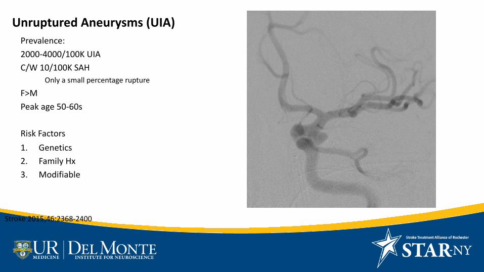
Unruptured Aneurysms (UIA)Prevalence:2000-4000/100K UIAC/W 10/100K SAH
Only a small percentage ruptureF>M Peak age 50-60s
Risk Factors1. Genetics2. Family Hx3. Modifiable
Stroke 2015;46:2368-2400

Genetic Risk Factors• At risk disorders (<10% of UIA):• Most common: AD PCKD (4-13%)• EDS type IV• Marfan• HHT
Family Risk Factors• Family occurrence 7-30%• Increased risk with first degree relatives with
h/o SAH– 4% by MRA– Siblings > children of affected
• Aneurysm in >=2 relatives
Modifiable Risk Factors• Smoking• Hypertension• Excessive Alcohol (> 3 drinks/day)
Factors for rupture of UIAs• Age > 60• Female sex• Japanese or Finnish descent• Size > 5mm• Posterior circulation• “Symptomatic” • Evidence of growth on serial imaging
Screening modalities• CTA and MRA are useful for detection and follow up– MRA avoids radiation– MRA better for detection of aneurysm > 3mm size– MRA is not as sensitive for infundibulum vs aneurysm
• Angiography can be useful compared with noninvasive modalities– If treatment is considered– The most sensitive modality for previously treated
aneurysms

Who to screen for UIA• >=2 family members with UIA or SAH– This is particularly high yield in combo with HTN,
smoking and female sex– Siblings > children of index SAH patient
• Certain conditions– AD PCKD, especially with Family H/O IA– Coarctation of aorta– Microcephalic osteoplastic primordial dwarfism

Arteriovenous Malformations (AVM)Abnormal connection between arterial and venous systems
Lack intervening capillary bedHemorrhage leads to significant morbidity/mortality
Incidence is 2-4% annuallyARUBA trial 2.2%
Many present with seizure or headacheNoninvasive imaging leads to increased diagnosis of unrupturedAVMAngiography is gold standard for evaluation

Arteriovenous Malformations (AVM)
• ARUBA 2014 concluded the natural history of unruptured brain AVMs is better than any form of treatment
• Lots of controversy– Wide range of treatment modalities– Lack of subgroup analysis – Small ”n”– Insufficient follow-up (<3years, only funded out to 5 years)
Mohr JP et al Lancet 2014;383:614-21Hong CS et al Clin Neuro Neurosurg 2016;;150:133-138

Not all AVMs are the same
• This is a “young person” issue– The longer you live, the more likely this will become symptomatic
• Treatments are often tailored to individual and involve multiple specialities– Open surgery– Stereotactic radiosurgery– Endovascular surgery
• Elements that increase risk of bleeding:– Associated aneurysms– Venous stenosis– Infratentorial location
Eur Concensus Statement Acta Neurochir 2017;159:1059-64Ajiboye N et al. Sci World J 2014;http://dx.doi.org/10.1155/2014/649036

CavernomasAKA:
Cavernous angioma
Cavernous hemangioma
Prevalence up to 0.5%
Annual detection of 0.56/100K adults
Clinical Manifestations
Seizures
ICH
Neurologic deficts w/o hemorrhage
Incidental 20-50%
Akers et al Neurosurg 2017;80: 665-680
2 forms of cavernomas• Genetic basis well established
– Mutations in CCM1-3• 20% familial
– AD, but incomplete penetrance, variable presentation– Usually multiple
• 80% sporadic– Usually solitary– Associated with a developmental venous anomaly (DVA)– No germline mutations of CCM genes– h/o brain XRT

Recommendations for Genetic Testing• 3 generation FHx at time of diagnosis, looking for– Headache– Seizure – Stroke – Abnormal MRI
• Consider genetic testing of CCM1-3 genes in – Multiple CCM without DVA– Without h/o brain XRT– Positive family hx
Imaging and f/u• Brain MRI is the gold-standard for diagnosis
and followup– Should include either a GRE or SWI sequence– Catheter angio where ddx includes AVM
• MRI repeated for new/worsened symptoms – Looking for new hemorrhage– Surveillance followup uncertain

Management overview• Since the presentation is variable, so is the
management– IVH and ICH are treated as per standard care– Asymptomatic incidental CCM – observe– Symptomatic easily accessible CCM- resect– Medically refractory seizures- resect– Radiosurgery - controversial