semanticknowledgeinpatienth.m.andother...
TRANSCRIPT
Semantic Knowledge in Patient H.M. and OtherPatients With Bilateral Medial and Lateral TemporalLobe LesionsHeike Schmolck,2 Elizabeth A. Kensinger,5
Suzanne Corkin,5 and Larry R. Squire1,2,3,4*
1Veterans Affairs Medical Center, San Diego, California2Department of Psychiatry, University of California,La Jolla, California3Department of Neurosciences, University of California,La Jolla, California4Department of Psychology, University of California,La Jolla, California5Department of Brain and Cognitive Sciences and theClinical Research Center, Massachusetts Institute ofTechnology, Cambridge, Massachusetts
ABSTRACT: We investigated the effects of damage to the medial temporallobe (MTL) and anterolateral temporal cortex on semantic knowledge. Westudied eight male controls, two patients with lesions limited to the hip-pocampal formation, three postencephalitic patients with extensive MTLlesions and variable damage to the lateral temporal cortex, and patient H.M.(whose lesion is limited mostly to the MTL, but who also has minimal damageto the anterolateral cortex). On 13 tests of semantic memory, patients withlesions limited to the hippocampal formation performed similarly to controls.Postencephalitic patients were mildly to moderately impaired on most tests.Patient H.M.’s performance was impaired on only a few tests and was lessseverely impaired overall than the three postencephalitic patients. A rankingof test scores showed a direct relationship between impairment and theextent of damage to lateral temporal cortex. These findings, and relatedfindings from other studies, point to the importance of anterolateral temporalcortex for semantic knowledge. Patient H.M. performed uniquely in certainrespects. For example, when providing definitions of objects, he made manygrammatical errors. In contrast, the other patients with large MTL lesionsmade no more errors than those made by controls. Considering that H.M.’slesion, both medially and laterally, is less extensive than the lesions in theseother patients, it appears unlikely that his shortcomings in language produc-tion are related to his temporal lobe lesion. Hippocampus 2002;12:520–533.Published 2002 Wiley-Liss, Inc.†
KEY WORDS: memory; hippocampus; perirhinal cortex; parahip-pocampal cortex
INTRODUCTION
Bilateral medial temporal lobe lesions cause severe andlasting memory impairment, affecting the ability both toacquire new information and to recall informationlearned previously (Scoville and Milner, 1957; Corkin,1984; Stefanacci et al., 2000; Manns and Squire, inpress). At the same time, information acquired early inlife is spared. For example, amnesic patient H.M. exhibitsintact grammatical processing and intact lexical informa-tion, such as intact information about words and wordforms (Kensinger et al., 2001). He also has preservedintellectual ability, as indicated by stable performance onfour subtests of standard intelligence tests, which wereadministered preoperatively and then on multiple occa-sions across five decades (Kensinger et al., 2001). Anotheramnesic patient (E.P.) exhibited intact memory for thespatial layout of the neighborhood where he grew up butmoved away from as a young adult (Teng and Squire,1999). Such findings provide support for the view thatmedial temporal lobe structures are required for the ac-quisition of new knowledge, but not for the retrieval anduse of remote, well-established semantic knowledge.
In contrast to these findings of preserved cognitiveabilities other than memory, it was recently reported thatH.M., E.P., and other patients with large medial tempo-ral lobe lesions were impaired at detecting and explainingambiguity in sentences (MacKay et al., 1998b; Schmolcket al., 2001; Squire et al., 2001). It is unclear how excep-tional this impairment is and whether it might be part ofa broader impairment in semantic knowledge. The ana-tomical basis of the impairment is also unclear. The pa-
Grant sponsor–Medical Research Service of the Department of VeteransAffairs; Grant sponsor–National Institute of Mental Health; Grant number–24600; Grant sponsor: Metropolitan Life Foundation; Grant sponsor: DFG;Grant number: Vo 770/1-1.*Correspondence to: Larry Squire, Department of Psychiatry 0603, Univer-sity of California, La Jolla, CA 92093.E-mail: [email protected] for publication 5 November 2001DOI 10.1002/hipo.10039Published online 30 May 2002 in Wiley InterScience (www.interscience.wiley.com).
HIPPOCAMPUS 12:520–533 (2002)
Published 2002 WILEY-LISS, INC. †This article is a US government work and, as such, isin the public domain in the United States of America.
tients who were tested and found to be impaired did have medialtemporal lobe lesions, but all the patients, including H.M., alsohave at least some damage to lateral temporal cortex. Accordingly,impaired appreciation of sentence ambiguity could be the result oflateral temporal damage. It was also of interest that H.M. haddeficits on this task that were not present in any of the otherpatients with large medial temporal lobe lesions (Schmolck et al.,2000), raising the possibility that some of H.M.’s difficulties mightbe unrelated to his lesion.
In the present study, we gave a large series of semantic knowl-edge tests to patients with medial temporal lobe lesions and vari-able damage to lateral temporal cortex (the same patients testedpreviously). In addition, we tested patients with lesions restrictedto the hippocampal formation within the medial temporal lobe.Finally, we tested H.M. and compared his performance with thatof the other patients. The purpose of the study was twofold: first, toassess the relation between test performance and the extent ofdamage to lateral temporal cortex and second, to determinewhether any aspects of H.M.’s performance were unique amongthe patients tested.
METHODS
Participants
We studied three patients who developed amnesia after herpessimplex encephalitis (E.P., G.P., and G.T.). These patients havelarge medial temporal lobe lesions, as well as variable damage toanterolateral temporal cortex (MTL�) (Fig. 1). We also studiedpatient H.M., who underwent bilateral medial temporal lobe re-section for relief of severe epilepsy (Scoville and Milner, 1957).Other participants included two amnesic patients with damagebelieved to be limited to the hippocampal formation (HF) andeight controls (CON) (Table 1). Magnetic resonance images(MRI) of the patients with MTL� lesions, and one of the twopatients with HF lesions, were acquired in a 1.5-tesla (T) SignaClinical scanner at the UCSD Medical Center. Descriptions of thelesions are based on axial T2-weighted, proton density fast spin-echo (FSE) images through the brain, matrix � 256 � 256, field ofview (FOV) � 20 � 22 cm (0.78 � 0.86 mm in-plane resolution),5-mm-thick sections; and on coronal oblique, T1-weighted imagesperpendicular to the long axis of the hippocampus, matrix �256 � 256, FOV � 16–20 cm (0.63–0.78 mm in-plane resolu-tion), 5-mm-thick, interleaved sections.
Patients with medial temporal lobe lesions andvariable damage to anterolateral temporal cortex(MTL�)
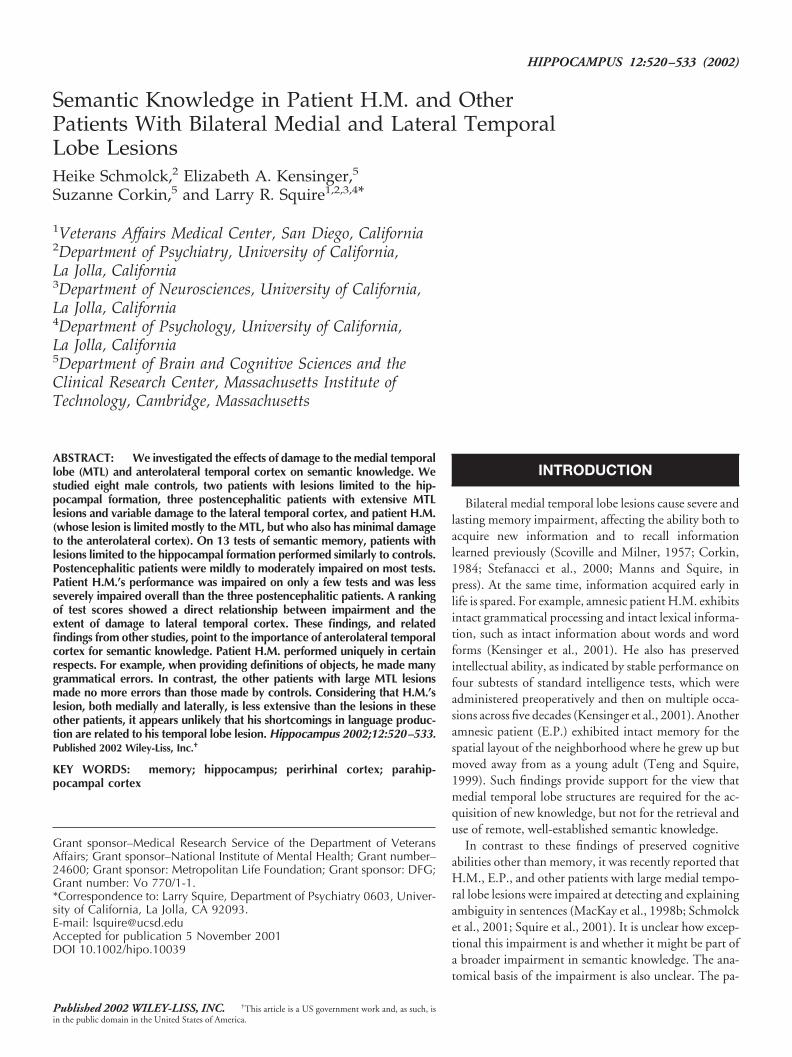
Patient E.P. E.P. developed profound amnesia in 1992 afterherpes simplex encephalitis. His lesion is primarily medial tempo-ral but also compromises the anterior portion of the fusiform gy-rus. The damage extends 7 cm caudally from the temporal polebilaterally, and includes all of the amygdala and all of the hip-
pocampal region (dentate gyrus, cell fields of the hippocampusproper, and subicular complex except for a small tag of abnormallyappearing vestigial tissue on each side that comprises about 10% ofhippocampal volume). In addition, the damage includes all of theentorhinal cortex, all of the perirhinal cortex, and much of theparahippocampal cortex (about 20% on the left and 60% on theright). Estimates of damage for the patients are based on quantita-tive analysis, following published procedures for segmenting thetemporal lobe (Insausti et al., 1998; Amaral and Insausti, 1990).The lesion also extends laterally to include the rostral portion of thefusiform gyrus (40% damage on the left, 53% on the right). Thelateral temporal cortex (inferior, middle, and superior temporalgyri) is reduced in volume bilaterally, particularly on the right side(left side � 10%; right side � 25%). The insula is also reduced insize bilaterally, with more substantial loss on the left side (19%)than on the right (11%) (Stefanacci et al., 2000).
Patient G.P. G.P. developed profound amnesia in 1987 afterherpes simplex encephalitis. Like E.P., G.P.’s damage is primarilymedial temporal, but his lesion extends further laterally. The dam-age extends through the anterior 7 cm of the left temporal lobe andthe anterior 6 cm of the right temporal lobe. The damage includesbilaterally all of the amygdala, all of the hippocampal region, all ofthe entorhinal and perirhinal cortices, and much of the parahip-pocampal cortex (77% on the left and 17% on the right). Lateraldamage is most severe in the anterior 1 cm of the temporal lobe,where it includes the fusiform gyrus as well as the inferior, middle,and superior temporal gyri, bilaterally. From 1 cm to 4.5 cm cau-dally, the lateral damage is restricted to the fusiform gyrus and theinferior temporal gyrus. The insular cortex is also damaged, withthe lesion extending caudally on the left side (3 cm) more than onthe right side (2.5 cm) (Schmolck et al., 2000; Fig. 2).
Patient G.T. G.T. developed profound amnesia in 1990 afterherpes simplex encephalitis. His lesion extends laterally to includemost of the temporal lobes bilaterally. The damage compromisesthe anterior 7 cm of the left temporal lobe, and the anterior 5 cm ofthe right temporal lobe, including bilaterally all of the amygdala, allof the hippocampal region, all of the entorhinal and perirhinalcortices, and much of the parahippocampal cortex (100% on theleft and about 43% on the right). Lateral cortical regions (fusiformgyrus; inferior, middle, and superior temporal gyri) are also dam-aged bilaterally at the level of the temporal pole. The damage to thefusiform gyrus continues caudally from the temporal pole for 6.0cm on the left and for 4.5 cm on the right. The damage to theinferior, middle, and superior temporal gyri extends caudally fromthe temporal pole for 4.5 cm on the left and 2.5 cm on the right.There is also bilateral insular damage, more extensive on the leftthan on the right (Schmolck et al., 2000; Fig. 3).
Patient H.M. The well-studied patient H.M (Scoville and Mil-ner, 1957; Corkin, 1984) has bilateral damage that is largely re-stricted to the medial temporal lobe. Within the medial temporallobe, he has some sparing of the ventrocaudal aspect of the perirhi-nal cortex, and almost complete sparing of the parahippocampalcortex. In addition, the posterior portion of the hippocampal for-
_____________________________________________________________________ SEMANTIC KNOWLEDGE 521
mation is present, although the tissue appears to be “somewhatatrophic bilaterally” and is likely deafferented due to removal of theentorhinal cortex (Corkin et al., 1997). Laterally, the lesion sparesthe fusiform gyrus, but there is damage at the temporal pole bilat-erally that compromises the rostralmost aspects of the middle andsuperior temporal gyri (Corkin et al., 1997) (Fig. 3K; see also Fig.5C,D). In addition, “the subcortical white matter associated withthe anterior portions of the superior, middle, and inferior temporalgyri may also have been compromised by the resection” (p. 3975).
Amnesic patients with damage limited to thehippocampal formation (HF)
Patient A.B. A.B. developed moderately severe amnesia after acardiac arrest in 1976. He is unable to participate in MRI studies
because he wears a pacemaker. In 2001, we obtained computedtomograph (CT) images in a GE Light Speed Plus Helical CTscanner (2 mm and 5 mm-thick axial sections, as well as 1.25 mmand 2.5 mm-thick coronal sections through the brain). The CTscan demonstrated some volume loss in the supraventricular por-tions of the frontal and parietal lobes, the medial occipital lobes,and the superior aspect of the cerebellar hemispheres. In contrast,temporal lobe volume appeared normal, and the temporal hornswere symmetric and normal in size. Thus there was no sign oftemporal lobe atrophy. Further, within the temporal lobe there wasno sign of stroke, encephalitis, abscess, or any large lesion. Thebasal ganglia and the thalamus also appeared normal. The onlyfocal lesions detected were small bilateral foci (maximum diame-ter � 1 cm) in the white matter lateral to the head of the caudatenucleus, which appeared to be old lacunar infarctions. The finer
FIGURE 1. Magnetic resonance images showing the extent of bilateral temporal lobe damage in patients E.P. (top row), G.P. (middle row),and G.T. (bottom row). A–C in each row are T2-weighted axial images through the temporal lobe. The images are continuous 5-mm sections(with 2.5-mm gaps) and are arranged from ventral (A) to dorsal (C). Damaged tissue is indicated by bright signal. D in each row is a coronalT1-weighted image at the level of the amygdala. Damaged tissue is indicated by dark signal. See text for detailed description of the lesions.
522 SCHMOLCK ET AL.

anatomy of the medial temporal lobe was not evident in the CTscan due to beam-hardening artifact in the coronal plane.
In view of the normal findings in the temporal lobe and a normalneurological exam (other than memory impairment), the extent ofA.B.’s damage appears to be quite limited. Within the medialtemporal lobe, the region most vulnerable to anoxic damage is thehippocampal formation (Caine and Watson, 2000). Further, an-oxic damage limited largely to the hippocampal formation, in theabsence of damage to basal ganglia, diencephalon, or basal fore-brain (except the medial septal nuclei), has been described in anamnesic patient where detailed neuropsychological and neurohis-tological information were available. (patient L.M.; Rempel-Clower et al., 1996). It therefore seems likely that A.B.’s memoryimpairment is due to damage within the hippocampal formation.
Patient L.J. L.J. developed moderately severe amnesia during a6-month period beginning in late 1988, and her memory impair-ment has remained stable since that time. MRI identified hip-pocampal formation damage bilaterally (Reed and Squire, 1998).Measured against three age- and sex-matched controls, her hip-pocampal region relative to the temporal lobe is reduced in area by46%. The size of the parahippocampal gyrus and of the lateraltemporal lobe is within control values.
Healthy controls (CON)
Eight healthy men were recruited from volunteers at the SanDiego Veterans Affairs Medical Center and the UCSD retirementcommunity. They were matched to the older patients with respectto age (74.0 years) and education (12.4 years; see Table 1).
Materials
All participants were given nine tests on three to five separateoccasions. Seven of the tests are from the Semantic Test Battery, asoriginally introduced by Hodges et al. (1992a) and subsequently
amended (Garrard et al., 1997; see also Hodges et al., 1996, 1999).We constructed two additional tests (tests 2 and 9; see below). Allnine tests were based on the same line drawings (Snodgrass andVanderwart, 1980) of 24 animals and 24 objects (or their names).Each of the 48 items could further be assigned to one of 8 catego-ries: 6 domestic land animals, 6 foreign land animals, 6 watercreatures, 6 birds, 6 electrical household items, 6 nonelectricalhousehold items, 6 vehicles, and 6 musical instruments. Unlessstated otherwise, there was no time limit for the tests.
1. Pointing to Picture (cue: Name): Participants were given thename of each of 48 items as a cue and were asked to identify theappropriate picture from among eight pictures in the same category.2. Pointing to Picture (cue: Description): Participants were given averbal description of each of 48 items as a cue (without mention ofphysical attributes) and asked to identify the appropriate picturefrom among eight pictures in the same category.3. Naming (cue: Picture): Participants were shown a picture ofeach of 48 items as a cue and asked to name it.4. Naming (cue: Description): Participants were given a verbal de-scription of each of 48 items as a cue and asked to name it.5. Semantic Features: Participants were asked eight yes/no ques-tions about each of 24 items, 4 questions about an item’s physicalfeatures and 4 questions about an item’s associative (nonphysical)features, e.g., Is a toaster round? Does a zebra live in Africa?6. Category Fluency: Participants were asked to name as many ex-amples as they could from each of 8 categories: 4 categories ofliving things (Animals, Birds, Water Creatures, Breeds of Dogs)and 4 categories of nonliving things (Household Items, Vehicles,Musical Instruments, and Types of Boat). For each of the eightcategories, participants were given 1 min to respond.7. Category Sorting: Participants were first asked to sort pictures ofall 48 items into one of two categories (living/manmade). Thenthey sorted the 24 items from each of these two categories intonarrower categories (sort the “living” items into land animals,
TABLE 1.
Characteristics of the Participants*
Name Year of birth Education
WAISR
Bostonnaming test
WMSIII Indices
Full-scaleIQ
Informationsubtest
Vocabularysubtest Working memory
Generalmemory
E.P. 1922 12 101 17 33 63.1 99 54G.P. 1946 16 99 20 39 70.2 99 57G.T. 1936 12 92 4 28 25.0 108 49H.M. 1926 12 101 18 39 82.5 87a 55a
A.B. 1937 20 104 27 65 88.1 81 47L.J. 1937 12 98 17 50 90.5 96 66
CON 1–8 1921–1929 12.4 — 23.6 58.2 87.2 — —Mean
*L.J. is female; the other participants are male. Indices of the the Wechsler Adult Intelligence Scale-Revised (WAIS-R) and the Wechsler MemoryScale-III (WMS-III) yield a mean score of 100 in the normal population with a SD of 15.aH.M.’s scores are for the Attention/Concentration and Delayed Memory indices from the Wechsler Memory Scale-Revised (WMS-R).
_____________________________________________________________________ SEMANTIC KNOWLEDGE 523

birds, or water creatures; sort the manmade things into householditems, vehicles, or musical instruments). Then, they sorted 12 landanimals and 12 household items three different times into stillnarrower categories (e.g., sort the land animals into foreign/domes-tic animals, fierce/nonfierce animals, and animals larger/smallerthan a German Shepherd dog).8. Definitions to Name: Participants were given the name of each ofthe 24 least common items and were asked to define it (i.e., as ifhe/she were explaining the item to someone who does not knowwhat it is and has never seen it before). A card with the name of theitem was in view, and 1 minute was allowed for each definition.9. Definitions to Picture: Participants were shown the picture ofeach of the 24 least common items and asked to define it. A linedrawing of the item was in view, and 1 min was allowed for eachdefinition.
Scoring
For all but tests 6, 8, and 9, performance was measured aspercentage correct. For test 6, the score was the total number of
FIGURE 2. The same 48 items were used for four different tests. A: Participants were given the name of an item and were asked to identifythe appropriate picture from among eight pictures of the same category (test 1). B: Participants were given a verbal description of an item andasked to identify the appropriate picture from among eight pictures of the same category (test 2). C: Participants were shown a picture of anitem and asked to name it (test 3). D: Participants were given a verbal description of an item and asked to name it (test 4). CON, 8 healthycontrols; HF, 2 amnesic patients with hippocampal formation damage; MTL�, 3 patients with large medial temporal lobe lesions and variableadditional damage to the anterolateral temporal lobe. Circles show individual scores within each group. Squares show scores for patient H.M.
FIGURE 3. Participants were asked eight yes/no questions abouteach of 24 items (test 5). Circles show the individual scores withineach group. Squares show scores for patient H.M. Abbreviations as inFig. 2.
524 SCHMOLCK ET AL.
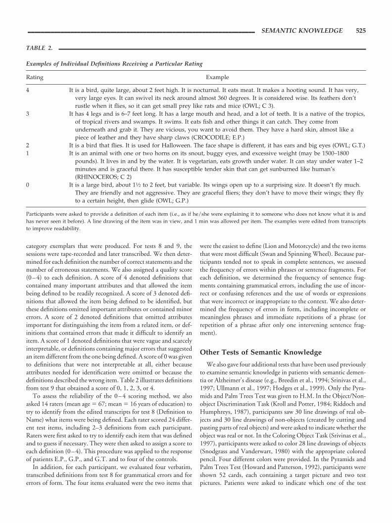
category exemplars that were produced. For tests 8 and 9, thesessions were tape-recorded and later transcribed. We then deter-mined for each definition the number of correct statements and thenumber of erroneous statements. We also assigned a quality score(0–4) to each definition. A score of 4 denoted definitions thatcontained many important attributes and that allowed the itembeing defined to be readily recognized. A score of 3 denoted defi-nitions that allowed the item being defined to be identified, butthese definitions omitted important attributes or contained minorerrors. A score of 2 denoted definitions that omitted attributesimportant for distinguishing the item from a related item, or def-initions that contained errors that made it difficult to identify anitem. A score of 1 denoted definitions that were vague and scarcelyinterpretable, or definitions containing major errors that suggestedan item different from the one being defined. A score of 0 was givento definitions that were not interpretable at all, either becauseattributes needed for identification were omitted or because thedefinitions described the wrong item. Table 2 illustrates definitionsfrom test 9 that obtained a score of 0, 1, 2, 3, or 4.
To assess the reliability of the 0–4 scoring method, we alsoasked 14 raters (mean age � 67; mean � 16 years of education) totry to identify from the edited transcripts for test 8 (Definition toName) what items were being defined. Each rater scored 24 differ-ent test items, including 2–3 definitions from each participant.Raters were first asked to try to identify each item that was definedand to guess if necessary. They were then asked to assign a score toeach definition (0–4). This procedure was applied to the responseof patients E.P., G.P., and G.T. and to four of the controls.
In addition, for each participant, we evaluated four verbatim,transcribed definitions from test 8 for grammatical errors and forerrors of form. The four items evaluated were the two items that
were the easiest to define (Lion and Motorcycle) and the two itemsthat were most difficult (Swan and Spinning Wheel). Because par-ticipants tended not to speak in complete sentences, we assessedthe frequency of errors within phrases or sentence fragments. Foreach definition, we determined the frequency of sentence frag-ments containing grammatical errors, including the use of incor-rect or confusing references and the use of words or expressionsthat were incorrect or inappropriate to the context. We also deter-mined the frequency of errors in form, including incomplete ormeaningless phrases and immediate repetitions of a phrase (orrepetition of a phrase after only one intervening sentence frag-ment).
Other Tests of Semantic Knowledge
We also gave four additional tests that have been used previouslyto examine semantic knowledge in patients with semantic demen-tia or Alzheimer’s disease (e.g., Breedin et al., 1994; Srinivas et al.,1997; Ullmann et al., 1997; Hodges et al., 1999). Only the Pyra-mids and Palm Trees Test was given to H.M. In the Object/Non-object Discrimination Task (Kroll and Potter, 1984; Riddoch andHumphreys, 1987), participants saw 30 line drawings of real ob-jects and 30 line drawings of non-objects (created by cutting andpasting parts of real objects) and were asked to indicate whether theobject was real or not. In the Coloring Object Task (Srivinas et al.,1997), participants were asked to color 28 line drawings of objects(Snodgrass and Vanderwart, 1980) with the appropriate coloredpencil. Four different colors were provided. In the Pyramids andPalm Trees Test (Howard and Patterson, 1992), participants wereshown 52 cards, each containing a target picture and two testpictures. Patients were asked to indicate which one of the test
TABLE 2.
Examples of Individual Definitions Receiving a Particular Rating
Rating Example
4 It is a bird, quite large, about 2 feet high. It is nocturnal. It eats meat. It makes a hooting sound. It has very,very large eyes. It can swivel its neck around almost 360 degrees. It is considered wise. Its feathers don’trustle when it flies, so it can get small prey like rats and mice (OWL; C 3).
3 It has 4 legs and is 6–7 feet long. It has a large mouth and head, and a lot of teeth. It is a native of the tropics,of tropical rivers and swamps. It swims. It eats fish and other things it can catch. They come fromunderneath and grab it. They are vicious, you want to avoid them. They have a hard skin, almost like apiece of leather and they have sharp claws (CROCODILE; E.P.)
2 It is a bird that flies. It is used for Halloween. The face shape is different, it has ears and big eyes (OWL; G.T.)1 It is an animal with one or two horns on its snout, buggy eyes, and excessive weight (may be 1500–1800
pounds). It lives in and by the water. It is vegetarian, eats growth under water. It can stay under water 1–2minutes and is graceful there. It has susceptible tender skin that can get sunburned like human’s(RHINOCEROS; C 2)
0 It is a large bird, about 11⁄2 to 2 feet, but variable. Its wings open up to a surprising size. It doesn’t fly much.They are friendly and not aggressive. They are graceful fliers; they don’t have to move their wings; they flyto a certain height, then glide (OWL; G.P.)
Participants were asked to provide a definition of each item (i.e., as if he/she were explaining it to someone who does not know what it is andhas never seen it before). A line drawing of the item was in view, and 1 min was allowed per item. The examples were edited from transcriptsto improve readability.
_____________________________________________________________________ SEMANTIC KNOWLEDGE 525
pictures “goes with” the target picture. For example, a saddle waspresented above drawings of a horse and a goat, and the participantwas asked: “Which one goes with the saddle?” In the “word ver-sion” of this test, participants performed the same task with threewords instead of three pictures. In the Nouns and Verbs Test(Ullmann and Corkin, 1997; Ullmann et al., 1997; Kensinger etal., 2001), participants were asked to produce 64 irregular and 64regular past tenses, and 16 regular and 16 irregular plurals. Eachtest item was presented as two short sentences, and participantswere asked to fill in the blank in the second sentence. For example,“Every day I take a banana. Just like every day, yesterday I abanana.” or “The hoof is hard. In fact, most are hard.”
RESULTS
Semantic Test Battery
Figure 2 shows performance on the two pointing tasks and thetwo naming tests. Controls (CON) and amnesic patients (HF)performed nearly without error on all four tests. By contrast, thethree patients with large medial temporal lobe lesions (MTL�:E.P., G.P., and G.T.) were mildly but significantly impaired oneach task (P � 0.005). Overall, the controls scored 98.9% correct,the HF patients scored 100% correct, and the three MTL� pa-tients scored 78.1% correct. H.M. performed close to the controllevel on three of the four tests (Fig. 2A–C, 96.5% correct for H.M.;99.5% correct for controls). On the test that asked him to nameliving things when given their descriptions, however, he scoredonly 66.7% correct (Fig. 2D, left), well outside the range of controlscores. The three patients with MTL� lesions and H.M. scoredmore poorly on questions about living things than on questionsabout nonliving things (74.0% vs 80.9% for MTL�; 87.5 vs96.9% for H.M; for all four patients, t[3] � 3.3; P � 0.05). It isimportant to note that the impairment exhibited by these patients
is not limited to a difficulty in naming. For example, performancewas similar on the two pointing tasks (Fig. 2A,B), even though thename of the item was relevant in only one of the tasks (A).
Figure 3 shows performance on test 5 (Semantic Features). Aswas the case for tests 1–4, the two HF patients performed well(96.9% correct vs 91.9% correct for the controls). In contrast, thethree patients with MTL� lesions were significantly impaired(80.9% vs 91.9%; t[9] �3.7, P � 0.005). For nonliving thingsalone (Figure 3, right panel), the impairment was marginal (85.8%vs 92.8%, P � 0.10; living things alone, 76.0% vs 91.0% correct;t[9] � 5.2, P � 0.001). H.M. performed similarly to the poorestcontrol on questions about living items (85.4% correct), and heobtained a normal score on questions about nonliving items(95.8% correct).
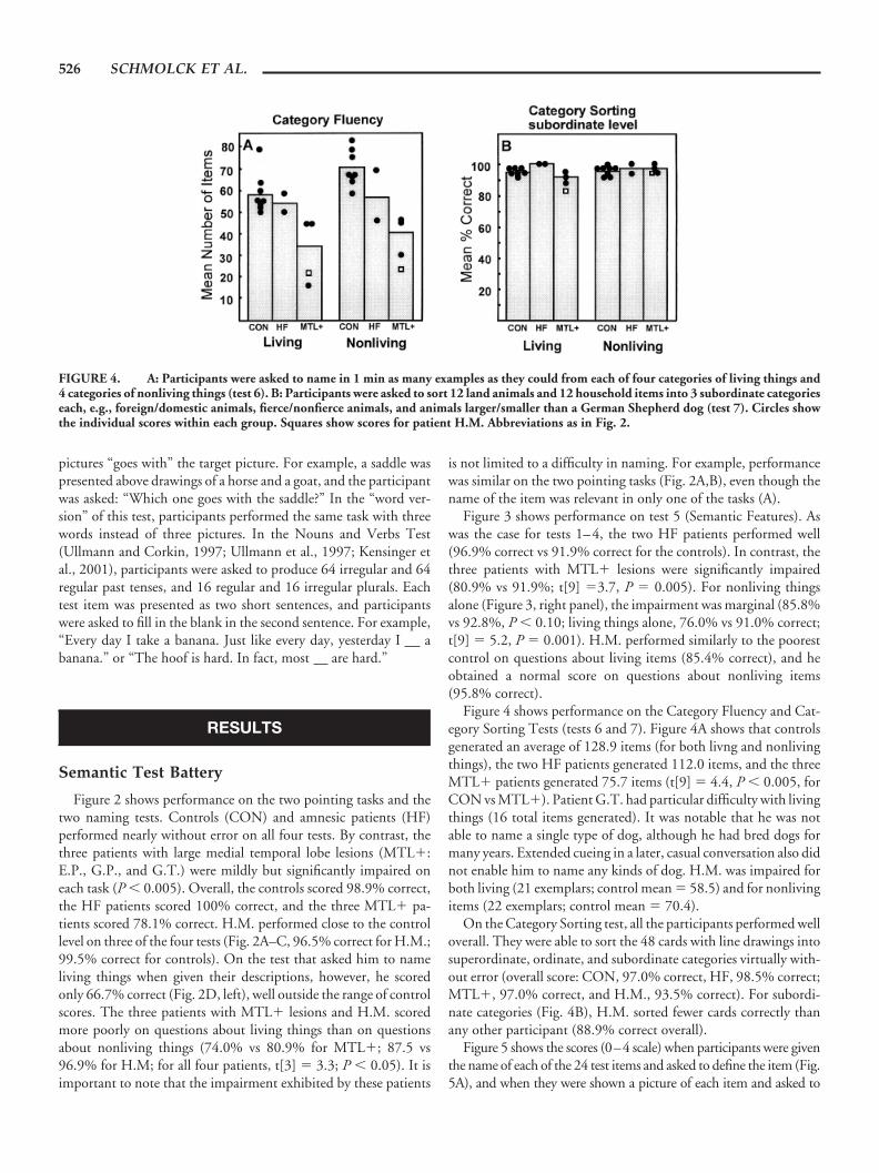
Figure 4 shows performance on the Category Fluency and Cat-egory Sorting Tests (tests 6 and 7). Figure 4A shows that controlsgenerated an average of 128.9 items (for both livng and nonlivingthings), the two HF patients generated 112.0 items, and the threeMTL� patients generated 75.7 items (t[9] � 4.4, P � 0.005, forCON vs MTL�). Patient G.T. had particular difficulty with livingthings (16 total items generated). It was notable that he was notable to name a single type of dog, although he had bred dogs formany years. Extended cueing in a later, casual conversation also didnot enable him to name any kinds of dog. H.M. was impaired forboth living (21 exemplars; control mean � 58.5) and for nonlivingitems (22 exemplars; control mean � 70.4).
On the Category Sorting test, all the participants performed welloverall. They were able to sort the 48 cards with line drawings intosuperordinate, ordinate, and subordinate categories virtually with-out error (overall score: CON, 97.0% correct, HF, 98.5% correct;MTL�, 97.0% correct, and H.M., 93.5% correct). For subordi-nate categories (Fig. 4B), H.M. sorted fewer cards correctly thanany other participant (88.9% correct overall).
Figure 5 shows the scores (0–4 scale) when participants were giventhe name of each of the 24 test items and asked to define the item (Fig.5A), and when they were shown a picture of each item and asked to
FIGURE 4. A: Participants were asked to name in 1 min as many examples as they could from each of four categories of living things and4 categories of nonliving things (test 6). B: Participants were asked to sort 12 land animals and 12 household items into 3 subordinate categorieseach, e.g., foreign/domestic animals, fierce/nonfierce animals, and animals larger/smaller than a German Shepherd dog (test 7). Circles showthe individual scores within each group. Squares show scores for patient H.M. Abbreviations as in Fig. 2.
526 SCHMOLCK ET AL.
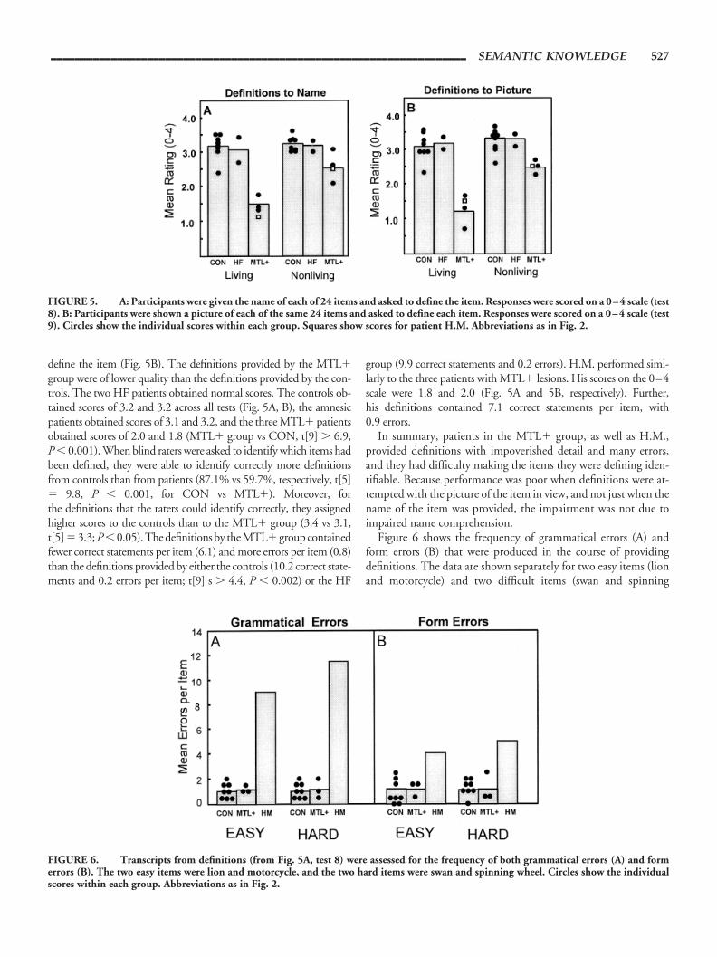
define the item (Fig. 5B). The definitions provided by the MTL�group were of lower quality than the definitions provided by the con-trols. The two HF patients obtained normal scores. The controls ob-tained scores of 3.2 and 3.2 across all tests (Fig. 5A, B), the amnesicpatients obtained scores of 3.1 and 3.2, and the three MTL� patientsobtained scores of 2.0 and 1.8 (MTL� group vs CON, t[9] � 6.9,P � 0.001). When blind raters were asked to identify which items hadbeen defined, they were able to identify correctly more definitionsfrom controls than from patients (87.1% vs 59.7%, respectively, t[5]� 9.8, P � 0.001, for CON vs MTL�). Moreover, forthe definitions that the raters could identify correctly, they assignedhigher scores to the controls than to the MTL� group (3.4 vs 3.1,t[5] � 3.3; P � 0.05). The definitions by the MTL� group containedfewer correct statements per item (6.1) and more errors per item (0.8)than the definitions provided by either the controls (10.2 correct state-ments and 0.2 errors per item; t[9] s � 4.4, P � 0.002) or the HF
group (9.9 correct statements and 0.2 errors). H.M. performed simi-larly to the three patients with MTL� lesions. His scores on the 0–4scale were 1.8 and 2.0 (Fig. 5A and 5B, respectively). Further,his definitions contained 7.1 correct statements per item, with0.9 errors.
In summary, patients in the MTL� group, as well as H.M.,provided definitions with impoverished detail and many errors,and they had difficulty making the items they were defining iden-tifiable. Because performance was poor when definitions were at-tempted with the picture of the item in view, and not just when thename of the item was provided, the impairment was not due toimpaired name comprehension.
Figure 6 shows the frequency of grammatical errors (A) andform errors (B) that were produced in the course of providingdefinitions. The data are shown separately for two easy items (lionand motorcycle) and two difficult items (swan and spinning
FIGURE 5. A: Participants were given the name of each of 24 items and asked to define the item. Responses were scored on a 0–4 scale (test8). B: Participants were shown a picture of each of the same 24 items and asked to define each item. Responses were scored on a 0–4 scale (test9). Circles show the individual scores within each group. Squares show scores for patient H.M. Abbreviations as in Fig. 2.
FIGURE 6. Transcripts from definitions (from Fig. 5A, test 8) were assessed for the frequency of both grammatical errors (A) and formerrors (B). The two easy items were lion and motorcycle, and the two hard items were swan and spinning wheel. Circles show the individualscores within each group. Abbreviations as in Fig. 2.
_____________________________________________________________________ SEMANTIC KNOWLEDGE 527
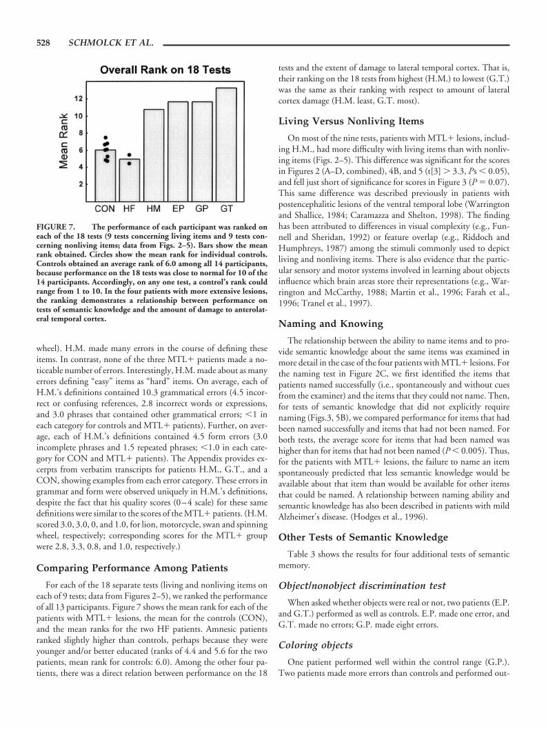
wheel). H.M. made many errors in the course of defining theseitems. In contrast, none of the three MTL� patients made a no-ticeable number of errors. Interestingly, H.M. made about as manyerrors defining “easy” items as “hard” items. On average, each ofH.M.’s definitions contained 10.3 grammatical errors (4.5 incor-rect or confusing references, 2.8 incorrect words or expressions,and 3.0 phrases that contained other grammatical errors; �1 ineach category for controls and MTL� patients). Further, on aver-age, each of H.M.’s definitions contained 4.5 form errors (3.0incomplete phrases and 1.5 repeated phrases; �1.0 in each cate-gory for CON and MTL� patients). The Appendix provides ex-cerpts from verbatim transcripts for patients H.M., G.T., and aCON, showing examples from each error category. These errors ingrammar and form were observed uniquely in H.M.’s definitions,despite the fact that his quality scores (0–4 scale) for these samedefinitions were similar to the scores of the MTL� patients. (H.M.scored 3.0, 3.0, 0, and 1.0, for lion, motorcycle, swan and spinningwheel, respectively; corresponding scores for the MTL� groupwere 2.8, 3.3, 0.8, and 1.0, respectively.)
Comparing Performance Among Patients
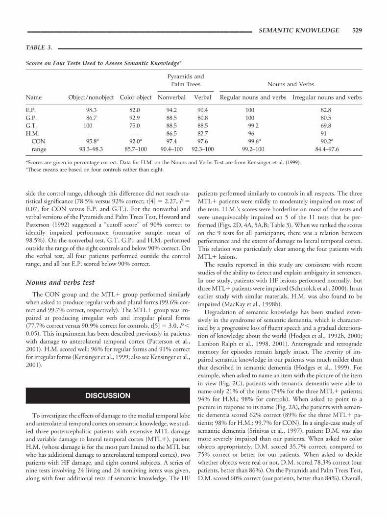
For each of the 18 separate tests (living and nonliving items oneach of 9 tests; data from Figures 2–5), we ranked the performanceof all 13 participants. Figure 7 shows the mean rank for each of thepatients with MTL� lesions, the mean for the controls (CON),and the mean ranks for the two HF patients. Amnesic patientsranked slightly higher than controls, perhaps because they wereyounger and/or better educated (ranks of 4.4 and 5.6 for the twopatients, mean rank for controls: 6.0). Among the other four pa-tients, there was a direct relation between performance on the 18
tests and the extent of damage to lateral temporal cortex. That is,their ranking on the 18 tests from highest (H.M.) to lowest (G.T.)was the same as their ranking with respect to amount of lateralcortex damage (H.M. least, G.T. most).
Living Versus Nonliving Items
On most of the nine tests, patients with MTL� lesions, includ-ing H.M., had more difficulty with living items than with nonliv-ing items (Figs. 2–5). This difference was significant for the scoresin Figures 2 (A–D, combined), 4B, and 5 (t[3] � 3.3, Ps � 0.05),and fell just short of significance for scores in Figure 3 (P � 0.07).This same difference was described previously in patients withpostencephalitic lesions of the ventral temporal lobe (Warringtonand Shallice, 1984; Caramazza and Shelton, 1998). The findinghas been attributed to differences in visual complexity (e.g., Fun-nell and Sheridan, 1992) or feature overlap (e.g., Riddoch andHumphreys, 1987) among the stimuli commonly used to depictliving and nonliving items. There is also evidence that the partic-ular sensory and motor systems involved in learning about objectsinfluence which brain areas store their representations (e.g., War-rington and McCarthy, 1988; Martin et al., 1996; Farah et al.,1996; Tranel et al., 1997).
Naming and Knowing
The relationship between the ability to name items and to pro-vide semantic knowledge about the same items was examined inmore detail in the case of the four patients with MTL� lesions. Forthe naming test in Figure 2C, we first identified the items thatpatients named successfully (i.e., spontaneously and without cuesfrom the examiner) and the items that they could not name. Then,for tests of semantic knowledge that did not explicitly requirenaming (Figs.3, 5B), we compared performance for items that hadbeen named successfully and items that had not been named. Forboth tests, the average score for items that had been named washigher than for items that had not been named (P � 0.005). Thus,for the patients with MTL� lesions, the failure to name an itemspontaneously predicted that less semantic knowledge would beavailable about that item than would be available for other itemsthat could be named. A relationship between naming ability andsemantic knowledge has also been described in patients with mildAlzheimer’s disease. (Hodges et al., 1996).
Other Tests of Semantic Knowledge
Table 3 shows the results for four additional tests of semanticmemory.
Object/nonobject discrimination test
When asked whether objects were real or not, two patients (E.P.and G.T.) performed as well as controls. E.P. made one error, andG.T. made no errors; G.P. made eight errors.
Coloring objects
One patient performed well within the control range (G.P.).Two patients made more errors than controls and performed out-
FIGURE 7. The performance of each participant was ranked oneach of the 18 tests (9 tests concerning living items and 9 tests con-cerning nonliving items; data from Figs. 2–5). Bars show the meanrank obtained. Circles show the mean rank for individual controls.Controls obtained an average rank of 6.0 among all 14 participants,because performance on the 18 tests was close to normal for 10 of the14 participants. Accordingly, on any one test, a control’s rank couldrange from 1 to 10. In the four patients with more extensive lesions,the ranking demonstrates a relationship between performance ontests of semantic knowledge and the amount of damage to anterolat-eral temporal cortex.
528 SCHMOLCK ET AL.
side the control range, although this difference did not reach sta-tistical significance (78.5% versus 92% correct; t[4] � 2.27, P �0.07, for CON versus E.P. and G.T.). For the nonverbal andverbal versions of the Pyramids and Palm Trees Test, Howard andPatterson (1992) suggested a “cutoff score” of 90% correct toidentify impaired performance (normative sample mean of98.5%). On the nonverbal test, G.T, G.P., and H.M. performedoutside the range of the eight controls and below 90% correct. Onthe verbal test, all four patients performed outside the controlrange, and all but E.P. scored below 90% correct.
Nouns and verbs test
The CON group and the MTL� group performed similarlywhen asked to produce regular verb and plural forms (99.6% cor-rect and 99.7% correct, respectively). The MTL� group was im-paired at producing irregular verb and irregular plural forms(77.7% correct versus 90.9% correct for controls, t[5] � 3.0, P �0.05). This impairment has been described previously in patientswith damage to anterolateral temporal cortex (Patterson et al.,2001). H.M. scored well: 96% for regular forms and 91% correctfor irregular forms (Kensinger et al., 1999; also see Kensinger et al.,2001).
DISCUSSION
To investigate the effects of damage to the medial temporal lobeand anterolateral temporal cortex on semantic knowledge, we stud-ied three postencephalitic patients with extensive MTL damageand variable damage to lateral temporal cortex (MTL�), patientH.M. (whose damage is for the most part limited to the MTL butwho has additional damage to anterolateral temporal cortex), twopatients with HF damage, and eight control subjects. A series ofnine tests involving 24 living and 24 nonliving items was given,along with four additional tests of semantic knowledge. The HF
patients performed similarly to controls in all respects. The threeMTL� patients were mildly to moderately impaired on most ofthe tests. H.M.’s scores were borderline on most of the tests andwere unequivocably impaired on 5 of the 11 tests that he per-formed (Figs. 2D, 4A, 5A,B; Table 3). When we ranked the scoreson the 9 tests for all participants, there was a relation betweenperformance and the extent of damage to lateral temporal cortex.This relation was particularly clear among the four patients withMTL� lesions.
The results reported in this study are consistent with recentstudies of the ability to detect and explain ambiguity in sentences.In one study, patients with HF lesions performed normally, butthree MTL� patients were impaired (Schmolck et al., 2000). In anearlier study with similar materials, H.M. was also found to beimpaired (MacKay et al., 1998b).
Degradation of semantic knowledge has been studied exten-sively in the syndrome of semantic dementia, which is character-ized by a progressive loss of fluent speech and a gradual deteriora-tion of knowledge about the world (Hodges et al., 1992b, 2000;Lambon Ralph et al., 1998, 2001). Anterograde and retrogradememory for episodes remain largely intact. The severity of im-paired semantic knowledge in our patients was much milder thanthat described in semantic dementia (Hodges et al., 1999). Forexample, when asked to name an item with the picture of the itemin view (Fig. 2C), patients with semantic dementia were able toname only 21% of the items (74% for the three MTL� patients;94% for H.M.; 98% for controls). When asked to point to apicture in response to its name (Fig. 2A), the patients with seman-tic dementia scored 62% correct (89% for the three MTL� pa-tients; 98% for H.M.; 99.7% for CON). In a single-case study ofsemantic dementia (Srinivas et al., 1997), patient D.M. was alsomore severely impaired than our patients. When asked to colorobjects appropriately, D.M. scored 35.7% correct, compared to75% correct or better for our patients. When asked to decidewhether objects were real or not, D.M. scored 78.3% correct (ourpatients, better than 86%). On the Pyramids and Palm Trees Test,D.M. scored 60% correct (our patients, better than 84%). Overall,
TABLE 3.
Scores on Four Tests Used to Assess Semantic Knowledge*
Name Object/nonobject Color object
Pyramids andPalm Trees Nouns and Verbs
Nonverbal Verbal Regular nouns and verbs Irregular nouns and verbs
E.P. 98.3 82.0 94.2 90.4 100 82.8G.P. 86.7 92.9 88.5 80.8 100 80.5G.T. 100 75.0 88.5 88.5 99.2 69.8H.M. — — 86.5 82.7 96 91
CON 95.8a 92.0a 97.4 97.6 99.6a 90.2a
range 93.3–98.3 85.7–100 90.4–100 92.3–100 99.2–100 84.4–97.6
*Scores are given in percentage correct. Data for H.M. on the Nouns and Verbs Test are from Kensinger et al. (1999).aThese means are based on four controls rather than eight.
_____________________________________________________________________ SEMANTIC KNOWLEDGE 529

the pattern of impairment in our patients is consistent with a loss ofspecific information from a network of conceptual knowledge(Murre et al., 2001), which results in a “blurring” of some conceptsand overlap among concepts that are closely related.
Semantic Knowledge and the Medial and LateralTemporal Lobes
Several lines of evidence suggest that impaired semantic knowl-edge is related to damage in anterolateral temporal cortex and notmedial temporal lobe structures, including perirhinal cortex. First,patients with semantic dementia typically have atrophy of the lat-eral temporal cortex and the temporal pole, with relative sparing ofthe medial temporal lobe and other cortical areas (Hodges et al.,1992; Garrard et al., 1997). In a recent morphometric MRI studyof six patients, the most consistent locus of atrophy was the leftpolar and inferior temporal lobe (Mummery et al., 2000). Moremedial cortex appears not to be compromised, at least in the earlystages of the disease. Although the status of the polar portion ofperirhinal cortex in semantic dementia is uncertain, there is astrong correlation between the severity of semantic dementia andthe degree of anterolateral temporal lobe atrophy (Simons et al.,1999; Mummery et al., 2000). A recent study found that thevolume of the left fusiform gyrus best correlated with measures ofsemantic knowledge and that measures of naming were addition-ally correlated with the volumes of the left temporal pole and theinferior and middle temporal gyri (Galton et al. 2001; for theimportance of anterior temporal lobe structures in semantic de-mentia, see also Chan et al., 2001). It is also noteworthy thatrecognition memory is impaired by damage to perirhinal cortex(Meunier et al., 1993; Buffalo et al., 1998, 1999). Yet patients withsemantic dementia exhibit intact recognition memory, eventhough they exhibit severe impairment on tests of semantic knowl-edge about the same stimuli (Simons et al., 1999).
Other evidence about the anatomy of semantic knowledge comesfrom functional neuroimaging studies. Within the temporal lobe,making semantic judgments about either words or pictures activatedthe left inferior temporal gyrus, the left middle temporal gyrus, and theleft fusiform gyrus (Vandernberghe et al., 1996). Similarly, lexico-semantic processing of words and word meanings was associated withactivity in the left middle and left inferior temporal gyri (Demonet etal., 1992). Finally, when volunteers generated appropriate colornames or action words in response to either line drawings of objects orwritten names of the objects, activity was observed in the temporallobe in the fusiform gyrus (for color words) and in the left posteriormiddle and superior temporal gyri (for action words) (Martin et al.,1995). For additional findings, see the comprehensive review by Ca-beza and Nyberg (2000).
Another source of evidence comes from brain stimulation stud-ies of patients who are candidates for unilateral temporal lobec-tomy. Stimulation in the inferior temporal lobe (the basal temporallanguage area) caused speech arrest and impaired confrontationnaming (Luders et al., 1986). The area in which language distur-bance could be produced included the inferior temporal gyrus,fusiform gyrus, and parahippocampal gyrus (Burnstine et al.,1990; Schaffler et al., 1994). The area in which stimulation most
consistently produced speech errors was the fusiform gyrus (Schaf-fler et al., 1994).
A final line of evidence comes from studies of confrontationnaming. Impaired confrontation naming (dysnomia) is related todamage lateral to the medial temporal lobe. First, patient H.M.,who has large medial temporal lobe lesions, performed within 1 SDof the control mean on a standard test of picture naming (theBoston Naming Test) (Kensinger et al., 2001). Second, impairedconfrontation naming is found following left anterior temporallobectomy (Langfitt and Rausch, 1996; Hermann et al., 1999;Hermann et al., 1994). In a recent study that assessed 217 patientsacross eight centers, the severity of postoperative naming problemswas related to the lateral extent of the resection (Hermann et al.,1999). Notably, language impairment following left anterior tem-poral lobectomy is not limited to difficulty with naming but alsoincludes difficulty in other tasks of semantic knowledge, such asthose tasks that assess the ability to comprehend nouns and makejudgments about synonyms (Glosser and Donofrio, 2001).
In summary, findings from semantic dementia, neuroimaging,brain stimulation, and unilateral temporal lobectomy point to theimportance of lateral temporal cortex for semantic knowledge.Moreover, the severity of the impairment in semantic knowledgein our patients was related to the extent of damage to lateral tem-poral cortex. Accordingly, we propose that the impairments exhib-ited by our three postencephalitic patients, and the milder impair-ments exhibited by H.M., are due to damage to anterolateraltemporal cortex, lateral to the medial temporal lobe. While it isdifficult to exclude entirely a possible contribution of damage to struc-tures within the parahippocampal gyrus (entorhinal, perirhinal, andparahippocampal cortex), this possibility seems unlikely. Thus, pa-tients G.T., G.P., and E.P. all have complete damage to the perirhinaland entorhinal cortices; yet G.T. performed overall more poorly onthe tests of semantic knowledge than did G.P. or E.P.
These considerations appear to rule out a contribution of theperirhinal and entorhinal cortices to deficits in semantic knowl-edge. Nevertheless, it was the case that damage within the parahip-pocampal cortex itself did relate to the severity of the impairmentin semantic knowledge in our three patients (E.P. � 40% damagebilaterally, G.P. � 42%, and G.T. � 72%). However, H.M. hadimpairments in semantic knowledge despite the fact that his para-hippocampal cortex was largely intact. Thus, while additional caseswill be useful to settle this point, damage to structures within theparahippocampal gyrus do not appear to be a significant factor inimpaired semantic knowledge (also see Galton et al., 2001).
The Special Case of Patient H.M.
In a recent, comprehensive study of H.M.’s language capacities,no evidence was found for impaired lexical knowledge or impairedproduction of regular and irregular word forms (Kensinger et al.,2001). On the tests of semantic knowledge studied here, H.M.performed within the range of control scores on most of the teststhat we gave him. He performed outside the control range when hehad to name items in response to their descriptions (Fig. 2D),when he had to generate category exemplars (Fig. 4A) (see alsoKensinger et al., 2001), and when he had to provide definitions
530 SCHMOLCK ET AL.

either in response to names of items (Fig. 5A) or pictures of items(Fig. 5B). He was also mildly impaired on the Pyramids and PalmTrees Test (Table 3). Kensinger et al. (2001) suggested that H.M.’spoor performance on tests of fluency may be related to motorslowing secondary to cerebellar degeneration. As for the other testson which he had reduced scores, it is not straightforward to iden-tify the basis for the impairment. It is notable that his shortcomingson tests of definitions resembled the difficulties that the threepostencephalitic patients had on these same tests. Like them, hefailed to supply the details that would make it possible to identifyitems and to differentiate items from other, semantically relateditems. Accordingly, in the case of the tests of definitions (andpossibly the other tests that he performed poorly), H.M.,’s impair-ment may have the same anatomic basis as in the patients in theMLT� group: damage lateral to the medial temporal lobe. InH.M.’s case, this lateral damage is limited, but it may be sufficientto impair performance on sensitive tests of semantic knowledge.
H.M.’s performance was also unique in important ways. First, asdescribed previously (Milner et al., 1968), H.M.’s intonation wasmonotone and little modulated. Second, while providing defini-tions, he made a large number of grammatical errors. For example,he made frequent errors in the use of pronouns and referents (seealso MacKay et al., 1998b). (For additional examples of difficultiesunique to H.M., see Schmolck et al., 2000.) In contrast, thepostencephalitic patients (E.P., G.P., and G.T.), all of whom havemore extensive medial and lateral lesions than H.M., spoke withnormal intonation and made no more grammatical errors than didcontrol subjects (Fig. 6). Accordingly, H.M.’s shortcomings inlanguage production, as described in previous reports (MacKay etal., 1998a, b; MacKay and James, 2001), are unlikely to be relatedto his medial or lateral temporal lobe damage. H.M. had seizuresbeginning at age 10 (which raises the question whether his lan-guage development was fully normal), his schooling was inter-rupted, and he came from a low socioeconomic background. Anyof these factors could be important.
In summary, patients with damage limited to the hippocampalformation (HF) performed normally in every respect on tests ofsemantic knowledge. In contrast, three postencephalitic patientswith large medial temporal lobe lesions and variable damage toanterolateral temporal cortex (MTL�) exhibited mild to moderateimpairment on these tests. Patient H.M. was impaired on five ofthe tests and was less severely impaired overall than were the threepostencephalitic patients. Accordingly, we suggest that the deficitsin semantic knowledge reported here are most likely related tocortical damage lateral to the medial temporal lobe. Finally,H.M.’s language production, including his use of grammar, distin-guished him from the other patients. These features of his perfor-mance are unlikely to be related to his temporal lobe lesion.
REFERENCES
Amaral DG, Insausti R. 1990. The human hippocampal formation. In:Paxinois G, editor. The human nervous system. p 711–755.
Buffalo EA, Ramus SJ, Clark RE, Teng E, Squire LR, Zola SM. 1999.Dissociation between the effects of damage to perirhinal cortex andarea TE. Learn Mem 6:572–599.
Buffalo EA, Reber PJ, Squire LR. 1998. The human perirhinal cortex andrecognition memory. Hippocampus 8:330–339.
Burnstine TH, Lesser RP, Hart J Jr, Uematsu S, Zinreich SJ, Krauss GL,Fisher RS, Vining EPG, Gordon B. 1990. Characterization of the basaltemporal language area in patients with left temporal lobe epilepsy.Neurology 40:966–970.
Cabeza R, Nyberg L. 2000. Imaging cognition II: an empirical review of275 PET and fMRI studies. J Cogn Neurosci 12:1–47.
Caine D, Watson J. 2000. Neuropsychological and neuropathologicalsequelae of cerebral anoxia: a critical review. J Int Neuropsychol Soc6:86–99.
Caramazza A, Shelton JR. 1998. Domain specific knowledge systems inthe brain: the animate–inanimate distinction. J Cogn Neurosci10:1–34.
Chan D, Fox NC, Scahill RI, Crum WR, Whitwell JL, Leschziner G,Rossor AM, Stevens JM, Cipolotti L, Rossor MN. 2001. Patterns oftemporal lobe atrophy in semantic dementia and Alzheimer’s disease.Ann Neurol 49:433–442
Corkin S. 1984. Lasting consequences of bilateral medial temporal lobec-tomy: clinical course and experimental findings in H.M. Semin Neu-rol 4:249–259.
Corkin S, Amaral DG, Gonzalez RG, Johnson KA, Hyman BT. 1997.H.M.’s medial temporal lobe lesion: findings from magnetic resonanceimaging. J Neurosci 17:3964–3980.
Demonet J, Chollet F, Ramsay S, Cardebat D, Nespoulous J, Wise R,Rascol A, Frackowiak RS. 1992. The anatomy of phonological andsemantic processing in normal subjects. Brain 115:1753–1768.
Farah MJ, Meyer MM, McMullen PA. 1996. The living/nonliving dis-tinction is not an artifact: giving an a priori implausible hypothesis astrong test. Cogn Neuropsychol 13:137–154.
Funnell E, Sheridan J. 1992. Categories of Knowledge? Unfamiliar aspectsof living and nonliving things. Cogn Neuropsychol 9:135–153.
Galton CJ, Patterson K, Graham K, Lambon Ralph MA, Williams G,Antoun N, Sahakian BJ, Hodges JR. 2001. Differing patterns of tem-poral atrophy in Alzheimer’s disease and semantic dementia. Neurol-ogy 57:216–225.
Garrard P, Perry R, Hodges JR. 1997. Disorders of semantic memory.J Neurol, Neurosurg Psychiatry 62:431–435.
Glosser G, Donofrio N. 2001. Differences between nouns and verbs afteranterior temporal lobectomy. Neuropsychology 15:39–47.
Hermann BP, Perrine K, Chelune GJ, Barr W, Loring DW, Strauss E,Trenerry MR, Weisterveld M. 1999. Visual confrontation namingfollowing left anterior temporal lobectomy: a comparison of surgicalapproaches. Neuropsychology 13:3–9.
Hermann BP, Wyler AR, Somes G, Clement L. 1994. Dysnomia after leftanterior temporal lobectomy without functional mapping: frequencyand correlates. Neurosurgery 35:52–57.
Hodges JR, Bozeat S, Lambon Ralph MA, Patterson K, Spatt J. 2000. Therole of conceptual knowledge in object use Evidence from semanticdementia. Brain 123:1913–1925.
Hodges JR, Patterson K, Graham N, Dawson K. 1996. Naming andknowing in dementia of Alzheimer’s type. Brain Lang 2:11–24.
Hodges JR, Salmon DP, Butters N. 1992a. Semantic memory impair-ment in Alzheimer’s disease: failure of access or degraded knowledge?Neuropsychologia 30:301–314.
Hodges JR, Patterson K, Oxbury S, Funnell E. 1992b. Semantic demen-tia: progressive fluent aphasia with temporal lobe atrophy. Brain 115:1783–1806.
Hodges JR, Patterson K, Ward R, Garrard P, Bak T, Perry R, Gregory C.1999. The differentiation of semantic dementia and frontal lobe de-mentia from early Alzheimer’s disease: a comparative neuropsycholog-ical study. Neuropsychology 13:31–40.
_____________________________________________________________________ SEMANTIC KNOWLEDGE 531

Howard D, Patterson K. 1992. Pyramids and Palm Trees: a test of seman-tic access from pictures and words. Bury St. Edmunds: Thames ValleyTest Company.
Insausti R, Juottonen K, Soininen H, Insausti A, Partanen K, Vainio P,Laakso M, Pitkanen. 1998. MR Volumetric analysis of the humanentorhinal, perirhinal, and temporopolar cortices. Am J Neuroradiol19:659–671.
Kensinger EA, Ullman MT, Locascio JJ, Corkin S. 1999. What is therelation between medial temporal lobe structures and lexical memory?Evidence from amnesic patient H.M. Soc Neurosci Abs 25:357.
Kensinger EA, Ullman MT, Corkin S. 2001. Bilateral medial temporallobe damage does not affect lexical or grammatical processing: evi-dence from amnesic patient H.M. Hippocampus 11:347–360.
Lambon Ralph MA, Graham KS, Ellis AW, Hodges JR. 1998. Naming insemantic dementia—what matters? Neuropsychologia 8:775–784.
Lambon Ralph MA, McClelland JL, Patterson K, Galton CJ, Hodges JR.2001. No right to speak? The relationship between object naming andsemantic impairment: neuropsychological evidence and a computa-tional model. J Cogn Neurosci 13:341–356.
Langfitt JT, Rausch R. 1996. Word finding deficits persist after left an-terotemporal lobectomy. Arch Neurol 53:72–76.
Luders H, Lesser RP, Hahn J, Dinner DS, Morris H, Resor S, HarrisonM. 1986. Basal temporal language area demonstrated by electricalstimulation. Neurology 36:505–510.
MacKay DG, James LE. 2001. The binding problem for syntax, seman-tics, and prosody: H.M.’s selective sentence-reading deficits under thetheoretical-syndrome approach. Lang Cogn Processes 16:419–460.
MacKay DG, Burke DM, Stewart R. 1998a. H.M.’s language productiondeficits: implications for relations between memory, semantic binding,and the hippocampal system. J Mem Lang 38:28–69.
MacKay DG, Stewart, R Burke DM. 1998b. H.M. Revisited: relationsbetween language comprehension, memory, and the hippocampal sys-tem. J Cogn Neurosci 10:377–394.
Manns JR, Squire LR (in press) The medial temporal lobe and memory forfacts and events. In: Baddeley A, Wilson B, Kopelman M, editors.Handbook of memory disorders. 2nd ed. New York: John Wiley & Sons.
Martin A, Haxby JV, Lalonde FM, Wiggs CL, Ungerleider LG. 1995.Discrete cortical regions associated with knowledge of color andknowledge of action. Science 270:102–105.
Martin A, Wiggs CL, Ungerleider LG, Haxby JV. 1996 Neural correlatesof category-specific knowledge. Nature 379:649–652.
Meunier M, Bachevalier J, Mishkin M, Murray EA. 1993. Effects onvisual recognition of combined and separate ablations of the entorhinaland perirhinal cortex in rhesus monkeys. J Neurosci 13:5418–5432.
Milner B, Corkin S, Teuber HL. 1968. Further analysis of the hippocam-pal amnesic syndrome: l4 year follow-up study of H.M. Neuropsycho-logia 6:215–234.
Mummery CJ, Patterson K, Price CJ, Ashburner J, Frackowiak RS,Hodges JR. 2000. A voxel based morphometric study of semantic
dementia: the relationship between temporal lobe atrophy and seman-tic memory. Ann Neurol 47:36–45.
Murre JM, Graham KS, Hodges JR. 2001. Semantic dementia: relevanceto connectionist models of long-term memory. Brain 124:647–675.
Patterson K, Lambon Ralph MA, Hodges JR, McClelland JL. 2001. Def-icits in irregular past-tense verb morphology associated with degradedsemantic knowledge. Neuropsychologia 39:709–724.
Reed JM, Squire LR. 1998. Retrograde amnesia for facts and events:findings from four new cases. J Neurosci 18:3943–3954.
Rempel-Clower N, Zola-Morgan S, Squire LR, Amaral DG. 1996. Threecases of enduring memory impairment after bilateral damage limitedto the hippocampal formation. J Neurosi 16:5233–5255.
Riddoch J, Humphreys GW. 1987. A case of integrative visual agnosia.Brain 110:1431–1462.
Schaffler L, Luders H, Morris H, Wyllie E. 1994. Anatomic distributionof cortical language sites in the basal temporal language areas in pa-tients with left temporal lobe epilepsy. Epilepsia 35:525–528.
Schmolck H, Stefanacci L, Squire LR. 2000. Detection and explanation ofsentence ambiguity are unaffected by hippocampal lesions but areimpaired by larger temporal lobe lesions. Hippocampus 10:759–770.
Scoville WB, Milner B. 1957 Loss of recent memory after bilateral hip-pocampal lesions. J Neurol Neurosurg Psychiatry 20:11–21.
Simons JS, Graham KS, Hodges JR. 1999. What does semantic dementiareveal about the functional role of the perirhinal cortex. Trends CognSci 3:248–249.
Snodgrass JG, Vanderwart M. 1980. A standardized set of 260 pictures:norms for name agreement, image agreement, familiarity, and visualcomplexity. J Exp Psychol Hum Learn Mem 6:174–215.
Squire LR, Schmolck H, Stefanacci L. 2001. Memory, language, andneuroanatomy: a reply. Brain Lang 78:273–275.
Srinivas K, Breedin SD, Branch-Coslett H, Saffran EM. 1997. Intactperceptual priming in a patient with damage to the anterior inferiortemporal lobes. Neuroscience 9:490–511.
Stefanacci L, Buffalo EA, Schmolck H, Squire LR. 2000. Profound am-nesia following damage to the medial temporal lobe–a neuroanatomi-cal and neuropsychological profile of patient E.P. J Neurosci20:7024–7036.
Teng E, Squire LR. 1999 Memory for places learned long ago is intactafter hippocampal damage. Nature 400:675–677.
Tranel D, Damasio H, Damasio AR. 1997. A neural basis for the retrievalof conceptual knowledge. Neuropsychologia 35:1319–1327.
Vandernberghe R, Price C, Wise R, Josephs O, Frackowiak RS. 1996.Functional anatomy of a common semantic system for words andpictures. Nature 383:254–256.
Warrington EK, McCarthy RA. 1988. The fractionation of retrogradeamnesia. Brain Cogn 7:184–200.
Warrington EK, Shallice T. 1984. Category specific semantic impair-ments. Brain 107:829–854.
APPENDIX
Excerpts from Verbatim Transcripts (Test 8,Definitions to Name) for Patients H.M. G.T.,and Control No. 4
Experimenter interjections are in italics. Grammatical (G) andform (F) errors are underlined.
Patient H.M., motorcycle
…well…it can be (F)…uh, uh…a motorcycle is (F)uh…maybe…4 (F), it’s on 2 wheels…. And it could be have (G) a
sidecar to it…(uh huh)…and uh, it’s very fast too…and it canca…carry a person in back of the driver (uh huh), …and also asidecar (G)…(uh huh)… PAUSE…(what does it look like, how does itwork)…it’s got 2 wheels (um hm) and a motor then (G) and youhave to pump it once to start it up…(um hm) and you start it (F)and it goes around and around and right at the point of where thedriver is (G), is a big tank (right) for gasoline (yup), the fuel forit…and it goes very fast…oh I don’t know how uh many miles anhour (fast)…very fast, ’cause (G) the policemen have to use ituh…you want to know…how they use it (G)? to chasespeeders…(right, very good)…and they (G) also have it the otherway (G)…(what do you mean, the other way)…for in races (G) (oh,in races, yes in races, they use it in races)…yeah…’cause I know
532 SCHMOLCK ET AL.

(F)…’cause (G) my father used to ride one at one time…and hestopped himself (G) because the doctor told him not to (G).
Patient G.T., motorcycle
…motorcycle, yes, I’ve rode them (G) many times…,[laughing]…PAUSE…(imagine I don’t know what it is…explain amotorcycle to me.)…motorcycle is like a large bicycle…(uh huh)…that has a motor in it and you ride it just likeyou do…an…automobile…(umhmm)…gasoline…(ok)…and it’sbig…PAUSE…(ok, what else?)…very powerful…(uh huh…what elseabout it?)…you need a driver’s license to drive it…(what else isimportant about it?)…gasoline, the oil…PAUSE…(uh huh…what’sthe cool thing about a motorcycle?)…cool thing?…(uh huh)…wellyou ride it…you have to sit on top of it and you ride out in theopen…and just have nothin’ to protect you…(that soundsright…I’ve never ridden one)…[laughs]…like a horse…(that’s whatpeople say)…yeah…you haven’t ridden one?…(uh uh)…never rid-den on a motorcycle?…I have plenty of times…they’re a lot offun…there are different kinds too…(right…what do you mean,different kinds?)…well, you have the motorcycle you just ride outdown the streets…you know…driving and all that…youknow…and a sport motorcycle…(right…that’s important.)…yougo out to the mountains and stuff with it…and then racingmotorcycles…(right) different kinds…(good).
Control 4, motorcycle
Motorcycle…I used to own motorcycles…in fact, I used to buythem and sell them…motorcycles…ah…similar to a bicycle, 2wheels…engine, between, or…within the frame…from, ah 10horsepower on up to uh…about 70, 60 horsepower…uh…chain,most of them used to have chain drive between the uh transmissionand the rear wheel…right, right hand on most Harley Davidsonscontrolled your gas, the left was your advance and retard, your,your spark…one headlight forward, one taillight in therear…uh…used to have a sailseat or a pinion seat, or a kingsize seatfor carrying an extra passenger…most gas tanks carried, uh 2 uh(F)…regular gas tank…a gallon and a half, auxiliary approximatelya little over a gallon…one quart of oil…one battery, uh…topspeed on most bikes back then was about 75 miles per hour…thenewest go up to 150 miles…um….
Patient H.M., swan
Well…a swan…flies around sometime (um hmm)…but it goeson the water too (um hmm)…and…goes after fish in a way(G)…(um hmm) and…PAUSE…(what else does it do, what does it
look like)…and it’s got a very long neck…(um hmm) and uh…wellthese (G) is just scooped over a certain area (G) and they (G) findthe fish…a lot of fish (F) in there (G) and they could swallow them(G)…(um hmm) and…are…and…(F)…by doin’ that (G) justsometimes (G) they can keep them stored in their mouth. (umhmm)…and uh, and the throat (F)…(um hmm)…so (G) they canget everything down that way (G)…(exp what, what is theircolor)…white…some of them are white, some of them arebrown…(ok)…but you mostly think of a white swan…(ok).
Patient G.T., swan
…a swan is a…uh. a large bird who lands on thewater…PAUSE…(uh huh, ok…what else?)…swims…(uh huh…whatelse?)…PAUSE…(what does it look like?)…giant turtle…(what does itlook like?)…giant turtle, it’s, it’s like a turtle…swan…big…it’shuge…it’s big (F2)…(uh huh, tell me more about what it lookslike)…(try to describe it)…umm…well…(what color is it?)…um Idunno…[laughing]…(you’re not really sure?…what do youthink?)…probably a blackish brown color…(ok…ok).
Control 4, swan
Swan…it’s a long neck (G)…like an S curve…basically most arewhite…uh, big beak…they dive for their fish…uh,when I say dive,they stick their neck way down and they will follow through(G)…they have webbed feet, uh,…very fast swimmers, verygraceful…mate for life…uh, they eat most anything, bread, food,seeds, they’ll eat that along with smaller fish…um…that’s aboutit…they grow, I’m gonna guess, in weight, they should grow up toabout maybe 15 pounds, 10–15 pounds…they’re lightweight re-ally, when you get down to it, ’cause all feathers (G)…and theyshed water, their…their feathers will not absorb water…and I didsay webbed feet….
Acknowledgments
This research was supported by the Medical Research Service ofthe Department of Veterans Affairs, NIMH grant 24600, the Met-ropolitan Life Foundation, DFG grant Vo 770/1-1 (to H.S.), anda Howard Hughes Medical Institute Predoctoral Fellowship (toE.A.K.) We thank Lisa Stefanacci, Joseph Manns, Gary Press,Joyce Zouzounis, Shauna Stark, and Jennifer Frascino for assis-tance; Michael Ullman for providing the Nouns and Verbs test;and John Hodges for providing tests 1 and 3–8. Heike Schmolckis now at the Department of Neurology, Baylor College of Medi-cine, Houston, Texas.
_____________________________________________________________________ SEMANTIC KNOWLEDGE 533