september, 2015 sultana sharmin graduate school of
TRANSCRIPT

Analysis of Thiosulfate Metabolism in a Marine Acidophilic
Sulfur-Oxidizing Bacterium,
Acidithiobacillus thiooxidans strain SH
September, 2015
SULTANA SHARMIN
Graduate School of Environmental and Life Science
(Doctor's Course)
OKAYAMA UNIVERSITY

CONTENTS
Pages
CHAPTER 1
1. GENERAL INTRODUCTION……………………………. 1
1.1. Biomining………………………………………... ......... 1
1.2. Fundamentals of Biomining……………………………. 2
1.2.1.Industrial Biomining…………………………............ 3
1.2.2.Metal Sulfide oxidation- the two pathways……………… 4
1.3. Applications of Biomining………………………………. 5
CHAPTER 2
2.1. INTRODUCTION……………………………………….. 12
2.2 MATERIALS AND METHODS………………………… 15
2.2.1. Bacterial strains, media, and growth conditions……… 15
2.2.2. Enzyme assay…………………………………………. 15
2.2.3. Purification of TSD from At.thiooxidans strain SH…… 16
2.2.4. Protein analysis………………………………………… 17
2.2.5. Analysis of sulfur compound………………………….. 17
2.3. RESULTS AND DISCUSSION…………………………… 19
2.3.1. Detection of TSD activity in thiosulfate-grown
At.thiooxidans strain SH………………………………………..19
2.3.2. Purification of TSD from thiosulfate-grown

At.thiooxidans strain SH……………………………………... 21
2.3.3. Biochemical Properties of TSD from strain SH………….26
2.3.4. Stoichiometry of thiosulfate oxidation……………………31
2.3.5. Substrate specificity and electron acceptor……………….33
2.3.6. Inhibitors………………………………………………….35
2.3.7. Identification of the gene encoding TSD………………….35
2.3. SUMMARY...……………………………………………………..38
CHAPTER 3
3.1. INTRODUCTION………………………………………………39
3.2. MATERIALS AND METHODS……………………………….41
3.2.1. DNA preparation………………………………………….41
3.2.2. Genome sequencing and draft assembly………………….41
3.2.3. Gene prediction and annotation………………………...41
3.2.4. At. thiooxidans genome sequences………………………..41
3.2.5. Comparative genome analysis…………………………….42
3.2.4. Accession number…………………………………………42
3.3 RESULTS AND DISCUSSION………………………………….43
3.3.1 Genomic analysis…………………………………………….43
3.3.2 Putative genes involved in sulfur oxidation………………….45
3.3.3. Construction of sulfur oxidation model in
At. thiooxidans strain SH……………………………………..53
3.3.4. Determination of a gene encoding TSD
in At. thiooxidans strain SH………………………………...56
3.4. SUMMARY…...…………………………………………………… 61

CHAPTER 4
CONCLUSION………………………………………………62
REFERENCES………………………………………………………….66
ACKNOWLEDGEMENTS………………………………………74

1
CHAPTER 1
1. GENERAL INTRODUCTION
1.1. Biomining
Continuous depletion of Earth’s high-grade ore reserves or deposits used for
increasing metal demand necessitates the need for cost-effective and innovative ways
of recovering metals from low-grade deposits. Traditional extraction methods involve
high cost and application of toxic chemicals in the presence of extremely high
temperatures such as, roasting and smelting which have deleterious effects on the
environment. The sustainable development challenge facing the mineral and mining
industry is to provide the supply of minerals, metals and material required to sustain
social and economic growth without causing long term degradation of the environment.
In this context, biomining allows economically viable and environmentally friendly
ways of extracting metals from low-grade ores. Biomining is a general term also known
as bioleaching is one of the most promising and revolutionary biotechnologies which
exploit microbiological processes to recover metals and minerals from sulfide or iron
containing ores, concentrates and waste materials (Rawlings, 1997). Now a day,
biomining is an increasingly applied biotechnological procedure for processing of ores
in the mining industry (Schippers et al., 2014). The technique is also receiving much
attention worldwide to detoxify sediments and soils contaminated with heavy metals
and toxic organic chemicals to make mining more sustainable.
Biomining or bioleaching processes employ microbial consortia that are dominated
by a variety of mineral-decomposing iron- and sulfur-oxidizing chemolithotrophic,
acidophilic, autotrophic prokaryotes. These chemolithotrophic acidophiles are
generally represented by sulfur- and iron-oxidizing bacteria and archaea that are
responsible for the extraction or mobilization of metals from sulfide ores. They are

2
found in a wide range of habitats including metal sulfide deposits all over the world
(Karavaiko et al., 2003), fresh waters associate with sulfide deposits (Gonzalez-Toril
et al., 2003), acid mine drainage (Brett et al., 2003) sites and very few in seawater
(Kamimura et al., 2003). These microorganisms interact with metals and minerals in
various ways and are involved in global cycling of elements. The most important
metal leaching microorganisms use ferrous iron and reduced inorganic sulfur
compounds (RISCs) as electron donors and fix carbon di-oxide (CO2). Metals for
which the biomining technique is mainly employed include copper, cobalt, nickel, zinc
and uranium. For recovery of gold and silver, the activity of mining bacteria is
applied only to remove interfering metal sulfides from ores bearing the precious metals
(Rohwerder,T et al.,2003). More recently, bioleaching is being used to also recover
metals from higher-grade ores as these processes are frequently more economic than
alternate methods.
1.2. Fundamentals of Biomining
The mechanism by which microorganisms enhance oxidation of metal sulfides and
leaching kinetics is still surrounded by controversy (Rossi, 1990). Some argue that the
mechanism may involve the bacterial action which could be the consequence of a
physicochemical alteration of the mineral surface produced by physical contact with
the bacteria, the so-called “direct attack”. Some other maintain that the bioleaching
could be simply a chemical process, the dominating factor being the presence in the
mineral suspension of iron or sulfur compounds, which in an acidic environment
oxidizes the mineral crystals.
The basis of microbial extraction is that the metal sulfides, the principal component
in many ores, are not soluble but when oxidized to sulfate become soluble so that the
metal salt can be extracted.
3
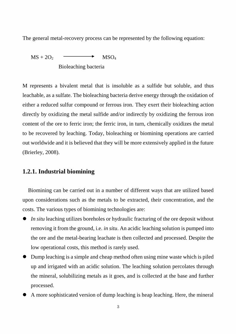
The general metal-recovery process can be represented by the following equation:
MS + 2O2 MSO4
Bioleaching bacteria
M represents a bivalent metal that is insoluble as a sulfide but soluble, and thus
leachable, as a sulfate. The bioleaching bacteria derive energy through the oxidation of
either a reduced sulfur compound or ferrous iron. They exert their bioleaching action
directly by oxidizing the metal sulfide and/or indirectly by oxidizing the ferrous iron
content of the ore to ferric iron; the ferric iron, in turn, chemically oxidizes the metal
to be recovered by leaching. Today, bioleaching or biomining operations are carried
out worldwide and it is believed that they will be more extensively applied in the future
(Brierley, 2008).
1.2.1. Industrial biomining
Biomining can be carried out in a number of different ways that are utilized based
upon considerations such as the metals to be extracted, their concentration, and the
costs. The various types of biomining technologies are:
In situ leaching utilizes boreholes or hydraulic fracturing of the ore deposit without
removing it from the ground, i.e. in situ. An acidic leaching solution is pumped into
the ore and the metal-bearing leachate is then collected and processed. Despite the
low operational costs, this method is rarely used.
Dump leaching is a simple and cheap method often using mine waste which is piled
up and irrigated with an acidic solution. The leaching solution percolates through
the mineral, solubilizing metals as it goes, and is collected at the base and further
processed.
A more sophisticated version of dump leaching is heap leaching. Here, the mineral

4
ore is crushed, often agglomerated to attach smaller particles to larger ore pieces,
and carefully piled onto a waterproof base to form a heap. The heap is then aerated
from underneath and irrigated from the top (as for dump bioleaching).
Reactor leaching is the most expensive biomining technology and is usually
utilized for precious metals, such as gold. Pulverized mineral is mixed with an
acidic, leaching solution in large tanks and microorganisms added. The tanks are
stirred and aerated, and have the potential to control variables such as pH, oxygen,
and temperature. The optimized conditions results in increased leaching rates. This
technique is exploited in the Barberton gold mines, South Africa, which produces
approximately 2,800 kg of gold annually (Pan African annual report, 2011).
1.2.2. Metal sulfide oxidation - the two pathways
The mechanism of how metals are solubilized from mineral ores has been
extensively investigated and has been the subject of many deliberations. It was
previously debated whether metal solubilization was a result of direct enzymatic
oxidation of the mineral or if ferric iron contained in the leaching solution oxidizes the
metal sulfide bond. The latter proved to be correct and in fact, sulfide minerals can be
abiotically leached without the contribution of microorganisms. However, this process
is extremely slow. The role of microorganisms is to re-generate the mineral attacking
agents, protons and ferric iron (Sand et al., 1995; Schippers et al., 1996; Schippers and
Sand, 1999). As microbial catalysis increases the rate-limiting step of ferrous iron
oxidation by 500-fold, the microorganisms considerably enhance the leaching rate
(Singer and Stumm, 1970). Metal sulfide minerals are principally leached via two
independent mechanisms, depending on the acid solubility of the mineral, which in
turn depends on the electron configuration (Schippers et al., 1996; Schippers and Sand,
1999). If the valence electrons in the metal sulfide bond are derived only from the metal

5
entity, they are acid-insoluble. In contrast, minerals with a contribution of valence
electrons from both the metal and the sulfide entity are acid-soluble.
Acid-insoluble metal sulfides are leached via the so-called “Thiosulfate pathway”.
The name derives from thiosulfate being the first inorganic sulfur compound (ISC)
intermediate formed. ISC-oxidizing microorganisms then oxidize thiosulfate via
tetrathionate, disulfane-monosulfonic acid, and trithionate into sulfuric acid. Further,
and most importantly, iron-oxidizing microorganisms oxidize the ferrous iron into
ferric iron, which can attack the chemical bond in the mineral and the catalytic cycle is
closed. Hence, only iron-oxidizing microorganisms are responsible for catalytic
dissolution of the mineral. Examples of metal sulfides that are leached via the
thiosulfate pathway are pyrite, molybdenite, and tungstenite. In contrast, acid-soluble
metal sulfides are leached via the “Polysulfide mechanism”. This mechanism is
characterized by the formation of hydrogen sulfide, which is rapidly oxidized into
polysulfides, which decompose into elemental sulfur in acidic solutions. Hence,
leaching of acid-soluble metal sulfides is characterized by the accumulation of large
amounts of elemental sulfur. The ferrous iron and the elemental sulfur are oxidized by
iron- and ISC-oxidizing microbes into ferric iron and sulfuric acid, respectively. Since
these types of minerals are acid-soluble, the protons produced by ISC oxidation can
also attack the mineral. Examples of minerals that are leached via the polysulfide
mechanism are chalcopyrite, sphalerite, and galena. (Schippers et al., 1996, Schippers
and Sand, 1999).
1.3. Applications of Biomining
The potential and current applications of biomining include the recovery of copper,
gold, cobalt, nickel, zinc, uranium and other heavy metals. Furthermore, it can be
applied for desulfurization of coal and oil, tertiary recovery of oil and biosorption of
metal ions. For biomining specialized microorganisms are used in order to recover
6
valuable metals from ores via bioleaching. At present, at least 11 putative prokaryote
divisions can be related to this phenomenon (Rohwerder et al., 2003). Bacteria
belonging to the genus Acidithiobacillus are by far the most studied acidophiles often
associated with biomining. Acidithiobacillus spp. are Gram-negative proteobacteria
that are subdivided into 5 distinct species: Acidithiobacillus albertensis,
Acidithiobacillus caldus, Acidithiobacillus ferrivorans, Acidithiobacillus ferrooxidans,
and Acidithiobacillus thiooxidans. Each species consists of several strains with various
characteristics. These acidophilic microorganisms play a central role by catalyzing
aerobic oxidation of sulfides. Acidithiobacillus spp. are active in several steps of the
sulfur cycle but their most important role in sulfur mobilization is to catalyse metal
sulphide oxidation at low pH, thereby releasing sulfate into solution in bioleaching or
biomining process (Schrenk et al., 1998). The principle bacteria in ore leaching are At.
ferrooxidans and At. thiooxidans. At. thiooxidans (formerly called as Thiobacillus
thiooxidans) was the first bacterium of the genus to be discovered (Kelly and Wood,
2000; Waksman, 1922). It was shown not to oxidize Fe2+ but was able to metabolize
various RISCs such as elemental sulfur (Sº), thiosulfate (S2O32-), tetrathionate (S4O6
2-)
and sulfite (SO32-) with different pH optimums for certain compounds. On the other
hand, At. ferroxidans is the best studied representative of all acidithiobacilli. It can
utilize both Fe2+ and RISCs as energy source. (Harrison, 1982; Harrison, 1984;
Karavaiko, 2003).
Ferric iron chemically attack chalcopyrite to solubilize cupric iron, ferrous iron and
elemental sulfur. To continue this reaction, ferrous ion should be oxidized to ferric ion.
Therefore, iron-oxidizing bacteria, such as At. ferrooxidans is used. On the contrary,
elemental sulfur covers the surface of ores and inhibits the chemical attack of ferric
iron, so sulfur-oxidizing bacteria, such as At. thiooxidans is used to oxidize elemental
sulfur to sulfate. The technique can also be applied for bioremediation of sediments
and soils containing salts and polluted with heavy metals (Fig. 1-1).

7
Fig. 1-1. The mechanism of biomining and its application for bioremediation. At.
ferrooxidans and At. thiooxidans are acidophilic iron- or sulfur-oxidizing bacterium,
and used for biomining or bacterial leaching. The technique is used to recover metals
from low-grade of sulfide ores. Bacterial leaching can also be applied for
bioremediation of sediments and soils polluted with heavy metals.

8
Because ocean deposits are rich in chalcopyrite, pyrite or marcasite, biomining
technique are useful for the recovery of metals from these sulfide ores containing NaCl.
Although At. ferroxidans and At. thiooxidans including other acidithiobacilli like At.
caldus, At. albertensis, At. ferrivorans have essential role in metal mobilizing
process of biomining, almost all of these bacteria cannot grow in media supplemented
with a high concentrations of NaCl (Huber and Stetter, 1989). Therefore, terrestrial
iron or sulfur-oxidizing bacteria cannot be applicable to the bioleaching process for
salt-containing samples. However, there have been some reports on NaCl-tolerant or
NaCl–requiring sulfur-oxidizing bacteria such as Halothiobacillu shalophilus (Wood
and Kelly, 1991), Halothiobacillus hydrothermalis (Durand et al., 1993),
Halothiobacillus neapolitanus (Kelly and Harrison, 1989), Halothiobacillus kellyi
(Sievert et al., 2000), and Thiomicrospira sp. (Kuenen and Veldkamp, 1972; Ruby et
al., 1981; Ruby Jannasch, 1982; Jannasch et al., 1985), these bacteria were not
acidophiles.
Acidophilic chemolithotrophic sulfur-oxidizers are generally soil, freshwater or acid
mine water bacteria and their ability to live in saline environments is limited.
However, acidophilic sulfur-oxidizing bacteria that are tolerant to NaCl or require NaCl
for growth may be useful in developing remedial technologies for salt-containing ores
and environments contaminated with heavy metals. Hence halophilic (salt requiring)
acidophiles have gained increasing interest because of their importance in biomining
oprations in salt containing environments. Some exceptional acidophilic isolates that
require or are tolerant to NaCl include the sulfur-oxidizer Thiobacillus prosperus
(Huber and Stetter, 1989), the iron-oxidizing bacterium KU2-11 (Kamimura et al.,
2001) and the sulfur-oxidizing halophilic At. thiooxidans strain SH (Kamimura et al.,
2003).
At. thiooxidans strain SH is particularly important as it is a marine bacterium that
can be used to remediate sulfur-polluted salt containing environment. The strain SH
grows and survives by autotrophically utilizing energy derived from the oxidation of

9
elemental sulfur and reduced inorganic sulfur compounds (RISCs). The bacterium
grows optimally in sulfur medium in the presence of 2% NaCl. NaCl stimulates the
sulfur- and sulite-oxidizing activity in resting cells. Strain SH was identified as At.
thiooxidans based on the 16S rRNA gene sequence (Fig. 1-2).
At. thiooxidans strain SH is able to oxidize elemental sulfur, sulfide and other sulfur
compounds to sulfate through the oxidation pathway. The oxidation pathway for
elemental sulfur and RISCs in At. thiooxidans strain SH has already been reported
(Kamimura et al., 2005) (Fig. 1-3). Since it has been assumed that thiosulfate
dehydrogenase can play a significant role in the oxidation of elemental sulfur and
RISCs in acidophilic bacterium At. thiooxidans strain SH, the current investigations
focus on the purification, characterization and genetic analysis of thiosulfate
dehydrogenase enzyme from At. thiooxidans strain SH.

10
Fig. 1-2. Effect of NaCl on the growth of acidophilic sulfur-oxidizing bacteria.
An acidoophilic sulfur-oxidizing bacterium (At. thiooxidans ON106) isolated from
terrestrial environments cannot grow in salt containing environments. However, strain
SH autotrophically grows on elemental sulfur, thiosulfate and tetrathionate as growth
substrates and requires NaCl for growth. Phylogenetic tree based on 16S rRNA gene
sequences of strain SH and other sulfur oxidizing bacteria has been shown in the figure.
Strain SH was identified as Acidithiobacillus thiooxidans based on the 16S rRNA
gene sequence .

11
Fig. 1-3. The putative oxidation pathway for elemental sulfur (Sº) and reduced
inorganic sulfur compounds (RISCs) in At. thiooxidans strain SH. In this pathway,
it is proposed that a metabolism of thiosulfate to tetrathionate by thiosulfate
dehydrogenase (TSD) is a key pathway for bacterial leaching. Sulfide quinone
reductase (SQR), cytochrome c oxidase (COX), bd-type ubiquinol oxidase (CYD)
enzymes were also thought to involve in sulfur compound oxidation pathway in strain
SH.

12
CHAPTER 2
Purification and characterization of thiosulfate
dehydrogenase from a marine
Acidithiobacillus thiooxidans strain SH
2.1. INTRODUCTION
Acidophilic chemoautotrophic iron- and sulfur-oxidizing bacteria, such as
Acidithiobacillus ferrooxidans and Acidithiobacillus thiooxidans, are utilized for the
bioleaching of sulfide minerals. NaCl is a crucial factor in this process, as it inhibits
the activities and growth of bioleaching microorganisms (Wang et al., 2012). As ocean
deposits are rich in pyrite, chalcopyrite, and marcasite, marine microorganisms show
advantages in the bioleaching of marine sulfide minerals. An acidophilic marine sulfur-
oxidizing bacterium, At. thiooxidans strain SH has been isolated from Seto Inland Sea,
Japan, to develop a bioleaching process for NaCl-containing sulfide minerals
(Kamimura et al., 2003 and 2005). The bacterium grows chemoautotrophically by
deriving energy from the oxidation of elemental sulfur and reduced inorganic sulfur
compounds (RISCs), such as thiosulfate and tetrathionate. Thiosulfate-metabolizing
activity is most important for bioleaching, as the sulfur moiety of sulfide minerals is
thought to be metabolized via thiosulfate as an intermediate as mentioned in Chapter 1
(Rohwerder et al., 2003). Some enzymes or proteins thought to be involved in the
aerobic oxidation of RISCs by At. thiooxidans include sulfite dehydrogenase
(Nakamura et al.,1995), thiosulfate dehydrogenase (Nakamura K et al., 2001), b-type
cytochrome (Tano et al.,1982) and tetrathionate hydrolase (Tano et al., 1996). A
mechanism of oxidation of RISCs by thiosulfate-grown At. thiooxidans has been
proposed (Masau et al., 2001).

13
A tentative scheme of the oxidation pathway for RISCs has also been proposed in
At. thiooxidans strain SH (Kamimura et al., 2005). However, since RISCs are
chemically reactive, and some reactions can occur non-enzymatically, the mechanism
of the biological sulfur oxidation in At. thiooxidans remains elusive.
At the genomic level, three draft genome sequences of At. thiooxidans have been
released, including those from ATCC 19377 (Valdes et al., 2011), Licanantay
(Travisany et al., 2014), and A01 (Yin et al., 2014). Based on the documented models
in other Acidithiobacillus species, such as At. ferrooxidans and At. caldus, hypothetical
models for sulfur oxidation in At. thiooxidans have been proposed (Valdes et al., 2011;
Yin et al., 2014). In these models, extracellular elemental sulfur is activated to form
thiol-bound sulfane sulfur atoms and is then transferred to the periplasm. Thiol-bound
sulfane sulfur atoms are oxidized by a sulfur dioxygenase to produce sulfite, which
reacts with the sulfur atom to produce thiosulfate without enzymatic catalysis
(Rohwerder et al., 2003; Suzuki, 1999). Because a gene homologous to doxDA
encoding a thiosulfate:quinone oxidoreductase (TQO) (Müller et al., 2004) was
detected in the At. thiooxidans genome, thiosulfate is thought to be catalyzed by TQO
to generate tetrathionate in the periplasm (Valdes et al., 2011). Alternatively, because
two incomplete sulfur oxidizing (SOX) systems have been found in the genome of At.
thiooxidans, thiosulfate may be catalyzed by the SOX system to generate sulfate and
elemental sulfur as described for the thiosulfate-oxidizing system in Allochromatium
vinosum (Hensen et al., 2006).
In this research, I tried to characterize the enzyme involving the thiosulfate oxidation.
At least four enzymes or four biochemical pathways associated with thiosulfate
oxidation have been identified in microorganisms (Kikumoto et al., 2013). Among
these, comparatively little is known about the enzymes catalyzing the oxidative

14
condensation of two molecules of thiosulfate to tetrathionate. As described above,
TQO from the archaeon Acidianus ambivalens has been characterized on the molecular
genetic level (Table 2-1) (Müller et al., 2004). Homologous genes have been identified
in At. thiooxidans (Valdes et al., 2011; Trasivany et al., 2014; Yin et al., 2014), At.
ferrooxidans (Valdes et al., 2008) and At. caldus (Mangold et al., 2011) genomes. The
other thiosulfate dehydrogenase characterized on the molecular genetic level is TsdA
from the purple sulfur bacterium Al. vinosum (Hensen et al., 2006). Database searches
have revealed the presence of tsdA-related genes in several different Proteobacteria,
such as Rhodopseudomonas palustris, Pseudomonas fluorescens, Thiobacillus
denitrificans, and Thiomonus intermedia; however, tsdA-related genes were not found
in the genomes of At. thiooxidans, At. caldus and At. ferrooxidans (Denkmann et al.,
2012). The tetrathionate-forming thiosulfate dehydrogenase (TSD) from At.
ferrooxidans ATCC 23270 has already been characterized on the molecular genetic
level (Kikumoto et al., 2013). Even though database searches revealed the occurrence
of homologous proteins to TSD in other bacteria, such as Acidiphilium multivorum,
Thiomonas intermedis, Hyphomicrobium sp., Bulkholderia cenocepacia, and
Hydrogenobaculum sp., homologous proteins to TSD were not found in At. caldus and
At.thiooxidans (Kikumoto et al., 2013). These results indicate that At. caldus and At.
thiooxidans use different tetrathionate-forming thiosulfate dehydrogenases from TSD
in At. ferrooxidans or Al.vinosum.
Although thiosulfate dehydrogenase containing c-type heme has been purified from
At. thiooxidans JCM 7814 (Table 2-1), the assignment of a gene to this enzyme has not
been achieved (Nakamura, 2001). In this study, I describe the purification and
characterization of a novel tetrathionate-forming TSD from At. thiooxidans strain SH.

15
2.2 MATERIALS AND METHODS
2.2.1. Bacterial strains, media, and growth conditions
Acidithiobacillus thiooxidans strain SH (NBRC 101132) was grown on minimal
salts medium (pH 5.0) containing 10 mM Na2S2O3 and 0.35 M NaCl . The mineral
salts medium was composed of the following basal salts (in g/L): (NH4)2SO4 (3.0),
Na2SO4·10H2O (3.2), KCl (0.1), K2HPO4 (0.05), MgSO4·7H2O (0.5), FeSO4 (0.02),
and Ca(NO3)2 (0.01). One milliliter of the following trace element solution (in mg/L)
was added to the medium: FeCl3·6H2O (11.0), CuSO4·5H2O (0.5), HBO3 (2.0),
MnSO4·H2O (2.0), Na2MoO4·2H2O (0.8), CoCl2·6H2O (0.6), and ZnSO4·7H2O (0.9).
Cells were grown with continuous aeration at 30°C.
2.2.2. Enzyme assay
TSD activity was measured by monitoring the reduction of ferricyanide as an
electron acceptor. The reaction mixture contained 50 mM citrate buffer (pH 4.0), 1
mM K-ferricyanide, 10 mM Na-thiosulfate, 200 mM NaCl, and the enzyme
preparation. The reaction was initiated by adding thiosulfate at 40°C. The reduction of
ferricyanide was monitored by measuring the absorbance of the reaction mixture at 420
nm (1.02 mM-1cm-1). One unit of activity (U) is defined as 1 μmol ferricyanide
reduced per minute. TQO activity was also measured by monitoring the decrease in
absorbance at 275 nm (12.25 mM-1cm-1) due to the reduction of ubiquinone. The
reaction mixture contained 50 mM citrate buffer (pH 4.0), 30 μM ubiquinone-2 (Eizai
Co., Tokyo, Japan), 10 mM Na-thiosulfate, 200 mM NaCl, and the enzyme
preparation. Thiosulfate:cytochrome c oxidoreductase activity was measured as the
increase in absorbance at 550 nm. The reaction mixture contained 50 mM citrate buffer
(pH 4.0), 0.1 mg/mL of an oxidized horse-heart cytochrome c, 10 mM Na-thiosulfate,
200 mM NaCl, and the enzyme preparation. All reaction rates were corrected for the
non-enzymatic reaction by heat-inactivated enzyme (10 min at 100ºC). All

16
measurements were performed in triplicate. Each data point is shown as the arithmetic
mean value.
2.2.3. Purification of TSD from At. thiooxidans strain SH
After 7 days of cultivation in basal salts medium supplemented with thiosulfate
and NaCl, cells were harvested by centrifugation at 6,000 × g for 10 min. The cells
were washed 3 times with 0.1 M K-phosphate buffer (pH 6.3) and disrupted by three
passages through a French press cell at 1,500 kg/cm, followed by sonication on ice
(Ultrasonic homogenizer VP-300; Taitec, Tokyo, Japan; 23% intensity cycles of 30 s
on, 60 s off for a total of 30 min). Unbroken cells and cellular debris were removed by
centrifugation at 10,000 × g for 10 min. The resulting supernatant (cell-free extract)
was further centrifuged at 110,000 × g for 60 min to prepare the membrane and
soluble (cytoplasmic and periplasmic) fractions. Since TSD activity was detected in
the membrane fraction, the membrane fraction was treated with n-dodecyl--D-
maltopyranoside (DM) at a concentration of 3mg of DM per mg of protein at 4°C
overnight with stirring. The resulting suspension was centrifuged at 110,000 × g for
60 min to obtain the solubilized membrane fraction. The fraction was dialyzed to 20
mM Bis-Tris buffer (pH 6.4) before loading on an anion-exchange column
chromatography by using a Q-sepharose (Tosoh, Tokyo, Japan) equilibrated with the
same buffer. TSD activity was detected in the unadsorbed fraction. The fraction was
dialyzed against 20 mM citrate buffer (pH 4.2), concentrated using a Vivapore
10/20 (Sartorius Stedim Biotech, Germany), and applied to a prepacked TSKgel
G3000 column (Tosoh) equilibrated with 20 mM citrate buffer (pH 4.2). Fractions
showing TSD activity were pooled and stored at -20°C. All chromatography steps were
carried out using an ÄKTAprime plus chromatography system (GE Healthcare, Little
Chalfont, UK).

17
2.2.4. Protein analysis
SDS-polyacrylamide gel electrophoresis (PAGE) was performed in 12.5% (w/v)
polyacrylamide gel with Tris-glycine buffer. Proteins were detected by Coomassie
Brilliant Blue staining. The apparent molecular mass of the native enzyme was
estimated by gel permeation chromatography as described above. Aldolase (158
kDa), albumin (67 kDa), ovalbumin (43 kDa), chymotrypsinogen A (25
kDa),myoglobin (17 kDa), and ribonuclease A (13.7 kDa) were used as molecular mass
standards. To determine the N-terminal amino acid sequence, proteins separated by
SDS-PAGE were electroblotted onto a polyvinylidene difluoride membrane(Hybond-
P, GE Healthcare) using a blotting apparatus (Semidry Blot AE- 6677, Atto Co., Tokyo,
Japan). The N-terminal amino acid sequence was analyzed by Edman sequencing
analysis using an automatic protein sequencer (Model 270, PE Biosystems, Waltham,
MA, USA). Alternatively, the Coomassie Brilliant Blue-stained protein band was
excised from gel and subjected to in-gel trypsin digestion. After extraction of the
peptide using an In-Gel Tryptic Digestion Kit (Pierce, Rockford, IL, USA), the peptide
solution was analyzed by HPLC-Chip/QTOF (G6520 and G4240, Agilent
Technologies, Santa Clara, CA, USA). The results obtained were analyzed using the
MASCOT software (Matrix Science Inc., Boston, MA, USA). The spectra of the
oxidized and reduced enzyme were recorded using a UV/visible spectrophotometer
(V-530, JASCO Co., Tokyo, Japan). The reduced enzyme was prepared by adding
dithionite to the enzyme solution.
2.2.5. Analysis of sulfur compounds
Thiosulfate and tetrathionate were analyzed by an ion chromatographic method
developed by Miura and Kawaoi, 2000). Briefly, thiosulfate oxidation by TSD was
carried out at pH 4.0 and 40°C in a reaction mixture containing 1 mM thiosulfate, 1
mM ferricyanide, 200 mM NaCl, and 5 μg enzyme. Alanine buffer (pH 4.0) was
used rather than citrate buffer (pH 4.0), as citrate buffer interferes with the detection of

18
thiosulfate by HPLC analysis. Thirty minutes after the addition of thiosulfate to the
reaction mixture, 100 L of the reaction mixture was used to analyze reaction
products. An ion chromatographic system composed of a Model LC-20A pump
(Shimadzu, Kyoto, Japan), a Model SPD-20AV photometric detector (Shimadzu), and
a Sunfire C18 separating column (Waters, Co., Milford, MA, USA) was used for
analyses. An acetonitrile-water (20:80, v/v) solution containing 6 mM
tetrapropylammonium hydroxide was used as the mobile phase.

19
2.3. RESULTS AND DISCUSSION
2.3.1. Detection of TSD activity in thiosulfate-grown At. thiooxidans
strain SH
The presence of TSD activity in cell-free extracts of At. thiooxidans strain SH cells
aerobically grown on thiosulfate was analyzed. When TSD activity was measured in a
reaction mixture containing a cell-free extract, ferricyanide, and thiosulfate, low
activities were detected within the pH range of 1.5–5.0 at 40°C by subtracting the non-
enzymatic values (obtained by using the heat-inactivated enzyme). The addition of
NaCl at the concentration up to 300 mM to the reaction mixture activated TSD activity.
The highest TSD activity in the cell-free extract was obtained in the reaction mixture
at 200 mM NaCl. Since the previous research on the TSD from At. ferrooxidans
ATCC 23270 has demonstrated the requirement of sulfate ion for enzyme activity
(Kikumoto et al., 2013), the effect of 100 mM Na2SO4 on enzyme activity was
examined. However, Na2SO4 did not have an activating effect on TSD from strain
SH. To our knowledge, there is no report of NaCl-stimulated TSD activity. The
activities of TSD in a membrane fraction and a soluble fraction prepared from cell-
free extract were analyzed to determine the localization of TSD. TSD activity (0.76
U/mg) was detected in the membrane fraction (Table 2-1). No activity was detected in
the soluble fraction. Although TQO in Ac. ambivalens has been reported to be localized
in the membrane (Müller et al., 2004), TSDs in the other bacteria, such as
Thiobacillus acidophilus (Meulenberg et al., 1993), Thiobacillus sp.W5 (Visser et al.,
1997), Al. vinosum ( Hensen et al., 2006), At. thiooxidans JCM7814 (Nakamura et
al., 2001), and At. ferrooxidans (Kikumoto et al., 2013; Janiczek O et al., 2007) have
been purified from the soluble fraction (Table 2-1). These results suggest that At.
thiooxidans strain SH possesses a unique TSD for thiosulfate metabolism.

20
Table 2-1. Comparison of structural and catalytic properties of thiosulfate
oxidizing enzymes in prokaryotes.

21
2.3.2. Purification of TSD from thiosulfate-grown At. thiooxidans
strain SH
The enzyme was successfully solubilized from the membrane fraction with DM and
purified from the solubilized membrane fraction in a two-step chromatographic
procedure using a Q-Sepharose anion-exchange fast flow column and gel filtration
chromatography. Approximately 71-fold enrichment with a recovery of 4.3% of the
total activity was obtained from the cell-free extract (Table 2-2). SDS-PAGE analysis
of the final preparation showed a major band with an apparent molecular mass of 44
kDa (Fig. 2-1). The apparent molecular mass of the native enzyme was calculated to
be 99 kDa by gel permeation chromatography (TSK gel G3000), suggesting that TSD
in strain SH is a homodimer (Fig. 2-2).
Some thiosulfate dehydrogenases catalyzing the oxidation of thiosulfate to
tetrathionate have been reported to contain a c-type heme molecule in their proteins
(Table 2-1) (Hensen et al., 2006 ; Nakamura et al., 2001; Meulenberg et al., 1993;
Visser et al., 1997). UV-visible absorption spectra of the TSD showed absorption peaks
for the reduced form at 275, 340, and 415 nm (Fig. 2-3). The absence of heme groups
in the protein was suggested because distinct absorption peaks at approximately 550
nm were not detected. TQO from Ac. ambivalens, which catalyzes the conversion of
thiosulfate to tetrathionate by using quinone as an electron acceptor, involves a mixture
of caldariella quinone, sulfolobus quinone, and menaquinone non-covalently bound to
the protein (Müller et al., 2004). Involvement of quinone in the TSD from strain SH
was not clearly revealed because the amount of the final preparation was very low. This
should be evaluated in future studies.

22
Table 2-2. Purification of thiosulfate dehydrogenase from the membrane fraction
of thiosulfate-grown At. thiooxidans strain SH
Enzyme activity was measured at pH 4.0 and 40C in a reaction mixture containing
50 mM citrate buffer, 200 mM NaCl, 1 mM ferricyanide and 10 mM thiosulfate.
Purification
step
Total
protein
(mg)
Specific activity
(μmol·min-1·mg-1)
Total
activity
(μmol·min-1)
Recovery
(%)
Purification
(fold)
Cell-free
extract 347.6 0.63 219.0 100 1
Membrane 277.6 0.75 208.2 95 1.2
DM-solubilized 29.6 3.42 101.2 46 5.4
Q-sepharose 9.65 10.3 99.4 45 16.3
Gel filtration 0.21 45.0 9.45 4.3 71.4

23
Fig. 2-1. SDS-PAGE analysis of thiosulfate dehydrogenase from At. thiooxidans
strain SH.

24
Fig.2-2. Determination of molecular mass of native thiosulfate dehydrogenase
from At. thiooxidans strain SH. Blue circle indicates thiosulfate dehydrogenase from
a gel permeation chromatography using a TSKgel G3000. Aldolase (158 kDa), albumin
(67 kDa), ovalbumin (43 kDa), chymotrypsinogen A (25 kDa), myoglobin (17 kDa),
and ribonuclease A (13.7 kDa) were used as molecular mass standards.

25
Wavelength (nm)
Fig. 2-3. UV/Visible absorption spectra of thiosulfate dehydrogenase from A.
thiooxidans strain SH. The reduced spectrum is represented by solid line (left axis),
and dotted line indicates (right axis) the spectrum of the enzyme obtained by gel
filtration.

26
2.3.3. Biochemical properties of TSD from strain SH
Maximum enzyme activity was observed at pH 4.0 (Fig. 2-4A) and a temperature of
40ºC (Fig. 2-4B). Since the TSD activity in cell-free extract was activated in the
reaction mixture supplemented with NaCl, the effect of NaCl on purified TSD was
analyzed. TSD showed the maximum activity at the same concentration of NaCl (200
mM) as observed in the cell-free extract (Fig. 2-4C). The effect of several other ions
on TSD activity was examined. The activity was stimulated by KCl and MgCl2
although the stimulatory effects of these compounds were lower than that of NaCl. A
slight stimulatory effect on the activity was observed with MgSO4, while Na2SO4 at
concentration of 100 mM showed no stimulation effect on the activity (Table 2-3). The
effect of Na2SO4 on TSD activity in the reaction mixture containing 200 mM NaCl was
tested. TSD activity was inhibited by increasing concentrations of Na2SO4 (Table 2-4).
These results suggested that chloride ion (Cl-) and magnesium ion (Mg2+) stimulated
TSD activity. Sodium ion (Na+) and sulfate ion (SO42-) appeared to have no effect on
TSD activity. High concentrations of Na2+ inhibited enzyme activity (Table 2-4).
Previous study revealed that NaCl at an optimum concentration of 0.35 M stimulated
the growth of strain SH on elemental sulfur, and that LiCl and KCl could not sustain
the growth (Kamimura et al., 2003). When the thiosulfate-oxidizing activity in strain
SH grown on sulfur was measured by the consumption of O2, NaCl did not affect the
activity. Although thiosulfate dehydrogenase activity at pH 4.0 was also detected in a
cell-free extract of strain SH grown on sulfur, the activity was not also affected by
NaCl (Kamimura et al., 2005). The results obtained in the present study contradict the
results of previous studies. Although the source of this contradiction remains unclear,
the difference in the substrate used for growth may explain the results. Since thiosulfate
is thought to be generated as one of the intermediates in sulfur metabolism in At.
thiooxidans (Rohwerder et al., 2003; Suzuki, 1999), a thiosulfate metabolizing enzyme
is required for sulfur-grown cells to metabolize the resulting thiosulfate. In present
study, because thiosulfate was used as the substrate for growth, a thiosulfate-

27
metabolizing enzyme must catalyze the first catabolic step of thiosulfate. Although
additional studies are required to clarify these issues, strain SH grown on sulfur may
use a different thiosulfate-metabolizing system. It has been proposed that an
incomplete SOX system similar to the system in Al. vinosum (Hensen et al., 2006) is
involved in thiosulfate metabolism in At. thiooxidans grown on sulfur (Bobadilla et
al., 2013), suggesting that the enzyme system, which does not require NaCl to activate
enzyme activity, is mainly used in sulfur-grown cells. To confirm this hypothesis, tsd
and sox gene expression should be examined in cells grown on sulfur, thiosulfate, or
tetrathionate.
The kinetic parameters of thiosulfate dehydrogenase were determined by examining
the effect of thiosulfate concentration in the reaction mixture. The relationship between
substrate concentration and reaction rate was represented by a sigmoidal curve, which
showed similar characteristics to an allosteric enzyme. The apparent Km was estimated
to be 0.4 mM (Fig. 2-4D). The Km value showed a similar level of substrate specificity
to the substrate specificity of other thiosulfate-oxidizing enzymes (Table 2-1).

28
Fig. 2-4. Effect of pH (A), temperature (B), NaCl (C) on enzyme activity and
determination of Km value (D) for the enzyme thiosulfate dehydrogenase.

29
Table 2-3. Effect of different chemicals on thiosulfate dehydrogenase activity.
The enzyme activity was measured in reaction mixture (20 mM citrate buffer, 10 mM
thiosulfate and 1 mM ferricyanide) supplemented with different chemicals at the
concentration indicated in table. The activities were indicated as relative activities to
the activity detected in the reaction mixture with 200 mM NaCl.
Chemicals Concentration
(mM)
Relative activity
(%)
NaCl 200 100
KCl 200 86.5
MgCl2 200 68.6
Na2SO4 100 4.5
MgSO4 200 24.4

30
Table 2-4. Inhibition of thiosulfate dehydrogenase activity by various chemicals.
Chemicals Concentration Relative activity (%)
Control
(NaCl)
200 mM 100
Na2SO4 50 mM 83
100 mM 57
200 mM 46
Na2SO3 0.1 mM 45
1 mM 30
NEM 1 mM 84
HQNO 1 M 12
NEM, N-ethylmeimide; HQNO, 2- heptyl-4-hydroxyquinoline N-oxide.
The enzyme activity was measured in reaction mixture (20 mM citrate buffer, 10 mM
thiosulfate and 1 mM ferricyanide) supplemented with various chemicals at the
concentration indicated in table. The activities were indicated as relative activities to
the activity detected in the reaction mixture with 200 mM NaCl.

31
2.3.4. Stoichiometry of thiosulfate oxidation
The end-products of thiosulfate oxidation in the presence of ferricyanide as the
artificial electron acceptor by TSD were analyzed by HPLC. HPLC analyses revealed
a new peak, which showed the same retention time and shape as tetrathionate, which
was used as a standard (Fig. 2-5). The analysis revealed the conversion of 1mM
thiosulfate to 0.437 mM tetrathionate, indicating that TSD catalyzed the conversion of
two molecules of thiosulfate to one molecule of tetrathionate.

32
Fig. 2-5. HPLC analysis of thiosulfate and tetrathionate. 100 L of sample was
analyzed by HPLC. (A) 1 mM thiosulfate in 50 mM -alanine buffer (pH 4.0); (B)
0.5 mM tetrathionate in 50 mM -alanine buffer; (C) 100 L of samples from reaction
mixtures with enzyme (yellow line) or with boiled enzyme (red line) was analyzed after
30 min incubation; (D) combined graph of A, B and C, blue line indicates 0.5 mM
tetrathionate in 50 mM -alanine buffer. The line with boiled enzyme shown in Fig. 2-
4C is omitted.

33
2.3.5. Substrate specificity and electron acceptors
TSD activity was evaluated using Na-sulfite and Na-tetrathionate as the substrates
at concentrations of 10 mM. Reduction of ferricyanide was not observed when these
sulfur compounds were used as substrates. Sulfide was not tested because it chemically
reduces ferricyanide. Since cytochrome c or ubiquinone is thought to be the electron
acceptor in vivo, TSD activity was measured using a ubiquinone (Q2) or a horse heart
cytochrome c as an electron acceptor. Horse heart cytochrome c was not used as an
electron acceptor, whereas ubiquinone (Q2) was used as the electron acceptor with the
specific activity of 7.38 mM (min/mg) (Table 2-5). These results suggest that the native
electron acceptor of TSD from strain SH is ubiquinone.

34
Table 2-5. Purification of thiosulfate dehydrogenase from the membrane fraction
of thiosulfate-grown At. thiooxidans strain SH (using ubiquinone)
Enzyme activity was measured at pH 3.0 and 30C in a reaction mixture containing
50 mM citrate buffer, 200 mM NaCl, 5 mM ubiquinone and 10 mM thiosulfate.
Purification
step
Total
protein
(mg)
Specific activity
(μmol·min-1·mg-1)
Total
activity
(μmol·min-1)
Recovery
(%)
Purification
(fold)
Cell-free
extract
347.6 0.30 104.28 100 1
Membrane 277.6 0.35 97.16 93 1.16
DM-solubilized 29.6 2.20 65.12 62 7.3
Q-sepharose 9.65 5.98 57.7 55 19.9
Gel filtration 0.21 7.38 1.55 1.48 24.6

35
2.3.6. Inhibitors
Sulfite has been reported to be a strong inhibitor of thiosulfate dehydrogenase
activities from Al. vinosum, T. acidophilus, and Thiobacillus sp. W5 (Table 2-1)
(Hensen et al., 2006; Meulenberg et al., 1993; Visser et al; 1997). TQO activity from
Ac. ambivalens is also strongly inhibited by sulfite (Müller et al., 2004). At micromolar
concentrations, sulfite was a potent inhibitor of TSD activity from strain SH (Table 2-
4). Although N-ethylmaleimide, which reacts with cysteine residues, moderately
inhibits TQO activity from Ac. ambivalens (54% inhibition at 1 mM) (Müller et al.,
2004), it had no effect on TSD activities from T. acidophilus and Thiobacillus sp W5
(Meulenberg et al., 1993; Visser et al., 1997). TSD activity from strain SH was reduced
to 84% at 1 mM, suggesting the absence of cysteine residues in the catalytic site of
TSD. 2-heptyl-4-hydroquinoline N-oxide, which inhibits the activity of enzymes using
quinone as the coenzyme for the reaction, reduced TSD activity to 12% at 1 M
(Table2- 4). Since the TSD from strain SH was able to use Q2 as an electron acceptor,
this result suggests that TSD from strain SH has TQO activity. Therefore, further
detailed investigations using ubiquinone are necessary to clarify properties of this
enzyme and will be the subject of a future communication.
2.3.7. Identification of the gene encoding TSD
Three draft genome sequences of At. thiooxidans strains are now available. I
hypothesized that a homologous protein with TSD in strain SH could be found in the
database; thus, I initially attempted to determine the gene from the N-terminal amino
acid sequence of the 44-kDa protein. However, the amino acid sequence was not
determined by Edman sequencing analysis. Therefore, I analyzed peptide fragments
produced from in-gel trypsin digestion using HPLC-Chip/QTOF. Amino acid
sequences for ten peptide fragments of the 44-kDa protein were determined (Table 2-
6). Analysis using MASCOT software revealed no protein with a high score and
coverage in the databases, indicating that a homologous protein to the 44-kDa protein

36
is not present in other At. thiooxidans strains whose genome sequences have been
determined. Recently, a comparative genomic analysis of three At. thiooxidans strains
revealed that the At. thiooxidans Licanantay genome contains additional elements that
may be associated with adaptation to the environment, although there is a large core of
genes shared by all species strains (Travisany et al., 2014). Since several of the genes
have been identified in a unique genomic region, the authors have suggested that they
are a part of genomic islands acquired by horizontal transfer. Similarly, we
hypothesized that strain SH acquires a unique gene encoding the 44-kDa protein by
horizontal transfer, as the protein with a high score and coverage was not found in other
At. thiooxidans genomes. To evaluate this hypothesis, whole genome sequence analysis
of strain SH is currently underway.

37
Table 2- 6. Amino acid sequences of peptide fragments from
in-gel digestion of the 44-kDa protein
VEVGLIR
HSYLVPGAANLGTSGYR
NPGIMVGDWFR
LQEAYVLSGK
LLGHGYDMNR
KAPLYNSQIYPGIANK
NIEMTLR
YDLYNR
LPNDPAQNR
IMAGYYFR

38
2.4. SUMMARY
Tetrathionate-forming TSD was purified and characterized for the first time from
the solubilized membrane fraction of a marine sulfur-oxidizing bacterium, At.
thiooxidans strain SH. Comparison with thiosulfate metabolizing enzymes from other
microorganisms showed that TSD from strain SH was structurally different from those
of previously reported thiosulfate-oxidizing enzymes. To our knowledge, this is the
first report of thiosulfate dehydrogenase whose activity is stimulated by NaCl. TSD
from strain SH was able to use ubiquinone (Q2) as the electron acceptor, suggesting
that TSD had TQO activity. TQO from Ac. ambivalens is the only TQO consisting of
two subunits (DoxDA) and that has been characterized on the molecular genetic level
(Müller et al., 2004). Although further detailed investigations are necessary to clarify
the properties of TSD from strain SH, the enzyme may be a novel TQO.

39
CHAPTER 3
Identification of a gene encoding tetrathionate-forming
thiosulfate dehydrogenase in a marine
Acidithiobacillus thiooxidans strain SH
3.1. INTRODUCTION
Acidithiobacillus thiooxidans obtains energy by the oxidation of elemental sulfur
and reduced inorganic sulfur compounds (RISCs). Several enzymatic activities, such
as sulfite dehydrogenase (Nakamura, 1995), thiosulfate dehydrogenase (Nakamura,
2001), b-type cytochrome (Tano, 1982), and tetrathionate hydrolase (Tano, 1996), have
been detected, but some of these activities were not associated with specific genes. The
studies of electron transport pathways for elemental sulfur or RISCs oxidation are also
complicated because some steps spontaneously take place without enzymatic catalysis.
As described in chapter 2, tetrathionate-forming thiosulfate dehydorenase (TSD)
was purified and characterized for the first time from a marine sulfur-oxidizing
bacterium At. thiooxidans strain SH. Comparison with thiosulfate metabolizing
enzymes from other microorganisms showed that TSD from strain SH was structurally
different from those of previously reported thiosulfate-oxidizing enzymes. TSD from
strain SH was able to use quinone as the electron acceptor, suggesting that TSD had
thiosulfate:quinone oxidoreductase (TQO) activity.
Three draft genome sequences of At. thiooxidans strains, ATCC 19377 (Valdes et al.,
2011), Licanantay (Travisany et al., 2014), and A01 (Yin et al., 2014) are now available.
Therefore, I hypothesized that a homologous protein with TSD from strain SH could
be found in database. I initially attempted to determine the gene from the N-terminal
amino acid sequence of TSD. However, the N-terminus was blocked and its sequence
could not be determined. Therefore, analyses of peptide fragments produced by in-gel

40
digestion with trypsin using HPLC-chip/QTOF and Mascot Server search were carried
out. However, unfortunately, we could not found any homologous proteins with a high
score and coverage in the database. These results strongly suggested that a homologous
protein to TSD in strain SH is not present in other At. thiooxidans strains whose genome
sequences have been determined.
Fortunately, with the ongoing and rapid development of sequencing technologies
and the continuous improvement of bioinformatics-based analytical methods, effective
tools have been offered for investigating metabolic and regulatory models. The method
based on genome sequence analysis could provide the opportunities to predict some of
these missing assignments, and also to suggest novel genes involved in the oxidation
of elemental sulfur or RISCs such as sulfide, thiosulfate, and tetrathionate (González
et al., 2014). To get a better understanding of thiosulfate metabolism in At. thiooxidans
strain SH, analyses of a draft genome sequence of strain SH were carried out.

41
3.2 MATERIALS AND METHODS
3.2.1 DNA preparation
At. thiooxidans SH was cultured as described in chapter 2. Cells were harvested by
centrifugation at 10,000 × g for 10 min. DNA was prepared by extraction with phenol
and chloroform (Marmur, 1961).
3.2.2 Genome sequencing and draft assembly
Whole genome sequencing was performed using Roche Genome Sequencer FLX
Titanium (for 8 kb long paired-end sequencing) and FLX+ (for shotgun sequencing)
technology provided by Operon Biotechnologies K.K. (Tokyo, Japan). A shotgun
library was prepared and subsequently sequenced, generating 245,865 reads in 161.68
Mbp of sequencing data. An 8 kb long paired-end library was also prepared and
sequenced, generating 226,288 reads in 36.18 Mbp of sequencing data. A total raw
coverage of 66.8X was obtained. Co-assembly of the results of both shotgun and
paired-end sequencing was performed by newbler 2.6, and were fully assembled into
73 large contigs defined as >1 kb. These contigs were then integrated 13 scaffolds.
3.2.3 Gene prediction and annotation
The prediction of putative coding sequences and gene annotation was performed
using the Microbial Genome Annotation Pipeline (http://www.migap.org/).
3.2.4 At. thiooxidans genome sequences
Draft genome sequences for AT. thiooxidans strains ATCC 19377 (GenBank
accession AFOH00000000), A01 (GenBank accession AZMO00000000, and
Licanantay (GenBank accession JMEB00000000) were downloaded from NCBI
database (http://www.ncbi.nlm.nih.gov/ ).

42
3.2.5 Comparative genome analysis
To compare the whole genome of At. thiooxidans SH with those of ATCC 19377
(Valdes et al., 2011), Licanantay (Travisany et al., 2014), and A01 (Yin et al., 2014),
corresponding gene clusters were manually found from each genome, and the gene
arrangements in the clusters were illustrated from the alignment data.
3.2.4 Accession number
The draft genome of At. thiooxidans SH is now depositing in the
DBJ/EMBL/GenBank database.

43
3.3 RESULTS AND DISCUSSION
3.3.1 Genomic analysis
The draft genome of At. thiooxidans strain SH contained 2,913,718 total base pairs
with GC content of 54.3% distributed in 73 contigs (Table 3-1). The maximum contig
length is 285,678 bp, and the minimum length is 1,259 bp. The contig length has an
N50 length of 100,856 bp. The sequence analysis revealed that 45 tRNAs, one 5S-16S-
23S operon, and 3,034 protein-coding sequences (CDSs) were present in the genome.
In addition, strain SH was confirmed to belong At. thiooxidans based on the 16S rRNA
gene (Fig. 3-1). Draft genome sequences of three At. thiooxidans strains, strain A01,
ATCC19377, and Licanantay, have been already reported (Valdes et al., 2011; Yin et
al., 2014; Travisany et al., 2014). The phylogenetic relationship between these strains
based on the 16S rRNA gene sequences is shown in Fig. 3-1. Comparative genomic
analysis of these three strains has been already reported (Travisany et al., 2014). Taking
advantage of the availability of other three At. thiooxidans draft genome sequences, the
genomic comparison analysis of strain SH was carried out. The main features of At.
thiooxidans draft genomes from strain SH, Licanantay, ATCC 19377, and A01 are
summarized in Table 3-1. Total length of genomes from strains Licanantay and A01
showed similar size. On the other hand, the length of SH showed shorter than those of
Licanantay and A01 and similar to that of ATCC 19377. In general, bacterial genome
size is directly related to the number of genes and the possibility to adapt its metabolism
under different conditions (Nuñez et al., 2013). Total number of CDS in strain SH
genome is also smaller than those in strain Licanantay and A01 and similar to that of
ATCC 19377. At. thiooxidans Licanantay was isolated from a copper mine and
currently used in bioleaching industrial process (Travisany et al., 2014). The strains
ATCC 19377 and A01 were non-bio-mining strains, isolated from Kimmeridge clay in
England and from a coal heap dump wastewater in China, respectively (Valdes et al.,
2011; Yin et al.,2014).

44
Fig. 3-1. Phylogenetic tree based on the 16S rRNA gene sequences
from iron- or sulfur-oxidizing Acidithiobacillus strains.
Table 3-1. Summary of draft genomes features for four At. thiooxidans
strains
Characteristic SH Licanantay ATCC 19377 A01
Total contigs 73 345 164 213
Total length (Mb) 2.91 3.94 3.02 3.82
GC (%) 54.3 52.8 53.2 53.1
Total number of
tRNA gene
45 84 43 111
Total number of
rRNA operon
1 1 1 1
Total number of
CDS
3034 3898 2841 3660

45
As strain SH is a marine acidophilic sulfur-oxidizing bacterium, it must acquire
genes necessary for the adaptation to marine acidic environments. Therefore, the total
number of CDS in strain SH is expected to be larger than that in terrestrial acidophilic
sulfur-oxidizing bacterium. Unexpectedly, the number was relatively smaller than
those of Licanantay and A01 and similar to that of ATCC 19377. Further detailed
genomic comparison analysis of strain SH with other At. thiooxidans strains is
necessary to elucidate the reason.
3.3.2 Putative genes involved in sulfur oxidation
Based on genome sequence analysis of strain SH, putative genes involved in the
oxidation of inorganic sulfur compounds and the electron transfer were detected and
were summarized in Table 3-2. Two sulfide quinone reductase (SQR) genes, 2
thiosulfate:quinone oxidoreductase (TQO) genes, tetrathionate hydrolase (TTH) gene,
2 sulfur oxidizing complex (SOX) gene clusters, heterodisulfide reductase (HDR) gene,
sulfur oxygenase reductase (SOR) gene, thiosulfate sulfotransferase (TST) gene,
phosphoadenosine phosphosulfate reductase (PPR) gene, 2 cytochrome c oxidase
(COX) gene clusters, and 4 bd-type ubiquinol oxidase (CYD) gene clusters were found
in the genome.
Comparative genome analysis revealed that most of putative genes for sulfur
oxidation detected in previously analyzed At. thiooxidans strains also existed in At.
thiooxidans strain SH (Fig. 3-2). Interestingly, the sequence analysis revealed the
existence of two homologs of doxD (doxD1 and doxD2) in the draft genome of At.
thiooxidans SH, despite only one doxD is found in genomes of other At. thiooxidans
strains. doxD is annotated due to the amino acid sequence similarity to the small
subunit (DoxD) of thiosulfate:quinone oxidoreductase (TQO). doxD1 was found in all
genomes from At. thiooxidans strains and clustered with tetrathionate hydrolase (TTH)
as shown in Fig. 3-2. On the other hand, homologous genes to doxD2 were not found
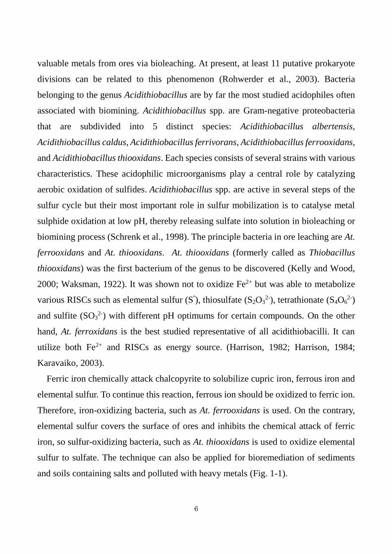
in previously released At. thiooxidans genomes. A result of sequence analysis of genes

46
around doxD2 is shown in Table 3-3. Corresponding gene products of At. thiooxidans
strain A01 are also shown in Table 3-3. doxD2 was found in a gene cluster containing
a hypothetical proteins and transposases (Fig. 3-3). Although the doxD1 gene
clustering with tth gene was found in genomes of all At. thiooxidans strains (Fig. 3-2),
the same gene arrangement as the cluster containing doxD2 of strain SH was not found
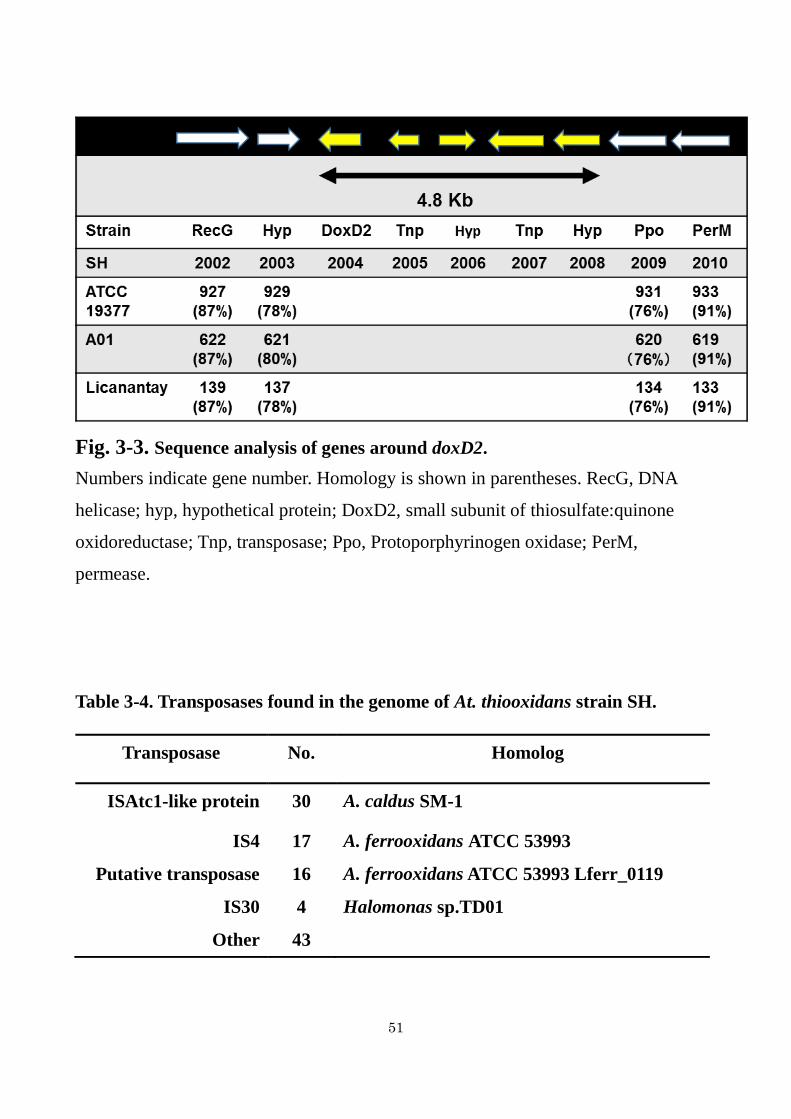
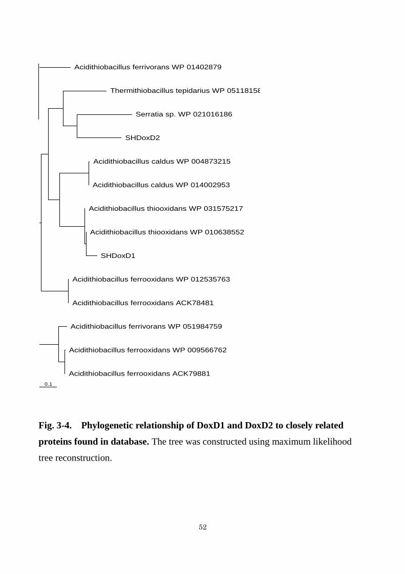
in genomes of other strains (Fig. 3-3). As shown in Fig. 3-4, although DoxD1 showed
relatively high amino acid sequence similarity (83%) to WP_010638552 (annotated as
DoxD and clustered with TTH) from At. thiooxidans ATCC 19377, homologous
proteins to DoxD2 were not found in other At. thiooxidans strains. A protein with the
highest homology (44%) to DoxD2 was found in Serratia sp. ATCC 39006 (Fig. 3-4).
The function is unknown.
The comparison of genome sequence of At. thiooxidans Licanantay with those of
strains ATCC 19377 and A01 revealed that there was a large core of genes shared by
all the species strains, while additional elements that can be associated with adaptation
to its environment have been found in Licanantay genome. Several of these genes were
located in unique genomic regions (Travisany et al., 2014). The analyses of genome
sequence from At. thiooxidans Licanantay have also revealed that most of these
genomic regions included several features that were characteristic of potential genomic
islands containing genetic mobility genes, such as integrase, transposase, phage-related
gene, and genes related to conjugation systems. Particularly, 69 non-shared unique
putative transposases have been found in the genome from Licanantay (Travisany et
al., 2014). The analysis of genome sequence form strain SH also revealed that about
110 transposase genes which showed relatively high sequence similarity to At. caldus
or At. ferrooxidans existed in the genome as shown in Table 3-4. Two transposase genes
existed in the gene cluster containing doxD2 gene and the cluster was absent in genome
sequences from other At. thiooxidans strains. These results strongly suggested that
strain SH acquired the doxD2 gene through a horizontal gene transfer for the adaptation
to marine acidophilic environments.

47
Table 3-2. Genes found in the draft genome of At. thiooxidans SH and predicted
to be involved in the sulfur oxidation
Abbreviation Enzyme name No. of
gene Position Reaction
SOR Sulfur dioxygenase 1 Periplasm S SO3
2−
SQR Sulfide quinone reductase 2 Inner
Membrane H
2S S
0
TQO Thiosulfate:quinone
oxidoreductase (DoxD) 2
Inner
Membrane S
2O
3
2− S
4O
6
2−
TTH Tetrathionate hydrolase 1 Outer
membrane S
4O
6
2− S
2O
3
2− + SO
4
2− + S
0
SOX Sulfur oxidition complex 2 Periplasm S2O
3
2− SO
4
2− + S
0
HDR Heterodisulfide reductase 1 Cytoplasm RSSH RSH + SO3
2−
SOR Sulfur oxygenase
reductase 1 S
0 H
2S + SO
3
2− + S
2O
3
2−
TST Thiosulfate
sulfurtransferase 1 Cytoplasm S
2O
3
2− SO
3
2− + S
0
PPR Phosphoadenosine
phosphosulfate reductase 1 Cytoplasm SO
3
2− PAPS
COX Cytochrome c oxidase 2 Inner
membrane
Cyt c (red) + 2H++ ½ O
2
Cyt c (ox) + H2O
CYD Ubiquinol oxidase 4 Inner
membrane QH2 + 2H
++ ½ O
2 Q + H2O

48
Fig. 3-2. Comparison of sox, tth, sqr, and hdr gene clusters between At.
thiooxidans SH and other three At. thiooxidans strains containing identified or
putative genes. sox, sulfur oxidation complex; tr, transcriptional regulator; orf, open
reading frame; tth, tetrathionate hydrolase; doxD, small subunit of thiosulfate:quinone
oxidoreductase; hpn, hypothetical protein; sqr, sulfide:quinone reductase; scp,
segregation and condensation protein; rho, rhodanese; hdr, heterodisulfide reductase.
The homologous proteins are indicated by the same color. The direction of
transcription is represented by the arrows.

49
Table 3-3. Sequence analysis of genes around doxD2

50
Table 3-3. Sequence analysis of genes around doxD2 (continue)
51
Fig. 3-3. Sequence analysis of genes around doxD2.
Numbers indicate gene number. Homology is shown in parentheses. RecG, DNA
helicase; hyp, hypothetical protein; DoxD2, small subunit of thiosulfate:quinone
oxidoreductase; Tnp, transposase; Ppo, Protoporphyrinogen oxidase; PerM,
permease.
Table 3-4. Transposases found in the genome of At. thiooxidans strain SH.
Transposase No. Homolog
ISAtc1-like protein 30 A. caldus SM-1
IS4 17 A. ferrooxidans ATCC 53993
Putative transposase 16 A. ferrooxidans ATCC 53993 Lferr_0119
IS30 4 Halomonas sp.TD01
Other 43
52
Fig. 3-4. Phylogenetic relationship of DoxD1 and DoxD2 to closely related
proteins found in database. The tree was constructed using maximum likelihood
tree reconstruction.
0.1
Acidithiobacillus ferrivorans WP 01402879
Thermithiobacillus tepidarius WP 051181587
Serratia sp. WP 021016186
SHDoxD2
Acidithiobacillus caldus WP 004873215
Acidithiobacillus caldus WP 014002953
Acidithiobacillus thiooxidans WP 031575217
Acidithiobacillus thiooxidans WP 010638552
SHDoxD1
Acidithiobacillus ferrooxidans WP 012535763
Acidithiobacillus ferrooxidans ACK78481
Acidithiobacillus ferrivorans WP 051984759
Acidithiobacillus ferrooxidans WP 009566762
Acidithiobacillus ferrooxidans ACK79881

53
3.3.3. Construction of sulfur oxidation model in At. thiooxidans strain
SH
Sulfur oxidation model of At. thiooxidans has already been constructed based on
previous studies from other sulfur-oxidizing bacteria and on sequence analyses (Valdes
et al., 2011; Yin et al., 2014). In the model, the first and critical step of sulfur oxidation
could be an opening of the S8 ring by the thiol groups of cysteine residues, resulting in
the formation of thiol-bound sulfane sulfur atoms (R-S-SnH). Subsequently, the R-S-
SnH is proposed to be transported into the periplasm and then oxidized by sulfur
dioxygenase (SDO) to produce sulfite, although sdo gene has not yet been determined
(Bobadilla et al., 2013). As sulfite thus produced spontaneously reacts with sulfur to
generate thiosulfate, thiosulfate has been detected as the main product of SDO reaction
(Rohwerder and Sand, 2003). Thiosulfate thus generated could be metabolized by
thisufate:quinone oxidoreductase (TQO), sulfur oxidizing protein (SOX), or thiosulfate
sulfurtransferase (TST). These enzymes catalyze the conversion of thiosulfate to
tetrathionate, to sulfate and elemental sulfur, or to sulfite and elemental sulfur,
respectively as shown in Table 3-2. As TST is a cytoplasmic enzyme, the periplasmic
thiosulfate can be metabolized to tetrathionate by TQO, which has been characterized
in Ac. ambivalens (Müller et al., 2004). As mentioned before, two doxD genes (doxD1
and doxD2) were present in strain SH. One of them is thought to be a candidate for
TQO. Alternatively, thiosulfate is oxidized to sulfate by sulfur oxidizing (Sox) system.
Two incomplete Sox gene clusters, in which Sox CD was absent, were found in the SH
genome. Sox system is located in the periplasm and comprised of SoxXA (both c type
cytochromes), SoxYZ (covalently sulfur-binding protein and sulfur compound
chelating protein, respectively), SoxB (dimanganese-containing protein considered to
act as the sulfate thiol esterase) (Ghosh and Dam, 2009). As strain SH does not have a
complete Sox system, thiosulfate would not be oxidized to sulfate. Tetrathionate
produced by TQO reaction or used as a substrate can be hydrolyzed by tetrathionate
hydrolase (TTH) to produce thiosulfate, elemental sulfur, and sulfate. TTH was studied

54
previously in At. thiooxidans (Tano et al; 1996)) and the gene has been identified in At.
ferrooxidans for the first time (Kanao et al; 2007).
Other RISCs oxidation enzymes were identified in Acidithiobacillus sp. including:
rhodanese (RHD) or thiosulfate sulfurtransferase (TST) and heterodisulfide reductase
(HDR). The TST widely exists in the cytoplasm of both prokaryotes and eukaryotes. It
cleaves the sulfur-sulfur bond of thiosulfate to yield sulfur and sulfite, and then the
former is transferred to a thiophilic acceptor such as cyanide and thiol compounds
(Gardener and Rawlings, 2000). The cytoplasmic HDR complex (HdrABC) has been
reported to catalyze the reversible reaction of the disulfide bond X-SS-X reduction
(Quantrini et al., 2009). Sulfide generated during sulfur oxidation is proposed to be
metabolized by sulfide:quinone reductase (SQR). An enzyme catalyzing the reaction
in the acidophilic sulfur oxidizing bacterium has been purified for the first time from
At. ferrooxidans (Wakai et al., 2007).
Based on the documented models in other At. thiooxidans strains (Valdes et al., 2011,
Yin et al., 2014) and genome sequence analysis of strain SH in this study, a hypothetical
model was developed for sulfur oxidation in At. thiooxidans SH (Fig. 3-5). In the model,
thiosulfate could be metabolized by TQO (DoxD1 or DoxD2) or an incomplete SOX
system. TSD purified in this study used a quinone as an electron acceptor and did not
contain a heme in the protein, suggesting that TSD encoded in doxD1 or doXD2 gene
was used for thiosulfate metabolism in strain SH.

55
Fig. 3-5. The sulfur oxidation model in At. thiooxidans strain SH based on
bioinformatics analysis of draft genome sequence. Abbreviations: SDO, sulfur
dioxygenase; SQR, sulfide quinone reductase; TQO, thiosulfate:quinone
oxidoreductase; TTH, tetrathionate hydrolase; Sox, sulfur oxidizing protein; HDR,
heterodisulfide reductase; SOR, sulfur oxygenase reductase; TST, thiosulfate
sulfurtransferase; bo3-cyt-ox, bo3-type cytochrome c oxidase; bd-ubi-ox, bd-type
ubiquinol oxidase. The sulfur metabolism in At. thiooxidans SH contains various sulfur
oxidation systems and the electron transfer pathways in different cellular compartments.
Elemental sulfur (S8) is activated and transported into the periplasm as thiol-bound
sulfane sulfur atoms (R-S-SnH). In the periplasm, R-S-SnH is oxidized by SDO to
produce sulfite. Thiosulfate generates spontaneously from sulfur and sulfite is
catalyzed by Sox complex or TQO. Tetrathionate is metabolized by TTH to produce
thiosulfate, sulfur, and sulfate. Sulfide is catalyzed by SQR. Two types of terminal
oxidases are also involved in sulfur compounds oxidation as shown. In the cytoplasm,
the particular enzymes perform the catalytic reaction by a sequence of steps that
eventually produce sulfate.

56
3.3.4. Determination of a gene encoding TSD in At. thiooxidans strain
SH
As described in chapter 2, the determination of a gene encoding TSD from At.
thiooxidans strain SH was tried analyzing sequences of peptide fragments produced
from in-gel trypsin digestion of the TSD protein. However, no gene with high
homology to the TSD was found in genome sequences of At. thiooxidans strains, ATCC
19377, A01, and Licanantay. The results suggested that TSD in At. thiooxidans strain
SH would be encoded in a unique gene. Thus, the analysis was carried out using
genome sequence of strain SH. The analyses resulted in the detection of a gene
encoding TSD in strain SH genome. As shown in Fig. 3-6, all 10 amino acid sequences
of peptides generated by TSD digestion with trypsin were found in an amino acid
sequence encoded in/by gene THioSH_02008 in contig2 of strain SH genome. The
gene encoded 444 amino acids with the signal peptide of 29 amino acids (Fig. 3-6),
indicating the translocation to periplasm. As TSD was purified from the solubilized
membrane fraction, TSD protein translocated to periplasm associates with membrane.
The molecular mass without the signal peptide was calculated to be 45,971 Da. It was
similar to that estimated by SDS-PAGE. A heme-binding motif (CxxCH) was not found
in the amino acid sequence. NEM showed no effect on the enzyme activity as described
in chapter 2, suggesting the absence of cysteine residues in the catalytic site. No
cysteine residue was found in the amino acid sequence. From the sulfur oxidation
pathway in Fig. 3-5, thiosulfate was suggested to be metabolized by DoxD1 or DoxD2.
However, TSD was encoded in different genes from doxD1 and doxD2. Interestingly,
the gene (739) was located in the genomic region containing doxD2 (Fig. 3-3). This
genomic region was specific for strain SH and was suggested to be acquired through a
horizontal gene transfer.
When a Blast search was performed using DDBJ database, some homologous
proteins to TSD were found in sulfur-oxidizing bacteria, such as At. thiooxidans, At.
caldus, At. ferroxidans, and At. ferrivorans, though the homologies were relatively low.

57
Surprisingly, a protein with the highest homology was not found in At. thiooxidans
strains. It was found in At. caldus (68%) as shown in Fig. 3-7. The protein
(WP_004873267) in At. caldus was annotated to be a phosphate porin, outer membrane
protein acting as a pore through which molecules can diffuse. Porin is usually consisted
of 3 subunits (Nikaido, 2003). TSD had an apparent molecular mass of 43 kDa and
consisted of homodimer having the apparent molecular mass of 99 kDa, indicating that
TSD was structurally different from typical porins. Thus, although TSD has an amino
acid sequence similarity to porins, I think that TSD has a different function from porin.
A Blast search using the deduced amino acid sequence of TSD from strain SH
revealed that some DoxD-like proteins also showed significant similarity to TSD.
DoxD is a small subunit of thiosulfate:quinone oxidoreductase (TQO), as described
previously. Among them, DoxD-like protein (ACK_78737) in At. ferrooxidans showed
47% sequence similarity to TSD. The protein is encoded in a gene clustered with tth
gene (tetrathionate hydrolase gene) in At. ferrooxidans. As TSD could use a ubiquinone
as the electron acceptor, the amino acid sequence was compared with Ac. ambivalens
TQO. As shown in Fig. 3-8, although some conserved amino acids were found, the
homology was relatively low (15%). Therefore, I could not determine TSD as a novel
TQO on the basis of the amino acid sequence.
Three types of tetrathionate-forming thiosulfate dehydrogenases have been
reported on the molecular genetic level; TQO from Ac. ambivarens (Müllar et al.,
2004), TsdA from Al. vinosum (Hensen et al., 2006), and TSD from At. ferrooxidans
(Kikumoto et al., 2013). The phylogenetic relationship of TSD from strain SH to
those enzymes was also shown in Fig. 3-7. TSD from strain SH did not show a close
relationship to other tetrathionate-forming thiosulfate dehydrogenase.

58
Fig. 3-6. Mascot search of peptide sequences determined by LC/MS of peptide
fragments produced by in-gel digestion of TSD with trypsin.
Amino acid sequences of peptide fragments determined by LC/MS were indicated by
red letters. A signal peptide was indicated by blue letters.
1 MIQKKPLLLSFFLSASLGAGTLFTQPAEAANWLAVQMVSPPKAPLFTVSG
51 FIEPTIWAQNGTVASAVGETPHINLIAPGFSQSTTAGIMRARIMFRGNLN
101 HHISYFFGGEFGNNGFTHIRKGYQPGLIDGHVTFSYIPGARVEVGLIRTP
151 GALGAIEGVFAYNYVLSPTMRLQLLNQHMIQGNSNYIIRPNAKHSYLVPG
201 AANLGTSGYRNPGIMVGDWFRSGHWETSYYAMLGMYGTVAAGNQSGAPME
251 AVRLQEAYVLSGKGPFRSDIQGGIWYQHARPRLLGHGYDMNRYGVDASYS
301 QGYMHPWGRMLRFSYVHGSGWIFAPAPFNETPAIAKKAPLYNSQIYPGIA
351 NKAWGYMVEGGLFFTKNIEMTLRYDLYNRLPNDPAQNRIFKDFAVGLQYH
401 FTPKTKIMAGYYFRTLDVPHPNDVSSSVASSVDNLFAMQAIISF 444

59
Fig. 3-7. Phylogenetic relationship of TSD from At. thiooxidans SH to related
proteins in database. The tree was constructed using maximum likelihood tree
reconstruction. Acidianus (Ac) ambivalens TQO was used as an outgroup.

60
Fig. 3-8. Alignment of the At. thiooxidans TSD and the combined Ac. ambivalens
TQO (DoxD and DoxA).

61
3.4. SUMMARY
Bioinformatics analysis of the genome sequence of At. thiooxidans strain SH
provides a valuable platform for gene discovery and functional prediction that is much
important given the difficulties in performing standard genetic research in this
microorganism. Based on our analysis and available documented data, a hypothetical
model for sulfur oxidation and electron transportation is proposed. The elemental sulfur
(S8) is activated and transported into the periplasm as thiol-bound sulfane sulfur atoms
(R-S-SnH). And then, the R-S-SnH is further oxidized in the periplasm where SDO,
TTH, and Sox system perform their functions. The cytoplasmic membrane involving
SQR and TQO is the third region with electrons transferring. Thiosulfate
dehydrogenase (TSD) was suggested to be encoded in doxD1 or doxD2, because it
encodes a protein with sequence similarity to TQO. However, TSD from strain SH was
encoded in a novel gene. The gene was located near genetic motility genes (e.g.
transposases) in genomic region of 4.8 kbp absent in other At. thiooxidans strains,
suggesting that strain SH acquired the gene by horizontal gene transfer. Thus, this study
provides novel insights and more instructive guides into thiosulfate metabolism in
sulfur-oxidizing microorganisms. However, many fundamental questions remain
unanswered. For example, some genes suggested to be involved in the sulfur oxidation
in At. thiooxidans, such as sor, doxD, and sox genes, need to be further verified via
biochemical experiments. Researches of TSD on the molecular genetic level also
remained as one of the important research subjects.

62
CHAPTER 4
CONCLUSION
Biomining also known as bioleaching is used to recover metals from low-grade of
sulfide ores. Acidophilic iron- and sulfur-oxidizing bacteria are used in the process.
The technique can also be applied for bioremediation of sediments and soils polluted
with heavy metals. Because ocean deposits are also rich in pyrite, chalcopyrite, and
marcasite, marine microorganisms show advantage in the bioleaching of marine sulfide
minerals. An acidophilic sulfur-oxidizing bacterium ––At. thiooxidans strain SH–– was
isolated from Seto Inland Sea, Japan, to develop a bioleaching process for NaCl-
containing sulfide minerals. The bacterium grows chemoautotrophically by deriving
energy from the oxidation of elemental sulfur and reduced inorganic sulfur compounds
(RISCs), such as thiosulfate and tetrathionate. Thiosulfate-metabolizing activity is
most important for bioleaching, as the sulfur moiety of sulfide minerals is thought to
be metabolized via thiosulfate as an intermediate. Thiosulfate has been suggested to be
metabolized by thiosulfate dehydrogenase, TSD. The oxidation pathway for elemental
sulfur and reduced inorganic sulfur compounds (RISCs) in strain SH has been
previously proposed. However, little information on the biochemical and genetic
properties of enzymes involved in elemental sulfur and RISCs is now available.
In chapter 2, to get a better understanding of sulfur metabolism in strain SH, the
purification of TSD from strain SH was carried out. TSD activity measured using
ferricyanide as the electron acceptor was detected in the cell-free extract of strain SH
cells grown on thiosulfate. The enzyme was localized in the membrane fraction and
solubilized by n-dodecyl--D-maltopyranoside. A three-step procedure resulted in
approximately 71-fold purification of the enzyme. An SDS-PAGE analysis of the final
preparation revealed a major protein with an apparent molecular mass of 44 kDa. The
maximum enzyme activity (45 U·mg-1) was observed at pH 4.0, 40C, and 200 mM

63
NaCl. Ubiquinone could be used as an electron acceptor, while horse heart cytochrome
c could not be used. The activity was strongly inhibited with 2-n-heptyl-4-hydroxy-
quinolone N-oxide and sulfite. To my knowledge, this is the first report of TSD whose
activity is stimulated by NaCl.
To identify the gene encoding TSD, the determination of the N-terminal amino acid
sequence was carried out. However the N-terminus was blocked and the sequence
could not be determined. Therefore, the analysis of peptide fragments produced by in-
gel digestion with trypsin has been done using HPLC-chip/QTOF and Mascot Server
search. Unfortunately, we could not found any homologous proteins with a high score
and coverage in the database.
In chapter 3, an analysis of a draft genome sequence of strain SH was carried out to
determine the gene encoding TSD. The draft genome of At. thiooxidans SH contained
a total of 2.91 Mbp distributed in 73 contigs with a G+C content of 54.3%. The
annotation results revealed one 5S-16S-23S operon, 45 tRNAs, and 3,034 CDSs. Draft
genome sequences of three At. thiooxidans strains, A01, ATCC19377 and Licanantay,
have been already released. Taking advantage of the availability of the other three At.
thiooxidans draft genome sequences, the genomic comparison analysis of strain SH
was carried out. Sulfur oxidizing complex (sox), tetrathionate hydrolase (tet), sulfide
quinone reductase (sqr), and heterodisuifide reductase (hdr), found in strain SH were
also found in other genomes with the similar gene arrangement. Two sqr genes, 2 sox
gene clusters, 2 cytochrome c oxidase (cox) gene clusters, and 4 bd-type ubiquinol
oxidase (cyd) gene clusters were found in SH genome. Interes
Based on the documented models in other Acidithiobacillus species, such as A.
ferrooxidans, At. caldus and At. thiooxidans, a hypothetical model for sulfur oxidation
pathway in strain SH was developed from the genome sequence analysis. In this model,
thiosulfate spontaneously generated from sulfite and elemental sulfur in periplasm is
metabolized by thiosufate:quinone oxidoreductase (TQO) or incomplete SOX system
(without SoxCD). TQO catalyzes the conversion of two molecules of thiosulfate to

64
tetrathionate by using a quinone as the electron acceptor. SOX catalyzes the conversion
of thiosulfate to sulfate and elemental sulfur. Thiosulfate in cytoplasm may be
metabolized to sulfite and elemental sulfur by thiosulfate sulfurtransferase. At.
thiooxidans SH draft genome sequence provides new insights into the genomic
diversity of members of the genus Acidithiobacillus and reveals a closer functional
relatedness of At. thiooxidans to sulfur-oxidizing-bacterium, Acidithiobacillus caldus
than to iron-oxidizing bacteria, such as Acidithiobacillus ferrooxidans and
Acidithiobacillus ferrivorans. It also provides new opportunities for experimental
research and contributes to a better understanding of the ecophysiology of extreme
acidic environments.
Using sequence data obtained by HPLC-chip/QTOF analysis of peptide fragments
produced in-gel trypsin digestion of TSD, a Mascot Server search was carried out to
determine a gene encoding TSD in strain SH genome. The protein was encoded in gene
THioSH_02008 in contig2 of strain SH genome. In the sulfur oxidation pathway
proposed on the basis of genome analyses of strain SH and other At. thiooxidans strains,
thiosulfate is suggested to be oxidized by TQO or SOX system. However, a gene
encoding TSD was different from the tqo or the sox gene in strain SH genome. A Blast
search revealed some homologous proteins (porins) from sulfur-oxidizing bacteria,
such as At. caldus, At. ferroxidans and At. ferrivorans, but the homologies were
relatively low. TSD found in strain SH also showed a low homology to the TQO from
Ac. ambivalens. Surprisingly, any homologous proteins with TSD from strain SH were
not found in other At. thiooxidans strains. To clarify the reason why genes homologous
to tsd of strain SH was not found in genomes of other At. thiooxidans strains, the gene
cluster containing tsd of strain SH was compared with those of other three At.
thioxidans strains. The tsd gene cluster of strain SH involved genes encoding one
hypothetical protein, two transposes and DoxD. DoxD is a homolog of the subunit of
the putative thiosulfate:quinone oxidoreductase (TQO). This gene cluster was not
found in the corresponding region of the other At. thiooxidans genomes.

65
Sequence analysis also showed that strain SH contained about 110 transposase
genes which showed relatively high sequence similarity to At. caldus or At.
ferrooxidans. tsd gene newly determined in strain SH was found in a genomic island
with 4.8 kb containing transposases showing the similarity to IS4 or Lferr from At.
ferrooxidans, suggesting that strain SH acquired this gene through an horizontal gene
transfer. Thus, I concluded that this is one of the reasons why homologous proteins
were not found in other three At. thiooxidans strains than strain SH.
In this research, TSD was purified for the first time from a marine sulfur-oxidizing
bacterium. Comparison with thiosulfate metabolizing enzymes from the other
microorganisms showed that TSD from strain SH was structurally different from the
thiosulfate-oxidizing enzymes reported so far. TSD from strain SH was able to use
quinone (Q2) as the electron acceptor, suggesting that the TSD had TQO activity. The
TSD from strain SH showed a low homology to the TQO from Ac. ambivalens, which
is the only TQO characterized on the molecular genetic level. Although further detailed
investigations are necessary to clarify the properties of TSD from strain SH, the TSD
from strain SH may be a novel thiosulfate:quinone oxidoreductase.

66
REFERENCES
Baker BJ, Banfield JF. (2003) Minireview. Microbial communities in acid mine
drainage. FEMS Microbiology Ecology. 44: 139-152.
Bobadilla FRA, Cortés MP, Padilla L, Maturana D, Budinich M, Maass A, Parada P.
(2013) Stoichiometric modeling of oxidation of reduced inorganic sulfur compounds
(RISCs) in Acidithiobacillus thiooxidans. Biotechnol Bioeng. 110:2242-2251.
Brierley CL. (2008) How will biomining be applied in future. T Nonferr Metal Soc.
18: 1302-1310.
Denkmann K, Grein F, Zigann R, Siemen A, Bergmann J, van Helmont S, Nicolai A,
Pereira IAC, Dahl C. (2012) Thiosulfate dehydrogenase: a widespread unusual
acidophilic c-type cytochrome. Environ Microbiol. 14:2673-88.
Durand P, Reysenbach AL, Prieur D, Pace N. (1993) Isolation and characterization of
Thiobacillus hydrothermalis sp. nov., a mesophilic obligately chemolithotrophic
bacterium isolated from a deep-sea hydrothermal vent in Fiji Basin. Arch Microbiol.
159: 39-44.
Gardner MN, Rawlings DE. (2000) Production of rhodanese by bacteria present in bio-
oxidation plants used to recover gold from arsenopyrite concentrates. J Appl Microbiol.
89:185-190.
Ghosh W, Dam B. (2009) Biochemistry and molecular biology of lithotrophic sulfur
oxidation by taxonomically and ecologically diverse bacteria and archaea. FEMS
Microbiol Rev. 33:999-1043.

67
González C, Yanquepe M, Cardenas JP, Valdes J, Quatrini R, Holmes DS, Dopson M.
(2014) Genetic variability of psychrotolerant Acidithiobacillus ferrivorans revealed by
(meta)genomic analysis. Res Microbiol. 165:726-734.
Harrison AP. (1982) Genomic and physiological diversity amongst strains of
Thiobacillus ferrooxidans, and genomic comparison with Thiobacillus thiooxidans.
Arch Microbiol. 131: 68-76.
Harrison AP. (1984) The acidophilic thiobacilli and other acidophilic bacteria that share
their habitat. Ann Rev Microbiol. 38: 265-292.
Hensen D, Sperling D, Trüper HG, Brune DC, Dahl C. (2006) Thiosulphate oxidation
in the phototrophic sulphur bacterium Allochromatium vinosum. Mol Microbiol.
62:794-810.
Hober H, Stetter KO. (1989) Thiobacillus prosperus sp. nov., represents a new group
of halotolerant metal-mobilizing bacteria isolated from a marine geothermal field. Arch
Microbiol. 151: 479-485.
Jannasch HW, Wirsen CO, Nelson DC, Robertson LA. (1985) Thiomicrospira
crunogena sp. nov., a colorless sulfuroxidizing bacterium from a deep-sea
hydrothermal vent. Int J Syst Bacteriol. 35: 422-424.
Kamimura K, Kunomura K, Wakai S, Murakami K. Sugio T. (2001) Some properties
of a novel obligately autotrophic iron-oxidizing bacterium isolated from seawater.
Hydrometallurgy. 59(2-3):373-381.
Kamimura K, Higashino E, Moriya S, Sugio T. (2003) Marine acidophilic sulfur-
oxidizing bacterium requiring salts for the oxidation of reduced inorganic sulfur

68
compounds. Extremophiles. 7:95-99.
Kamimura K, Higashino E, Kanao T, Sugio T. (2005) Effects of inhibitors and NaCl
on the oxidation of reduced inorganic sulfur compounds by a marine acidophilic,
sulfur-oxidizing bacterium, Acidithiobacillus thiooxidans strain SH. Extremophiles.
9:45-51.
Kanao T, Kamimura K, Sugio T. (2007) Identification of a gene encoding a
tetrathionate hydrolase in Acidithiobacillus ferrooxidans. J. Biotechnol. 132:16-22.
Karavaiko GI, Turova TP, Kodratéva TF, Lysenko AM , Kolganova TV, Ageeva
SN. (2003) Phylogenetic heterogeneity of the species Acidithiobacillus
ferrooxidans.Int J Syst Evol Microbiol. 53: 113-119
Kelly DP and Harrison AH. (1989) Genus Thiobacillus. In Bergey's Manual of
Systematic Bacteriology, 1st edn. 3:1842-1858. Edited by J. T. Staley, M. P. Bryant,
N. Pfennig &J. G. Holt. Baltimore: Williams & Wilkins.
Kelly DP and Wood AP. (2000) Reclassification of some species of Thiobacillus to the
newly designated genera Acidithiobacillus gen. nov.,Halothiobacillus gen. nov. and
Thermithiobacillus gen. nov. Int. J. Syst. Evol. Microbiol. 50: 511-516.
Kikumoto M, Nogami S, Kanao T, Takada J, Kamimura K. (2013) Tetrathionate-
forming thiosulfate dehydrogenase from the acidophilic, chemolithoautotrophic
bacterium Acidithiobacillus ferrooxidans. Appl Environ Microbiol. 79:113-20.

69
Kuenen JG, Boonstra J, Schröder HGJ, Veldkamp H. (1977) Competition for inorganic
substrates among chemoorganotrophic and chemolithotrophic bacteria. Microb Ecol.
3: 119-130.
Masau RJY, Oh JK, Suzuki I. (2001) Mechanism of oxidation of inorganic sulfur
compounds by thiosulfate-grown Thiobacillus thiooxidans. Can J Microbiol. 47:348-
358.
Marmur, J. (1961) A procedure for the isolation of deoxyribonucleic acid from
microorganisms. J Mol Biol. 3:208-218.
Meulenberg R, Pronk, JT, Hazeu W, van Duken JP, Frank J, Bos p, Kuenen JG. (1993)
Purification and partial characterization of thiosulphate dehydrogenase from
Thiobacillus acidophilus. J Gen Microbiol. 139: 2033-2039.
Miura Y and Kawaoi A. (2000) Determination of thiosulfate, thiocyanate and
polythionates in a mixture by ion-pair chromatography with ultraviolet absorbance
detection. J Chromato A. 884:81-87.
Müller FH, Bandeiras TM, Urich T, Teixeira M, Gomes CM, Kletzin A. (2004)
Coupling of the pathway of sulphur oxidation to dioxygen reduction: characterization
of a novel membrane-bound thiosulphate:quinone oxidoreductase. Mol Microbiol.
53:1147-1160.
Nakamura K, Yoshikawa H, Okubo S, Kurosawa H, Amano Y. (1995) Purification and
properties of membrane-bound sulfite dehydrogenase from Thiobacillus thiooxidans
JCM7814. Biosci Biotechnol Biochem. 59:11-15.

70
Nakamura K, Nakamura M, Yoshikawa H, Amano Y. (2001) Purification and
properties of thiosulfate dehydrogenase from Acidithiobacillus thiooxidans JCM7814.
Biosci Biotechnol Biochem. 65:102108.
Nikaido H. (2003) Molecular basis of bacterial outer membrane permeability revisited.
Microbiol. Mol Biol Rev. 67;593-656.
Nuñez PA, Romero H, Farber MD, Rocha EP. (2013) Natural selection for operons
depends on genome size. Genome Biol. Evol. 5:2242-2254.
Pan African resources PLC. 2011. Pan African Annual Report.
Quatrini R, Appia-Ayme C, Denis Y, Jedlicki E, Holmes DS, Bonnefoy V. (2009)
Extending the models for iron and sulfur oxidation in the extreme acidophile
Acidithiobacillus ferrooxidans. BMC Genomics. doi: 10.1186/1471-2164-10-394.
Rawlings DE. (1997) Biomining: Theory, Microbes and Industrial Processes. Berlin:
Springer-Verlag.
Rohwerder T, Gehrke T, Kinzler K, Sand W. (2003) Bioleaching review part A:
Progress in bioleaching: fundamentals and mechanisms of bacterial metal sulfide
oxidation. Appl Microbiol Biotech. 63: 239-248.
Rohwerder T, Sand W. (2003) The sulfane sulfur of persulfides is the actual substrates
of the sulfur-oxidizing enzymes from Acidithiobacillus and Acidiphilium spp.
Microbiology. 149:1699-1710.
Rossi G. (1990) Biohydrometallurgy. Hamburg: McGraw-Hill, ISBN 3-89028-781-6.

71
Ruby EG, Wirsen CO , Jannasch HW. (1981) Chemolithoautotrophic sulfur-oxidizing
bacteria from the Galapagos Rift hydrothermal vents. Appl Environ Microbiol. 42:
317-342.
Ruby EG and Jannasch HW. (1982) Physiological characteristics of Thiomicrospira sp.
strain L-12 isolated from deep-sea hydrothermal vents. J Bacteriol. 149: 161-165.
Sand W, Gerke T, Hallmann R, Schippers A. (1995) Sulfur chemistry, biofilm, and the
(in)direct attack mechanism - a critical evaluation of bacterial leaching. Appl Microbiol
Biotechnol. 43: 961-966.
Schippers A, Jozsa PG, Sand W. (1996) Sulfur chemistry in bacterial leaching of pyrite.
Appl Environ Microbiol. 62: 3424-3431.
Schippers A, Sand W. (1999) Bacterial leaching of metal sulfides proceeds by two
indirect mechanisms via thiosulfate or via polysulfides and sulfur. Appl Environ
Microbiol. 65: 319-321.
Schippers A, Hedrich S, Vasters J, Drobe M, Sand W, Willscher S. (2014) Biomining:
metal recovery from ores with microorganisms. Adv Biochem Eng Biotechnol. 141:1-
47.
Sievert SM, Heidorn T, Kuever J. (2000) Halothiobacillus kellyi sp. nov., a
mesophilic,obligately chemolithoautotrophic, sulfuroxidizing bacterium isolated from
a shallowwater hydrothermal vent in the Aegean Sea, and emended description of the
genus Halothiobacillus.International J Syst Evol Microbiol. 50: 1229–1237.

72
Singer PC and Stumm W. (1970) Acidic mine drainage - the rate limiting step. Science.
167: 1121-1123.
Suzuki I. (1999) Oxidation of inorganic sulfur compounds: chemical and enzymatic
reactions. Can J Microbiol. 45:97-105.
Tano T, Takesue H, Sugio T, Imai K. (1982) B-type cytochrome, an electron carrier in
the sulfite-oxidation system of Thiobacillus thiooxidans. J Ferment Technol. 160:181-
187.
Tano T, Kitaguchi H, Harada K, Nagasawa T, Sugio T. (1996) Purification and some
properties of tetrathionate decomposing enzyme from Thiobacillus thiooxidans. Biosci
Biotechnol Biochem. 60:224-227.
Travisany D, Cortés MP, Latorre M, Di Genova A, Budinich M, Bobadilla-Fazzini RA,
Parada P, González M, Maass A. (2014) A new genome of Acidithiobacillus
thiooxidans provides insights into adaptation to a bioleaching environment. Res
Microbiol. 165:743-752.
Valdes J, Ossandon F, Quatrini R, Dopson M, Holmes DS. (2011) Draft genome
sequence of the extremely acidophilic biomining bacterium Acidithiobacillus
thiooxidans ATCC 19377 provides insights into the evolution of the Acidithiobacillus
genus. J Bacteriol. 193:7003-7004.
Visser JM, de Jong GAH, Robertson LA, Kuenen JG. (1997) Purification and
characterization of a periplasmic thiosulfate dehydrogenase from the obligately
autotrophic Thiobacillus sp. W5. Arch Microbiol. 166:372-378.

73
Wakai S, Tsujita M, Kikumoto M, Manchur MA, Kanao T, Kamimura K. (2007)
Purification and characterization of sulfide:quinone oxidoreductase from an
acidophilic iron-oxidizing bacterium, Acidithiobacillus ferrooxidans. Biosci
Biotechnol Biochem. 71:2735-2742.
Waksman SA, Joffe JS. (1922 ) Microorganisms concerned in the oxidation of sulfur
in the soil.II. Thiobacillus thiooxidans,a new sulfur-oxidizing organism isolated from
the soil. J Bacteriol. 7:239-256.
Wood AP and Kelly DP. (1991) Isolation and characterization of Thiobacillus
halophilus sp. nov., a sulphur-oxidising autotrophic eubacterium from a Western
Australian hypersaline lake. Arch Microbiol. 156: 277-280.
Yin H, Zhang X, Li X, He Z, Liang Y, Guo X, Hu Q, Xiao Y, Cong J, Ma L, Niu J,
Liu X. (2014) Whole-genome sequencing reveals novel insights into sulfur oxidation
in the extremophile Acidithiobacillus thiooxidans. MBC Microbiol. 14:179-193.
74
ACKNOWLEDGEMENTS
The study presented in this thesis paper has been carried out under the supervision
of Professor Kazuo KAMIMURA, at the Laboratory of “ Microbial Function” under
the Graduate School of Environmental and Life Science, Okayama University, Japan,
since October, 2012 to September, 2015.
First of all, I would like to express my sincerest gratitude and gratefulness to my
PhD supervisor Professor Kazuo KAMIMURA, for his constant guidance and
immense support, valuable suggestions, criticism and motivation throughout my PhD
course. His guidance and patience helped me in all the time of research and writing of
this thesis. Without his kind assistance and support, it would not be possible to conduct
this research. I am deeply grateful to him.
I would like to express my humble thanks and sincere gratitude to my Co-Supervisor
Dr. Tadayoshi Kanao, Associate Professor, Okayama University, for his valuable
suggestions, continual encouragement and insightful comments that benefitted me to
widen my research from various perspectives.

75
I would like to express my sincere honor to Kenji INAGAKI, Professor, Okayama
University, my Co-Supervisor for his fruitful guidelines and instructions throughout
the period of my PhD study.
I take this opportunity to acknowledge Mr. Nogami, Ms. Sadakuni, Mr. Akita, Mr.
Hamasaki, Ms. Takada, Ms. Yoshino and all the members, present and past of this
laboratory for their kind support, encouragement and friendly manner.
I would like thank Okayama University for providing me the financial support by
JASSO-Scholarship during my study.
I wish to thank Dr. Tsugumi Shiokawa (Advanced Science Research Center,
Okayama University) for their assistance in the analyses of the N-terminal amino acid
sequence and peptide fragments produced by in-gel trypsin digestion of the 44-kDa
protein.
I am grateful to all my family members for their unceasing encouragement, support
and attention.

76
Finally, I would like to express my deepest appreciation and heartfelt thanks to my
adorable little daughter and husband for their enormous support, sacrifice, patience and
heartwarming encouragement.
2015.9.11
SULTANA SHARMIN
Laboratory of Microbial Function
Department of Biofunctional Chemistry
Graduate School of Environmental and Life Science
Okayama University, Japan.