severe myocarditis: a 2012 update… alain combes service de réanimation ican, institute of...
TRANSCRIPT

Severe Myocarditis:A 2012 update…
Alain CombesService de RéanimationiCAN, Institute of Cardiometabolism and NutritionHôpital Pitié-Salpêtrière, AP-HP, ParisUniversité Pierre et Marie Curie, Paris 6www.reamedpitie.com

Definition - Etiologies
«Myocarditis» is defined as inflammation of the heart muscle
Histology: cellular infiltrate and myocyte necrosis Etiologies:
Infectious diseases• Viruses: Coxsackie, Adenovirus, HIV,
Parvovirus B19, HHV6, (H1N1)• Bacteria• Parasites (Toxoplasma, Chagas)• Fungi
Hypersensitivity (Drugs) Autoimmune and systemic diseases
• Lupus, Wegener, Eosinophilic, Sarcoidosis, Giant cell Myocardial toxins (Cocaine, chemotherapy) Peripartum
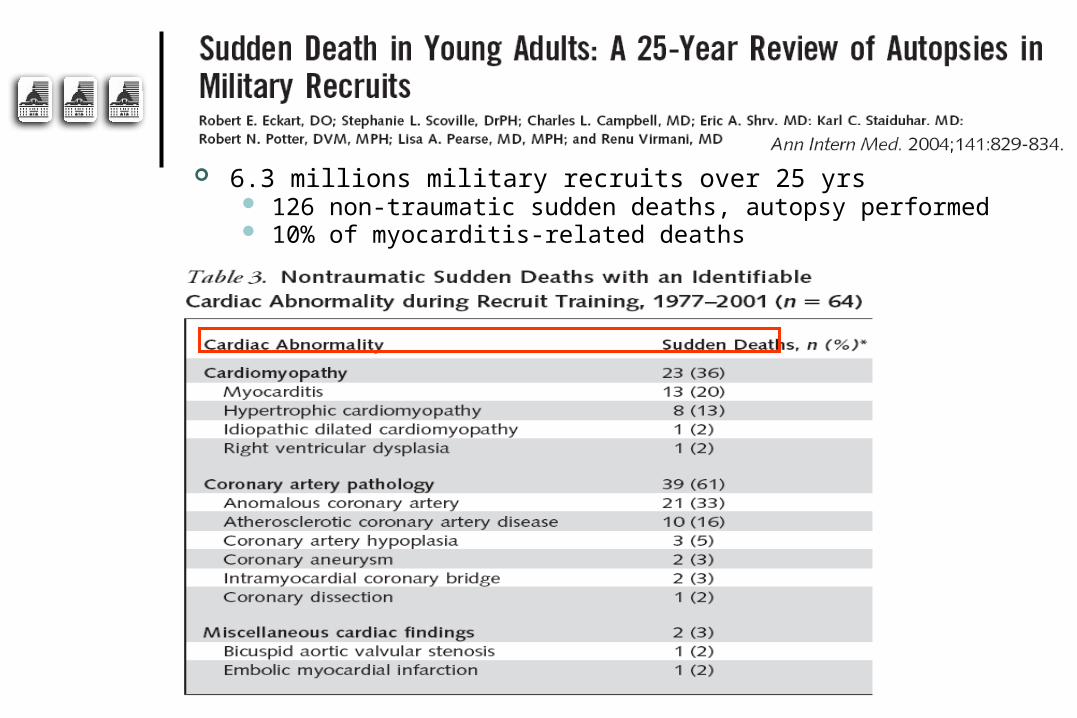
6.3 millions military recruits over 25 yrs 126 non-traumatic sudden deaths, autopsy performed 10% of myocarditis-related deaths

Clinical manifestations
From asymptomatic EKG abnormalities to overt cardiac failure…
Clinical features: Preceding viral illness, flu-like syndrome Fever Chest pain, mimicking acute coronary syndromes Tachycardia Arrhythmia Sudden death:
• 10% of cases (Eckart, AnnIntMed, O4) Clinical signs of heart failure
• Minimal, slow evolution • Fulminant, leading to refractory cardiogenic shock
in a few days

EKG
EKG findings Sinus tachycardia Diffuse ST-T wave abnormalities Prolonged QT interval Bundle branch block (LBBB++) Myocardial infarction pattern Complete heart block Supraventricular tachyarrhythmias Ventricular tachyarrhythmias
May be normal…

EKG mimicking AMI

EKG: LBBB

Laboratory Findings

Biology: Troponin (Smith, Circ, 97)

Many patients with acute/fulminant myocarditis will undergo coronary artery angiography…

Other laboratory findings
Non specific tests Leucocytosis/leucopenia, Eosinophilia+++ Mononucleosic syndrome Sedimentation rate, CRP, PCT…
Specialized tests Virological diagnosis
• Serology (limited value)• Cultures: throat and stools• PCR (blood, CSF, tissues)
Inflammation:• Antinuclear Ab, ANCA, Angiotensin Conversion Enzyme
Research tests Autoantibodies (mitochondria, myosin, b-receptor) Immunohistochemical myocardial studies (research)

Doppler Echocardiography

Fulminant myocarditisMarkedly decreased LV EFNear normal LV dimensionIncreased septal thickness
Acute myocarditisMarkedly decreased LV EFDilated LVNormal septal thickness

Doppler Echocardiography
Other findingsRegional wall motion abnormalities Diastolic dysfunctionChange in echocardiographic image
texture:• Increased brightness• Heterogeneity
ThrombiPericardial effusion

Cardiac MRI…
The new diagnostic gold standard?

Cardiac MRI
Combination of T1-weighted and T2-weighted Gadolinium contrast-enhanced MRI +++
Visualize localization, activity and extent of inflammation One or several foci in the myocardium Foci most frequently located in lateral free wall Frequent subepicardial lesions Can guide myocardial biopsies+++


Still indication for myocardial biopsies?

Histology: Dallas Criteria
3 histological grades (Aretz, Hum Pathol, 87)
Active Myocarditis : • Cellular infiltrate +, myocyte necrosis +
Borderline Myocarditis : • Cellular infiltrate +, myocyte necrosis -
Negative Biopsy : • Cellular infiltrate -, myocyte necrosis -
Distribution and diffusion of the cellular infiltrate Focal, confluent or diffuse Mild, moderate, severe
However, low to moderate sensitivity/specificity

Histology: Dallas Criteria
Borderline Active


Grade 1, Level B:
.

Prognosis

PrognosisMcCarthy, NEJM, 2000
Survie sans transplantation
P=0.05
(15 patients)
(132 patients)


2003 - 2009 41 patients refractory cardiogenic
shock due to fulminant myocarditisMean age 38±12 years66%, women
Mechanical assistanceThoratec BiVAD (n=6) or ECMO (n=35)


Long term survival: 68%4 (10%) patients had heart transplantation
Independent predictors of ICU death determined at admission:SAPS II >56 (OR, 10.23) and troponin Ic >12 microg/L (OR, 7.49)

Complete follow-up for 26 survivors Median follow-up was 525 [92–2400] days Mean LVEF was 57±9%
≥60% for 12 non-transplant and all 4 transplanted 40–60% for 10 nontransplanted survivors
21 patients had percutaneous femoral ECMO 10 still complained of paresthesia/peripheral
neurological defect 2 had persistent leg ischemia requiring surgical repair
for 1 and amputation for the other

0
10
20
30
40
50
60
70
80
90
100
PhysicalFunctioning
Role-physical
BodilyPain
General Health
Vitality Social Functioning
Role-Emotional
Mental Health
* * * *

Relatives of dying ICU patients
ICU patients
Myocarditis
Type I I diabetics
10
20
30
40
50
60
HAD-A HAD-D
70
Sco
re ≥
8,
(%)


Treatment

Treatment
Supportive care always indicated Bed rest Diuretics, vasodilators ACE inhibitors, angiotensin-receptor blockers Aldosterone antagonists -b blockers, (with caution in the acute phase) Vasopressors/inotropic agents in case of shock
Mechanical assistance +++ May be urgently needed if fulminant form or rapid
deterioration of hemodynamic status Patients should rapidly be transferred to experienced
centers Bridge to recovery:
• ECMO+++, First line assistance (Heart transplantation)

ECMO vs. BiVAD for fulminant myocarditis?

BIVAD ECMO
n=5 n=6
Age 32 ±2 40 ±4
LVEF (%) 33 ±8 18 ±4
Creat(micromoL/L) 183 ±65 127 ±27
T Bili (micromoL/L) 14 ±3 37 ±11
Apache II: 11 ±9 24 ±18
Difference of effectivness in Fulminant Myocarditis?
P = ns

BIVAD ECMO
Output (L/min) 5,4 ±0,7 4,8 ±0,4
Duration (days) 21 ±5 13 ±4
Death 20% (4/5) 16,6% (5/6)
PRBC: 22 ±5 7 ±4 (p=0,03)
RESULTS

Creatinine mol/dlT bilirubin mol/dl


Long term survival: 68%, 4 (10%) patients had heart transplantationIndependent predictors of ICU death determined at admission:
SAPS II >56 (OR, 10.23) and troponin Ic >12 g/L (OR, 7.49)

Specific/Novel treatments
Immunosuppression First line therapy if
• Giant cell• Systemic autoimmune diseases
Corticosteroids Cyclosporine, Tacrolimus Azathioprine
Immunomodulation/Stimulation IV Immune globulins Interferon
Antiviral agents, vaccination

Myocarditis Treatment Trial 111 randomized patients, LVEF<45% Histologically proven myocarditis Immunosuppression protocol Placebo vs prednisone + Cyclosporine or
azathioprine
Mason, NEJM, 1995

IV immune globulinMcNamara, Circ, 2001
62 patients with DCM, randomized, LVEF<40%
Placebo vs IVIg P = NS

Tailored immune-modulating strategiesLiu, Circ, 2001
Phase I Phase II Phase III

Immunomodulation vs Immunosuppression
Interféron b chez les patients avec persistance virale entero-adénovirus Kuhl, Circ, 2003 22 patients, Dysfonction VG, génome viral + Interféron: Clearance virus, amélioration FE
Immunosuppression:prednisone+imurel Frustaci, Circ, 03
• 41 patients, myocardite active, Dysfonction VG• 21 répondeurs, 20 non rep• Répondeurs: AutoAC +, génome viral- (sauf Hep C)
Wojnicz, Circ 01• 84 patients CMD, HLA surexprimé sur myocytes• Traitement de 3 mois• Amélioration dans groupe traité: 71% vs 31%


Conclusion
Myocarditis is a rare and severe condition Especially the fulminant form
Diagnosis based on clinical features, EKG, Echo, Troponin, MRI Myocarditis can mimic acute coronary syndromes
Mechanical circulatory assistance may be urgently needed if rapid hemodynamic deterioration
Immunosuppression during the acute phase Giant cell Systemic autoimmune diseases
Significant progresses in the understanding of the pathophysiology of the disease in recent years Help design tailored immune-modulating strategies

La Pitié: Louis XIV, 1656…To 2012…
La Chapelle Institut de Cardiologie