specialissuepaper 1397 swallowablemedicaldevicesfordiagnosis andsurgery:thestateoftheart · ·...
TRANSCRIPT
SPECIAL ISSUE PAPER 1397
Swallowable medical devices for diagnosisand surgery: the state of the artJ L Toennies1, G Tortora2, M Simi2, P Valdastri2, and R J Webster III1∗1Department of Mechanical Engineering, Vanderbilt University, Nashville, Tennessee, USA2CRIM Lab, Scuola Superiore Sant’Anna, Pisa, Italy
The manuscript was received on 2 August 2009 and was accepted after revision for publication on 9 December 2009.
DOI: 10.1243/09544062JMES1879
Abstract: The first wireless camera pills created a revolutionary new perspective for engineersand physicians, demonstrating for the first time the feasibility of achieving medical objectivesdeep within the human body from a swallowable, wireless platform. The approximately 10 yearssince the first camera pill has been a period of great innovation in swallowable medical devices.Many modules and integrated systems have been devised to enable and enhance the diagnosticand even robotic capabilities of capsules working within the gastrointestinal (GI) tract. Thisarticle begins by reviewing the motivation and challenges of creating devices to work in thenarrow, winding, and often inhospitable GI environment. Then the basic modules of modernswallowable wireless capsular devices are described, and the state of the art in each is discussed.This article is concluded with a perspective on the future potential of swallowable medical devicesto enable advanced diagnostics beyond the capability of human visual perception, and even todirectly deliver surgical tools and therapy non-invasively to interventional sites deep within theGI tract.
Keywords: swallowable devices, capsules, gastrointestinal tract, wireless capsule endoscopy,robotic capsules, minimally invasive surgery, endoluminal devices, surgical endoscopy
1 INTRODUCTION
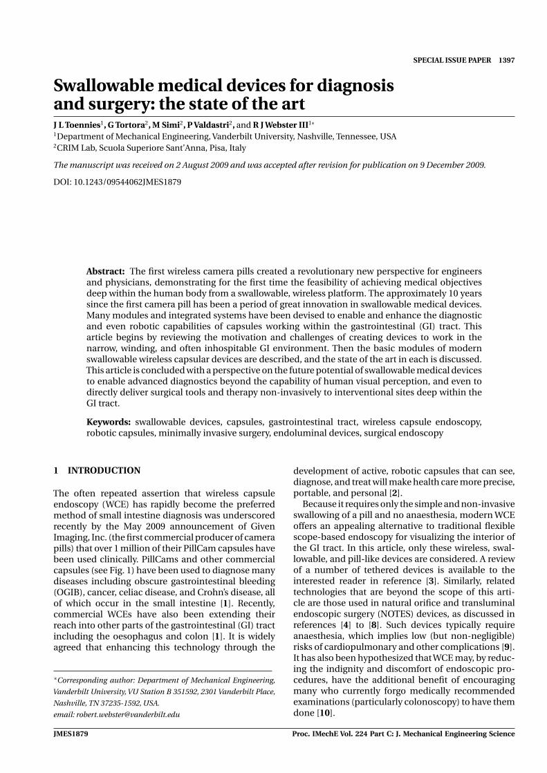
The often repeated assertion that wireless capsuleendoscopy (WCE) has rapidly become the preferredmethod of small intestine diagnosis was underscoredrecently by the May 2009 announcement of GivenImaging, Inc. (the first commercial producer of camerapills) that over 1 million of their PillCam capsules havebeen used clinically. PillCams and other commercialcapsules (see Fig. 1) have been used to diagnose manydiseases including obscure gastrointestinal bleeding(OGIB), cancer, celiac disease, and Crohn’s disease, allof which occur in the small intestine [1]. Recently,commercial WCEs have also been extending theirreach into other parts of the gastrointestinal (GI) tractincluding the oesophagus and colon [1]. It is widelyagreed that enhancing this technology through the
∗Corresponding author: Department of Mechanical Engineering,
Vanderbilt University, VU Station B 351592, 2301 Vanderbilt Place,
Nashville, TN 37235-1592, USA.
email: [email protected]
development of active, robotic capsules that can see,diagnose, and treat will make health care more precise,portable, and personal [2].
Because it requires only the simple and non-invasiveswallowing of a pill and no anaesthesia, modern WCEoffers an appealing alternative to traditional flexiblescope-based endoscopy for visualizing the interior ofthe GI tract. In this article, only these wireless, swal-lowable, and pill-like devices are considered. A reviewof a number of tethered devices is available to theinterested reader in reference [3]. Similarly, relatedtechnologies that are beyond the scope of this arti-cle are those used in natural orifice and transluminalendoscopic surgery (NOTES) devices, as discussed inreferences [4] to [8]. Such devices typically requireanaesthesia, which implies low (but non-negligible)risks of cardiopulmonary and other complications [9].It has also been hypothesized that WCE may, by reduc-ing the indignity and discomfort of endoscopic pro-cedures, have the additional benefit of encouragingmany who currently forgo medically recommendedexaminations (particularly colonoscopy) to have themdone [10].
JMES1879 Proc. IMechE Vol. 224 Part C: J. Mechanical Engineering Science
1398 J L Toennies, G Tortora, M Simi, P Valdastri, and R J Webster III
Fig. 1 Current commercially available swallowable camera capsules include (a) PillCam SB2, (b)PillCam ESO, (c) PillCam Colon, all by Given Imaging, Inc., as well as (d) EndoCapsuleof Olympus, Inc., (e) OMOM of Jinshan Science and Technology Group, and (f) MiRoof Intromedic Co. These capsules carry cameras into the GI tract for visual inspection.While most of the capsules shown are designed for the small intestine, PillCam ESO is anoesophageal capsule and the PillCam Colon carries two onboard cameras for use in the largeintestine. All capsules are 11 mm in diameter × 26 mm in length except for OMOM (13 mm× 27.9 mm), MiRo (10.8 mm × 24 mm) [14], and PillCam Colon (11 mm × 31 mm) [15]
Robotic systems including sensors and actua-tors have shown great promise in extending theinterventional and diagnostic capabilities of WCEs,and the purpose of this article is to review the stateof the art in such systems. Because a number ofrecent and thorough reviews of commercial passive(imaging-only) camera pills are already available tothe interested reader (see references [1], [3], [11],and [12]), the present authors treat them only brieflyhere, providing a complementary review of recentrobotic advancements in swallowable sensors andactuators, and the associated electronics, telemetry,and power systems that enable them to work. A briefsurvey of some (but not all) of these topics has beengiven in reference [13], and in the current paper anexpanded review of all aspects of state-of-the-art WCEtechnology is provided.
The sections that follow first discuss the uniquerequirements of the various areas of the GI tract andbriefly treat commercial capsules. Then, a high-leveloverview of the modules that can be included in arobotic WCE is given, after which each module is dis-cussed in depth. The article concludes in section 10with a perspective on the role robotic technologies canbe expected to play in the future of WCE.
2 CAPSULE ENVIRONMENT AND MODULES
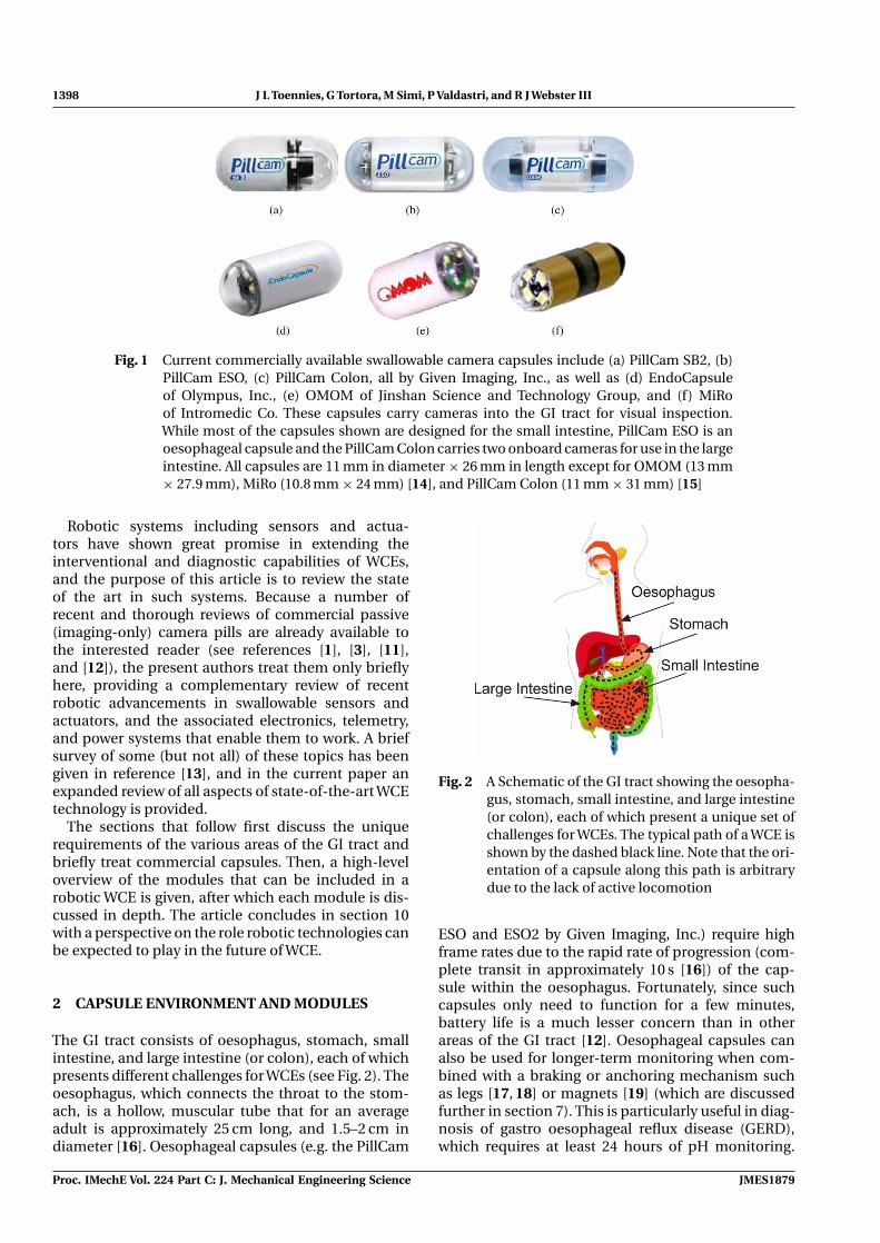
The GI tract consists of oesophagus, stomach, smallintestine, and large intestine (or colon), each of whichpresents different challenges for WCEs (see Fig. 2). Theoesophagus, which connects the throat to the stom-ach, is a hollow, muscular tube that for an averageadult is approximately 25 cm long, and 1.5–2 cm indiameter [16]. Oesophageal capsules (e.g. the PillCam
Fig. 2 A Schematic of the GI tract showing the oesopha-gus, stomach, small intestine, and large intestine(or colon), each of which present a unique set ofchallenges for WCEs. The typical path of a WCE isshown by the dashed black line. Note that the ori-entation of a capsule along this path is arbitrarydue to the lack of active locomotion
ESO and ESO2 by Given Imaging, Inc.) require highframe rates due to the rapid rate of progression (com-plete transit in approximately 10 s [16]) of the cap-sule within the oesophagus. Fortunately, since suchcapsules only need to function for a few minutes,battery life is a much lesser concern than in otherareas of the GI tract [12]. Oesophageal capsules canalso be used for longer-term monitoring when com-bined with a braking or anchoring mechanism suchas legs [17, 18] or magnets [19] (which are discussedfurther in section 7). This is particularly useful in diag-nosis of gastro oesophageal reflux disease (GERD),which requires at least 24 hours of pH monitoring.
Proc. IMechE Vol. 224 Part C: J. Mechanical Engineering Science JMES1879
Swallowable medical devices for diagnosis and surgery 1399
The Medtronic Bravo Capsule, which does not col-lect images, is designed for this purpose but requiresfixation to the oesophageal wall via an endoscopicprocedure [20].
After passing through the oesophagus, the capsulereaches the stomach, a large, typically deflated elas-tic sac that is lined with a thick mucous membraneand contains gastric juices. In its deflated state, thestomach contains many longitudinal folds, and atthe pylorus, the narrowest part of the stomach, it isabout 2 cm in diameter [16]. Owing to the challengeof maintaining traction on the stomach wall, few cap-sules have been purposely designed for this portion ofthe GI tract, although prototypes have been developedfor a propeller-driven ‘swimming’ capsule [21] (seesection 7) and a multi-module assemblable and recon-figurable robotic system consisting of several unitsintended to be independently swallowed [22].
A capsule exits the stomach into the small intestine,where WCEs have made their most significant clin-ical impact. The small intestine is an elastic lumen,3–4 cm in diameter, and is the longest portion of theGI tract (approximately 670–760 cm in length). Thistubular structure, which winds and curves many timesin the abdomen, has villi coating its internal surface.It normally contains semi-digested foods, pancreaticjuices, enzymes, and bile [16], although the food prod-ucts are typically removed via ingestible agents priorto capsule endoscopy. WCEs are pushed through thesmall intestine via peristalsis (the natural muscle activ-ity that normally moves decomposing food products).Peristalsis generally occurs over short segments andpropagates at a rate of 1–2 cm/min [16]. Commercialcapsules designed for the small intestine (see Fig. 1)include the PillCam SB and SB2 and Agile Patency Sys-tem (a self-disintegrating pill [12]) by Given Imaging,Inc., the EndoCapsule by Olympus, Inc., the OMOMcapsule by the Jinshan Science and Technology GroupCo., Ltd., and the MiRo capsule by Intromedic Co.The Agile capsule, an accessory to the PillCam, canbe used prior to the ingestion of the PillCam SB toverify adequate capsule passage through the GI tractin patients with known or suspected strictures. Thiscapsule assists in detecting the problem of capsuleretention, which occurs in 0.75 per cent of OGIBcases, but may occur at a rate of up to 6.7 per centin patients with Crohn’s disease [12]. While each ofthese commercial capsules is designed for use in thesmall intestine, they vary in the optical sensors (com-plementary metal oxide silicone (CMOS) or chargedcouple device (CCD)) used, their size, and their attain-able frame rates. See section 5 for further informationas well as reference [14] for an in-depth review ofcommercial camera capsule capabilities.
The last region of the GI tract encountered by a WCEis the large intestine or colon, which measures about150 cm in length and 6 cm in diameter [16, 23]. While itdoes not contain as many folds as the small intestine,
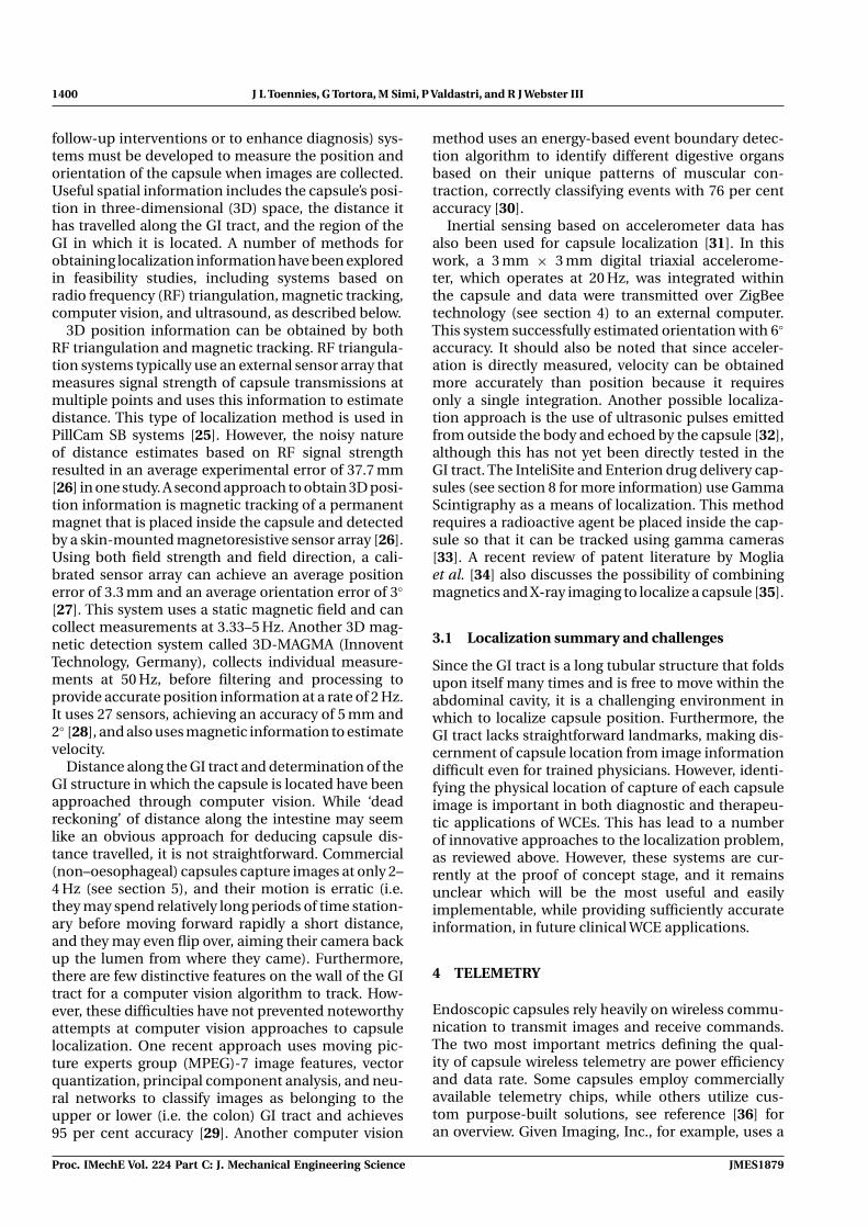
Fig. 3 Schematic diagram of possible modules in arobotic wireless capsule
the large intestine contains two flexures (corners) thata capsule must navigate around. The larger lumenmakes diagnosis difficult for capsules designed forthe small intestine because they tumble in the largerlumen and generally cannot view the entire internalsurface. A number of devices have been designed towork in the colon by entering through the rectum andtravelling against peristalsis (which is the approach ofstandard colonoscopy) as reviewed in reference [3].WCEs designed for the colon include PillCam Colonby Given Imaging, Inc. and the legged capsule ofValdastri et al. [10]. The time of passage of a capsulethrough the entire GI tract is generally about 8–10 h,with approximately 1 h spent in the stomach, 4 h inthe small intestine, and 5 h in the colon [24].
2.1 Robotic endoscopic capsule modules
There are a number of modules possible in a roboticWCE (see Fig. 3), although most capsules built todate include only a subset of these due to space con-straints.Whether it will ultimately be possible to createa single robotic device with all of these modules thatcan both diagnose and treat disease, or whether agroup of task-specific capsules will work coopera-tively (a more likely initial scenario), a large number ofrobotic modules will be required. The major modulesof a robotic capsule system are localization, telemetry,vision, non-visual sensors, locomotion, interventionalsystems, and power. Each of these modules individu-ally poses a variety of worthy engineering challenges,and integrating two or more of them is also challeng-ing. Subsequent sections of this article discuss thetechnical progress that has been made in each of thegeneral modules.
3 LOCALIZATION
To determine the location of lesions and otherstructures visualized in capsule images (for future
JMES1879 Proc. IMechE Vol. 224 Part C: J. Mechanical Engineering Science
1400 J L Toennies, G Tortora, M Simi, P Valdastri, and R J Webster III
follow-up interventions or to enhance diagnosis) sys-tems must be developed to measure the position andorientation of the capsule when images are collected.Useful spatial information includes the capsule’s posi-tion in three-dimensional (3D) space, the distance ithas travelled along the GI tract, and the region of theGI in which it is located. A number of methods forobtaining localization information have been exploredin feasibility studies, including systems based onradio frequency (RF) triangulation, magnetic tracking,computer vision, and ultrasound, as described below.
3D position information can be obtained by bothRF triangulation and magnetic tracking. RF triangula-tion systems typically use an external sensor array thatmeasures signal strength of capsule transmissions atmultiple points and uses this information to estimatedistance. This type of localization method is used inPillCam SB systems [25]. However, the noisy natureof distance estimates based on RF signal strengthresulted in an average experimental error of 37.7 mm[26] in one study. A second approach to obtain 3D posi-tion information is magnetic tracking of a permanentmagnet that is placed inside the capsule and detectedby a skin-mounted magnetoresistive sensor array [26].Using both field strength and field direction, a cali-brated sensor array can achieve an average positionerror of 3.3 mm and an average orientation error of 3◦
[27]. This system uses a static magnetic field and cancollect measurements at 3.33–5 Hz. Another 3D mag-netic detection system called 3D-MAGMA (InnoventTechnology, Germany), collects individual measure-ments at 50 Hz, before filtering and processing toprovide accurate position information at a rate of 2 Hz.It uses 27 sensors, achieving an accuracy of 5 mm and2◦ [28], and also uses magnetic information to estimatevelocity.
Distance along the GI tract and determination of theGI structure in which the capsule is located have beenapproached through computer vision. While ‘deadreckoning’ of distance along the intestine may seemlike an obvious approach for deducing capsule dis-tance travelled, it is not straightforward. Commercial(non–oesophageal) capsules capture images at only 2–4 Hz (see section 5), and their motion is erratic (i.e.they may spend relatively long periods of time station-ary before moving forward rapidly a short distance,and they may even flip over, aiming their camera backup the lumen from where they came). Furthermore,there are few distinctive features on the wall of the GItract for a computer vision algorithm to track. How-ever, these difficulties have not prevented noteworthyattempts at computer vision approaches to capsulelocalization. One recent approach uses moving pic-ture experts group (MPEG)-7 image features, vectorquantization, principal component analysis, and neu-ral networks to classify images as belonging to theupper or lower (i.e. the colon) GI tract and achieves95 per cent accuracy [29]. Another computer vision
method uses an energy-based event boundary detec-tion algorithm to identify different digestive organsbased on their unique patterns of muscular con-traction, correctly classifying events with 76 per centaccuracy [30].
Inertial sensing based on accelerometer data hasalso been used for capsule localization [31]. In thiswork, a 3 mm × 3 mm digital triaxial accelerome-ter, which operates at 20 Hz, was integrated withinthe capsule and data were transmitted over ZigBeetechnology (see section 4) to an external computer.This system successfully estimated orientation with 6◦
accuracy. It should also be noted that since acceler-ation is directly measured, velocity can be obtainedmore accurately than position because it requiresonly a single integration. Another possible localiza-tion approach is the use of ultrasonic pulses emittedfrom outside the body and echoed by the capsule [32],although this has not yet been directly tested in theGI tract. The InteliSite and Enterion drug delivery cap-sules (see section 8 for more information) use GammaScintigraphy as a means of localization. This methodrequires a radioactive agent be placed inside the cap-sule so that it can be tracked using gamma cameras[33]. A recent review of patent literature by Mogliaet al. [34] also discusses the possibility of combiningmagnetics and X-ray imaging to localize a capsule [35].
3.1 Localization summary and challenges
Since the GI tract is a long tubular structure that foldsupon itself many times and is free to move within theabdominal cavity, it is a challenging environment inwhich to localize capsule position. Furthermore, theGI tract lacks straightforward landmarks, making dis-cernment of capsule location from image informationdifficult even for trained physicians. However, identi-fying the physical location of capture of each capsuleimage is important in both diagnostic and therapeu-tic applications of WCEs. This has lead to a numberof innovative approaches to the localization problem,as reviewed above. However, these systems are cur-rently at the proof of concept stage, and it remainsunclear which will be the most useful and easilyimplementable, while providing sufficiently accurateinformation, in future clinical WCE applications.
4 TELEMETRY
Endoscopic capsules rely heavily on wireless commu-nication to transmit images and receive commands.The two most important metrics defining the qual-ity of capsule wireless telemetry are power efficiencyand data rate. Some capsules employ commerciallyavailable telemetry chips, while others utilize cus-tom purpose-built solutions, see reference [36] foran overview. Given Imaging, Inc., for example, uses a
Proc. IMechE Vol. 224 Part C: J. Mechanical Engineering Science JMES1879
Swallowable medical devices for diagnosis and surgery 1401
custom telemetry chip produced specifically for theirPillCams, made by Zarlink, Inc. The power consump-tion of this chip is 5.2 mW, and it has a data rate of2.7 Mbps with a carrier frequency of 403–434 MHz [36].This telemetry link is unidirectional and is used fortransmitting images out of the capsule only. Other cus-tom unidirectional modules for image transmissionalso exist. The system by Shen et al. [37] transmits on a416 MHz carrier frequency at slightly lower power lev-els (4 mW) and data rates (2 Mbps) than the Zarlinkchips described above. Thone et al. [36] developeda similar design that transmits at 2 Mbps at only2 mW of power consumption, using a 144 MHz carrierfrequency.
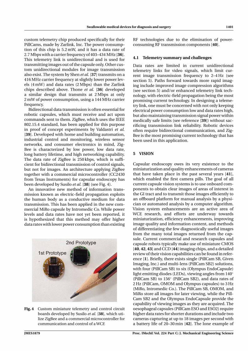
Bidirectional data transmission is often essential forrobotic capsules, which must receive and act uponcommands sent to them. ZigBee, which uses the IEEE802.15.4 standard, has been applied for this purposein proof of concept experiments by Valdastri et al.[39]. Developed with home and building automation,industrial control and monitoring, wireless sensornetworks, and consumer electronics in mind, Zig-Bee is characterized by low power, low data rate,long battery lifetime, and high networking capability.The data rate of ZigBee is 250 kbps, which is suffi-cient for bidirectional transmission of control signals,but not for images. An architecture applying ZigBeetogether with a commercial microcontroller (CC2430from Texas Instruments) for capsular endoscopy hasbeen developed by Susilo et al. [38] (see Fig. 4).
An innovative new method of information trans-mission known as electric-field propagation exploitsthe human body as a conductive medium for datatransmission. This has been applied in the new com-mercial MiRo capsule by Intromedic Co. While powerlevels and data rates have not yet been reported, itis hypothesized that this method may offer higherdata rates with lower power consumption than existing
Fig. 4 Custom miniature telemetry and control circuitboards developed by Susilo et al. [38], which uti-lize ZigBee and a commercial microcontroller forcommunication and control of a WCE
RF technologies due to the elimination of power-consuming RF transmission components [40].
4.1 Telemetry summary and challenges
Data rates are limited in current unidirectionaltelemetry links for video signals, which limit cur-rent image transmission frequency to 2–4 Hz (seesection 5). Paths forward towards more rapid imag-ing include improved image compression algorithms(see section 5) and/or enhanced telemetry link tech-nology, with electric-field propagation being the mostpromising current technology. In designing a teleme-try link, one must be concerned with not only keepingelectrical power consumption low and data rates high,but also maintaining transmission signal power withinmedically safe limits (see reference [39]) without sac-rificing transmission link reliability. Robotic devicesoften require bidirectional communication, and Zig-Bee is the most promising current technology that hasbeen used in this application.
5 VISION
Capsular endoscopy owes its very existence to theminiaturization and quality enhancements of camerasthat have taken place in the past several years [41],which enabled the first camera pills. The goal of allcurrent capsule vision systems is to use onboard com-ponents to obtain clear images of areas of interest inthe GI tract and to transmit those images efficiently toan offboard platform for manual analysis by a physi-cian or automated analysis by a computer algorithm.Vision system enhancements are an active area ofWCE research, and efforts are underway towardsminiaturization, efficiency enhancements, improvingimage quality and information content, and methodsof differentiating the few diagnostically useful imagesfrom the many total images returned from the cap-sule. Current commercial and research capsules andcapsule robots typically make use of miniature CMOS[40, 42, 43] and CCD [44] imaging chips, and a detailedreview of their vision capabilities can be found in refer-ence [1]. Briefly, there exists single (PillCam SB, GivenImaging, Inc.) and multi-lens (PillCam SB2) solutions,with four (PillCam SB) to six (Olympus EndoCapsule)light emitting diodes (LEDs), viewing angles from 140◦
(PillCam SB) to 156◦ (PillCam SB2), and data rates of2 Hz (PillCam, OMOM and Olympus capsules) to 3 Hz(MiRo, Intromedic Co.). The PillCam SB, OMOM, andMiRo store all images for later viewing, while the Pill-Cam SB2 and the Olympus EndoCapsule provide thecapability of viewing images as they are acquired. Theoesophageal capsules (PillCam ESO and ESO2) requirehigher data rates for shorter durations and include twocameras capturing at up to 18 images per second witha battery life of 20–30 min [42]. The lone example of
JMES1879 Proc. IMechE Vol. 224 Part C: J. Mechanical Engineering Science
1402 J L Toennies, G Tortora, M Simi, P Valdastri, and R J Webster III
a commercial capsule designed for the colon (PillCamColon) also has two cameras capturing images at 4 Hzeach. Because it can be switched off until the capsulereaches the colon, the device has a battery life quotedat 10 h [1], but its active period is shorter.
5.1 Image acquisition hardware
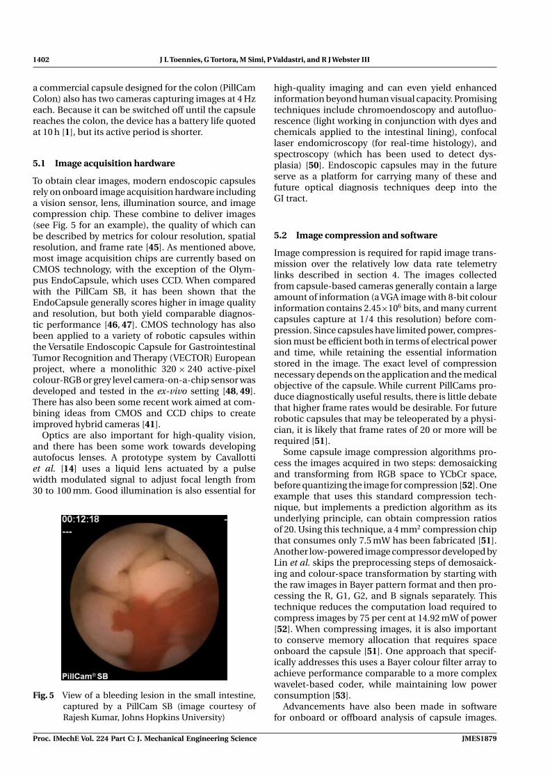
To obtain clear images, modern endoscopic capsulesrely on onboard image acquisition hardware includinga vision sensor, lens, illumination source, and imagecompression chip. These combine to deliver images(see Fig. 5 for an example), the quality of which canbe described by metrics for colour resolution, spatialresolution, and frame rate [45]. As mentioned above,most image acquisition chips are currently based onCMOS technology, with the exception of the Olym-pus EndoCapsule, which uses CCD. When comparedwith the PillCam SB, it has been shown that theEndoCapsule generally scores higher in image qualityand resolution, but both yield comparable diagnos-tic performance [46, 47]. CMOS technology has alsobeen applied to a variety of robotic capsules withinthe Versatile Endoscopic Capsule for GastrointestinalTumor Recognition and Therapy (VECTOR) Europeanproject, where a monolithic 320 × 240 active-pixelcolour-RGB or grey level camera-on-a-chip sensor wasdeveloped and tested in the ex-vivo setting [48, 49].There has also been some recent work aimed at com-bining ideas from CMOS and CCD chips to createimproved hybrid cameras [41].
Optics are also important for high-quality vision,and there has been some work towards developingautofocus lenses. A prototype system by Cavallottiet al. [14] uses a liquid lens actuated by a pulsewidth modulated signal to adjust focal length from30 to 100 mm. Good illumination is also essential for
Fig. 5 View of a bleeding lesion in the small intestine,captured by a PillCam SB (image courtesy ofRajesh Kumar, Johns Hopkins University)
high-quality imaging and can even yield enhancedinformation beyond human visual capacity. Promisingtechniques include chromoendoscopy and autofluo-rescence (light working in conjunction with dyes andchemicals applied to the intestinal lining), confocallaser endomicroscopy (for real-time histology), andspectroscopy (which has been used to detect dys-plasia) [50]. Endoscopic capsules may in the futureserve as a platform for carrying many of these andfuture optical diagnosis techniques deep into theGI tract.
5.2 Image compression and software
Image compression is required for rapid image trans-mission over the relatively low data rate telemetrylinks described in section 4. The images collectedfrom capsule-based cameras generally contain a largeamount of information (a VGA image with 8-bit colourinformation contains 2.45×106 bits, and many currentcapsules capture at 1/4 this resolution) before com-pression. Since capsules have limited power, compres-sion must be efficient both in terms of electrical powerand time, while retaining the essential informationstored in the image. The exact level of compressionnecessary depends on the application and the medicalobjective of the capsule. While current PillCams pro-duce diagnostically useful results, there is little debatethat higher frame rates would be desirable. For futurerobotic capsules that may be teleoperated by a physi-cian, it is likely that frame rates of 20 or more will berequired [51].
Some capsule image compression algorithms pro-cess the images acquired in two steps: demosaickingand transforming from RGB space to YCbCr space,before quantizing the image for compression [52]. Oneexample that uses this standard compression tech-nique, but implements a prediction algorithm as itsunderlying principle, can obtain compression ratiosof 20. Using this technique, a 4 mm2 compression chipthat consumes only 7.5 mW has been fabricated [51].Another low-powered image compressor developed byLin et al. skips the preprocessing steps of demosaick-ing and colour-space transformation by starting withthe raw images in Bayer pattern format and then pro-cessing the R, G1, G2, and B signals separately. Thistechnique reduces the computation load required tocompress images by 75 per cent at 14.92 mW of power[52]. When compressing images, it is also importantto conserve memory allocation that requires spaceonboard the capsule [51]. One approach that specif-ically addresses this uses a Bayer colour filter array toachieve performance comparable to a more complexwavelet-based coder, while maintaining low powerconsumption [53].
Advancements have also been made in softwarefor onboard or offboard analysis of capsule images.
Proc. IMechE Vol. 224 Part C: J. Mechanical Engineering Science JMES1879
Swallowable medical devices for diagnosis and surgery 1403
Onboard software can enable closed-loop control ofcamera orientation for improving image quality [54].It can also be used for in-situ capsule video analysisin order to detect and predict upcoming video imagesthat may be of concern to a clinician [55]. Externalpostprocessing software for capsule images is alsoextremely useful because a typical small intestine cap-sule will return approximately 50 000 total images froma single use [56], but only a very small fraction of theseimages will contain useful diagnostic information. Forthis reason, algorithms are under development to sortthese images and guide the physician to the mostimportant frames, with the future intent of aidingthe physician by automatically suggesting appropriatediagnoses for lesions identified [56].
5.3 Vision summary and challenges
The challenges in vision involve obtaining clear, sharpimages of diagnostically useful areas of the GI tract andcompressing them sufficiently (with minimal infor-mation loss) for fast transmission over the telemetrylink. While the current performance of capsule-basedcameras (typically 2–4 Hz at 1/4 VGA resolution) isgood enough for some diagnostic tasks and per-haps for some future automated robotic tasks that donot involve real-time human interaction, there is lit-tle debate that speed and resolution improvementswould be desirable. Such improvements will be nec-essary to enable some future teleoperated roboticcapsules envisioned by researchers, which will requireimaging at approximately 24 fps so that the imagesreturned have the appearance of video to the humanoperator. Unless there are dramatic breakthroughs intelemetry, advancements towards this goal are likely toinclude increasingly sophisticated image compressionalgorithms. It is also important to develop automaticimage analysis algorithms in both the onboard andoffboard contexts, to assist with real-time control andidentification of the few diagnostically useful imagesamong the thousands typically returned, respectively.
6 DIAGNOSTIC SYSTEMS BEYOND IMAGEACQUISITION
It is in principle possible to carry a wide rangeof biosensors onboard a capsule. Quantities thathave been measured to date by capsule-based sen-sors include pH, pressure, oxygenation, electricalconductivity, temperature, and blood detection. Anexample of a capsule with integrated pressure, tem-perature, and pH sensing capabilities is presented inreference [45].
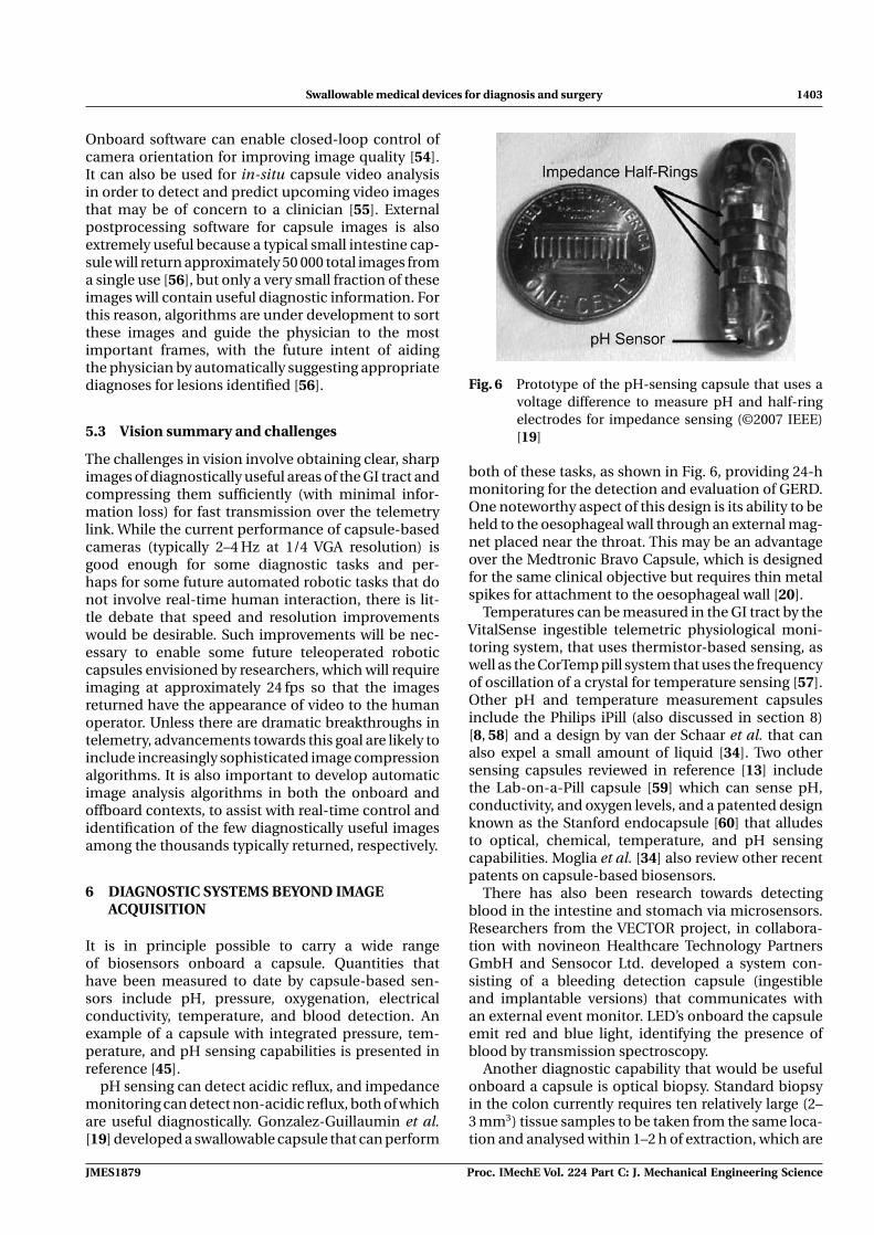
pH sensing can detect acidic reflux, and impedancemonitoring can detect non-acidic reflux, both of whichare useful diagnostically. Gonzalez-Guillaumin et al.[19] developed a swallowable capsule that can perform
Fig. 6 Prototype of the pH-sensing capsule that uses avoltage difference to measure pH and half-ringelectrodes for impedance sensing (©2007 IEEE)[19]
both of these tasks, as shown in Fig. 6, providing 24-hmonitoring for the detection and evaluation of GERD.One noteworthy aspect of this design is its ability to beheld to the oesophageal wall through an external mag-net placed near the throat. This may be an advantageover the Medtronic Bravo Capsule, which is designedfor the same clinical objective but requires thin metalspikes for attachment to the oesophageal wall [20].
Temperatures can be measured in the GI tract by theVitalSense ingestible telemetric physiological moni-toring system, that uses thermistor-based sensing, aswell as the CorTemp pill system that uses the frequencyof oscillation of a crystal for temperature sensing [57].Other pH and temperature measurement capsulesinclude the Philips iPill (also discussed in section 8)[8, 58] and a design by van der Schaar et al. that canalso expel a small amount of liquid [34]. Two othersensing capsules reviewed in reference [13] includethe Lab-on-a-Pill capsule [59] which can sense pH,conductivity, and oxygen levels, and a patented designknown as the Stanford endocapsule [60] that alludesto optical, chemical, temperature, and pH sensingcapabilities. Moglia et al. [34] also review other recentpatents on capsule-based biosensors.
There has also been research towards detectingblood in the intestine and stomach via microsensors.Researchers from the VECTOR project, in collabora-tion with novineon Healthcare Technology PartnersGmbH and Sensocor Ltd. developed a system con-sisting of a bleeding detection capsule (ingestibleand implantable versions) that communicates withan external event monitor. LED’s onboard the capsuleemit red and blue light, identifying the presence ofblood by transmission spectroscopy.
Another diagnostic capability that would be usefulonboard a capsule is optical biopsy. Standard biopsyin the colon currently requires ten relatively large (2–3 mm3) tissue samples to be taken from the same loca-tion and analysed within 1–2 h of extraction, which are
JMES1879 Proc. IMechE Vol. 224 Part C: J. Mechanical Engineering Science
1404 J L Toennies, G Tortora, M Simi, P Valdastri, and R J Webster III
challenging (and perhaps impossible) requirementsfor a mobile swallowable capsule. Optical biopsy pro-vides a potential alternative using the properties oflight for diagnosis. Promising imaging techniques foroptical biopsy include fluorescence endoscopy, opti-cal coherence tomography, confocal microendoscopy,light-scattering spectroscopy, raman spectroscopy,and molecular imaging, all of which are discussed indetail in reference [61].
6.1 Sensing summary and challenges
Significant challenges in integrating sensing onboardWCEs are in developing and miniaturizing the biosen-sors themselves. Additional challenges arise in pack-aging them onboard the capsule where they must beshielded from unwanted environmental contact andexposed when desired. As with all other capsule com-ponents, sensors must also be power efficient andable to be integrated with other modules. A numberof promising sensing methods have been developedincluding sensors for pressure, temperature, electri-cal conductivity, and pH, and optical biopsy tech-niques hold great promise for extending the diagnosticcapabilities of endoscopic capsules.
7 LOCOMOTION
Current commercial capsules move through the GItract by means of peristalsis alone. However, suchmovement is unpredictable and inconsistent, whichresults in incomplete evaluations approximately 20per cent of the time [62]. Performance is even worsein the large intestine, where the sensitivity of capsuleendoscopes for detecting colonic lesions is low com-pared with the use of conventional colonoscopy [15].Active locomotion has the potential to improve cap-sule imaging consistency and coverage, by enablingthe capsule to suspend itself in the middle of the lumenand move forward, backward, and to stop in place atareas of interest [10]. Two main approaches have beendeveloped for WCE locomotion: (a) internal and (b)external. Internal locomotion is defined as the useof micromechanisms integrated solely onboard thecapsule platform for locomotion. In contrast, exter-nal locomotion utilizes propulsive forces transmittedby an external system, typically a magnetic field.
7.1 Internal locomotion
Advantages of internal locomotion are the ability toobtain precise motion relative to the lumen due tolocal contact, and the fact that no external magneticfield generators are required. Another advantage ofsome mechanisms (such as legs) is the ability to simul-taneously distend tissue away from the camera lens
[10]. The major drawback is the need to supply rel-atively large actuators with a significant amount ofpower. A number of different internal locomotionstrategies have been developed over the past few years,as discussed below.
Vibratory actuation can aid the forward progressionof a capsule along the GI tract by reducing frictionwith its environment [63]. Here, the capsule containsa micromotor with an assymetric mass on the rotor tocreate vibrations around its central axis.The advantageof this method of locomotion is its mechanical sim-plicity. Challenges include lack of active orientationcontrol and difficulty obtaining good images duringvibration. Furthermore, the capsule cannot activelyhold a given position or reverse direction and travelback up the intestine.
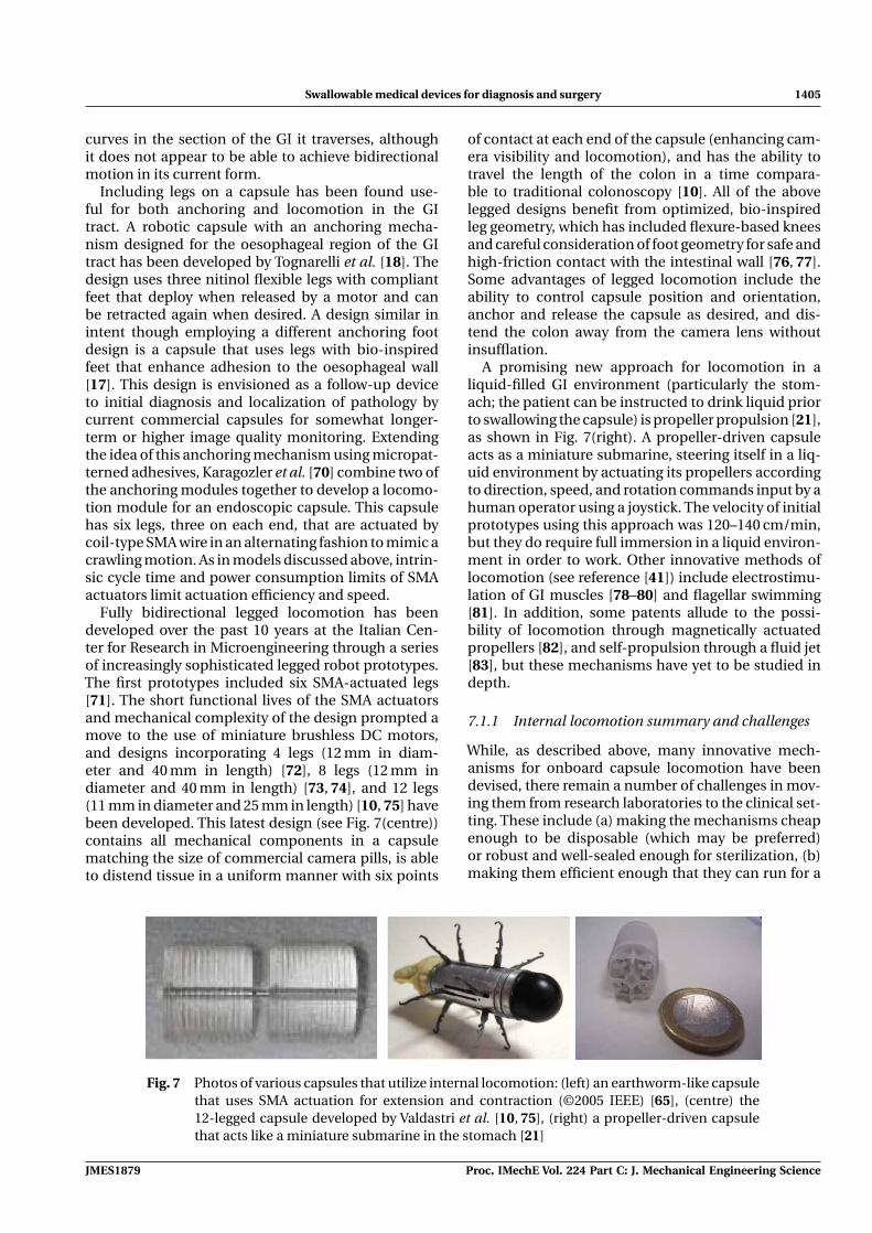
Another means of locomotion inspired by biologythat has been applied to WCEs is earthworm-likemotion. Kim and colleagues [64] have developedtwo prototypes, one 13 mm in diameter by 33 mmin length, and another 15 mm in diameter by 30 mmin length [65, 66], as shown in Fig. 7 (left). The firstdevice propels itself by cyclic compression/extensionof shape memory alloy (SMA) spring actuators, whilealternately anchoring its ends to a surface using direc-tional spines (micro needles) mounted at each end.The locomotion method of the second prototype isthe same as the first, but it consists of two bel-lows joined by a custom impact-based piezo actuator.Both designs were tested in ex-vivo porcine intestineand achieved velocities of 0.85 cm/min (one pointof contact) and 1.47 cm/min (three points of con-tact) for the first prototype, and 13.38 cm/min with an11 mm stroke for the second prototype. The first pro-totype moved slower than the second due to the timeconstants associated with heating and cooling SMAactuators. Directional spines provide the advantage ofa passive anchoring mechanism that does not requirepower or additional actuators, but likely at the expenseof precluding bidirectional motion.
Another bio-inspired solution is a cilia system forlocomotion [67]. The 15 mm diameter by 35 mm longcapsule contains six units (intended to mimic biologi-cal cells), with two appendages that extend from eachunit (mimicking cilia extensions). Each unit is drivenby two SMA actuators to enable bidirectional motionat a maximum velocity of 2.4 cm/min. However, thepower consumption of this design is currently high dueto numerous SMA actuators. In order to increase fric-tion with the intestinal wall, design optimization of theleg-like cilia extensions has also been performed [68].
It is also possible to produce locomotion in the GItract using a ‘paddling’ technique where leg-like finstravel the length of the capsule [69]. At the back of thecapsule, the fins retract before recycling to the front ofthe capsule for the next paddle stroke. An advantageof the paddling motion is the rapid velocity it enablesof up to 18–36 cm/min depending on the number of
Proc. IMechE Vol. 224 Part C: J. Mechanical Engineering Science JMES1879
Swallowable medical devices for diagnosis and surgery 1405
curves in the section of the GI it traverses, althoughit does not appear to be able to achieve bidirectionalmotion in its current form.
Including legs on a capsule has been found use-ful for both anchoring and locomotion in the GItract. A robotic capsule with an anchoring mecha-nism designed for the oesophageal region of the GItract has been developed by Tognarelli et al. [18]. Thedesign uses three nitinol flexible legs with compliantfeet that deploy when released by a motor and canbe retracted again when desired. A design similar inintent though employing a different anchoring footdesign is a capsule that uses legs with bio-inspiredfeet that enhance adhesion to the oesophageal wall[17]. This design is envisioned as a follow-up deviceto initial diagnosis and localization of pathology bycurrent commercial capsules for somewhat longer-term or higher image quality monitoring. Extendingthe idea of this anchoring mechanism using micropat-terned adhesives, Karagozler et al. [70] combine two ofthe anchoring modules together to develop a locomo-tion module for an endoscopic capsule. This capsulehas six legs, three on each end, that are actuated bycoil-type SMA wire in an alternating fashion to mimic acrawling motion. As in models discussed above, intrin-sic cycle time and power consumption limits of SMAactuators limit actuation efficiency and speed.
Fully bidirectional legged locomotion has beendeveloped over the past 10 years at the Italian Cen-ter for Research in Microengineering through a seriesof increasingly sophisticated legged robot prototypes.The first prototypes included six SMA-actuated legs[71]. The short functional lives of the SMA actuatorsand mechanical complexity of the design prompted amove to the use of miniature brushless DC motors,and designs incorporating 4 legs (12 mm in diam-eter and 40 mm in length) [72], 8 legs (12 mm indiameter and 40 mm in length) [73, 74], and 12 legs(11 mm in diameter and 25 mm in length) [10, 75] havebeen developed. This latest design (see Fig. 7(centre))contains all mechanical components in a capsulematching the size of commercial camera pills, is ableto distend tissue in a uniform manner with six points
of contact at each end of the capsule (enhancing cam-era visibility and locomotion), and has the ability totravel the length of the colon in a time compara-ble to traditional colonoscopy [10]. All of the abovelegged designs benefit from optimized, bio-inspiredleg geometry, which has included flexure-based kneesand careful consideration of foot geometry for safe andhigh-friction contact with the intestinal wall [76, 77].Some advantages of legged locomotion include theability to control capsule position and orientation,anchor and release the capsule as desired, and dis-tend the colon away from the camera lens withoutinsufflation.
A promising new approach for locomotion in aliquid-filled GI environment (particularly the stom-ach; the patient can be instructed to drink liquid priorto swallowing the capsule) is propeller propulsion [21],as shown in Fig. 7(right). A propeller-driven capsuleacts as a miniature submarine, steering itself in a liq-uid environment by actuating its propellers accordingto direction, speed, and rotation commands input by ahuman operator using a joystick. The velocity of initialprototypes using this approach was 120–140 cm/min,but they do require full immersion in a liquid environ-ment in order to work. Other innovative methods oflocomotion (see reference [41]) include electrostimu-lation of GI muscles [78–80] and flagellar swimming[81]. In addition, some patents allude to the possi-bility of locomotion through magnetically actuatedpropellers [82], and self-propulsion through a fluid jet[83], but these mechanisms have yet to be studied indepth.
7.1.1 Internal locomotion summary and challenges
While, as described above, many innovative mech-anisms for onboard capsule locomotion have beendevised, there remain a number of challenges in mov-ing them from research laboratories to the clinical set-ting. These include (a) making the mechanisms cheapenough to be disposable (which may be preferred)or robust and well-sealed enough for sterilization, (b)making them efficient enough that they can run for a
Fig. 7 Photos of various capsules that utilize internal locomotion: (left) an earthworm-like capsulethat uses SMA actuation for extension and contraction (©2005 IEEE) [65], (centre) the12-legged capsule developed by Valdastri et al. [10, 75], (right) a propeller-driven capsulethat acts like a miniature submarine in the stomach [21]
JMES1879 Proc. IMechE Vol. 224 Part C: J. Mechanical Engineering Science
1406 J L Toennies, G Tortora, M Simi, P Valdastri, and R J Webster III
sufficient length of time on miniature batteries (forexample, see reference [10] for a discussion of thepower requirements of the dual brushless motorleggeddesign and associated electronics and image captureequipment), (c) making complex mechanisms smallenough to fit within a swallowable package (manyexisting initial prototypes have been larger than thisfor proof of concept), and (d) integrating locomo-tion mechanisms with the other modules described inFig. 3, while maintaining overall dimensions at a swal-lowable size. Some of these issues are addressed, albeitat the cost of a significantly more complex overall sys-tem, by the external locomotion techniques discussedin the next section.
7.2 External locomotion
The primary approach to imparting forces and torquesto a capsule through a system external to the patienthas been through the use of magnetic fields. Mag-netic actuation of internal devices has previously beendemonstrated in guiding catheters (e.g. the Niobesystem of Stereotaxis, Inc.) [84] and in wireless micro-robots designed for use in the human eye [85, 86] andin capillaries [87].
The main advantage of using magnetic fields to con-trol capsule position and orientation is the reductionin onboard space requirements for actuators, actua-tion mechanisms, and batteries. Primary challengesinclude determining the direction and strength ofthe magnetic field that should be applied to achievedesired capsule forces and torques, and dynamicallymodeling the capsule and intestine sufficiently well toenable control of positions and velocities via appliedforces and torques. A trade-off in magnetic systemdesign is the choice of coils versus permanent mag-nets for field generation. Coils can provide bettercontrol of field strength and direction, whereas perma-nent magnets can generate higher fields in a smallerform factor. Another critical limitation of externalmagnetic locomotion is that it is challenging to imple-ment in a deflated lumen, although wireless insuffla-tion systems have been proposed to overcome thisobstacle [88].
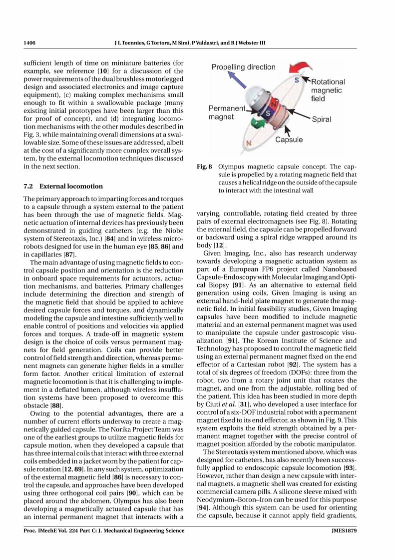
Owing to the potential advantages, there are anumber of current efforts underway to create a mag-netically guided capsule. The Norika Project Team wasone of the earliest groups to utilize magnetic fields forcapsule motion, when they developed a capsule thathas three internal coils that interact with three externalcoils embedded in a jacket worn by the patient for cap-sule rotation [12, 89]. In any such system, optimizationof the external magnetic field [86] is necessary to con-trol the capsule, and approaches have been developedusing three orthogonal coil pairs [90], which can beplaced around the abdomen. Olympus has also beendeveloping a magnetically actuated capsule that hasan internal permanent magnet that interacts with a
Fig. 8 Olympus magnetic capsule concept. The cap-sule is propelled by a rotating magnetic field thatcauses a helical ridge on the outside of the capsuleto interact with the intestinal wall
varying, controllable, rotating field created by threepairs of external electromagnets (see Fig. 8). Rotatingthe external field, the capsule can be propelled forwardor backward using a spiral ridge wrapped around itsbody [12].
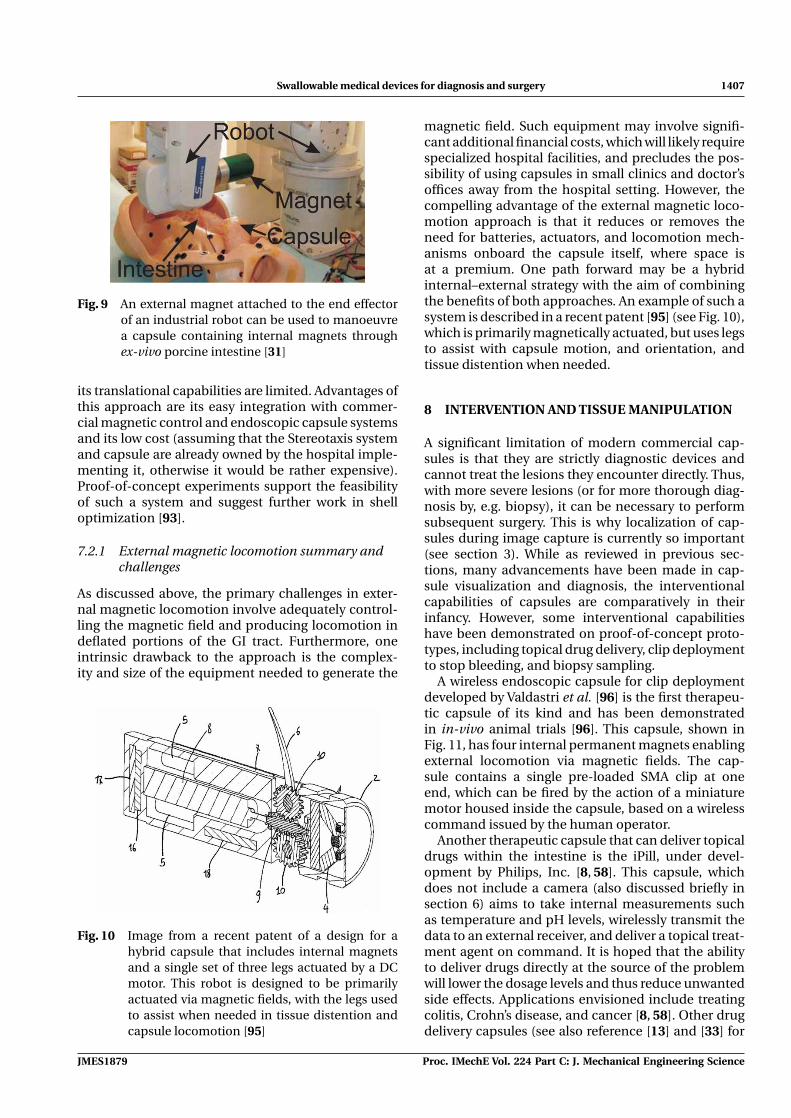
Given Imaging, Inc., also has research underwaytowards developing a magnetic actuation system aspart of a European FP6 project called NanobasedCapsule-Endoscopy with Molecular Imaging and Opti-cal Biopsy [91]. As an alternative to external fieldgeneration using coils, Given Imaging is using anexternal hand-held plate magnet to generate the mag-netic field. In initial feasibility studies, Given Imagingcapsules have been modified to include magneticmaterial and an external permanent magnet was usedto manipulate the capsule under gastroscopic visu-alization [91]. The Korean Institute of Science andTechnology has proposed to control the magnetic fieldusing an external permanent magnet fixed on the endeffector of a Cartesian robot [92]. The system has atotal of six degrees of freedom (DOFs): three from therobot, two from a rotary joint unit that rotates themagnet, and one from the adjustable, rolling bed ofthe patient. This idea has been studied in more depthby Ciuti et al. [31], who developed a user interface forcontrol of a six-DOF industrial robot with a permanentmagnet fixed to its end effector, as shown in Fig. 9. Thissystem exploits the field strength obtained by a per-manent magnet together with the precise control ofmagnet position afforded by the robotic manipulator.
The Stereotaxis system mentioned above, which wasdesigned for catheters, has also recently been success-fully applied to endoscopic capsule locomotion [93].However, rather than design a new capsule with inter-nal magnets, a magnetic shell was created for existingcommercial camera pills. A silicone sleeve mixed withNeodymium–Boron–Iron can be used for this purpose[94]. Although this system can be used for orientingthe capsule, because it cannot apply field gradients,
Proc. IMechE Vol. 224 Part C: J. Mechanical Engineering Science JMES1879
Swallowable medical devices for diagnosis and surgery 1407
Fig. 9 An external magnet attached to the end effectorof an industrial robot can be used to manoeuvrea capsule containing internal magnets throughex-vivo porcine intestine [31]
its translational capabilities are limited. Advantages ofthis approach are its easy integration with commer-cial magnetic control and endoscopic capsule systemsand its low cost (assuming that the Stereotaxis systemand capsule are already owned by the hospital imple-menting it, otherwise it would be rather expensive).Proof-of-concept experiments support the feasibilityof such a system and suggest further work in shelloptimization [93].
7.2.1 External magnetic locomotion summary andchallenges
As discussed above, the primary challenges in exter-nal magnetic locomotion involve adequately control-ling the magnetic field and producing locomotion indeflated portions of the GI tract. Furthermore, oneintrinsic drawback to the approach is the complex-ity and size of the equipment needed to generate the
Fig. 10 Image from a recent patent of a design for ahybrid capsule that includes internal magnetsand a single set of three legs actuated by a DCmotor. This robot is designed to be primarilyactuated via magnetic fields, with the legs usedto assist when needed in tissue distention andcapsule locomotion [95]
magnetic field. Such equipment may involve signifi-cant additional financial costs, which will likely requirespecialized hospital facilities, and precludes the pos-sibility of using capsules in small clinics and doctor’soffices away from the hospital setting. However, thecompelling advantage of the external magnetic loco-motion approach is that it reduces or removes theneed for batteries, actuators, and locomotion mech-anisms onboard the capsule itself, where space isat a premium. One path forward may be a hybridinternal–external strategy with the aim of combiningthe benefits of both approaches. An example of such asystem is described in a recent patent [95] (see Fig. 10),which is primarily magnetically actuated, but uses legsto assist with capsule motion, and orientation, andtissue distention when needed.
8 INTERVENTION AND TISSUE MANIPULATION
A significant limitation of modern commercial cap-sules is that they are strictly diagnostic devices andcannot treat the lesions they encounter directly. Thus,with more severe lesions (or for more thorough diag-nosis by, e.g. biopsy), it can be necessary to performsubsequent surgery. This is why localization of cap-sules during image capture is currently so important(see section 3). While as reviewed in previous sec-tions, many advancements have been made in cap-sule visualization and diagnosis, the interventionalcapabilities of capsules are comparatively in theirinfancy. However, some interventional capabilitieshave been demonstrated on proof-of-concept proto-types, including topical drug delivery, clip deploymentto stop bleeding, and biopsy sampling.
A wireless endoscopic capsule for clip deploymentdeveloped by Valdastri et al. [96] is the first therapeu-tic capsule of its kind and has been demonstratedin in-vivo animal trials [96]. This capsule, shown inFig. 11, has four internal permanent magnets enablingexternal locomotion via magnetic fields. The cap-sule contains a single pre-loaded SMA clip at oneend, which can be fired by the action of a miniaturemotor housed inside the capsule, based on a wirelesscommand issued by the human operator.
Another therapeutic capsule that can deliver topicaldrugs within the intestine is the iPill, under devel-opment by Philips, Inc. [8, 58]. This capsule, whichdoes not include a camera (also discussed briefly insection 6) aims to take internal measurements suchas temperature and pH levels, wirelessly transmit thedata to an external receiver, and deliver a topical treat-ment agent on command. It is hoped that the abilityto deliver drugs directly at the source of the problemwill lower the dosage levels and thus reduce unwantedside effects. Applications envisioned include treatingcolitis, Crohn’s disease, and cancer [8, 58]. Other drugdelivery capsules (see also reference [13] and [33] for
JMES1879 Proc. IMechE Vol. 224 Part C: J. Mechanical Engineering Science
1408 J L Toennies, G Tortora, M Simi, P Valdastri, and R J Webster III
Fig. 11 Clip deployment capsule of Valdastri et al.(©2008 Georg Thieme Verlag KG) [96]. (Left)The capsule is oriented toward the lesion usingexternal magnetic locomotion. (Right) At thecommand of the human operator a wirelesssignal triggers clip deployment, clamping thelesion and stopping bleeding
good discussions on this topic) include the InteliSite(Innovative Devices) capsule that uses SMA wires toline up perforated inner and outer sleeves to dispersea drug through the holes [33], and the Enterion cap-sule (Phaeton Research and Pharmaceutical Profiles)that can deliver a treating agent (liquid, powder, semi-solid) using a piston/spring actuation system [33].
A biopsy device consisting of a rotational razor forcutting tissue attached to a torsion spring has alsobeen implemented onboard a capsule platform [97].This device, tested ex-vivo in animal intestine, wassuccessful in extracting a tissue sample. However, itis not yet clear whether such a mechanism will beable to collect a sufficient number or volume of sam-ples for accurate external histological analysis. Someadditional ideas for intervention and tissue interactionhave also recently been the subject of patent disclo-sures [34], and one concept consists of a dual capsulecamera-biopsy system, where the two capsules areconnected by a tether [98]. Capsules housing ultra-sonic transducer modules have also been sketched inpatents by Miyake [99] and Taniguchi [100].
8.1 Intervention and tissue manipulationsummary and challenges
Interventional capabilities are one of the most promis-ing areas of endoscopic capsule research. Developinga system that can see, diagnose, and treat lesions itfinds is compelling from the perspectives of reduc-ing invasiveness for the patient and reducing thefinancial costs of treating GI diseases. Endowingcapsules with interventional capabilities is challeng-ing due to the intelligence and miniature mecha-nisms required in diagnosis and treatment. However,providing clinical capsules with the interventionalcapabilities mentioned above or others yet to be devel-oped has the potential to make WCEs a much more
powerful weapon with which physicians can combatdisease.
9 POWER
Since nearly every other module relies on it, thepower source is a critical issue for robotic endoscopiccapsules. Commercial endoscopic capsules rely onsilver-oxide watch batteries, which are the only bat-teries of their kind approved for clinical use [1]. Thesebatteries provide 3V at 55 mA-hr for approximately 8 h,which implies an average power delivery of approxi-mately 20 mW [101]. However, these traditional bat-teries are neither energy dense enough nor able tosource the peak currents needed to power roboticcapsules–particularly those with internal locomotioncapabilities.
In general, large-scale battery technologies do notscale down well to capsule dimensions [88]. One pos-sible exception to this rule are Lithium Ion Polymerbatteries (LiPo), such as those available from Plantracoand PowerStream, which are close to the size neededfor capsule integration. LiPo batteries have the highestenergy density (approximately 200Wh/kg) available inoff-the-shelf batteries, are capable of supplying peakcurrents up to 20 times their nominal current [41], andare miniaturizable, making them good candidates fora robotic endoscopic capsule power source.
Valdastri et al. elaborate on the power requirementsof their legged capsule design, noting that the aver-age power required for their 12-legged capsule wasmeasured to be 430 mW, plus an additional 180 mWfor the real-time vision system incorporated on theirprior prototypes. They come to the conclusion thata 100 mA-h battery would be sufficient for poweringtheir robotic capsule that is actuated by two brushlessDC motors. The smallest (10 mm diameter and 30 mmlength), battery they were able to identify provid-ing a capacity above 100 mA-h is the TLM-1030 fromTadiran, Israel [10]. Thus, advancements in batterytechnology are clearly needed, together with capsulemechanism efficiency enhancements. Note, however,that it is in principle possible to manufacture custom-shaped LiPo batteries, so that it may be possible toshape the battery to fill whatever space is availablewithin a capsule prototype. Thus, while it is not impos-sible to include batteries onboard robotic WCEs, it isclear that alternative methods of power would also bevaluable, and some are being developed.
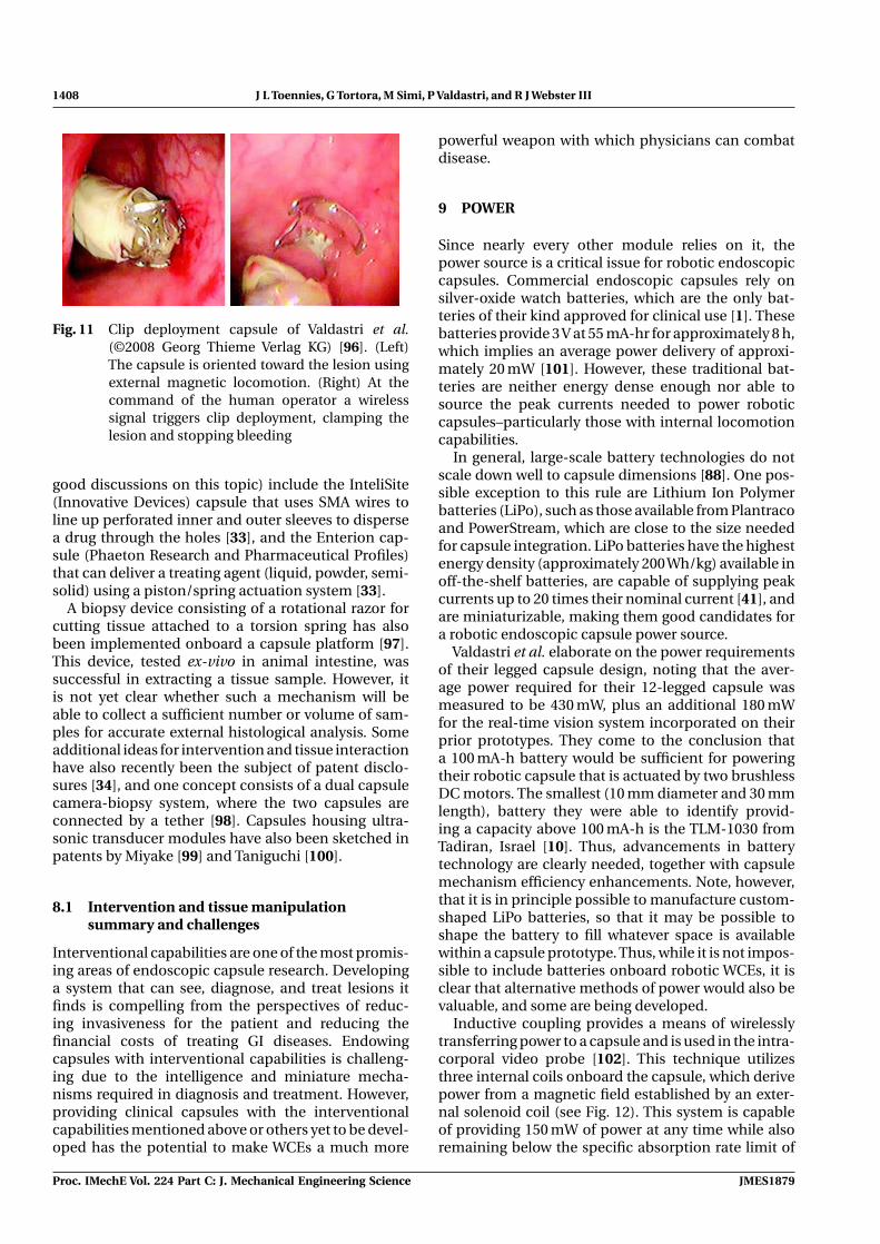
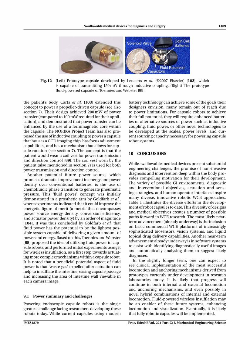
Inductive coupling provides a means of wirelesslytransferring power to a capsule and is used in the intra-corporal video probe [102]. This technique utilizesthree internal coils onboard the capsule, which derivepower from a magnetic field established by an exter-nal solenoid coil (see Fig. 12). This system is capableof providing 150 mW of power at any time while alsoremaining below the specific absorption rate limit of
Proc. IMechE Vol. 224 Part C: J. Mechanical Engineering Science JMES1879
Swallowable medical devices for diagnosis and surgery 1409
Fig. 12 (Left) Prototype capsule developed by Lenaerts et al. (©2007 Elsevier) [102], whichis capable of transmitting 150 mW through inductive coupling. (Right) The prototypefluid-powered capsule of Toennies and Webster [88]
the patient’s body. Carta et al. [103] extended thisconcept to power a propeller-driven capsule (see alsosection 7). Their design achieved 200 mW of powertransfer (compared to 100 mW required for their appli-cation), and demonstrated that power transfer can beenhanced by the use of a ferromagnetic core withinthe capsule. The NORIKA Project Team has also pro-posed the use of inductive coupling to power a capsulethat houses a CCD imaging chip, has focus adjustmentcapabilities, and has a mechanism that allows for cap-sule rotation (see section 7). The concept is that thepatient would wear a coil vest for power transmissionand direction control [89]. The coil vest worn by thepatient (also mentioned in section 7) is used for bothpower transmission and direction control.
Another potential future power source, whichpromises dramatic improvement in energy and powerdensity over conventional batteries, is the use ofchemofluidic phase transition to generate pneumaticpressure. This ‘fluid power’ concept was initiallydemonstrated in a prosthetic arm by Goldfarb et al.,where experiments indicated that it could improve theenergetic figure of merit (a metric that encompassespower source energy density, conversion efficiency,and actuator power density) by an order of magnitude[104]. It was thus concluded by Goldfarb et al. thatfluid power has the potential to be the lightest pos-sible system capable of delivering a given amount ofpower and energy. Based on this,Toennies andWebster[88] proposed the idea of utilizing fluid power in cap-sule robots, and performed initial experiments using itfor wireless insufflation, as a first step towards actuat-ing more complex mechanisms within a capsule robot.It is noted that a beneficial potential aspect of fluidpower is that ‘waste gas’ expelled after actuation canhelp to insufflate the intestine, easing capsule passageand increasing the area of intestine wall viewable ineach camera image.
9.1 Power summary and challenges
Powering endoscopic capsule robots is the singlegreatest challenge facing researchers developing theserobots today. While current capsules using modern
battery technology can achieve some of the goals theirdesigners envision, many remain out of reach dueto power limitations. For capsule robots to achievetheir full potential, they will require enhanced batter-ies or alternative sources of power such as inductivecoupling, fluid power, or other novel technologies tobe developed at the scales, power levels, and cur-rent sourcing capacity necessary for powering capsulerobot systems.
10 CONCLUSIONS
While swallowable medical devices present substantialengineering challenges, the promise of non-invasivediagnosis and intervention deep within the body pro-vides compelling motivation for their development.The variety of possible GI environments, diagnosticand interventional objectives, actuation and sens-ing strategies, and human operator interfaces inspiremany diverse, innovative robotic WCE approaches.Table 1 illustrates the diverse efforts in the develop-ment of robot capsules to date.This diversity of designsand medical objectives creates a number of possiblepaths forward in WCE research. The most likely near-term advancement (already underway) is the inclusionon basic commercial WCE platforms of increasinglysophisticated biosensors, vision systems, and liquidtopical drug delivery capabilities. Another near-termadvancement already underway is in software systemsto assist with identifying diagnostically useful imagesand automatically analysing them to suggest likelydiagnoses.
In the slightly longer term, one can expect tosee clinical implementation of the most successfullocomotion and anchoring mechanisms derived fromprototypes currently under development in researchlaboratories today. It is likely that progress willcontinue in both internal and external locomotionand anchoring mechanisms, and even possibly innovel hybrid combinations of internal and externallocomotion. Fluid-powered wireless insufflation maybe an enabler of these future systems, enhancinglocomotion and visualization. Eventually, it is likelythat fully robotic capsules will be implemented.
JMES1879 Proc. IMechE Vol. 224 Part C: J. Mechanical Engineering Science
1410 J L Toennies, G Tortora, M Simi, P Valdastri, and R J Webster III
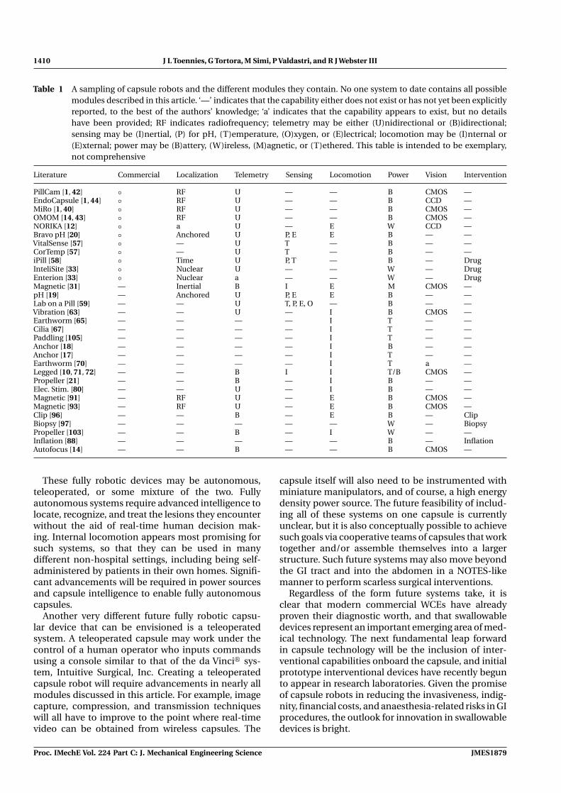
Table 1 A sampling of capsule robots and the different modules they contain. No one system to date contains all possiblemodules described in this article. ‘—’ indicates that the capability either does not exist or has not yet been explicitlyreported, to the best of the authors’ knowledge; ‘a’ indicates that the capability appears to exist, but no detailshave been provided; RF indicates radiofrequency; telemetry may be either (U)nidirectional or (B)idirectional;sensing may be (I)nertial, (P) for pH, (T)emperature, (O)xygen, or (E)lectrical; locomotion may be (I)nternal or(E)xternal; power may be (B)attery, (W)ireless, (M)agnetic, or (T)ethered. This table is intended to be exemplary,not comprehensive
Literature Commercial Localization Telemetry Sensing Locomotion Power Vision Intervention
PillCam [1, 42] ◦ RF U — — B CMOS —EndoCapsule [1, 44] ◦ RF U — — B CCD —MiRo [1, 40] ◦ RF U — — B CMOS —OMOM [14, 43] ◦ RF U — — B CMOS —NORIKA [12] ◦ a U — E W CCD —Bravo pH [20] ◦ Anchored U P, E E B — —VitalSense [57] ◦ — U T — B — —CorTemp [57] ◦ — U T — B — —iPill [58] ◦ Time U P, T — B — DrugInteliSite [33] ◦ Nuclear U — — W — DrugEnterion [33] ◦ Nuclear a — — W — DrugMagnetic [31] — Inertial B I E M CMOS —pH [19] — Anchored U P, E E B — —Lab on a Pill [59] — — U T, P, E, O — B — —Vibration [63] — — U — I B CMOS —Earthworm [65] — — — — I T — —Cilia [67] — — — — I T — —Paddling [105] — — — — I T — —Anchor [18] — — — — I B — —Anchor [17] — — — — I T — —Earthworm [70] — — — — I T a —Legged [10, 71, 72] — — B I I T/B CMOS —Propeller [21] — — B — I B — —Elec. Stim. [80] — — U — I B — —Magnetic [91] — RF U — E B CMOS —Magnetic [93] — RF U — E B CMOS —Clip [96] — — B — E B — ClipBiopsy [97] — — — — — W — BiopsyPropeller [103] — — B — I W — —Inflation [88] — — — — — B — InflationAutofocus [14] — — B — — B CMOS —
These fully robotic devices may be autonomous,teleoperated, or some mixture of the two. Fullyautonomous systems require advanced intelligence tolocate, recognize, and treat the lesions they encounterwithout the aid of real-time human decision mak-ing. Internal locomotion appears most promising forsuch systems, so that they can be used in manydifferent non-hospital settings, including being self-administered by patients in their own homes. Signifi-cant advancements will be required in power sourcesand capsule intelligence to enable fully autonomouscapsules.
Another very different future fully robotic capsu-lar device that can be envisioned is a teleoperatedsystem. A teleoperated capsule may work under thecontrol of a human operator who inputs commandsusing a console similar to that of the da Vinci� sys-tem, Intuitive Surgical, Inc. Creating a teleoperatedcapsule robot will require advancements in nearly allmodules discussed in this article. For example, imagecapture, compression, and transmission techniqueswill all have to improve to the point where real-timevideo can be obtained from wireless capsules. The
capsule itself will also need to be instrumented withminiature manipulators, and of course, a high energydensity power source. The future feasibility of includ-ing all of these systems on one capsule is currentlyunclear, but it is also conceptually possible to achievesuch goals via cooperative teams of capsules that worktogether and/or assemble themselves into a largerstructure. Such future systems may also move beyondthe GI tract and into the abdomen in a NOTES-likemanner to perform scarless surgical interventions.
Regardless of the form future systems take, it isclear that modern commercial WCEs have alreadyproven their diagnostic worth, and that swallowabledevices represent an important emerging area of med-ical technology. The next fundamental leap forwardin capsule technology will be the inclusion of inter-ventional capabilities onboard the capsule, and initialprototype interventional devices have recently begunto appear in research laboratories. Given the promiseof capsule robots in reducing the invasiveness, indig-nity, financial costs, and anaesthesia-related risks in GIprocedures, the outlook for innovation in swallowabledevices is bright.
Proc. IMechE Vol. 224 Part C: J. Mechanical Engineering Science JMES1879
Swallowable medical devices for diagnosis and surgery 1411
ACKNOWLEDGEMENTS
This work was supported in part by a Vanderbilt Uni-versity Discovery Grant, the NSF Graduate ResearchFellowship Programme, and the VECTOR FP6 Euro-pean Project EU/IST-2006-033970.
© Authors 2010
REFERENCES
1 Moglia, A., Menciassi, A., Dario, P., and Cuschieri, A.Capsule endoscopy: progress update and challengesahead. Nat. Rev. Gastroenterol. Hepatol., 2009, 6, 353–362.
2 Fantastic journey. The Economist Report, 18 April 2009,pp. 11–13.
3 Menciassi, A., Quirini, M., and Dario, P. Microroboticsfor future gastrointestinal endoscopy. Minim. InvasiveTherapy Allied Technol., 2007, 16, 91–100.
4 Sclabas, G., Swain, P., and Swanstrom, L. Endolumi-nal methods for gastrotomy closure in natural orificetransenteric surgery (NOTES). Surg. Innov., 2006, 13,23–30.
5 Spaun G. and Swanstrom, L. Quo vaids NOTES? Eur.Sur., 2008, 40, 211–219.
6 Scott, D., Tang, S., Fernandez, R., Bergs, R., Goova,M., Zeltser, I., Kehdy, F., and Cadeddu, J. Completelytransvaginal NOTES cholecystectomy using magneti-cally anchored instruments. Surg. Endosc., 2007, 21,2308–2316.
7 Lehman, A., Dumpert, J., Wood, N., Redden, L., Visty,A., Farritor, S., Varnell, B., and Oleynikov, D., Naturalorifice cholecystectomy using a miniature robot. Surg.Endosc., 2009, 23, 260–266.
8 Forgione, A. In vivo microrobots for natural orificetransluminal surgery. current status and future perspec-tives. Surg. Oncol., 2009, 18, 121–129.
9 Cotton, P. B. and Williams, C. B. Practical gastroin-testinal endoscopy the fundamentals, 5th edition, 2003,(Wiley-Blackwell, Oxford).
10 Valdastri, P., Webster, III R. J., Quaglia, C., Quirini,M., Menciassi, A., and Dario, P. A new mechanismfor mesoscale legged locomotion in compliant tubularenvironments. IEEE Trans. Robot., 2009, 25, 1047–1057.
11 Waterman, M. and Eliakim, R. Capsule enteroscopyof the small intestine. Abdom. Imaging, 2008, 34,452–458.
12 Moglia, A., Menciassis, A., Schurr, M., and Dario, P.Wireless capsule endoscopy: from diagnostic devicesto multipurpose robotic systems. Biomed. Microdevices,2007, 9, 235–243.
13 Twomey, K. and Marchesi, J. R. Swallowable capsuletechnology: current perspectives and future directions.Endoscopy, 2009, 41, 357–362.
14 Cavallotti, C., Piccigallo, M., Susilo, E., Valdastri, P.,Menciassi, A., and Dario, P. An integrated vision systemwith autofocus for wireless capsular endoscopy. Sens.Actuators A, 2009, 156, 72–78.
15 Gossum, A. V., Navas, M. M., Fernandez-Urien, I.,Carretero, C., Gay, G., Delvaux, M., Lapalus, M. G.,
Ponchon, T., Neuhaus, H., Philipper, M., Costamagna,G., Riccioni, M. E., Spada, C., Petruzziello, L., Fraser, C.,Postgate, A., Fitzpatrick, A., Hagenmuller, F., Keuchel,M., Schoofs, N., and Deviere, J. Capsule endoscopy ver-sus colonoscopy for the detection of polyps and cancer.N. Engl. J. Med., 2009, 361(3), 264–270.
16 Encyclopedia Britannica Online. Human digestive sys-tem, 2009, available from http://www.britannica.com/.
17 Glass, P., Cheung, E., and Sitti, M. A legged anchoringmechanism for capsule endoscopes using micropat-terned adhesives. IEEE Trans. Biomed. Eng., 2008, 55,2759–2767.
18 Tognarelli, S., Quaglia, C., Valdastri, P., Susilo, E., Men-ciassi, A., and Dario, P. Innovative stopping mechanismfor esophageal wireless capsular endoscopy. ProcediaChem., 2009, 1, 485–488.
19 Gonzalez-Guillaumin, J., Sadowski, D., Kaler, K., andMintchev, M. Ingestible capsule for impedance andpH monitoring in the esophagus. IEEE Trans. Biomed.Engng, 2007, 54, 2231–2236.
20 Kwiatek, M. A. and Pandolfino, J. E. The Bravo(TM) pHcapsule system. Dig. Liver Dis., 2008, 40, 156–160.
21 Tortora, G.,Valdastri, P., Susilo, E., Menciassi, A., Dario,P., Rieber, F., and Schurr, M. Propeller-based wirelessdevice for active capsular endoscopy in the gastric dis-trict. Minim. Invasive Therapy Allied Technol., 2009, 18,280–290.
22 Nagy, Z., Harada, K., Fluckiger, M., Susilo, E., Kaliakat-sos, I. K., Menciassi, A., Hawkes, E., Abbott, J. J., Dario,P., and Nelson, B. J. Assembling reconfigurable endolu-minal surgical systems: Opportunities and challenges.Int. J. Biomechatronics and Biomed. Robot., 2009, 1,3–16.
23 Hounnou,G.,Destrieux,C.,Desme,J.,Bertrand,P.,andVelut, S. Anatomical study of the length of the humanintestine. Surg. and Radiol. Anat., 2002, 24, 290–294.
24 Drossman, D. A., Grimm, I. S., and Shaheen, N.J. Handbook of gastroenterologic procedures, 4th edi-tion, 2005 (Lippincott Williams & Wilkins, Philadelphia,Pennsylvania).
25 Fischer, D., Schreiber, R., Levi, D., and Eliakim, R. Cap-sule endoscopy: the localization system. Gastrointest.Endos. Clin. N. Am., 2004, 14, 25–31.
26 Wang, X., Meng, M., and Hu, C. A localization methodusing 3-axis magnetoresistive sensors for tracking ofcapsule endoscope. In Proceedings of the IEEE/EMBS,New York, NY, USA, 2006, pp. 2522–2525.
27 Hu, C., Meng, M., and Mandal, M. The calibrationof 3-axis magnetic sensor array system for trackingwireless capsule endoscope. In Proceedings of theIEEE/RSJ International Conference on Intelligent robotsand systems, Beijing, P. R. China, 2006, pp. 162–167.
28 Hocke, M., Schone, U., Richert, H., Gornert, P., Keller,J., Layer, P., and Stallmach, A. Every slow-wave impulseis associated with motor activity of the human stomach.Am. J. Physiol. Gastrointest. Liver Physiol., 2008, 296,709–716.
29 Bulat, J., Duda, K., Duplaga, M., Fraczek, R., Skalski, A.,Socha, M., Turcza, P., and Zielinski, T. Data processingtasks in wireless GI endoscopy: image-based capsulelocalization & navigation with video compression. InProceedings of the IEEE/EMBS, Lyon, France, 2007,pp. 2815–2818.
JMES1879 Proc. IMechE Vol. 224 Part C: J. Mechanical Engineering Science
1412 J L Toennies, G Tortora, M Simi, P Valdastri, and R J Webster III
30 Lee, J., Oh, J., Shah, S., Yuan, X., and Tang, S. Automaticclassification of digestive organs in wireless endoscopyvideos. In Proceedings of the ACM Symposium onApplied computing, Seoul, Rep. of Korea, 2007.
31 Ciuti, G., Valdastri, P., Menciassi, A., and Dario,P. Robotic magnetic steering and locomotion ofmicrosystems for diagnostic and surgical endolumi-nal procedures. Robotica, 2010, 28(2), 199–207. DOI:10.1017/S0263574709990361.
32 Arshak, K. and Adepoju, F. Capsule tracking in the GItract: a novel microcontroller based solution. In Pro-ceedings of the IEEE Sensors Applications Symposium,Houston, Texas, USA, 2006, pp. 186–191.
33 Wilding, I., Hirst, P., and Connor, A. Developmentof a new engineering-based capsule for human drugabsorption studies. Pharm. Sci. Technol. Today, 2000, 3,385–392.
34 Moglia, A., Menciassi, A., and Dario, P. Recent patentson wireless capsule endoscopy. Recent Patents Biomed.Eng., 2008, 1, 24–33.
35 Kuth, R., Reinschke, J., and Roeckelein, R. Methodfor determining the position and orientation of anendoscopy capsule guided through an examinationobject by using a navigating magnetic field generated bymeans of a navigation device. DE102005032370, 2007.
36 Thone, J., Radiom, S., Turgis, D., Carta, R., Gielen, G.,and Puers, R. Design of a 2 Mbps FSK near-field trans-mitter for wireless capsule endoscopy. Sens. ActuatorsA, 2009, 156, 43–48.
37 Shen, M.-W., Lee, C.-Y., and Bor, J.-C. A 4.0-mW 2-Mbpsprogrammable BFSK transmitter for capsule endoscopeapplications. In Proceedings of the IEEE Asian Solid-State Circuits Conference, Hsinchu, Taiwan, 2005.
38 Susilo, E., Valdastri, P., Menciassi, A., and Dario, P.A miniaturized wireless control platform for roboticcapsular endoscopy using advanced pseudokernelapproach. Sens. Actuators A, Phys., 2009, 156, 49–58.
39 Valdastri, P., Menciassi, A., and Dario, P. Transmissionpower requirements for novel ZigBee implants in thegastrointestinal tract. IEEE Trans. Biomed. Eng., 2008,55, 1705–1710.
40 Bang, S., Park, J., Jeong, S., Kim, Y., Shim, H., Kim, T.,Lee, D., and Song, S. First clinical trial of the ‘‘MiRo’’capsule endoscope by using a novel transmission tech-nology: electric-field propagation. Clin. Endos., 2009,69, 253–259.
41 Swain, P. The future of wireless capsule endoscopy.World J. Gastroenterol. 2008, 14, 4142–4145.
42 Given Imaging, Inc.: www.givenimaging.com.43 Jinshan Science and Technology Group, Ltd., Co.
http://www.omom.us/main.php?sLAN=en.44 Olympus, Inc.: www.olympus.com.45 Kim, T., Song, S., Jung, H., Kim, J., and Yoon, E. Micro
capsule endoscope for gastro intestinal tract. In Pro-ceedings of the IEEE/EMBS, Lyon, France, 2007, pp.2823–2826.
46 Metzger, Y., Adler, S., Shitrit, A., Koslowsky, B., andBjarnason, I. Comparison of a new PillCam SB2 videocapsule versus the standard PillCam SB for detection ofsmall bowel disease. Med. Imaging, 2009, 2, 7–11.
47 Cave, D., Fleischer, D., Gostout, C., Faigel, D., Leighton,J., Heigh, R., Sharma, V., Mergener, K., Bhattacharya,K., Rajan, E., Foley, A., Lee, M., Knipschield, M., and
Hibberd, P. A multi-center randomized comparison ofthe EndoCapsule: Olympus Inc and the PillCam SB:Given Imaging in patients with obscure GI bleeding[abstract]. Gastrointest. Endos., 2007, 65, AB125.
48 Vatteroni, M., Covi, D., Cavallotti, C.,Valdastri, P., Men-ciassi, A., Dario, P., and Sartori, A. Smart optical CMOSsensor for endoluminal applications. Procedia Chem.,2009, 1, 1271–1274.
49 VECTOR: www.vector-project.com.50 Hasan, M. and Wallace, M. Image-enhanced
endoscopy. Am. Soc. Gastrointest. Endos., 2009, 16, 1–5.51 Turgis, D. and Puers, R. Image compression in video
radio transmission for capsule endoscopy. Sens. Actua-tors A, 2005, 123–124, 129–136.
52 Lin, M.-C., Dung, L.-R., and Weng, P.-K. An ultra-low-power image compressor for capsule endoscope.BioMed. Eng. OnLine, 2006, 5, 14.
53 Turcza, P., Zielinski, T., and Duplaga, M. Hardwareimplementation aspects of new low complexity imagecoding algorithm for wireless capsule endoscopy. Int.Conf. Computat. Sci., 2008, 5101, 476–485.
54 Zabulis, X., Argyros, A., and Tsakiris, D. Lumen detec-tion for capsule endoscopy. In Proceedings of theIEEE/RSJ International Conference on Intelligent robotsand systems, Nice, France, 2008, pp. 3921–3926.
55 Wang, X. and Meng, M. In situ analysis of capsuleendoscopy images. In Proceedings of the IEEE/RSJInternational Conference on Intelligent robots and sys-tems, St Louis, Missouri, USA, 2009.
56 Bejakovic, S., Kumar, R., Dassopoulos, T., Mullin, G.,and Hager, G. Analysis of crohn’s disease lesions incapsule endoscopy images. In Proceedings of the IEEEInternational Conference on robotics and automation,Kobe, Japan, 2009.
57 McKenzie, J. and Osgood, D. Validation of a new tele-metric core temperature monitor. J. Therm. Biol., 2004,29, 605–611.
58 Philips, Inc.: www.philips.com.59 Johannessen, E. A., Wang, L., Cui, L., Tang, T. B.,
Ahmadian, M., Astaras, A. Reid, S. W. J., Yam, P. S.,Murray, A. F., Flynn, B. W., Beaumont, S. P., Cumming,D. R. S., and Cooper, J. M. Implementation of multi-channel sensors for remote biomedical measurementsin a microsystems format. IEEE Trans. Biomed. Eng.,2004, 51, 525–535.
60 Allison, E., Kiraly, Z., Springer, G., and Dam, J. Endo-capsule. Patent number WO/2006/045011, 2006.
61 Wang, T. D. and Van Dam, J. Optical biopsy: a newfrontier in endoscopic detection and diagnosis. Clin.Gastroenterol. Hepatol., 2004, 2, 744–753.
62 Westerhof, J., Weersman, R., and Koornstra, J. Riskfactors for incomplete small-bowel capsule endoscopy.Gastrointest. Endos., 2009, 69, 74–80.
63 Zabulis, X., Sfakiotakis, M., and Tsakiris, D. Effects ofvibratory actuation on endoscopic capsule vision. InProceedings of the IEEE/EMBS Conference, Vancouver,British Columbia, Canada, 2008, pp. 5901–5904.
64 Kim, B., Lee, S., Park, J. H., and Park, J.-O. Design andfabrication of a locomotive mechanism for capsule-type endoscopes using shape memory alloys (SMAs).IEEE Trans. Mechatronics, 2005, 10, 77–86.
65 Kim, B., Park, S., Yoon, S.-J., and Jee, C. Y. Anearthworm-like locomotive mechanism for capsule
Proc. IMechE Vol. 224 Part C: J. Mechanical Engineering Science JMES1879
Swallowable medical devices for diagnosis and surgery 1413
endoscopes. In Proceedings of the IEEE/RSJ Interna-tional Conference on Intelligent robots and systems,Edmonton, Alberta, Canada, 2005, pp. 2997–3002.
66 Kwon, J., Park, S., Park, J., and Kim, B. Evalua-tion of the critical stroke of an earthworm-like robotfor capsule endoscopes. Proc. IMechE, Part H: J.Engineering in Medicine, 2007, 221, 397–405. DOI:10.1243/09544119JEIM134.
67 Li, W, Guo, W., Li, M., and Zhu, Y. A novel locomo-tion principle for endoscopic robot. In Proceedings ofthe IEEE International Conference on Mechatronics andautomation, Luoyang, P. R. China, 2006, pp. 1658–1662.
68 Feng,Y.,Li,W.,Li,M.,and Sun,L. Structure optimizationof the endoscopic robot ciliary leg based on dimen-sional analysis. In Proceedings of the IEEE InternationalConference on Robotics and biomimetics, Sanya, P. R.China, 2007, pp. 109–114.
69 Park, S., Park, H., Park, S., and Kim, B. A paddlingbased locomotive mechanism for capsule endoscopes.J. Mech. Sci. Technol., 2006, 20, 1012–1018.
70 Karagozler, M., Cheung, E., Kwon, J., and Sitti, M.Miniature endoscopic capsule robot using biomimeticmicro-patterned adhesives. In Proceedings of theIEEE/RAS-EMBS International Conference on Biomed-ical robotics and biomechatronics, Pisa, Italy, 2006, pp.105–111.
71 Gorini, S., Quirini, M., Menciassi, A., Pernorio, G., Ste-fanini, C., and Dario, P. A novel SMA-based actuatorfor a legged endoscopic capsule. In Proceedings of theIEEE/RAS-EMBS International Conference on Biomed-ical robotics and biomechatronics, Pisa, Italy, 2006, pp.443–449.
72 Quirini, M., Menciassi, A., Scapellato, S., Stefanini, C.,and Dario, P. Design and fabrication of a motor leggedcapsule for the active exploration of the gastrointestinaltract. IEEE/ASME Trans. Mechatronics, 2008, 13, 169–179.
73 Quirini, M., Scapellato, S., Valdastri, P., Menciassi, A.,and Dario, P. An approach to capsular endoscopy withactive motion. In Proceedings of the IEEE/EMBS, Lyon,France, 2007, pp. 2827–2830.
74 Quirini, M., Menciassi, A., Scapellato, S., Dario, P.,Rieber, F., Ho, C-N., Schostek, S., and Schurr, M. Fea-sibility proof of a legged locomotion capsule for the GItract. Gastrointest. Endos., 2008, 67, 1153–1158.
75 Quaglia, C., Buselli, E., Webster, III R. J., Valdastri,P., Menciassi, A., and Dario, P. An endoscopic cap-sule robot: a meso-scale engineering case study. J.Micromech. Microeng., 2009, 19, 105007.
76 Buselli, E., Valdastri, P., Quirini, M., Menciassi, A.,and Dario, P. Superelastic leg design optimization foran endoscopic capsule with active locomotion. SmartMater. Struct., 2009, 18, 015001 (8pp).
77 Buselli, E., Pensabene, V., Castrataro, P., Valdastri, P.Menciassi, A., and Dario, P. Micro hairs for frictionenhancement in active capsular endoscopy. In Proceed-ings of the Second European Conference on Tribology,Pisa, Italy, 2009, pp. 25–30.
78 Mosse, A., Mills, T., Appleyard, M., Kadirkamanathan,S., and Swain, P. Electrical stimulation for propellingendoscopes. Gastrointest. Endos., 2001, 54, 79–83.
79 Woo, S. H., Jang, J. Y., Jung, E. S., Lee, J. H., Moon Y.K., Kim, T. W., Won, C. H., Choi, H. C., and Cho, J. H.
Electrical stimuli capsule for control moving directionat the small intestine. In Proceedings of the IASTEDInternational Conference on Biomedical engineering,Innsbruck, Austria, 2006.
80 Woo, S. H., Kim, T. W., Lee, J. H., Kim, P. U., Won, C. H.,and Cho, J. H. Implemented edge shape of an electricalstimulus capsule. Int. J. Med. Robot. Comput. Assist.Surg., 2009, 5, 59–65.
81 Kosa, G., Jakb, P., Hata, N., Jolesz, F., Neubach,Z., Shoham, M. Zaaroor, M., and Szekely, G. Flag-ellar swimming for medical micro robots: Theory,experiments, and application. In Proceedings of theIEEE/RAS-EMBS International Conference on Biomed-ical robotics and biomechatronics, Scottsdale, Arizona,USA, 2008, pp. 258–263.
82 Iddan, G. and Gilad, Z. Motor for an in-vivo device.Patent number WO 2006/070350 A2, 2006.
83 Iddan, G. Self propelled device. US Patent number2006/0030754 A1, 2006.
84 Ramcharitar, S., Patterson, M. S., Geuns, R. J.,Meighem, C., and Serruys, P. W. Technology insight:magnetic navigation in coronary interventions. Nat.Clin. Pract. Cardiovasc. Med., 2008, 5, 148–156.
85 Ergeneman, O., Dogangil, G., Kummer, M. P., Abbott,J. J., Nazeeruddin, M. K., and Nelson, B. J. A mag-netically controlled wireless optical oxygen sensor forintraocular measurements. IEEE Sen. J. Am. Chem. Soc.,2008, 8, 29–37.
86 Abbott, J. J., Ergeneman, O., Kummer, M. P., Hirt, A.M.,and Nelson, B. J. Modeling magnetic torque and forcefor controlled manipulation of soft-magnetic bodies.IEEE Trans. Robot., 2007, 23, 1247–1252.
87 Martel, S., Felfoul, O., Mathieu, J.-B., Chanu, A.,Tamaz,S., Mohammadi, M., Mankiewicz, M., and Tabatabaei,N. MRI-based medical nanorobotic platform for thecontrol of magnetic nanoparticles and flagellated bac-teria for target interventions in human capillaries. Int.J. Robot. Res., 2009, 28, 1169–1182.
88 Toennies, J. L. and Webster, III R. J. A wireless insuf-flation system for capsular endoscopes. ASME J. Med.Devices, 2009, 3(2), 27514.
89 Uehara, A. and Hoshina, K. Capsule endoscope norikasystem. Minim. Invasive Therapy Allied Technol., 2003,1, 227–234.
90 Wang, X. and Meng, M. A magnetic stereo actuationmechanism for active capsule endoscope. In Proceed-ings of IEEE/EMBS, Lyon, France, 2007, pp. 2811–2814.
91 Volke, F., Keller, J., Schneider, A., Gerber, J., Reimann-Zawadzki, M., Rabinovitz, B., Mosse, C., and Swain,P. In-vivo remote manipulation of modified capsuleendoscopes using an external magnetic field [abstract].Gastrointest. Endos., 2008, 67, AB121–AB122.
92 Kim, B. K., Park, J. O.,and Hong,Y. S. Capsule type endo-scope control system. Patent Number WO/2005/122866,Korean Institute of Science and Technology,2005.
93 Carpi, F. and Pappone, C. Magnetic maneuvering ofendoscopic capsules by means of a robotic navigationsystem. IEEE Trans. Biomed. Eng., 2009, 56, 1482–1490.
94 Carpi, F., Galbiati, S., and Carpi, A. Magnetic shellsfor gastrointestinal endoscopic capsules as a means tocontrol their motion. Biomed. Pharmacother., 2006, 60,370–374.
JMES1879 Proc. IMechE Vol. 224 Part C: J. Mechanical Engineering Science
1414 J L Toennies, G Tortora, M Simi, P Valdastri, and R J Webster III
95 Menciassi, A., Dario, P., Valdastri, P., Quaglia, C.,and Buselli, E. Hybrid active locomotion teleoperatedendoscopic capsule. International patent applicationnumber PCT/IB2009/054491, 2009.
96 Valdastri, P., Quaglia, C., Susilo, E., Menciassi, A., Dario,P., Ho, C. N., Anhoeck, G., and Schurr, M. O. Wirelesstherapeutic endoscopic capsule: in vivo experiment.Endoscopy, 2008, 40, 979–982.
97 Kong, K., Cha, J., Jeon, D., and Cho, D. A rotationalmicro biopsy device for the capsule endoscope. InProceedings of the IEEE/RSJ International Conferenceon Intelligent robots and systems, Edmonton, Alberta,Canada, 2005, pp. 1839–1843.
98 Swain, C. P. Method, system and device for in-vivobiopsy. Patent number EP1757099, 2007.
99 Miyake, K. Capsule ultrasonic endoscope and capsuleultrasonic endoscope system. Patent number JP325874,2006.
100 Taniguchi, Y. Intracoelomic mobile body, andcapsule-type ultrasonic endoscope. Patent numberUS20050119577, 2005.
101 Carey, D. Camera-in-a-pill takes pictures of diges-tive tract, August 2006. Optical Electronics andDisplay, available from http://www.eetasia.com/ART_8800430920_480700_NP_6342156c.HTM.
102 Lenaerts, B. and Puers, R. An inductive power link fora wireless endoscope. Biosens. Bioelectron., 2007, 22,1390–1395.
103 Carta, R., Tortora, G., Lenaerts, B., Thone, J., Valdas-tri, P., Menciassi, A., Puers, R., and Dario, P. Wirelesspowering for a self-propelled and steerable endoscopiccapsule for stomach inspection. Biosens. Bioelectron,2009, 25, 845–851.
104 Goldfarb, M., Barth, E., Gongola, M., and Wehrmeyer,J., Design and energetic characterization of a liquid-propellant-powered actuator for self-powered robots.IEEE/ASME Trans. Mechatronics, 2003, 8, 254–262.
105 Park, H., Park, S., Yoon, E., Kim, B., Park, J., and Park,S. Paddling based microrobot for capsule endoscopes.In Proceedings of the IEEE International Conference onRobotics and automation, Roma, Italy, 2007, pp. 3377–3382.
Proc. IMechE Vol. 224 Part C: J. Mechanical Engineering Science JMES1879