spinal plasticity in stroke patients after botulinum neurotoxin … · spinal plasticity in stroke...
TRANSCRIPT
ORIGINAL RESEARCH
Spinal plasticity in stroke patients after botulinumneurotoxin A injection in ankle plantar flexorsClaire Aymard1,2, Louis-Solal Giboin1, Alexandra Lackmy-Vall�ee1 & V�eronique Marchand-Pauvert1
1 Service MPR, Centre Paris Sud, Fondation hospitali�ere Sainte Marie, Paris, France
2 UPMC Univ Paris 06, Er 6, F-75005, Paris, France
Keywords
Botulinum toxin, reciprocal inhibition, stroke.
Correspondence
V�eronique Marchand-Pauvert, ER 6 UPMC,
Service MPR Hopital Piti�e-Salpetri�ere, 47 bd
de l’Hopital, 75651 Paris Cedex 13, France.
Tel: +33-1-42-16-11-20
Fax: +33-1-42-16-11-02
E-mail: [email protected]
Funding Information
L. S. G. was supported by a grant from
UPMC Universit�e Paris 6 (Minist�ere de
l’Enseignement Sup�erieur et de la
Recherche). The study was supported by
UPMC Universit�e Paris 6, Assistance Publique-
Hopitaux de Paris (AP-HP), Institut pour la
Recherche sur la Moelle �epini�ere et
l’Enc�ephale (IRME), and INSERM.
Received: 29 October 2013; Accepted: 4
November 2013
doi: 10.1002/phy2.173
Physiol Rep, 1 (6), 2013, e00173, doi:
10.1002/phy2.173
Abstract
The effect of botulinum neurotoxin A (BoNT-A) in stroke patients’ upper
limbs has been attributed to its peripheral action only. However, BoNT-A
depressed recurrent inhibition of lumbar motoneurons, likely due to its retro-
grade transportation along motor axons affecting synapses to Renshaw cells.
Because Renshaw cells control group Ia interneurons mediating reciprocal
inhibition between antagonists, we tested whether this inhibition, particularly
affected after stroke, could recover after BoNT-A. The effect of posterior tibial
nerve (PTN) stimulation on tibialis anterior (TA) electromyogram (EMG) was
investigated in 13 stroke patients during treadmill walking before and 1
month after BoNT-A injection in ankle plantar flexors. Before BoNT-A, PTN
stimuli enhanced TA EMG all during the swing phase. After BoNT-A, the
PTN-induced reciprocal facilitation in TA motoneurons was depressed at the
beginning of swing and reversed into inhibition in midswing, but at the end
of swing, the reciprocal facilitation was enhanced. This suggests that BoNT-A
induced spinal plasticity leading to the recovery of reciprocal inhibition likely
due to the withdrawal of inhibitory control from Renshaw cells directly
blocked by the toxin. At the end of swing, the enhanced reciprocal facilitation
might be due to BoNT-induced modification of peripheral afferent inputs.
Therefore, both central and peripheral actions of BoNT-A can modify muscle
synergies during walking: (1) limiting ankle muscle co-contraction in the tran-
sition phase from stance to swing, to assist dorsiflexion, and (2) favoring it
from swing to stance, which blocks the ankle joint and thus assists the balance
during the single support phase on the paretic limb.
Introduction
Post-stroke walking is characterized by coactivation of
ankle flexors and extensors. Botulinum neurotoxin A
(BoNT-A), indicated to paralyze overactive ankle plantar
flexors, improves the temporal pattern of electromyo-
graphic (EMG) activity and the patients recover better
alternated ankle muscle activities (Hesse et al. 1996). It is
not known whether neurophysiological changes occur
after muscular injection and the extent to which central
phenomena participate in this functional improvement.
Indeed, besides its well-known action at peripheral level,
BoNT-A can affect central activity by influencing afferent
inputs through its action on gamma motor endings
(Filippi et al. 1993; Rosales et al. 1996), by inducing plas-
tic changes following the blockade of the neuromuscular
transmission (Abbruzzese and Berardelli 2006; Caleo et al.
2009), and finally through its retrograde transport along
the motor axons (Antonucci et al. 2008; Torii et al.
2011). However, because their spinal excitability did not
change after muscular injection in forearm muscles, it has
been claimed that BoNT-A clinical effect is only limited
to its peripheral action in stroke patients (Girlanda et al.
1997). However, it has been shown in arm muscles that
BoNT-A reduces spastic co-contraction of non-injected
antagonistic muscles (Gracies et al. 2009; Vinti et al.
2012). Moreover, we have recently shown in stroke
patients that BoNT-A reduces recurrent inhibition of
ª 2013 The Authors. Physiological Reports published by Wiley Periodicals, Inc. on behalf of
the American Physiological Society and The Physiological Society.
This is an open access article under the terms of the Creative Commons Attribution License,
which permits use, distribution and reproduction in any medium, provided the original work is properly cited.
2013 | Vol. 1 | Iss. 6 | e00173Page 1
Physiological Reports ISSN 2051-817X
lumbar motoneurons (Marchand-Pauvert et al. 2013).
These results raise again the question whether the clinical
effects of BoNT-A in stroke patients are really limited to
its peripheral action on overactive muscle or if it also
induces spinal plasticity improving muscle synergies and
functional recovery (Kaji 2013).
Stroke patients exhibit abnormal muscle synergies and
co-activation of antagonistic muscles in particular.
Accordingly, reciprocal inhibition between antagonists has
been found strongly depressed after stroke (Yanagisawa
et al. 1976; Crone et al. 2003). It has been shown in
healthy subjects that reciprocal inhibition is modulated
during the gait cycle and may help to inactivate antagonis-
tic motoneurones in the appropriate phases of the walking
cycle. Depression of the inhibition in the opposite phases
may help to ensure an unhindered activation of the moto-
neurones by descending and segmental excitatory inputs
(Petersen et al. 1999). Because reciprocal inhibition is
depressed after stroke (Yanagisawa et al. 1976; Crone
et al. 2003) and the ankle muscles are co-activated during
post-stroke walking, one would expect abnormal modula-
tion of reciprocal inhibition between ankle muscles dur-
ing post-stroke walking. Moreover, triceps surae being
mostly injected in stroke patients, we investigated recipro-
cal inhibition in ankle dorsiflexors. First, because we
wanted to further investigate how changes in afferent
inputs from an injected muscle influences the activity of
motoneurons supplying a non-injected muscle. Second,
because Ia inhibitory interneurons mediating reciprocal
inhibition between ankle muscles are controlled by
Renshaw cells (Baret et al. 2003; Fig. 1), we wondered
whether BoNT-A may help to reactivate reciprocal group
Ia interneurons by blocking Renshaw cells (Marchand-
Pauvert et al. 2013). We therefore tested reciprocal inhibi-
tion from ankle extensors to flexors during post-stroke
walking, before and after BoNT-A muscular injection in
ankle extensors.
The so-called reciprocal inhibition between wrist mus-
cles was found unchanged after toxin injection in stroke
patients (Girlanda et al. 1997). However, the inhibition of
motoneurons supplying the injected muscle was investi-
gated, which makes it difficult to interpret the EMG
recordings due to the plastic changes at muscular and
motoneuron levels (Abbruzzese and Berardelli 2006; Caleo
et al. 2009). On the other hand, spastic co-contraction of
non-injected antagonistic muscle was reduced at elbow
level after BoNT-A, possibly due to plastic changes at
spinal level (Gracies et al. 2009; Vinti et al. 2012). There-
fore, it seems that the effects of BoNT-A may be different
depending on the target joint. We thus further addressed
the question whether BoNT-A influences activity of non-
injected antagonistic muscle and its repercussions on
spinal excitability in stroke patients, who were tested
while walking, to explore the effects of BoNT-A in a func-
tional context.
Methods
Ethical approval
The study conformed to the standards set by the latest
revision of the Declaration of Helsinki and has been
approved by the ethics committee of Piti�e-Salpetri�ere
Hospital (CPP-Ile-de-France VI). Seventeen stroke
patients (five females) were included in the protocol, all
of whom had given informed written consent to the
experimental procedures.
Patients
Inclusion criteria were spastic leg paresis, marked
increase in tone at ankle level, a minimum 1-month
interval since stroke, and clinical prescription for BoNT-
RenshawTA
PTN
BoNT-A
Group Ia
Sol
Ia interneuron
DPN
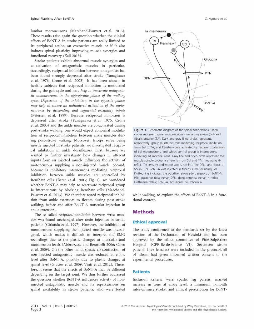
Figure 1. Schematic diagram of the spinal connections. Open
circles represent spinal motoneurons innervating soleus (Sol) and
tibialis anterior (TA). Dark and gray filled circles represent,
respectively, group Ia interneurons mediating reciprocal inhibition
from Sol to TA, and Renshaw cells activated by recurrent collaterals
of Sol motoneurons, and which control group Ia interneurons
inhibiting TA motoneurons. Gray line and open circle represent the
muscle spindle group Ia afferents from Sol and TA, mediating H-
reflex. TA sensory and motor axons run into the DPN, and those of
Sol in PTN. BoNT-A was injected in triceps surae including Sol.
Dotted line indicates the putative retrograde transport of BoNT-A.
PTN, posterior tibial nerve; DPN, deep peroneal nerve; H-reflex,
Hoffmann reflex; BoNT-A, botulinum neurotoxin A.
2013 | Vol. 1 | Iss. 6 | e00173Page 2
ª 2013 The Authors. Physiological Reports published by Wiley Periodicals, Inc. on behalf of
the American Physiological Society and The Physiological Society.
Spinal Plasticity After BoNT-A C. Aymard et al.
A injection only in ankle plantar flexors. Exclusion crite-
ria included BoNT-A injection within the previous 4
months and in other muscles than ankle plantar flexors,
previous alcohol or phenol blocks, surgical intervention,
or casting of the lower limb: fixed contractures in the
limb(s) or profound atrophy of muscle(s) to be injected.
We had to exclude 4/17 patients because soleus reflex
response was so large that it contaminated tibialis ante-
rior (TA) EMG activity, which was no longer interpret-
able due to concomitant compound potential with
similar shape in TA and soleus EMG (cross talk; Hutton
et al. 1988). The experimental protocol was thus possible
in only 13 patients (mean age: 52.8 � 3.0 years old;
range: 25–64), and mean interval since stroke was
29.3 � 14.8 months (range: 1–180). Ongoing treatments
(physical therapy and medication) remained unchanged.
Each patient was assessed 2 times: before (preBoNT-A)
and 1 month after BoNT-A injection (postBoNT-A), i.e.,
when clinical effects of BoNT-A injection are seen in
most patients (Kaji et al. 2010). BoNT-A was injected by
C. A. (among the authors) into triceps surae in all
patients, and into tibialis posterior in six patients,
according to the clinical prescription. The injection site
was guided by EMG to localize motor end plates and
muscle hyperactivity. Doses were established according
to the guidelines of the Worldwide Education and
Awareness for Movement Disorders (WEMOVE, www.
mdvu.org). Although the doses may be considered low,
muscle tone was reduced by two points in 12/13
patients, and by one point in the remaining patient
(Table 1).
Recordings
EMG was recorded with bipolar surface electrodes
(DE-2.1; Delsys Inc., Natick, MA) placed over the muscle
bellies of soleus (medial part of the posterior aspect of the
leg, 2–3 cm below the gastrocnemius muscles) and TA
(medial part of the anterior aspect of the leg, 10–15 cm
below the patella). EMG activity was amplified (10,0009;
Delsys Bagnoli System; Delsys Inc.), and filtered (band-
width 20–450 Hz), before being digitally stored (2-kHz
sampling rate) on a personal computer for off-line analysis
(Power 1401; CED, Cambridge, UK). Recordings were
undertaken during treadmill locomotion (Biodex Medical
Systems Inc., Shirley, NY). Because of foot drop on paretic
side and slow speed, the patients contacted the ground
with the forefoot. The pressure transducer, detecting the
ground contact, was thus placed at the middle and external
aspect of the sole to time the beginning of stance phase.
The patients first walked on the treadmill for 5–10 min
before recordings, to accustom themselves to treadmill
walking and to determine their comfortable speed (mean
walking speed 1.3 � 0.2 km h�1, range 0.6–2.0; Table 1).
This speed was not their maximum possible speed, but
they felt secure and were able to walk for 2–3 min
Table 1. Clinical features of the patients.
Lesion BONT-A Spasticity Inhibition
Patients Speed Time Type Site Type Soleus MG LG TP Pre Post Pre Post
1.F.62 0.6 5 Isch. L ABO 3/300 1/100 1/100 1/50 3 1 90.6 82.0
2.M.53 2.0 2 Isch. L ABO 3/300 1/100 1/100 – 2 0 99.2 76.8
3.M.64 0.6 3 Isch. R ABO 3/300 1/150 1/150 1/150 2 0 69.5 86.5
4.M.42 2.6 1 Hem. R ABO 3/300 1/100 1/100 – 2 0 83.2 80.0
5.F.53 1.0 5 Isch. R ABO 3/300 1/100 1/100 – 3 1 103.0 50.1
6.M.50 0.6 180 Isch. R ABO 3/300 1/100 1/100 1/150 3 1 74.9 40.4
7.M.63 1.8 7 Isch. R ABO 3/300 1/100 1/100 – 3 1 83.6 86.1
8.M.49 0.8 3.5 Isch. L ABO 3/300 1/100 1/100 1/150 2 0 101.8 80.4
9.M.61 0.8 16 Hem. L ABO 3/300 1/150 1/150 1/150 3 1 176.8 94.0
10.M.62 1.4 11 Isch. L ABO 3/300 1/100 1/100 – 3 1 114.6 77.7
11.M.25 1.6 108 Hem. L ONA 3/60 1/20 1/20 – 2 0 60.1 63.4
12.M.55 2.0 24 Hem. L ABO 3/300 1/100 1/100 – 3 2 72.7 75.3
13.M.47 1.0 15 Hem. L ABO 3/200 1/100 1/100 1/100 2 0 98.7 73.4
Patients: rank, gender (M, male; F, female), and age of the patients at the time of the investigation (years); Speed: walking speed before and
after BoNT-A; Lesion: Time = Time lapse between stroke and the first electrophysiological investigation, before BoNT-A (months); Type = origin
of the lesion (Isch., ischemia; Hem., hemorrhage); Site = cerebral hemisphere affected; BoNT-A: type indicates the type of toxin injected (ABO:
abobotulinumtoxinA; ONA: onabotulinumtoxinA); number of injection sites/dose (UI) in muscles receiving BoNT-A: soleus, MG (medial gasct-
rocnemius), LG (lateral gasctrocnemius), and TP (tibialis posterior); Spasticity: estimation of muscle tone (Ashworth score) in soleus before (Pre)
and 1 month after toxin injection (Post); Inhibition: maximal inhibition/less facilitation observed before (Pre) and 1 month after toxin injection
(Post), whatever the walking phase.
ª 2013 The Authors. Physiological Reports published by Wiley Periodicals, Inc. on behalf ofthe American Physiological Society and The Physiological Society.
2013 | Vol. 1 | Iss. 6 | e00173Page 3
C. Aymard et al. Spinal Plasticity After BoNT-A
(~recording duration) without fatigue. They were asked to
walk at the same speed after BoNT-A. All patients were
able to walk freely and investigations were performed
without bodyweight support.
Stimulation
One-millisecond rectangular electrical pulses were deliv-
ered through surface electrodes by constant current stim-
ulators (DS7A; Digitimer Ltd, Hertfordshire, UK). The
current crossed posterior tibial nerve (PTN) through a
7-cm2 brass hemispheric electrode placed in the popliteal
fossa (cathode) and a 21-cm2 brass plaque above the
patella. The deep peroneal nerve (DPN) was stimulated
using two 7-cm2 brass hemispheres: one placed behind
the head of the fibula (cathode) and the second one, on
the anterior aspect of the leg, 5–7 cm below the patella.
The optimal stimulation sites were determined clinically
by tendon palpation of soleus and TA. PTN stimulation
intensity was adjusted according to the threshold intensity
for direct motor response in soleus EMG activity (xMT,
motor threshold): stimulus intensity was 1 xMT in all
patients, and stimuli at 1.5 xMT were also tested in four
patients. DPN stimulation was adjusted to evoke a size-
able Hoffmann reflex (H-reflex) in TA EMG and to com-
pare its latency to that of soleus H-reflex (see below).
Experimental Procedures
The effect of PTN stimulation on TA EMGwas tested during
treadmill walking. Stimuli were triggered by the signal from
the pressure transducer, at four delays after foot contact dur-
ing the swing phase when TA was activated: at the onset of
TA activity (Early swing 1, ESw1), at maximal TA activity in
early swing (Early swing 2, ESw2), in mid-swing (MSw) and
late swing (LSw; Fig. 2). These delays were determined
according to the walking pattern and did not significantly
differ between the two experiments: (1) mean delay for
ESw1 was 1037 � 100 msec (600–1720 msec) before
BoNT-A versus 992 � 104 msec (600–1800 msec) after
BoNT-A (P = 0.47), (2) for ESw2, 1167 � 110 msec (650–1820 msec) versus 1115 � 116 msec (640–2000 msec;
P = 0.44), (3) for MSw, 1431 � 122 msec (870–2200 msec)
versus 1393 � 128 msec (710–2500 msec; P = 0.61), and
(4) for LSw, 1649 � 125 msec (1090–2700) versus
1651 � 150 ms (1020–2800 msec; P = 0.98). The speed and
the average step cycle time (1888 � 164 msec versus
1912 � 193 msec; P = 0.87) were similar before and after
BoNT-A, and PTN stimuli were thus delivered on average at
53 � 1 (ESw1), 60 � 2 (ESw2), 75 � 2 (MSw), and
91 � 2 (LSw)% the duration of the gait cycle during the two
experiments. One recording session consisted in delivering
20 PTN stimuli at one delay after ground contact. Two
recording sessions were performed at each delay: two ses-
sions for 1 delay 9 4 delays = eight recording sessions in
each patient. Recordings of control (without PTN stimula-
tion) and conditioned EMG (with PTN stimulation) were
randomly alternated during each session at a frequency
determined by foot contact (0.3–0.5 Hz).
EMG analysis
TA EMG activity was rectified and averaged: N = 40 traces
of control EMG and 40 of conditioned EMG for each delay
investigated. Usually, the analysis is calibrated according to
the arrival time of muscle spindle group Ia afferent fibers at
motoneuron level. For this study, the calculation should
have been based on the latency of TA H-reflex, the distance
between DPN and PTN stimulation sites, and the conduc-
tion velocity of their group Ia fibers. The mean latencies of
TA and soleus H-reflexes were similar (32.5 � 0.8 and
31.6 � 1.1 msec, respectively; P = 0.43). However, it was
possible to evoke an H-reflex in TA only in 9/13 patients,
and in soleus in all of them. Hence, for interindividual
comparisons, PTN-induced modulation of TA EMG was
analyzed within the window of analysis determined accord-
ing to patient soleus H-reflex latency. The central latency
for reciprocal inhibition is about 1 msec (Crone et al.
1987), and its duration is about 10 msec (Pierrot-Deseil-
ligny et al. 1981; Capaday et al. 1990; Petersen et al. 1999).
Therefore, the window of analysis started from the latency
of soleus H-reflex + 1 msec and lasted 10 msec in all
patients (vertical dashed lines, Fig. 3). Both control and
conditioned EMG were evaluated over the same window of
analysis, and the area of conditioning EMG was normalized
to the mean area of control EMG recorded during the same
session. The ratio gave a quantitative estimation of PTN-
induced modification of TA motoneuron excitability
(ratio > 100% indicates motoneuron facilitation and
<100%, motoneuron inhibition).
Statistical analysis
Two-tailed paired t-tests were performed to compare con-
trol and conditioned EMG in each individual, locomotor
parameters before and after BoNT-A and the H-reflex
latencies in the group. Because normality and homogene-
ity of variances were not respected in the group data,
nonparametric Friedman tests were performed to com-
pare pre- and post-BoNT-A recordings, background EMG
activity, conditioned TA and soleus EMG and intensity of
PTN stimulation. If the tests provided significant P val-
ues, Wilcoxon signed rank tests were performed for com-
parison of two means. Spearman’s rank correlation
coefficient was calculated to test the influence of the time
since stroke and the walking speed on reciprocal inhibi-
2013 | Vol. 1 | Iss. 6 | e00173Page 4
ª 2013 The Authors. Physiological Reports published by Wiley Periodicals, Inc. on behalf of
the American Physiological Society and The Physiological Society.
Spinal Plasticity After BoNT-A C. Aymard et al.
tion and its changes after BoNT-A. The levels of reciprocal
facilitation and inhibition in the group, in each condition
(walking phase, preBoNT-A, postBoNT-A), were individ-
ually tested using one-sample t-tests. Tests were per-
formed using StatEL software (www.adscience.eu) and the
significance level was set at P < 0.05. The mean data are
indicated � 1 standard error of the mean (SEM).
Results
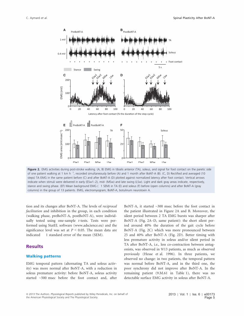
Walking patterns
EMG temporal pattern (alternating TA and soleus activ-
ity) was more normal after BoNT-A, with a reduction in
soleus premature activity: before BoNT-A, soleus activity
started ~500 msec before the foot contact and, after
BoNT-A, it started ~300 msec before the foot contact in
the patient illustrated in Figure 2A and B. Moreover, the
silent period between 2 TA EMG bursts was sharper after
BoNT-A (Fig. 2A–D, same patient): the short silent per-
iod around 40% the duration of the gait cycle before
BoNT-A (Fig. 2C) which was more pronounced between
25 and 40% after BoNT-A (Fig. 2D). Better timing with
less premature activity in soleus and/or silent period in
TA after BoNT-A, i.e., less co-contraction between antag-
onists, was observed in 9/13 patients, as much as observed
previously (Hesse et al. 1996). In three patients, we
observed no change: in two patients, the temporal pattern
was normal before BoNT-A, and in the third one, the
poor synchrony did not improve after BoNT-A. In the
remaining patient (9.M.61 in Table 1), there was no
detectable surface EMG activity in soleus after BoNT-A.
A B
C D
E F
EMG
(mV)
ESw1
ESw2
MSw LSw ESw1
ESw2
MSw LSw
EMG
area
(mV.
mse
c–1 )
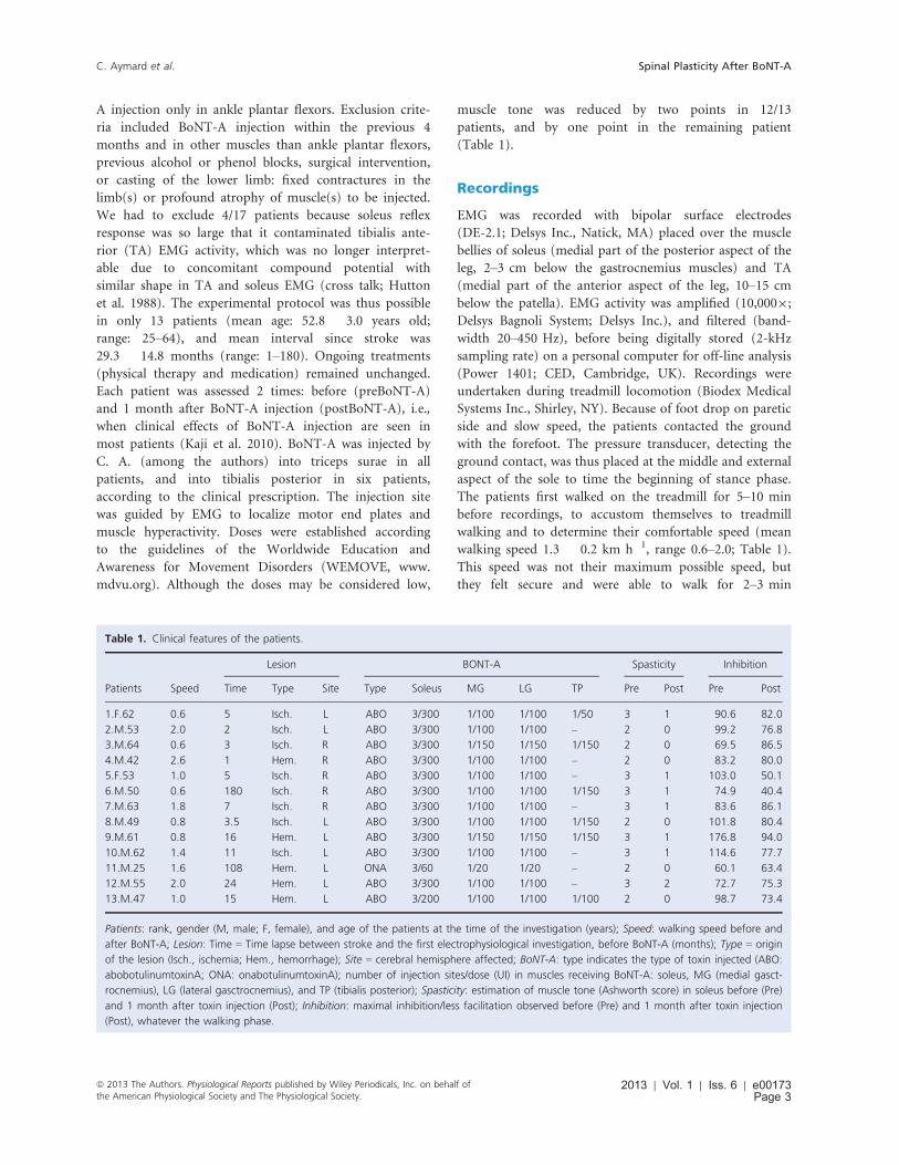
Figure 2. EMG activities during post-stroke walking. (A, B) EMG in tibialis anterior (TA), soleus, and signal for foot contact on the paretic side
of one patient walking at 1 km h�1, recorded simultaneously before (A) and 1 month after BoNT-A (B). (C, D) Rectified and averaged (10
steps) TA EMG in the same patient before (C) and after BoNT-A (D) plotted against normalized latency after foot contact. Vertical arrows
indicate when stimuli were delivered in early (ESw1–2), mid- (MSw) and late swing (LSw). Light and dark gray areas indicate, respectively,
stance and swing phase. (EF) Mean background EMG (�1 SEM) in TA (E) and soleus (F) before (open columns) and after BoNT-A (gray
columns) in the group of 13 patients. EMG, electromyogram; BoNT-A, botulinum neurotoxin A.
ª 2013 The Authors. Physiological Reports published by Wiley Periodicals, Inc. on behalf ofthe American Physiological Society and The Physiological Society.
2013 | Vol. 1 | Iss. 6 | e00173Page 5
C. Aymard et al. Spinal Plasticity After BoNT-A
On average (13 patients), the level of TA and soleus
EMG did not change between pre- and post-BoNT-A
recordings (Fig. 2E and F; P = 0.28 and 0.42, respec-
tively). In a previous study, EMG was greater after BoNT-
A (Hesse et al. 1996) but this was probably due to an
increase in walking speed. Most of our patients could
walk faster after BoNT-A, but the speed was similar to
compare spinal excitability under the same conditions.
PTN-induced modifications in TA EMG
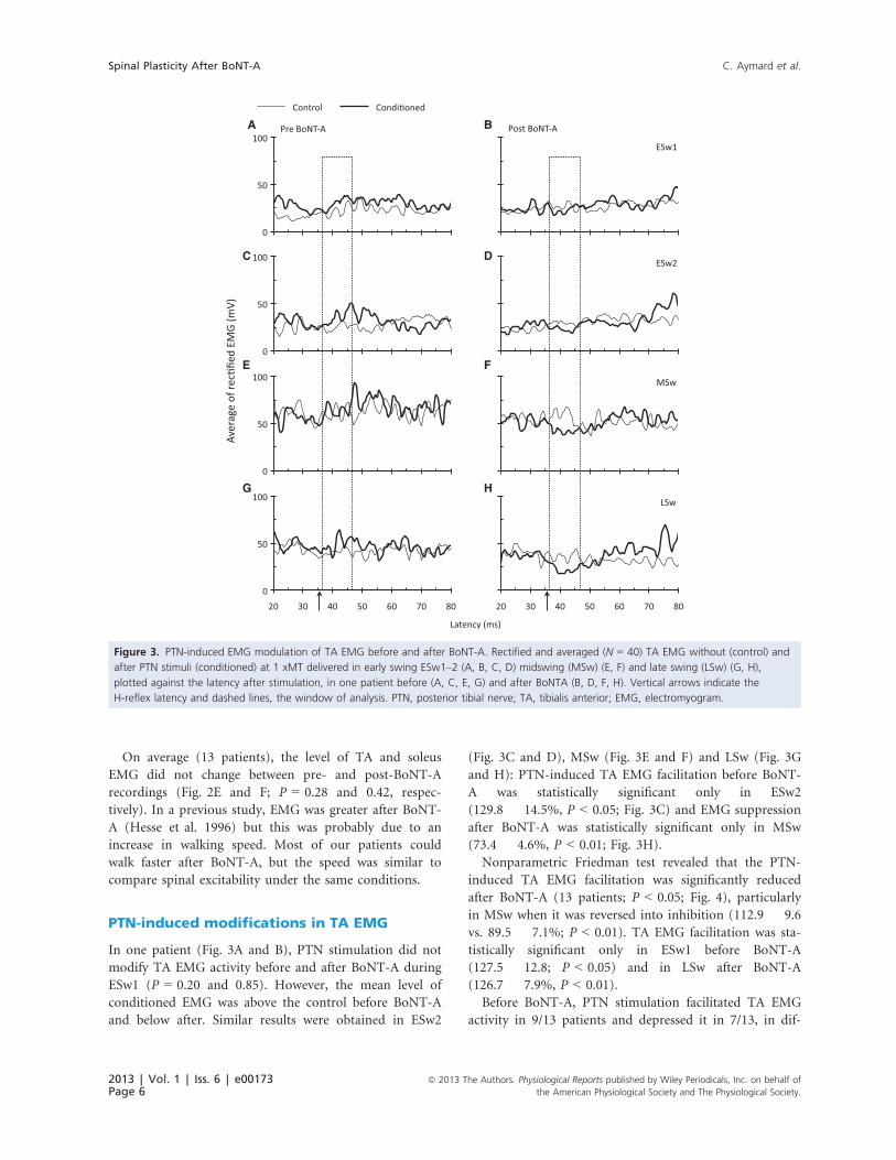
In one patient (Fig. 3A and B), PTN stimulation did not
modify TA EMG activity before and after BoNT-A during
ESw1 (P = 0.20 and 0.85). However, the mean level of
conditioned EMG was above the control before BoNT-A
and below after. Similar results were obtained in ESw2
(Fig. 3C and D), MSw (Fig. 3E and F) and LSw (Fig. 3G
and H): PTN-induced TA EMG facilitation before BoNT-
A was statistically significant only in ESw2
(129.8 � 14.5%, P < 0.05; Fig. 3C) and EMG suppression
after BoNT-A was statistically significant only in MSw
(73.4 � 4.6%, P < 0.01; Fig. 3H).
Nonparametric Friedman test revealed that the PTN-
induced TA EMG facilitation was significantly reduced
after BoNT-A (13 patients; P < 0.05; Fig. 4), particularly
in MSw when it was reversed into inhibition (112.9 � 9.6
vs. 89.5 � 7.1%; P < 0.01). TA EMG facilitation was sta-
tistically significant only in ESw1 before BoNT-A
(127.5 � 12.8; P < 0.05) and in LSw after BoNT-A
(126.7 � 7.9%, P < 0.01).
Before BoNT-A, PTN stimulation facilitated TA EMG
activity in 9/13 patients and depressed it in 7/13, in dif-
A B
C D
E F
G H
Aver
age
of re
cfie
d EM
G (m
V)
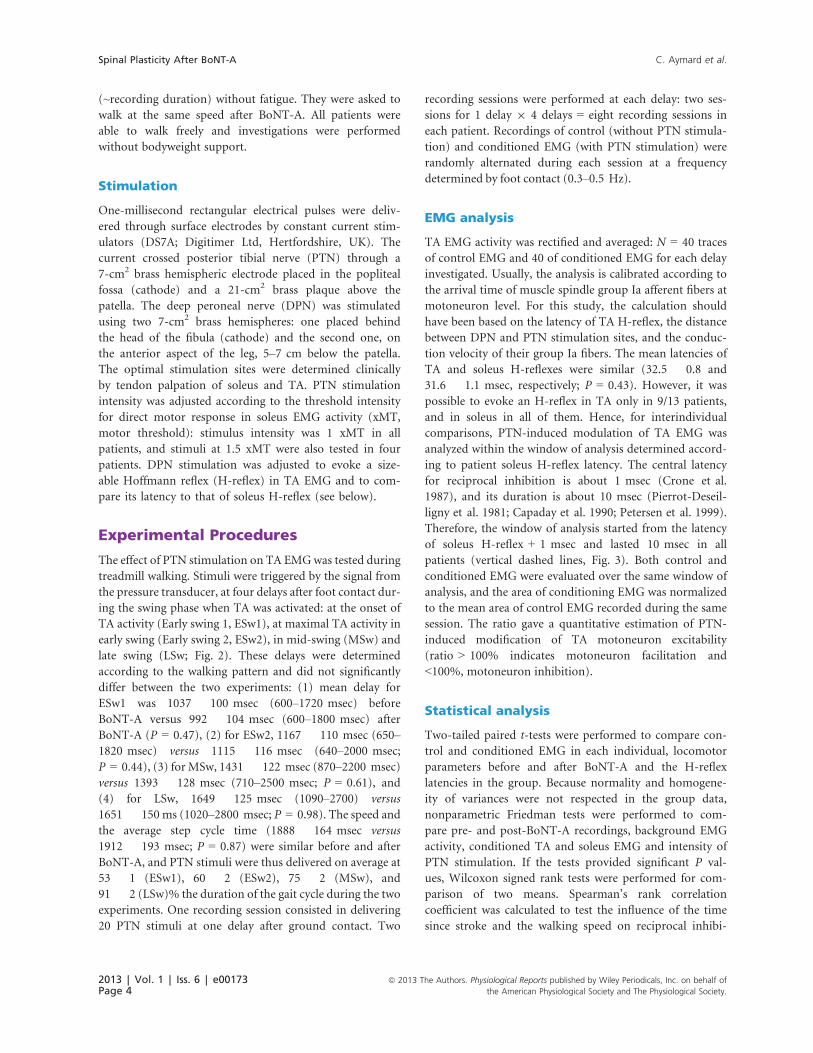
Figure 3. PTN-induced EMG modulation of TA EMG before and after BoNT-A. Rectified and averaged (N = 40) TA EMG without (control) and
after PTN stimuli (conditioned) at 1 xMT delivered in early swing ESw1–2 (A, B, C, D) midswing (MSw) (E, F) and late swing (LSw) (G, H),
plotted against the latency after stimulation, in one patient before (A, C, E, G) and after BoNTA (B, D, F, H). Vertical arrows indicate the
H-reflex latency and dashed lines, the window of analysis. PTN, posterior tibial nerve; TA, tibialis anterior; EMG, electromyogram.
2013 | Vol. 1 | Iss. 6 | e00173Page 6
ª 2013 The Authors. Physiological Reports published by Wiley Periodicals, Inc. on behalf of
the American Physiological Society and The Physiological Society.
Spinal Plasticity After BoNT-A C. Aymard et al.
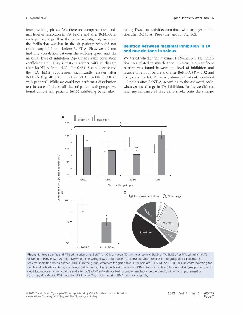
ferent walking phases. We therefore compared the maxi-
mal level of inhibition in TA before and after BoNT-A in
each patient, regardless the phase investigated, or when
the facilitation was less in the six patients who did not
exhibit any inhibition before BoNT-A. First, we did not
find any correlation between the walking speed and the
maximal level of inhibition (Spearman’s rank correlation
coefficient r = �0.08, P = 0.77) neither with it changes
after Bo-NT-A (r = �0.21, P = 0.46). Second, we found
the TA EMG suppression significantly greater after
BoNT-A (Fig. 4B: 94.5 � 8.1 vs. 74.3 � 4.1%; P < 0.05;
9/13 patients). While we could not perform a distribution
test because of the small size of patient sub-groups, we
found almost half patients (6/13) exhibiting better alter-
nating TA/soleus activities combined with stronger inhibi-
tion after BoNT-A (Pre-/Post+ group, Fig. 4C).
Relation between maximal inhibition in TAand muscle tone in soleus
We tested whether the maximal PTN-induced TA inhibi-
tion was related to muscle tone in soleus. No significant
relation was found between the level of inhibition and
muscle tone both before and after BoNT-A (P = 0.32 and
0.61, respectively). Moreover, almost all patients exhibited
�2 points after BoNT-A, according to the Ashworth scale,
whatever the change in TA inhibition. Lastly, we did not
find any influence of time since stroke onto the changes
A
B C
Figure 4. Reverse effects of PTN stimulation after BoNT-A. (A) Mean area (% the mean control EMG) of TA EMG after PTN stimuli (1 xMT)
delivered in early (ESw1–2), mid- (MSw) and late swing (LSw), before (open columns) and after BoNT-A in the group of 13 patients. (B)
Maximal inhibition (mean surface <100%) in the group, whatever the gait phase. Error bars are �1 SEM. *P < 0.05. (C) Pie chart indicating the
number of patients exhibiting no change (white and light gray portions) or increased PTN-induced inhibition (black and dark gray portions) and
good locomotor synchrony before and after BoNT-A (Pre+/Post+) or bad locomotor synchrony before (Pre-/Post+) or no improvement of
synchrony (Pre-/Post-). PTN, posterior tibial nerve; TA, tibialis anterior; EMG, electromyography.
ª 2013 The Authors. Physiological Reports published by Wiley Periodicals, Inc. on behalf ofthe American Physiological Society and The Physiological Society.
2013 | Vol. 1 | Iss. 6 | e00173Page 7
C. Aymard et al. Spinal Plasticity After BoNT-A
in reciprocal inhibition after BoNT-A (Spearman’s rank
correlation coefficient r = 0.15, P = 0.61).
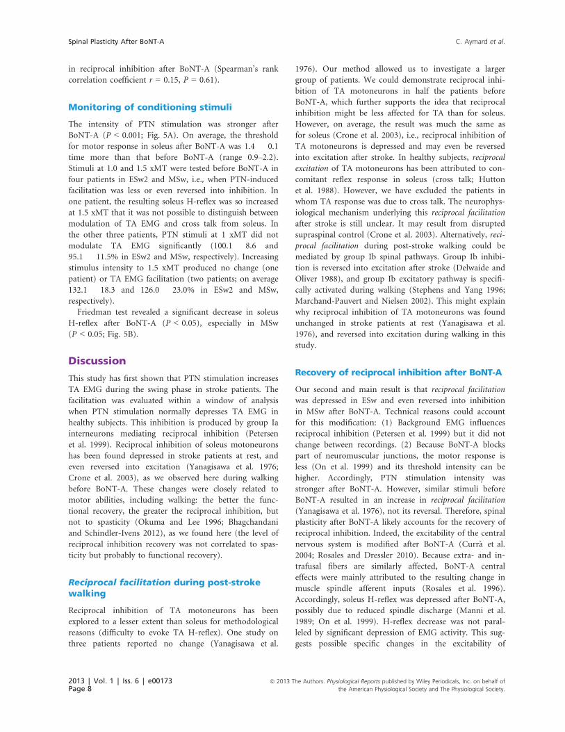
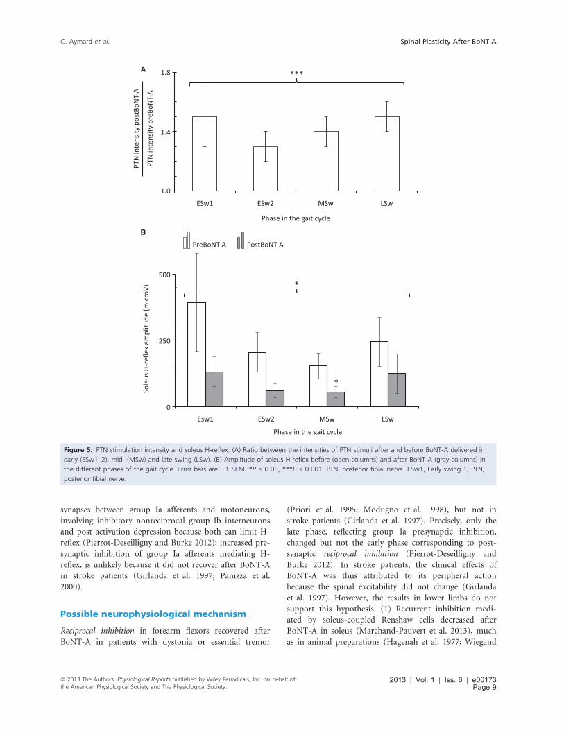
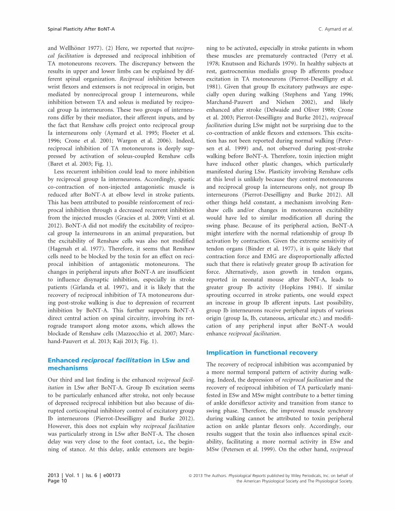
Monitoring of conditioning stimuli
The intensity of PTN stimulation was stronger after
BoNT-A (P < 0.001; Fig. 5A). On average, the threshold
for motor response in soleus after BoNT-A was 1.4 � 0.1
time more than that before BoNT-A (range 0.9–2.2).Stimuli at 1.0 and 1.5 xMT were tested before BoNT-A in
four patients in ESw2 and MSw, i.e., when PTN-induced
facilitation was less or even reversed into inhibition. In
one patient, the resulting soleus H-reflex was so increased
at 1.5 xMT that it was not possible to distinguish between
modulation of TA EMG and cross talk from soleus. In
the other three patients, PTN stimuli at 1 xMT did not
modulate TA EMG significantly (100.1 � 8.6 and
95.1 � 11.5% in ESw2 and MSw, respectively). Increasing
stimulus intensity to 1.5 xMT produced no change (one
patient) or TA EMG facilitation (two patients; on average
132.1 � 18.3 and 126.0 � 23.0% in ESw2 and MSw,
respectively).
Friedman test revealed a significant decrease in soleus
H-reflex after BoNT-A (P < 0.05), especially in MSw
(P < 0.05; Fig. 5B).
Discussion
This study has first shown that PTN stimulation increases
TA EMG during the swing phase in stroke patients. The
facilitation was evaluated within a window of analysis
when PTN stimulation normally depresses TA EMG in
healthy subjects. This inhibition is produced by group Ia
interneurons mediating reciprocal inhibition (Petersen
et al. 1999). Reciprocal inhibition of soleus motoneurons
has been found depressed in stroke patients at rest, and
even reversed into excitation (Yanagisawa et al. 1976;
Crone et al. 2003), as we observed here during walking
before BoNT-A. These changes were closely related to
motor abilities, including walking: the better the func-
tional recovery, the greater the reciprocal inhibition, but
not to spasticity (Okuma and Lee 1996; Bhagchandani
and Schindler-Ivens 2012), as we found here (the level of
reciprocal inhibition recovery was not correlated to spas-
ticity but probably to functional recovery).
Reciprocal facilitation during post-strokewalking
Reciprocal inhibition of TA motoneurons has been
explored to a lesser extent than soleus for methodological
reasons (difficulty to evoke TA H-reflex). One study on
three patients reported no change (Yanagisawa et al.
1976). Our method allowed us to investigate a larger
group of patients. We could demonstrate reciprocal inhi-
bition of TA motoneurons in half the patients before
BoNT-A, which further supports the idea that reciprocal
inhibition might be less affected for TA than for soleus.
However, on average, the result was much the same as
for soleus (Crone et al. 2003), i.e., reciprocal inhibition of
TA motoneurons is depressed and may even be reversed
into excitation after stroke. In healthy subjects, reciprocal
excitation of TA motoneurons has been attributed to con-
comitant reflex response in soleus (cross talk; Hutton
et al. 1988). However, we have excluded the patients in
whom TA response was due to cross talk. The neurophys-
iological mechanism underlying this reciprocal facilitation
after stroke is still unclear. It may result from disrupted
supraspinal control (Crone et al. 2003). Alternatively, reci-
procal facilitation during post-stroke walking could be
mediated by group Ib spinal pathways. Group Ib inhibi-
tion is reversed into excitation after stroke (Delwaide and
Oliver 1988), and group Ib excitatory pathway is specifi-
cally activated during walking (Stephens and Yang 1996;
Marchand-Pauvert and Nielsen 2002). This might explain
why reciprocal inhibition of TA motoneurons was found
unchanged in stroke patients at rest (Yanagisawa et al.
1976), and reversed into excitation during walking in this
study.
Recovery of reciprocal inhibition after BoNT-A
Our second and main result is that reciprocal facilitation
was depressed in ESw and even reversed into inhibition
in MSw after BoNT-A. Technical reasons could account
for this modification: (1) Background EMG influences
reciprocal inhibition (Petersen et al. 1999) but it did not
change between recordings. (2) Because BoNT-A blocks
part of neuromuscular junctions, the motor response is
less (On et al. 1999) and its threshold intensity can be
higher. Accordingly, PTN stimulation intensity was
stronger after BoNT-A. However, similar stimuli before
BoNT-A resulted in an increase in reciprocal facilitation
(Yanagisawa et al. 1976), not its reversal. Therefore, spinal
plasticity after BoNT-A likely accounts for the recovery of
reciprocal inhibition. Indeed, the excitability of the central
nervous system is modified after BoNT-A (Curr�a et al.
2004; Rosales and Dressler 2010). Because extra- and in-
trafusal fibers are similarly affected, BoNT-A central
effects were mainly attributed to the resulting change in
muscle spindle afferent inputs (Rosales et al. 1996).
Accordingly, soleus H-reflex was depressed after BoNT-A,
possibly due to reduced spindle discharge (Manni et al.
1989; On et al. 1999). H-reflex decrease was not paral-
leled by significant depression of EMG activity. This sug-
gests possible specific changes in the excitability of
2013 | Vol. 1 | Iss. 6 | e00173Page 8
ª 2013 The Authors. Physiological Reports published by Wiley Periodicals, Inc. on behalf of
the American Physiological Society and The Physiological Society.
Spinal Plasticity After BoNT-A C. Aymard et al.
synapses between group Ia afferents and motoneurons,
involving inhibitory nonreciprocal group Ib interneurons
and post activation depression because both can limit H-
reflex (Pierrot-Deseilligny and Burke 2012); increased pre-
synaptic inhibition of group Ia afferents mediating H-
reflex, is unlikely because it did not recover after BoNT-A
in stroke patients (Girlanda et al. 1997; Panizza et al.
2000).
Possible neurophysiological mechanism
Reciprocal inhibition in forearm flexors recovered after
BoNT-A in patients with dystonia or essential tremor
(Priori et al. 1995; Modugno et al. 1998), but not in
stroke patients (Girlanda et al. 1997). Precisely, only the
late phase, reflecting group Ia presynaptic inhibition,
changed but not the early phase corresponding to post-
synaptic reciprocal inhibition (Pierrot-Deseilligny and
Burke 2012). In stroke patients, the clinical effects of
BoNT-A was thus attributed to its peripheral action
because the spinal excitability did not change (Girlanda
et al. 1997). However, the results in lower limbs do not
support this hypothesis. (1) Recurrent inhibition medi-
ated by soleus-coupled Renshaw cells decreased after
BoNT-A in soleus (Marchand-Pauvert et al. 2013), much
as in animal preparations (Hagenah et al. 1977; Wiegand
A
B
Figure 5. PTN stimulation intensity and soleus H-reflex. (A) Ratio between the intensities of PTN stimuli after and before BoNT-A delivered in
early (ESw1–2), mid- (MSw) and late swing (LSw). (B) Amplitude of soleus H-reflex before (open columns) and after BoNT-A (gray columns) in
the different phases of the gait cycle. Error bars are �1 SEM. *P < 0.05, ***P < 0.001. PTN, posterior tibial nerve. ESw1, Early swing 1; PTN,
posterior tibial nerve.
ª 2013 The Authors. Physiological Reports published by Wiley Periodicals, Inc. on behalf ofthe American Physiological Society and The Physiological Society.
2013 | Vol. 1 | Iss. 6 | e00173Page 9
C. Aymard et al. Spinal Plasticity After BoNT-A
and Wellh€oner 1977). (2) Here, we reported that recipro-
cal facilitation is depressed and reciprocal inhibition of
TA motoneurons recovers. The discrepancy between the
results in upper and lower limbs can be explained by dif-
ferent spinal organization. Reciprocal inhibition between
wrist flexors and extensors is not reciprocal in origin, but
mediated by nonreciprocal group I interneurons, while
inhibition between TA and soleus is mediated by recipro-
cal group Ia interneurons. These two groups of interneu-
rons differ by their mediator, their afferent inputs, and by
the fact that Renshaw cells project onto reciprocal group
Ia interneurons only (Aymard et al. 1995; Floeter et al.
1996; Crone et al. 2001; Wargon et al. 2006). Indeed,
reciprocal inhibition of TA motoneurons is deeply sup-
pressed by activation of soleus-coupled Renshaw cells
(Baret et al. 2003; Fig. 1).
Less recurrent inhibition could lead to more inhibition
by reciprocal group Ia interneurons. Accordingly, spastic
co-contraction of non-injected antagonistic muscle is
reduced after BoNT-A at elbow level in stroke patients.
This has been attributed to possible reinforcement of reci-
procal inhibition through a decreased recurrent inhibition
from the injected muscles (Gracies et al. 2009; Vinti et al.
2012). BoNT-A did not modify the excitability of recipro-
cal group Ia interneurons in an animal preparation, but
the excitability of Renshaw cells was also not modified
(Hagenah et al. 1977). Therefore, it seems that Renshaw
cells need to be blocked by the toxin for an effect on reci-
procal inhibition of antagonistic motoneurons. The
changes in peripheral inputs after BoNT-A are insufficient
to influence disynaptic inhibition, especially in stroke
patients (Girlanda et al. 1997), and it is likely that the
recovery of reciprocal inhibition of TA motoneurons dur-
ing post-stroke walking is due to depression of recurrent
inhibition by BoNT-A. This further supports BoNT-A
direct central action on spinal circuitry, involving its ret-
rograde transport along motor axons, which allows the
blockade of Renshaw cells (Mazzocchio et al. 2007; Marc-
hand-Pauvert et al. 2013; Kaji 2013; Fig. 1).
Enhanced reciprocal facilitation in LSw andmechanisms
Our third and last finding is the enhanced reciprocal facil-
itation in LSw after BoNT-A. Group Ib excitation seems
to be particularly enhanced after stroke, not only because
of depressed reciprocal inhibition but also because of dis-
rupted corticospinal inhibitory control of excitatory group
Ib interneurons (Pierrot-Deseilligny and Burke 2012).
However, this does not explain why reciprocal facilitation
was particularly strong in LSw after BoNT-A. The chosen
delay was very close to the foot contact, i.e., the begin-
ning of stance. At this delay, ankle extensors are begin-
ning to be activated, especially in stroke patients in whom
these muscles are prematurely contracted (Perry et al.
1978; Knutsson and Richards 1979). In healthy subjects at
rest, gastrocnemius medialis group Ib afferents produce
excitation in TA motoneurons (Pierrot-Deseilligny et al.
1981). Given that group Ib excitatory pathways are espe-
cially open during walking (Stephens and Yang 1996;
Marchand-Pauvert and Nielsen 2002), and likely
enhanced after stroke (Delwaide and Oliver 1988; Crone
et al. 2003; Pierrot-Deseilligny and Burke 2012), reciprocal
facilitation during LSw might not be surprising due to the
co-contraction of ankle flexors and extensors. This excita-
tion has not been reported during normal walking (Peter-
sen et al. 1999) and, not observed during post-stroke
walking before BoNT-A. Therefore, toxin injection might
have induced other plastic changes, which particularly
manifested during LSw. Plasticity involving Renshaw cells
at this level is unlikely because they control motoneurons
and reciprocal group Ia interneurons only, not group Ib
interneurons (Pierrot-Deseilligny and Burke 2012). All
other things held constant, a mechanism involving Ren-
shaw cells and/or changes in motoneuron excitability
would have led to similar modification all during the
swing phase. Because of its peripheral action, BoNT-A
might interfere with the normal relationship of group Ib
activation by contraction. Given the extreme sensitivity of
tendon organs (Binder et al. 1977), it is quite likely that
contraction force and EMG are disproportionally affected
such that there is relatively greater group Ib activation for
force. Alternatively, axon growth in tendon organs,
reported in neonatal mouse after BoNT-A, leads to
greater group Ib activity (Hopkins 1984). If similar
sprouting occurred in stroke patients, one would expect
an increase in group Ib afferent inputs. Last possibility,
group Ib interneurons receive peripheral inputs of various
origin (group Ia, Ib, cutaneous, articular etc.) and modifi-
cation of any peripheral input after BoNT-A would
enhance reciprocal facilitation.
Implication in functional recovery
The recovery of reciprocal inhibition was accompanied by
a more normal temporal pattern of activity during walk-
ing. Indeed, the depression of reciprocal facilitation and the
recovery of reciprocal inhibition of TA particularly mani-
fested in ESw and MSw might contribute to a better timing
of ankle dorsiflexor activity and transition from stance to
swing phase. Therefore, the improved muscle synchrony
during walking cannot be attributed to toxin peripheral
action on ankle plantar flexors only. Accordingly, our
results suggest that the toxin also influences spinal excit-
ability, facilitating a more normal activity in ESw and
MSw (Petersen et al. 1999). On the other hand, reciprocal
2013 | Vol. 1 | Iss. 6 | e00173Page 10
ª 2013 The Authors. Physiological Reports published by Wiley Periodicals, Inc. on behalf of
the American Physiological Society and The Physiological Society.
Spinal Plasticity After BoNT-A C. Aymard et al.
facilitation in LSw after BoNT-A reinforces the co-contrac-
tion between antagonists, and possibly counteracts the
transition phase from swing to stance. Contrary to ESw,
the co-contraction between antagonists might be interest-
ing in LSw to block the ankle position when the body-
weight shifts onto the paretic leg, to ensure the upright
posture. Accordingly, co-contraction of ankle antagonists
occurs when the balance is perturbed during normal walk-
ing, especially in early stance (Misiaszek et al. 2000; Iles
et al. 2007). During such a co-contraction, reciprocal inhi-
bition is depressed (Nielsen and Kagamihara 1992). The
risk of falling is particularly increased after stroke (Weer-
desteyn et al. 2008), and co-contraction of antagonists
when the upright posture is particularly challenged at the
beginning of the single limb support on the paretic side
could minimize this. The role of co-contractions in move-
ment disorders after stroke is still debated (Knutsson and
Richards 1979; Knutsson and M�artensson 1980; Hidler
et al. 2007). Muscle weakness is the most limiting parame-
ter for motor recovery, but is not explained by excessive
antagonist activity (Newham and Hsiao 2001; Neckel et al.
2006). Therefore, reciprocal facilitation after BoNT-A in
LSw favors co-contraction at ankle level, and this might
contribute to the maintenance of upright posture.
Conclusions
BoNT-A muscular injection improves muscle synchrony
during post-stroke walking, not only by paralyzing par-
tially spastic muscles but also by influencing spinal excit-
ability through direct and indirect central actions: (1)
blockade of the synapse between motor axons and Ren-
shaw cells (Marchand-Pauvert et al. 2013), which facili-
tates reciprocal inhibition recovery, and (2) enhanced
group Ib excitations likely due to toxin-induced changes
in peripheral inputs (Curr�a et al. 2004; Rosales and
Dressler 2010). The resulting changes in spinal excitability
influences muscle synergies by (1) limiting co-contraction
between antagonistic muscles when necessary during the
transition phase from stance to swing, and (2) facilitating
the same co-contraction during the transition phase from
swing to stance to block the ankle joint and assist the
maintenance of upright posture during the supporting
phase on the paretic side. These effects are mainly due to
the spinal circuitry organization, which allows plasticity
and makes possible the activation of relevant neural
networks for the functional recovery. BoNTA muscular
injection does not partially paralyze spastic muscles only
but also promotes profitable post-lesional plasticity. Our
results show the importance of taking into account the
spinal circuitry behind the injected muscle because it may
condition the plasticity after BoNT-A injection and its
clinical effects.
Acknowledgments
The authors wish to express their gratitude to David
Burke for reading and for comments on the manuscript.
Conflict of Interest
None declared.
References
Abbruzzese, G., and A. Berardelli. 2006. Neurophysiological
effects of botulinum toxin type A. Neurotox. Res. 9:109–
114.
Antonucci, F., C. Rossi, L. Gianfranceschi, O. Rossetto, and
M. Caleo. 2008. Long-distance retrograde effects of
botulinum neurotoxin A. J. Neurosci. 28:3689–3696.
Aymard, C., L. Chia, R. Katz, C. Lafitte, and A. P�enicaud.
1995. Reciprocal inhibition between wrist flexors and
extensors in man: a new set of interneurones? J. Physiol.
487:221–235.
Baret, M., R. Katz, J. C. Lamy, A. P�enicaud, and I. Wargon.
2003. Evidence for recurrent inhibition of reciprocal
inhibition from soleus to tibialis anterior in man. Exp. Brain
Res. 152:133–136.
Bhagchandani, N., and S. Schindler-Ivens. 2012. Reciprocal
inhibition post-stroke is related to reflex excitability and
movement ability. Clin. Neurophysiol. 123:2239–2246.
Binder, M. D., J. S. Kroin, G. P. Moore, and D. G. Stuart.
1977. The response of Golgi tendon organs to single motor
unit contractions. J. Physiol. 271:337–349.
Caleo, M., F. Antonucci, L. Restani, and R. Mazzocchio. 2009.
A reappraisal of the central effects of botulinum neurotoxin
type A: by what mechanism? J. Neurochem. 109:15–24.
Capaday, C., F. W. Cody, and R. B. Stein. 1990. Reciprocal
inhibition of soleus motor output in humans during
walking and voluntary tonic activity. J. Neurophysiol.
64:607–616.
Crone, C., H. Hultborn, B. Jespersen, and J. Nielsen. 1987.
Reciprocal Ia inhibition between ankle flexors and extensors
in man. J. Physiol. 389:163–185.
Crone, C., J. Nielsen, N. Petersen, M. A. Tijssen, and J. G. van
Dijk. 2001. Patients with the major and minor form of
hyperekplexia differ with regards to disynaptic reciprocal
inhibition between ankle flexor and extensor muscles. Exp.
Brain Res. 140:190–197.
Crone, C., L. L. Johnsen, F. Biering-Sørensen, and
J. B. Nielsen. 2003. Appearance of reciprocal facilitation of
ankle extensors from ankle flexors in patients with stroke or
spinal cord injury. Brain 126:495–507.
Curr�a, A., C. Trompetto, G. Abbruzzese, and A. Berardelli.
2004. Central effects of botulinum toxin type A: evidence
and supposition. Mov. Disord. 19:S60–S64.
ª 2013 The Authors. Physiological Reports published by Wiley Periodicals, Inc. on behalf ofthe American Physiological Society and The Physiological Society.
2013 | Vol. 1 | Iss. 6 | e00173Page 11
C. Aymard et al. Spinal Plasticity After BoNT-A
Delwaide, P. J., and E. Oliver. 1988. Short-latency autogenic
inhibition (IB inhibition) in human spasticity. J. Neurol.
Neurosurg. Psychiatry 51:1546–1550.
Filippi, G. M., P. Errico, R. Santarelli, B. Bagolini, and
E. Manni. 1993. Botulinum A toxin effects on rat jaw
muscle spindles. Acta Otolaryngol. (Stockh) 113:400–404.
Floeter, M. K., F. Andermann, E. Andermann, M. Nigro, and
M. Hallett. 1996. Physiological studies of spinal inhibitory
pathways in patients with hereditary hyperekplexia.
Neurology 46:766–772.
Girlanda, P., A. Quartarone, S. Sinicropi, C. Nicolosi,
M. L. Roberto, G. Picciolo, et al. 1997. Botulinum toxin in
upper limb spasticity: study of reciprocal inhibition between
forearm muscles. NeuroReport 8:3039–3044.
Gracies, J.-M., M. Lugassy, D. J. Weisz, M. Vecchio,
S. Flanagan, and D. M. Simpson. 2009. Botulinum toxin
dilution and endplate targeting in spasticity: a double-blind
controlled study. Arch. Phys. Med. Rehabil. 90:9–16.
Hagenah, R., R. Benecke, and H. Wiegand. 1977. Effects of
type A botulinum toxin on the cholinergic transmission at
spinal Renshaw cells and on the inhibitory action at Ia
inhibitory interneurones. Naunyn Schmiedebergs Arch.
Pharmacol. 299:267–272.
Hesse, S., J. Krajnik, D. Luecke, M. T. Jahnke, M. Gregoric,
and K. H. Mauritz. 1996. Ankle muscle activity before and
after botulinum toxin therapy for lower limb extensor
spasticity in chronic hemiparetic patients. Stroke 27:455–
460.
Hidler, J. M., M. Carroll, and E. H. Federovich. 2007. Strength
and coordination in the paretic leg of individuals following
acute stroke. IEEE Trans. Neural Syst. Rehabil. Eng. 15:526–
534.
Hopkins, W. G. 1984. Axon growth in tendon organs of
botulinum-paralyzed neonatal mouse muscles. Exp. Neurol.
86:405–409.
Hutton, R. S., R. R. Roy, and V. R. Edgerton. 1988. Coexistent
Hoffmann reflexes in human leg muscles are commonly due
to volume conduction. Exp. Neurol. 100:265–273.
Iles, J. F., R. Baderin, R. Tanner, and A. Simon. 2007. Human
standing and walking: comparison of the effects of
stimulation of the vestibular system. Exp. Brain Res.
178:151–166.
Kaji, R. 2013. Direct central action of intramuscularly injected
botulinum toxin: is it harmful or beneficial? J. Physiol.
591:749.
Kaji, R., Y. Osako, K. Suyama, T. Maeda, Y. Uechi, and
M. Iwasaki. 2010. Botulinum toxin type A in post-stroke
lower limb spasticity: a multicenter, double-blind,
placebo-controlled trial. J. Neurol. 257:1330–1337.
Knutsson, E., and A. M�artensson. 1980. Dynamic motor
capacity in spastic paresis and its relation to prime mover
dysfunction, spastic reflexes and antagonist co-activation.
Scand. J. Rehabil. Med. 12:93–106.
Knutsson, E., and C. Richards. 1979. Different types of
disturbed motor control in gait of hemiparetic patients.
Brain 102:405–430.
Manni, E., B. Bagolini, V. E. Pettorossi, and P. Errico. 1989.
Effect of botulinum toxin on extraocular muscle
proprioception. Doc. Ophthalmol. 72:189–198.
Marchand-Pauvert, V., and J. B. Nielsen. 2002. Modulation of
heteronymous reflexes from ankle dorsiflexors to hamstring
muscles during human walking. Exp. Brain Res. 142:402–
408.
Marchand-Pauvert, V., C. Aymard, L.-S. Giboin, F. Dominici,
A. Rossi, and R. Mazzocchio. 2013. Beyond muscular effects:
depression of spinal recurrent inhibition after botulinum
neurotoxin A. J. Physiol. 591:1017–1029.
Mazzocchio, R., R. Spidalieri, F. Dominici, T. Popa,
M. Hallett, and A. Rossi. 2007. Putative central effects of
botulinum toxin, possibly mediated by changes in Renshaw
cell activity, following intramuscular injection in humans.
Mov. Disord. 22:S122.
Misiaszek, J. E., M. J. Stephens, J. F. Yang, and K. G. Pearson.
2000. Early corrective reactions of the leg to perturbations at
the torso during walking in humans. Exp. Brain Res.
131:511–523.
Modugno, N., A. Priori, A. Berardelli, L. Vacca, B. Mercuri,
and M. Manfredi. 1998. Botulinum toxin restores
presynaptic inhibition of group Ia afferents in patients with
essential tremor. Muscle Nerve 21:1701–1705.
Neckel, N., M. Pelliccio, D. Nichols, and J. Hidler. 2006.
Quantification of functional weakness and abnormal synergy
patterns in the lower limb of individuals with chronic
stroke. J. Neuroeng. Rehabil. 3:17.
Newham, D. J., and S. F. Hsiao. 2001. Knee muscle isometric
strength, voluntary activation and antagonist co-contraction
in the first six months after stroke. Disabil. Rehabil. 23:379–
386.
Nielsen, J., and Y. Kagamihara. 1992. The regulation of
disynaptic reciprocal Ia inhibition during co-contraction of
antagonistic muscles in man. J. Physiol. 456:373–391.
Okuma, Y., and R. G. Lee. 1996. Reciprocal inhibition in
hemiplegia: correlation with clinical features and recovery.
Can. J. Neurol. Sci. 23:15–23.
On, A. Y., Y. Kirazli, B. Kismali, and R. Aksit. 1999.
Mechanisms of action of phenol block and botulinus toxin
Type A in relieving spasticity: electrophysiologic
investigation and follow-up. Am. J. Phys. Med. Rehabil.
78:344–349.
Panizza, M., M. Castagna, A. di Summa, L. Saibene, G. Grioni,
and J. Nilsson. 2000. Functional and clinical changes in
upper limb spastic patients treated with botulinum toxin
(BTX). Funct. Neurol. 15:147–155.
Perry, J., R. L. Waters, and T. Perrin. 1978. Electromyographic
analysis of equinovarus following stroke. Clin. Orthop.
Relat. Res. 131:47–53.
2013 | Vol. 1 | Iss. 6 | e00173Page 12
ª 2013 The Authors. Physiological Reports published by Wiley Periodicals, Inc. on behalf of
the American Physiological Society and The Physiological Society.
Spinal Plasticity After BoNT-A C. Aymard et al.
Petersen, N., H. Morita, and J. Nielsen. 1999. Modulation of
reciprocal inhibition between ankle extensors and flexors
during walking in man. J. Physiol. 520:605–619.
Pierrot-Deseilligny, E., and D. Burke. 2012. Spinal and
corticospinal mechanisms of movement. Cambridge
University Press, New York, In The circuitry of the human
spinal cord.
Pierrot-Deseilligny, E., C. Morin, C. Bergego, and N. Tankov.
1981. Pattern of group I fibre projections from ankle flexor
and extensor muscles in man. Exp. Brain Res. 42:337–350.
Priori, A., A. Berardelli, B. Mercuri, and M. Manfredi. 1995.
Physiological effects produced by botulinum toxin: changes
in reciprocal inhibition between forearm muscles. Brain
118:801–807.
Rosales, R. L., and D. Dressler. 2010. On muscle spindles,
dystonia and botulinum toxin. Eur. J. Neurol. 17:71–80.
Rosales, R. L., K. Arimura, S. Takenaga, and M. Osame. 1996.
Extrafusal and intrafusal muscle effects in experimental
botulinum toxin-A injection. Muscle Nerve 19:488–496.
Stephens, M. J., and J. F. Yang. 1996. Short latency,
non-reciprocal group I inhibition is reduced during the
stance phase of walking in humans. Brain Res. 743:24–31.
Torii, Y., N. Akaike, T. Harakawa, K. Kato, N. Sugimoto,
Y. Goto, et al. 2011. Type A1 but not type A2 botulinum
toxin decreases the grip strength of the contralateral foreleg
through axonal transport from the toxin-treated foreleg of
rats. J. Pharmacol. Sci. 117:275–285.
Vinti, M., F. Costantino, N. Bayle, D. M. Simpson,
D. J. Weisz, and J.-M. Gracies. 2012. Spastic cocontraction
in hemiparesis: effects of botulinum toxin. Muscle Nerve
46:926–931.
Wargon, I., J. C. Lamy, M. Baret, Z. Ghanim, C. Aymard,
A. P�enicaud, et al. 2006. The disynaptic group I inhibition
between wrist flexor and extensor muscles revisited in
humans. Exp. Brain Res. 168:203–217.
Weerdesteyn, V., M. de Niet, H. J. R. van Duijnhoven, and
A. C. H. Geurts. 2008. Falls in individuals with stroke.
J. Rehabil. Res. Dev. 45:1195–1213.
Wiegand, H., and H. H. Wellh€oner. 1977. The action of
botulinum A neurotoxin on the inhibition by antidromic
stimulation of the lumbar monosynaptic reflex. Naunyn
Schmiedebergs Arch. Pharmacol. 298:235–238.
Yanagisawa, N., R. Tanaka, and Z. Ito. 1976. Reciprocal Ia
inhibition in spastic hemiplegia of man. Brain 99:555–574.
ª 2013 The Authors. Physiological Reports published by Wiley Periodicals, Inc. on behalf ofthe American Physiological Society and The Physiological Society.
2013 | Vol. 1 | Iss. 6 | e00173Page 13
C. Aymard et al. Spinal Plasticity After BoNT-A