standard definitions and polices
TRANSCRIPT
Standard Definitions and Polices
Institutional Kit of Specialty-Specific Summary Sheets

Introduction to Institutional Summary Kits of ITMIG Standard Definitions and Policies
Thymic malignancies are relatively uncommon, making it imperative to be able to pool data from
different institutions. This requires a consistent definition of terms, standardization of basic practices and
a consistent baseline for reporting outcomes. The International Thymic Malignancy Interest Group
(ITMIG) has developed a set of consensus definitions which have been widely endorsed by thymic
experts throughout the world. The full series of articles are available as a series of papers in the Journal
of Thoracic Oncology, 2011, Volume 7, Supplement 3 (http://www.itmig.org/?page_id=315).
In order to have the salient points available at the time they are needed, ITMIG has assembled a
series of summary sheets, specifically geared towards different specialties. This document is set up for
double-sided printing. Ideally the relevant portions would be distributed to the respective specialists at an
institution to facilitate clear communication within an institution as well as internationally. Alternatively
individual portions can be downloaded as needed.
ITMIG has also developed laminated plastic sheets designed so that the surgeon can place a
resected specimen on it, with an accompanying drawing on paper, in order to facilitate communication
with the pathologist. The instructions in the surgeon’s packet and the pathologist’s packet provide
guidance for consistent handling and reporting for those unfamiliar with the routine.
An entire Institutional Kit including 10 laminated sheets is available from ITMIG
(www.itmig.org by contacting Pam Bruce [email protected] ). We have attempted to anticipate the
needs of a moderate sized institution over the course of a year. Packets of laminated sheets are also
available separately from ITMIG on request. We hope you will find this useful, and we believe this will
foster better care and scientific advancement for patients with thymic malignancies.
With warm regards,
Jess Schwartz and Frank Detterbeck
Overview of Institutional Kits: ITMIG Standard Definitions and Policies
Cover Page
ITMIG Staging Kit
ITMIG Surgeon Kit
Mediastinal Diagram (paper)
Mediastinal Diagram (laminated plastic)
ITMIG Pathologist Kit
ITMIG CT Imaging Kit
ITMIG Radiation Oncologist Kit
ITMIG Medical Oncologist Kit


ITMIG Staging Kit: Masaoka-Koga Staging System (with ITMIG Definition of Details)
Stage Definition
I Grossly and microscopically completely encapsulated tumor This includes tumors with invasion into but not through the capsule, or …
Tumors in which the capsule is missing but without invasion into surrounding tissues
II a Microscopic transcapsular invasion Microscopic transcapsular invasion (not grossly appreciated)
b Macroscopic invasion into thymic or surrounding fatty tissue, or grossly adherent to but not
breaking through mediastinal pleura or pericardium Gross visual tumor extension into normal thymus or perithymic fat surrounding the thymoma
(microscopically confirmed), or …
Adherence to pleura or pericardium making removal of these structures necessary during resection,
with microscopic confirmation of perithymic invasion (but without microscopic extension into or
through the mediastinal pleura or into the fibrous layer of the pericardium)
III Macroscopic invasion into neighboring organ (i.e. pericardium, great vessel or lung)
This includes extension of the primary tumor to any of the following tissues:
Microscopic involvement of mediastinal pleura (either partial or penetrating the elastin layer); or …
Microscopic involvement of the pericardium (either partial in the fibrous layer or penetrating through
to the serosal layer); or …
Microscopically confirmed direct penetration into the outer elastin layer of the visceral pleura or into
the lung parenchyma; or …
Invasion into the phrenic or vagus nerves (microscopically confirmed, adherence alone is not
sufficient); or …
Invasion into or penetration through major vascular structures (microscopically confirmed);
Adherence (i.e. fibrous attachment) of lung or adjacent organs only if there is mediastinal pleural or
pericardial invasion (microscopically confirmed)
IV a Pleural or pericardial metastases Microscopically confirmed nodules, separate from the primary tumor, involving the visceral or
parietal pleural surfaces, or the pericardial or epicardial surfaces,
b Lymphogenous or hematogenous metastasis Any nodal involvement (e.g. anterior mediastinal, intrathoracic, low/anterior cervical nodes, any other
extrathoracic nodes)
Distant metastases (i.e. extrathoracic and outside the cervical perithymic region) or pulmonary
parenchymal nodules (not a pleural implant)

Reference: Detterbeck F, Nicholson A, Kondo K et al. The Masaoka-Koga Stage Classification for Thymic
Malignancies: Clarification and Definition of Terms. J Thorac Oncol 2011;6(7,Suppl 3):S1710-6
Available for download at: http://www.itmig.org/?page_id=315

ITMIG Surgeon Kit
Intraoperative Policies for the Surgeon during Resection:
Marking
Mark areas of concern immediately upon dissection, both on the specimen and in the patient
Routinely mark a representative area adjacent to the pericardium and innominate vein (or mark these
structures if resected)
Routinely mark right/left mediastinal pleural surfaces (if resected)
Mark a representative area adjacent to the SVC, if the tumor is nearby
Place marking stitches through loose tissue as well as into more substantial deeper tissue in order to
prevent tissue disruption
Orientation
The surgeon should be involved with orientation of the specimen
The surgeon should either orient the specimen together with the pathologist of use a system of
communicating the orientation of the specimen to the pathologist
Orienting the unfurled specimen on a mediastinal board or diagram is encouraged
A digital photo of the mounted specimen is encouraged
A sketch of the specimen with adjacent structures and marking stitches is encouraged
Lymph Nodes
Any suspicious nodes should be routinely removed in patients with a thymoma
For stage I,II thymoma removal of adjacent nodes and anterior mediastinal nodes is encouraged
For stage III thymoma a systematic anterior mediastinal node dissection is recommended, and a
systematic sampling of appropriate intrathoracic sites is encouraged (i.e. paratracheal,
aortopulmonary window, subcarinal etc).
For thymic carcinoma at least a systematic sampling of anterior mediastinal, intrathoracic,
supraclavicular and lower cervical nodes should be done (if the diagnosis is suspected or known).
Frozen Section
A frozen section for diagnosis should be interpreted cautiously, and should be limited to cases with
unexpected features or suspected to not be a thymic malignancy (e.g. lymphoma, germ cell
tumor). The clinical diagnosis of thymoma is generally at least as reliable as a frozen section
diagnosis.
Frozen section determination of adequacy of margins is difficult (high false negative and false
positive rates); the clinical impression should be carefully considered as well as the microscopic
impression.
Operative Note
The operative note should specifically mention the following:
Whether gross tumor was left behind, and if so, where
The extent of resection performed (i.e. complete thymectomy)
The presence and location of any adhesions that were simply divided (not suspicious for involvement)
Any additional structures (i.e. mediastinal pleura, pericardium, phrenic nerve, innominate vein) or
organs removed (i.e. lung)
Any sites of intraoperative concern, including how these were marked on the specimen and in the
patient
Which nodal areas were explored and the extent of assessment (i.e. sampling vs. complete dissection)
Whether the pleural and pericardial spaces were (able to be) inspected for metastases
Reference: Detterbeck F, Moran C, Huang J et al. Which Way is Up? Policies and Procedures for Surgeons and
Pathologists Regarding Resection Specimens of Thymic Malignancy. J Thorac Oncol 2011;6(7,Suppl 3):S1730-8
Available for download at: http://www.itmig.org/?page_id=315


General Principles for Minimally Invasive Resection
Overall the planned and or completed resection should not be diminished or compromised in any way in
order to accomplish the resection in a minimally invasive manner. Opening should be considered
standard expectation, and not a complication, if variation from the planned resection is encountered.
1- A minimally invasive resection of a thymic malignancy should involve no rib spreading or sternal
cutting. The intent should be to perform a complete resection and a significant portion should be
done with visualization on a video monitor.
2- Resection should involve the thymoma, thymus and mediastinal fat.
3- Dissection and visualization of innominate vein and both phrenic nerves should be done.
4- Conversion to open is required if oncologic principles are being compromised or violated: e.g.
perforation of the capsule, incomplete resection, risk of a discontinuous (not en bloc) resection or
disruption of the tissues exposing the tumor.
5- The access incision for retrieval should be large enough to prevent specimen disruption.
6- Exploration of pleura should be done if the thymoma invades the mediastinal pleura.
7- Retrieval in the bag.
8- Examination of the removed specimen to assess for completeness of the resection is required.
9- Communication with pathologist about suspicious areas is essential. The issues are orientation of the
specimen, marking of several routine areas both on the specimen and in the patient, and
identification of areas of tissue disruption that were not “close” during the dissection.
Details of Operative Report for Minimally Invasive Resection
1. The number, placement and size of incisions (e.g. cervical incision)
2. Was any sternal lifting used?
3. Was the xiphoid or a rib cartilage removed?
4. Was any rib spreading, sternal splitting or rib cutting done?
5. Is there invasion of adjacent structures of organs? These should be listed.
6. Which mediastinal structures were visualized (i.e. right/left phrenic nerves, innominate vein,
right/left mediastinal pleura, pericardium, SVC, major vessels, A-P window)?
7. Extent of exploration: were the right/left and pericardial cavities visually inspected?
8. Details of the resected tissue (i.e. thymus, with attached adjacent structures, adjacent fat, nodes)
9. Was the thymic tumor resected en bloc? Was the surface of the tumor exposed?
10. Which nodal areas were explored; was systematic sampling or lymphadenectomy performed?
11. Resection of fatty tissue (i.e. pericardiophrenic, mediastinal, cervical; en bloc or separately
resected?)
12. Suspected areas in close proximity to tumor and whether or not they were marked (on the specimen
and in the patient at the time of identification during resection)
13. Hemostatic material used (type, amount and where it was placed)
14. Reason of conversion to open approach
Reference: Toker S, Sonett J, Zielinski M et al. Standard Terms, Definitions, and Policies for Minimally Invasive
Resection of Thymoma. J Thorac Oncol 2011;6(7,Suppl 3):S1730-8
Available for download at: http://www.itmig.org/?page_id=315

Policies Regarding Surgical Incisional Biopsies of Mediastinal Lesions
Technical Aspects when Obtaining Incisional Biopsies
Frozen Section is useful to assess whether the tissue is representative
Frozen section diagnoses should be interpreted cautiously
Additional tissue not processed for frozen section should be obtained
Multiple biopsies are recommended due to frequent heterogeneity of mediastinal tumors
Biopsies that are deep rather than wide are suggested
Reference: Marchevsky A, Marx A, Ströbel P et al. Policies and Reporting Guidelines for Small Biopsy Specimens
of Mediastinal Masses. J Thorac Oncol 2011;6(7,Suppl 3):S1724-9
Available for download at: http://www.itmig.org/?page_id=315
Masaoka-Koga Staging System (with ITMIG Definition of Details)
Stage Definition
I Grossly and microscopically completely encapsulated tumor This includes tumors with invasion into but not through the capsule, or …
Tumors in which the capsule is missing but without invasion into surrounding tissues
II a Microscopic transcapsular invasion Microscopic transcapsular invasion (not grossly appreciated)
b Macroscopic invasion into thymic or surrounding fatty tissue, or grossly adherent to but not
breaking through mediastinal pleura or pericardium Gross visual tumor extension into normal thymus or perithymic fat surrounding the thymoma
(microscopically confirmed), or …
Adherence to pleura or pericardium making removal of these structures necessary during
resection, with microscopic confirmation of perithymic invasion (but without microscopic
extension into or through the mediastinal pleura or into the fibrous layer of the pericardium)
III Macroscopic invasion into neighboring organ (i.e. pericardium, great vessel or lung)
This includes extension of the primary tumor to any of the following tissues:
Microscopic involvement of mediastinal pleura (either partial or penetrating the elastin layer); or
Microscopic involvement of the pericardium (either partial in the fibrous layer or penetrating
through to the serosal layer); or …
Microscopically confirmed direct penetration into the outer elastin layer of the visceral pleura or
into the lung parenchyma; or …
Invasion into the phrenic or vagus nerves (microscopically confirmed, adherence alone is not
sufficient); or …
Invasion into or penetration through major vascular structures (microscopically confirmed);
Adherence (i.e. fibrous attachment) of lung or adjacent organs only if there is mediastinal pleural
or pericardial invasion (microscopically confirmed)
IV a Pleural or pericardial metastases Microscopically confirmed nodules, separate from the primary tumor, involving the visceral or
parietal pleural surfaces, or the pericardial or epicardial surfaces,
b Lymphogenous or hematogenous metastasis Any nodal involvement (e.g. anterior mediastinal, intrathoracic, low/anterior cervical nodes, any
other extrathoracic nodes)
Distant metastases (i.e. extrathoracic and outside the cervical perithymic region) or pulmonary
parenchymal nodules (not a pleural implant)

ITMIG Pathologist Kit
Recommended Surgeon’s Preparation of the Gross Specimen:
Marking
Mark areas of concern immediately upon dissection, both on the specimen and in the patient
Routinely mark a representative area adjacent to the pericardium and innominate vein (or mark these
structures if resected)
Routinely mark right/left mediastinal pleural surfaces (if resected)
Mark a representative area adjacent to the SVC, if the tumor is nearby
Place marking stitches through loose tissue as well as into more substantial deeper tissue in order to
prevent tissue disruption
Orientation
The surgeon should be involved with orientation of the specimen
The surgeon should either orient the specimen together with the pathologist of use a system of
communicating the orientation of the specimen to the pathologist
Orienting the unfurled specimen on a mediastinal board or diagram is encouraged
A digital photo of the mounted specimen is encouraged
A sketch of the specimen with adjacent structures and marking stitches is encouraged
Recommended Pathologist’s Preparation of the Gross Specimen:
Gross Preparation of the Excised Specimen
Resolution of ambiguities by communication between the surgical and pathological team immediately
at the time of resection
Identify areas of concern prior to sectioning
Identify areas of tissue disruption that occurred during handling
Anterior, posterior, right and left surfaces should be clearly distinguished (e.g. inked with different
colors or with a detailed block key)
Tumor bread-loafed (e.g. from superior to inferior) with sections serially ordered and submitted
One block per cm of tumor should be submitted
At least 5 representative sections should be taken regardless of the tumor diameter
Random sections from the remaining uninvolved thymus should be submitted
As much tissue as possible should be banked without compromising the diagnostic assessment;
adjacent sections in paraffin should be taken for comparison
Reference: Detterbeck F, Moran C, Huang J et al. Which Way is Up? Policies and Procedures for Surgeons and
Pathologists Regarding Resection Specimens of Thymic Malignancy. J Thorac Oncol 2011;6(7,Suppl 3):S1730-8
Available for download at: http://www.itmig.org/?page_id=315

Recommended Routine Policies for Microscopic Findings:
Reporting Policies for Margins
Capsular Integrity and Invasion
Thymoma, localized (encapsulated, although capsule may be partially absent)
Thymoma, minimally invasive (penetration through capsule but only minimally into adjacent fat,
i.e. <3 mm)
Thymoma, invasive (with infiltration of surrounding structures including mediastinal fat)
Margin Status
Negative
intact normal tissue overlying the tumor, or
invasion of structures bounded by a space (i.e. pleura or pericardium) or
inked outer surface of specimen consisting of intact capsule, or
tumor extending up to inked margin in an area of tissue disruption that was identified as not
grossly concerning intraoperatively (with additional text identifying this situation)
Positive (tumor extending to an inked cut margin)
Distance to closest margin
Distance in mm reported whenever <3 mm
If ≤ 1 mm (or ≤ 1 hpf) at least 3 additional levels should be examined
Processing and Reporting Policies after Neoadjuvant Therapy
Gross preparation should follow the same principles as a primarily resected specimen
At least 5 representative sections should be taken regardless of the tumor diameter
At least one block per cm of tumor should be submitted;
Careful sampling is required according to the policies defined for a primary specimen before a
complete pathologic response can be defined
The percent of viable tumor (in 10% increments) should be reported based on an aggregate
assessment of multiple representative sections of the resected tumor.
Reference: Detterbeck F, Moran C, Huang J et al. Which Way is Up? Policies and Procedures for Surgeons and
Pathologists Regarding Resection Specimens of Thymic Malignancy. J Thorac Oncol 2011;6(7,Suppl 3):S1730-8
Available for download at: http://www.itmig.org/?page_id=315

Policies Regarding FNA Biopsies of Mediastinal Lesions Technical Aspects when Obtaining FNA (Fine Needle Aspiration) Biopsies
22-guage needle (or larger)
Either ROSE or at least 3 passes
Either ROSE or at least 6 smears (2 smears per pass). and collection of materials in CYTORICH® red
collection fluid or similar solution
Preparation of a cell block is suggested
A sample for flow cytometry is recommended if lymphoma is suspected
Interpretation and Reporting of FNA Biopsies
Interpretation should be correlated with clinical and radiologic findings
Specimen adequacy should be reported*
Immunostains should be used as suggested by the differential diagnosis
Consultation with an experienced second pathologist is recommended whenever there is any diagnostic
difficulty
*No general criteria are possible, but should be assessed relative to the clinically applicable differential diagnosis
FNA, Fine Needle Aspiration; ROSE, Real-time On-Site Examination
Policies Regarding Needle Core Biopsies of Mediastinal Lesions Technical Aspects when Obtaining Needle Core Needle Biopsies
19-guage needle (or larger)
3 passes (or more)
Interpretation and Reporting of Needle Core Biopsies
Interpretation should be correlated with clinical and radiologic findings
Immunostains should be used as suggested by the differential diagnosis
Consultation with an experienced second pathologist is recommended whenever there is any diagnostic
difficulty
Policies Regarding Surgical Incisional Biopsies of Mediastinal Lesions Technical Aspects when Obtaining Incisional Biopsies
Frozen Section is useful to assess whether the tissue is representative
Frozen section diagnoses should be interpreted cautiously
Additional tissue not processed for frozen section should be obtained
Multiple biopsies are recommended due to frequent heterogeneity of mediastinal tumors
Biopsies that are deep rather than wide are suggested
Policies in Interpretation and Reporting of Surgical Incisional Biopsies
Interpretation should be correlated with clinical and radiologic findings
Consultation with an experienced second pathologist is recommended whenever there is any diagnostic
difficulty
Immunostains may be helpful in addressing issues related to subtyping of thymic malignancies and
differentiation from other mediastinal malignancies
Reference: Marchevsky A, Marx A, Ströbel P et al. Policies and Reporting Guidelines for Small Biopsy Specimens
of Mediastinal Masses. J Thorac Oncol 2011;6(7,Suppl 3):S1724-9
Available for download at: http://www.itmig.org/?page_id=315

Selected Immunohistochemical Markers Used in the Differential Diagnosis of Mediastinal Lesions
Epithelial
markers
Miscellaneous
markers of
Thymic
carcinoma
Neuroendocrine
markers
Germ cell
tumor markers
Markers of
pulmonary
origin
Lymphoid
markers of
mature T
phenotype
Lymphoid
markers of
immatureT
phenotype
Lymphoid
markers:
CD20
Cyto-
keratin
CD117, CD5*,
CD70, EMA*
Synaptophysin,
chromogranin,
CD56
Oct 3 / 4,
fetoprotein,
CD30, PLAP
TTF-1, Napsin,
Surfactant
apoprotein
CD3,
CD45
CD99, Tdt,
CD1a LY EC
Thymoma + - - - - + + - -/+
Thymic hyperplasia + - - - - + + + -
Thymic carcinoma + + +/- - - + - - -
Thymic neuroendocrine
tumors + - + - - - - - -
Lymphoma - - - + (CD30°) - + + + -
Germ cell tumors +/- - - + - - - - -
Metastases +/- -* +/- - + - - - -
Legend: among several antibodies (Ab) useful in the evaluation of anterior mediastinal masses, it should be remembered that some markers have to be evaluated
for the epithelial cell (EC) component or for putative Germ cells, whereas other are useful in the evaluation of the Lymphoid cell (LY) component. Moreover,
some few Ab originally established to characterize hematolymphoid cells (CD5, CD20, CD117) proved to be of value in the diagnosis of thymic epithelial
tumors, because aberrantly expressed in selected thymoma subtypes or in thymic carcinomas. Morphological criteria and antibody panels should be applied in
selected cases in order to establish the diagnosis.27-32
CD30°: in mediastinal lymphomas, CD30 is expressed (in different settings ) in Hodgkin lymphoma and sometimes in Primary mediastinal B cell lymphoma.
*Adenocarcinomas of extrathymic origin frequently express CD5 and EMA immunoreactivity.

ITMIG CT Imaging Kit
Documentation of Primary Tumor Characteristics
VARIABLE MENU OPTION
Size (cm) X-axis (longest dimension on axial slice)
Y-axis (perpendicular to longest dimension)
Z-axis (cranio-caudal dimension)
Contour Smooth
Lobulated
Internal density Homogenous
Heterogeneous
Cystic
Calcification Yes
No
Infiltration of surrounding fat Yes
No
Abutment of ≥50% of mediastinal structure with loss of
fat plane
Yes (list which structure/s)
No
Additional mediastinal structures tumor abuts Yes (list)
No
Direct vascular endoluminal invasion Yes (list vessel name)
No
Documentation of Involvement of Surrounding Structures
VARIABLE MENU OPTION
Abnormalities in adjacent lung parenchyma Yes
No
Presence of a pleural effusion Unilateral
Bilateral
No
Presence of a pleural nodule No
Unilateral/bilateral
1
2-5
>5/diffuse
Mediastinal lymph node enlargement (> 1 cm in short
axis on an axial image)
Yes (location according to node map26
)
No
Abutment of expected location of phrenic nerve Yes
No
Elevated hemidiaphragm Yes
No
Presence of a pulmonary nodule Yes
No
Extrathoracic suspected metastases Yes (location)
No
Reference: Marom E, Rosado-de-Christenson M, Bruzzi J et al. Standard Report Terms for Chest Computed Tomography
Reports of Anterior Mediastinal Masses Suspicious for Thymoma. J Thorac Oncol 2011;6(7,Suppl 3):S1717-23
Available for download at: http://www.itmig.org/?page_id=315

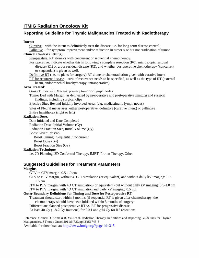
ITMIG Radiation Oncology Kit
Reporting Guideline for Thymic Malignancies Treated with Radiotherapy
Intent:
Curative – with the intent to definitively treat the disease, i.e. for long-term disease control
Palliative – for symptom improvement and/or reduction in tumor size but not eradication of tumor
Clinical Context (Setting):
Preoperative, RT alone or with concurrent or sequential chemotherapy.
Postoperative, indicate whether this is following a complete resection (R0), microscopic residual
disease (R1) or gross residual disease (R2), and whether postoperative chemotherapy (concurrent
or sequential) is given as well.
Definitive RT (i.e. no plans for surgery) RT alone or chemoradiation given with curative intent
RT for recurrent disease – area of recurrence needs to be specified, as well as the type of RT (external
beam, endobronchial brachytherapy, intraoperative)
Area Treated:
Gross Tumor with Margin: primary tumor or lymph nodes
Tumor Bed with Margin: as delineated by preoperative and postoperative imaging and surgical
findings, including surgical clips
Elective Sites Beyond Initially Involved Area: (e.g. mediastinum, lymph nodes)
Sites of Pleural metastases: either postoperative, definitive (curative intent) or palliative
Entire hemithorax (right or left)
Radiation Dose:
Date Initiated and Date Completed
Radiation Dose, Initial Volume (Gy)
Radiation Fraction Size, Initial Volume (Gy)
Boost Given: yes/no
Boost Timing: Sequential/Concurrent
Boost Dose (Gy)
Boost Fraction Size (Gy)
Radiation Technique:
i.e. 2D Planning. 3D Conformal Therapy, IMRT, Proton Therapy, Other
Suggested Guidelines for Treatment Parameters Margins
GTV to CTV margin: 0.5-1.0 cm
CTV to PTV margin, without 4D CT simulation (or equivalent) and without daily kV imaging: 1.0-
1.5 cm
ITV to PTV margin, with 4D CT simulation (or equivalent) but without daily kV imaging: 0.5-1.0 cm
ITV to PTV margin, with 4D CT simulation and daily kV imaging: 0.5 cm
Outer Boundary Definitions for Timing and Dose for Postoperative RT
Treatment should start within 3 months (if sequential RT is given after chemotherapy, the
chemotherapy should have been initiated within 3 months of surgery
Differentiate planned postoperative RT vs. RT for progressive disease
At least 40 Gy (1.8-2 Gy fractions) for R0,1 and ≥54 Gy for R2 resections
Reference: Gomez D, Komaki R, Yu J et al. Radiation Therapy Definitions and Reporting Guidelines for Thymic
Malignancies. J Thorac Oncol 2011;6(7,Suppl 3):S1743-8
Available for download at: http://www.itmig.org/?page_id=315

Definition of Recurrences Tumor regrowth after palliative RT should be classified as progression of disease
Distant recurrence – Outside the thorax or Intraparenchymal pulmonary nodules
Regional recurrence – intrathoracic, but not contiguous with original tumor or thymus (this includes
pleural or pericardial nodules)
Local Recurrence – at site of original tumor (including curatively treated pleural implants), or in thymic
bed including adjacent nodes. This should be further classified according to the RT treatment field:
Out of field recurrence – Outside the RT field; i.e. center lies outside of the 50% isodose field
Marginal Miss – geographic center of the recurrence lies in a region receiving 50-100% of the
prescription dose
Toxicity Definition Define according to CTCAE v 4.02 (Common Toxicity Criteria for Adverse Events, available at
www.acrin.org/Portals/0/Administration/Regulatory/CTCAE_4.02_2009-09-
15_QuickReference... · PDF file)
Should include at least grade 3-5, and categories of Esophagus, Respiratory, Cardiac and Other
Include maximal toxicity, toxicity duration, and whether it represents a dose-limiting toxicity
Proposed Dosimetric Constraints for Treatment of Thymic Malignancies5
RT Alone Chemo and RT
Chemo and RT Before
Surgery
Spinal cord1
Dmax <45 Gy Dmax <45 Gy Dmax <45 Gy
Lung2
MLD ≤20 Gy
V20 ≤ 40%
MLD ≤ 20 Gy
V20 ≤ 35%
V10 ≤ 45%
V5 ≤ 65%
MLD ≤ 20 Gy
V20 ≤ 30%
V10 ≤ 40%
V5 ≤ 55%
Heart V30 ≤45%
Mean dose <26 Gy
V30 ≤ 45%
Mean dose <26 Gy
V30 ≤45%
Mean dose <26 Gy
Esophagus Dmax ≤ 80 Gy
V70 < 20%
V50 < 50%
Mean dose<34 Gy
Dmax ≤ 80 Gy
V70 < 20%
V50 < 40%
Mean dose<34 Gy
Dmax ≤ 80 Gy
V70 < 20%
V50 < 40%
Mean dose<34 Gy
Kidney3
20 Gy < 32% of bilateral
kidney
20 Gy < 32% of bilateral
kidney
20 Gy < 32% of bilateral
kidney
Liver
V30 ≤40%
Mean dose <30 Gy
V30 ≤40%
Mean dose <30 Gy
V30 ≤40%
Mean dose <30 Gy
RT, radiotherapy; chemo, chemotherapy; MLD, mean lung dose; Dmax = maximal dose 1 The size of the treated volume of the spinal cord should be considered; when PTV is close (<1 cm) to spinal cord, the cord may
receive a dose higher than the recommended threshold in order to maintain adequate dose to the GTV target volume, but should
be <60 Gy, even in a very limited volume, and ~ 40 Gy if large fractions (i.e. 3-Gy) are used. 2 V20 = the effective lung volume (total lung volume – gross tumor volume) receiving 20 Gy or more. For patients who undergo
pneumonectomy before RT, we recommend an MLD of < 8 Gy, a V20 of < 10% and V5 <60%. Note that in the setting of
postoperative treatment in which a gross total resection has been achieved, there is no GTV, so the lung constraint will be
representative of solely the total lung, not the total lung minus the CTV. 3 Consider a kidney scan if a large volume of one kidney will be treated with a high dose
Reference: Marks LB, Yorke ED, Jackson A, et al: Use of normal tissue complication probability models in the
clinic. Int J Radiat Oncol Biol Phys 2010;76:S10-9

ITMIG Medical Oncologist Kit
Reporting Guidelines for Thymic Malignancies Treated with Chemotherapy
CHEMOTHERAPY STRATEGIES
Initial treatment
Curative-intent Primary
chemotherapy
Chemotherapy prior to another focal treatment – surgery or RT
Intent of the treatment should be documented, i.e. primary
chemotherapy prior to surgery or prior to RT
Final strategy has to be indicated: primary preoperative
chemotherapy or primary chemo-radiotherapy.
Post-operative
chemotherapy
Chemotherapy delivered following surgery.
Completeness of resection (R0, R1 or R2) should be noted.
Palliative-intent Palliative
chemotherapy
Chemotherapy alone in cases for which there is no plan for surgery
or radiotherapy.
Chemotherapy for recurrence Chemotherapy delivered for tumor recurrence appearing after
previous curative-intent treatment.
Chemotherapy for recurrence may be curative-intent (primary
preoperative/chemoradiation, post-operative) or palliative-
intent (chemotherapy alone).
Intent of the treatment and final strategy have to be documented as
for initial treatment.
CHEMOTHERAPY GENERAL REPORTING GUIDELINES
Modalities Chemotherapy regimen
Number of cycles administered
Dose intensity: > or < 70% of the planned dose-intensity
Analysis Treatment outcome evaluated separately for thymoma and thymic carcinoma.
Toxicities Grade 3-5 and dose limiting toxicities should be reported using the NCI-Common
Toxicity Criteria Adverse Event (CTCAE) v4.02.
Report both acute and late toxicities (especially late events such as cardiac toxicities).
Response Assessment of tumor response as described in “Standard Outcome Measures for
Thymic Malignancies” paper.
Whether the tumor contains a substantial lymphocyte component should be noted
Octreoscan results should be reported for patients treated with octreotide.
Report effect of antitumor treatment on associated paraneoplastic manifestations
Report Corticosteroid treatment doses (equivalent to prednisone doses above
0.5mg/kg/day) and durations.
Follow-Up After R0 surgical resection – annual CT (with contrast) for 5 years, then annual CXR
alternating with CT for 5 years is suggested as a minimum
After curative intent treatment for stage III, IVa – CT every 6 months for 3 years, then
schedule noted above
Recurrence Tumor regrowth should be classified as progression if treatment was palliative.
Recurrence should denote regrowth after complete resection or radiographic
complete response to curative intent therapy.
Time of recurrence should be defined as when clinical suspicion of recurrence first
occurred, regardless of whether a biopsy was done (unless the finding is
subsequently demonstrated not to be a recurrence).
Rebound hyperplasia must be considered when tumor re-growth occurs within 15
months following treatment cessation.
Local Recurrence – at site of original tumor, or in thymic bed including adjacent nodes
Regional recurrence – intrathoracic, but not contiguous with original tumor or thymus
(this includes pleural or pericardial nodules)
Distant recurrence – outside of the thorax, or Intraparenchymal nodules

Schematic Diagram of Treatment Strategies Involving Chemotherapy in Thymic Malignancies
Reference: Girard N, Lal R, Wakelee H et al. Chemotherapy Definitions and Policies for Thymic Malignancies.. J Thorac Oncol 2011;6(7,Suppl 3):S1749-55
Available for download at: http://www.itmig.org/?page_id=315