structure and axon outgrowth inhibitor binding of the …wabarton/pdf/barton_et_al_embo.pdfomgp...
TRANSCRIPT

William A.Barton, Betty P.Liu1,Dorothea Tzvetkova, Philip D.Jeffrey,Alyson E.Fournier1, Dinah Sah2,Richard Cate2, Stephen M.Strittmatter1,3
and Dimitar B.Nikolov3
Cellular Biochemistry and Biophysics Program, MemorialSloan-Kettering Cancer Center, 1275 York Avenue, New York,NY 10021, 1Department of Neurology, Yale University School ofMedicine, 333 Cedar Street, New Haven, CT 06520 and2Biogen, Inc., 14 Cambridge Center, Cambridge, MA 02142, USA
3Corresponding authorse-mail: [email protected] [email protected]
W.A.Barton and B.P.Liu contributed equally to this work
The myelin-derived proteins Nogo, MAG and OMgplimit axonal regeneration after injury of the spinalcord and brain. These cell-surface proteins signalthrough multi-subunit neuronal receptors thatcontain a common ligand-binding glycosylphospha-tidylinositol-anchored subunit termed the Nogo-66receptor (NgR). By deletion analysis, we show that thebinding of soluble fragments of Nogo, MAG and NgRto cell-surface NgR requires the entire leucine-richrepeat (LRR) region of NgR, but not other portions ofthe protein. Despite sharing extensive sequence simi-larity with NgR, two related proteins, NgR2 andNgR3, which we have identi®ed, do not bind Nogo,MAG, OMgp or NgR. To investigate NgR speci®cityand multi-ligand binding, we determined the crystalstructure of the biologically active ligand-bindingsoluble ectodomain of NgR. The molecule is bananashaped with elongation and curvature arising fromeight LRRs ¯anked by an N-terminal cap and a smallC-terminal subdomain. The NgR structure analysis, aswell as a comparison of NgR surface residues not con-served in NgR2 and NgR3, identi®es potential proteininteraction sites important in the assembly of a func-tional signaling complex.Keywords: axon outgrowth/leucine-rich repeats/ligand binding/Nogo-66 receptor
Introduction
Many spinal cord and brain injuries damage axons withoutcausing prominent neuronal loss. The inability of axons tore-extend in the injured CNS produces profound andpersistent functional de®cits in numerous clinical cases.Nevertheless, the axons of the adult CNS are capable ofregeneration when provided with a suitable environment,such as a growth-permissive peripheral nerve grafts(Richardson et al., 1980; David and Aguayo, 1981).Multiple factors contribute to the observed lack of
spontaneous regeneration. Both astroglial scar tissue(Snow et al., 1990; Dou and Levine, 1994; Davies et al.,1997) and CNS myelin (Schwab and Caroni, 1988; Savioand Schwab, 1989; Bandtlow et al., 1990) have recognizedroles in blocking CNS axon growth.
Protein fractionation studies identi®ed three cell-surfacemolecules in CNS myelin with axon outgrowth inhibitoryactivity, Nogo-A (GrandPre et al., 2000; Huber andSchwab, 2000; Prinjha et al., 2000), MAG (McKerracheret al., 1994; Mukhopadhyay et al., 1994), and OMgp(Wang et al., 2002a). Each of these proteins has beenshown to directly collapse axonal growth cones and toinhibit axonal extension. Nogo-A is the longest isoform ofNogo, a large transmembrane protein member of thereticulon family. The molecule is divided into threeregions separated by two hydrophobic segments (McGeeand Strittmatter, 2003). Its large N-terminal domain(N-Nogo) resides in the cytoplasmic space, as does itssmall C-terminal domain (C-Nogo), whereas a shortstretch of 66 amino acids (aa), termed Nogo-66, formsan extracellular loop detectable on the oligodendrocytesurface (GrandPre et al., 2000; McGee and Strittmatter,2003). Independently, this Nogo-66 domain and theN-Nogo domain can inhibit axons in vitro (Fournieret al., 2001, 2003). Several lines of evidence demonstratethe relevance of Nogo to axon regeneration in vivo.Antibodies directed against Nogo-A can promote axongrowth and plasticity in the adult brain (Schnell andSchwab, 1990; Thallmair et al., 1998; Raineteau et al.,1999). A Nogo-66 peptide antagonist increases axonalsprouting and functional recovery after spinal cord injury(GrandPre et al., 2002; Li and Strittmatter, 2003).Expression of Nogo in peripheral myelinating Schwanncells slows the otherwise robust pace of axon regeneration(Pot et al., 2002; Kim et al., 2003a). Some strains of micelacking Nogo-A exhibit CNS regenerative axonal growthafter spinal cord section (Kim et al., 2003b).
MAG is a member of the immunglobulin superfamilyexpressed in myelinating cells (McKerracher et al., 1994;Mukhopadhyay et al., 1994). The protein inhibits in vitroaxonal growth in a fashion similar to Nogo-A, but does notshare sequence similarity to Nogo-A. Mice lacking MAGexhibit enhanced axonal regeneration under specializedcircumstances (Bartsch et al., 1995; Schafer et al., 1996).OMgp is the third myelin-derived axon outgrowthinhibitor (Wang et al., 2002a). It is a glycosylphos-phatidylinositol (GPI)-anchored leucine-rich repeat (LRR)protein without detectable sequence similarity to Nogo-Aor MAG. Its role has not been studied in vivo.
We identi®ed Nogo-66 receptor (NgR) as a high-af®nityNogo-66 binding site required for inhibition of axonextension (Fournier et al., 2001). Surprisingly, thisreceptor has also been shown to mediate the inhibitoryactivities of two other myelin-derived proteins, MAG and
Structure and axon outgrowth inhibitor binding ofthe Nogo-66 receptor and related proteins
The EMBO Journal Vol. 22 No. 13 pp. 3291±3302, 2003
ã European Molecular Biology Organization 3291

OMgp (Domeniconi et al., 2002; Liu et al., 2002; Wanget al., 2002a). This is particularly striking, since there is nosequence similarity between these NgR ligands. NgR is aGPI-anchored molecule that belongs to the family of LRRproteins (Fournier et al., 2001; McGee and Strittmatter,2003). It contains eight central LRRs ¯anked by acysteine-rich C-terminal subdomain (LRRCT) and by asmaller leucine-rich N-terminal subdomain (LRRNT).This 311 aa NgR region is necessary and suf®cient forNogo-66 binding (Fournier et al., 2002). The mostC-terminal 100 NgR residues are most likely unstructuredon their own, but may participate in the interactions of
Nogo with its co-receptors (Fournier et al., 2002; Wanget al., 2002b). NgR is concentrated in lipid rafts, andsoluble recombinant NgR has af®nity for cell-surface NgR(Fournier et al., 2002; Liu et al., 2002), but the exactvalence of the protein in vivo is not clear. Since NgR lacksa cytoplasmic domain, it was predicted early on that itsignals through the action of co-receptors (Fournier et al.,2001), one of which was recently identi®ed as theneurotrophin receptor p75NTR (Wang et al., 2002b; Wonget al., 2002). p75NTR was ®rst described as an NGF-bindingprotein and was subsequently shown to interact withmultiple ligands and to modulate the activity of several
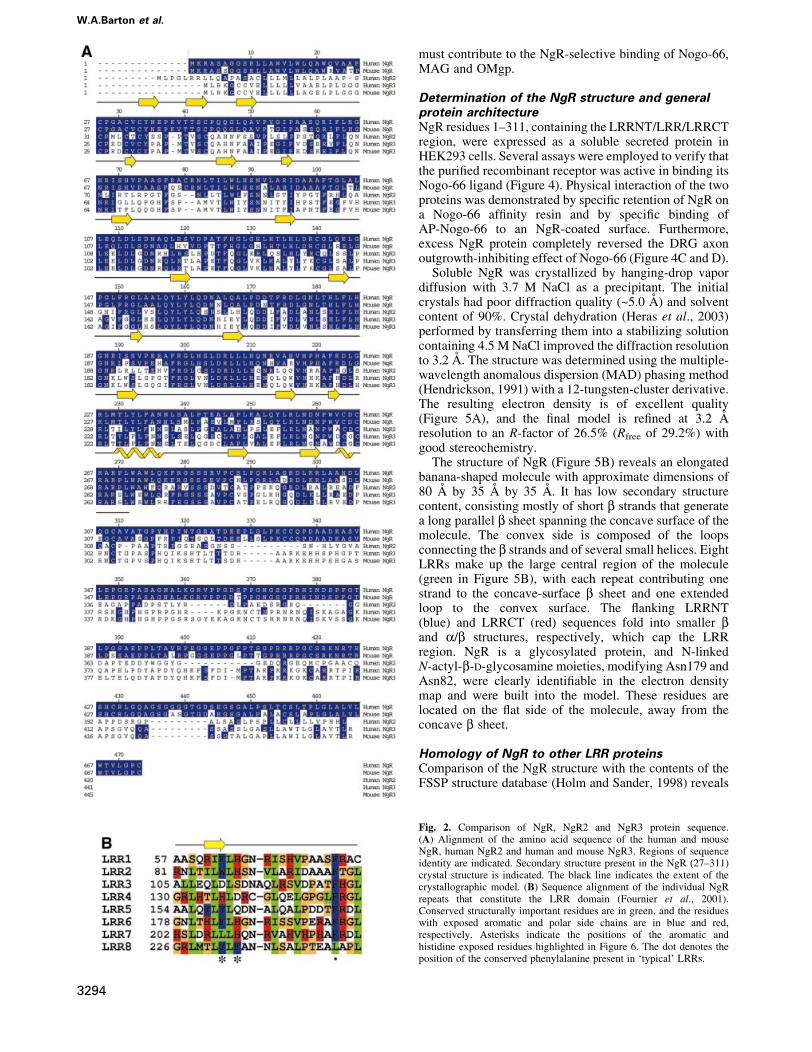
Fig. 1. MAG-binding region in NgR. COS-7 cells were transfected with wild-type (WT) NgR or NgR deletion mutant plasmids and tested forAP-MAG binding. WT NgR and LRRNT/LRR/LRRCT-expressing COS-7 cells bind AP-MAG, whereas other NgR deletion mutants do not. Table Isummarizes the ligand-binding attributes of the different NgR mutants from the present work and from Fournier et al. (2002). Deletions were asfollows: NgR-DNT, residues 27±57; NgR-D1±2, residues 58±105; NgR-D3±4, residues 106±154; NgR-D5±6, residues 155±202; NgR-D7±8,residues 203±250; NgR-DCT, residues 260±310; NgR-DLRR, residues 27±310; and NT/LRR/CT, residues 311±445.
W.A.Barton et al.
3292

Trk family tyrosine kinases (Dechant and Barde, 2002;Roux and Barker, 2002). It associates with NgR both aloneand in complex with some of the NgR ligands (Wang et al.,2002b; Wong et al., 2002). This association depends onboth the LRR domains and more carboxyl regions of theNgR (Wang et al., 2002b).
Since NgR mediates the signaling of all characterizedmyelin-derived inhibitory factors, an understanding of themolecular details of its receptor/ligand and receptor/co-receptor interactions is an essential step on the roadto developing clinically effective therapeutics to promoterecovery after adult CNS injury. Therefore, we identi®edand examined two proteins closely related to NgR formyelin inhibitor-binding activity. We also determined thecrystal structure of the N-terminal region of NgRcomprising the LRRCT, LRR and LRRNT subdomainsof the protein. A comparison of structural features andnon-conserved residues in the closely related but inactiveNgR-like proteins provides insight into the basis of NgRmolecular interactions.
Results and discussion
Identi®cation of the MAG-binding domain in NgRA series of NgR deletion mutants was previously gener-ated and screened to identify regions required for Nogo-66and NgR binding (Fournier et al., 2002). These deletionsremove speci®c modular domains in toto. Such studiesdemonstrated that deletion of the LRRNT subdomain, anytwo LRR subdomains or the LRRCT subdomain abrogatesbinding activity. To investigate the relationship betweenthe MAG, Nogo-66 and NgR (NgR self-association)binding sites, we examined the ability of soluble AP-MAG ligand to bind to each of the deletion mutants of
NgR. Previous results indicate that MAG interacts with theNgR LRR region (Liu et al., 2002), but the interactionswith the individual subdomains had not been studied. Thedata presented in Figure 1 shows that MAG binding is lostby deletion of the same regions as for Nogo-66 or NgR.Thus, the LRRNT/LRR/LRRCT domain as a whole isrequired for binding Nogo-66, MAG or soluble NgR.Despite this detailed similarity, the Nogo-66 and MAGbinding sites are probably not identical, since NEP1-40blocks Nogo-66 but not MAG activity (Liu et al., 2002).The localization of distinct binding sites within the sameLRRNT/LRR/LRRCT domain emphasizes the need forfurther and more detailed analysis to better understand themolecular basis of NgR function.
Identi®cation of NgR2 and NgR3Since there are numerous ligands for NgR, we consideredthe possibility that NgR might be part of a family ofproteins. Initial scans of Genbank cDNA and completedgenomic sequences at the time of NgR identi®cation didnot reveal any sequences predicted to encode proteins withgreater than ~35% aa identity within the LRR region. Thismoderate degree of similarity is unlikely to re¯ectfunctional homology, since all recognized LRR domains(>200 proteins) share 25±35% aa identity. Therefore, wescanned un®nished genomic sequence databases and foundtwo possible NgR-related sequences (Figure 2). Parts ofthese sequences are found in cDNA databases, consistentwith expression of the proteins in vivo. One humansequence predicts a protein (NgR-related protein-2, NgR2)with 55% LRR identity with the NgR, and a mousesequence predicts another protein (NgR-related protein-3,NgR3) with 55% LRR identity to both NgR and humanNgR2. Later genomic searches with mouse NgR3 allowedidenti®cation of the human NgR3 sequence. NgR2 andNgR3 sequences have identical overall architectures asNgR. They all encode a signal sequence, followed by anLRRNT, eight LRRs, an LRRCT, an ~100 aa unique linkerregion and a predicted GPI anchorage site. We obtainedfull-length cDNA for both human NgR2 and human andmouse NgR3 (Figure 2A). These two NgR-related proteinswere recently identi®ed independently by another group(Pignot et al., 2003), who has named them NgRH1 andNgRH2 (see Note added in proof).
NgR2 and NgR3 do not bind known NgR ligandsTo determine whether the sequence and the expectedstructural homology between NgR, NgR2 and NgR3extends to their ligand-binding speci®cities, each ofthese proteins was expressed in COS-7 cells and examinedfor binding to known NgR ligands (Figure 3). Cellstransfected with His-NgR2 or Myc-NgR3 clearly expressthe epitope-tagged protein on their surface, but do not bindAP-Nogo-66, AP-MAG or AP-OMgp under conditionsthat produce strong binding to NgR. The absence ofAP-NgR binding to NgR2 and NgR3 indicates that theyare unlikely to exist in a complex with NgR that wouldallow for indirect participation in myelin inhibitor signal-ing. Although these three proteins form a sequence-relatedfamily, their functions appear to have diverged duringevolution. We therefore conclude that a proportion of theresidues that differ between NgR and NgR2 plus NgR3
Table I. Summary of crystallographic data
Crystal Peak In¯ection Remote
Resolution (AÊ ) 3.2 3.2 3.2Wavelength (AÊ ) 1.210 1.212 1.186Anom. completeness (%) 99.6
(99.0)99.7(99.6)
99.9(100)
Redundancy (fold) 4.4 4.4 4.2I/sI 12 12 12Rmerge 7.1 6.2 5.7F.O.M. (pre/post-dm) 0.38/0.62Space group P3121Cell dimensions (AÊ ) a = b = 123.96 c =120.17
Re®nement
Resolution (AÊ ) 8.0±3.2Re¯ections (working/test) 29 483/3133Non-hydrogen atoms 2136Rcrys/Rfree 26.5/29.2R.m.s. deviations
Bonds (AÊ ) 0.017Angle (degrees) 2.27
Rmerge = S|I ± <I>|/SI, where I is the observed intensity and <I> is theaverage intensity obtained from multiple observations of symmetryrelated re¯ections.R.m.s. deviations in bond lengths and angles are the respective root-mean-square deviations from ideal values. r.m.s. thermal parameter isthe r.m.s. deviation between the B values of covalently bound atomicpairs.
Nogo-66 receptor and related proteins
3293
must contribute to the NgR-selective binding of Nogo-66,MAG and OMgp.
Determination of the NgR structure and generalprotein architectureNgR residues 1±311, containing the LRRNT/LRR/LRRCTregion, were expressed as a soluble secreted protein inHEK293 cells. Several assays were employed to verify thatthe puri®ed recombinant receptor was active in binding itsNogo-66 ligand (Figure 4). Physical interaction of the twoproteins was demonstrated by speci®c retention of NgR ona Nogo-66 af®nity resin and by speci®c binding ofAP-Nogo-66 to an NgR-coated surface. Furthermore,excess NgR protein completely reversed the DRG axonoutgrowth-inhibiting effect of Nogo-66 (Figure 4C and D).
Soluble NgR was crystallized by hanging-drop vapordiffusion with 3.7 M NaCl as a precipitant. The initialcrystals had poor diffraction quality (~5.0 AÊ ) and solventcontent of 90%. Crystal dehydration (Heras et al., 2003)performed by transferring them into a stabilizing solutioncontaining 4.5 M NaCl improved the diffraction resolutionto 3.2 AÊ . The structure was determined using the multiple-wavelength anomalous dispersion (MAD) phasing method(Hendrickson, 1991) with a 12-tungsten-cluster derivative.The resulting electron density is of excellent quality(Figure 5A), and the ®nal model is re®ned at 3.2 AÊ
resolution to an R-factor of 26.5% (Rfree of 29.2%) withgood stereochemistry.
The structure of NgR (Figure 5B) reveals an elongatedbanana-shaped molecule with approximate dimensions of80 AÊ by 35 AÊ by 35 AÊ . It has low secondary structurecontent, consisting mostly of short b strands that generatea long parallel b sheet spanning the concave surface of themolecule. The convex side is composed of the loopsconnecting the b strands and of several small helices. EightLRRs make up the large central region of the molecule(green in Figure 5B), with each repeat contributing onestrand to the concave-surface b sheet and one extendedloop to the convex surface. The ¯anking LRRNT(blue) and LRRCT (red) sequences fold into smaller band a/b structures, respectively, which cap the LRRregion. NgR is a glycosylated protein, and N-linkedN-actyl-b-D-glycosamine moieties, modifying Asn179 andAsn82, were clearly identi®able in the electron densitymap and were built into the model. These residues arelocated on the ¯at side of the molecule, away from theconcave b sheet.
Homology of NgR to other LRR proteinsComparison of the NgR structure with the contents of theFSSP structure database (Holm and Sander, 1998) reveals
Fig. 2. Comparison of NgR, NgR2 and NgR3 protein sequence.(A) Alignment of the amino acid sequence of the human and mouseNgR, human NgR2 and human and mouse NgR3. Regions of sequenceidentity are indicated. Secondary structure present in the NgR (27±311)crystal structure is indicated. The black line indicates the extent of thecrystallographic model. (B) Sequence alignment of the individual NgRrepeats that constitute the LRR domain (Fournier et al., 2001).Conserved structurally important residues are in green, and the residueswith exposed aromatic and polar side chains are in blue and red,respectively. Asterisks indicate the positions of the aromatic andhistidine exposed residues highlighted in Figure 6. The dot denotes theposition of the conserved phenylalanine present in `typical' LRRs.
W.A.Barton et al.
3294

that the overall fold resembles that of other LRR-containing proteins. The closest structural homologs ofNgR are the internalin B protein (InlB) of Listeriamonocytogenes (Marino et al., 1999), the platelet glyco-protein Iba (Huizinga et al., 2002) and the humanribonuclease inhibitor (RI) (Kobe and Deisenhofer, 1993;Kobe and Deisenhofer, 1995; Papageorgiou et al., 1997).The LRR domains of these proteins can be superimposedon the NgR LRR domain with root-mean-square deviationsbetween a-carbon positions of 2.5 AÊ for InlB (166 atoms),2.6 AÊ for platelet glycoprotein Iba (234 atoms) and 4.9 AÊ
for RI (216 atoms). However, only the model for theplatelet glycoprotein Iba contains LRRNT and LRRCTsubdomains homologous to those in NgR.
The NgR b strands, although relatively short, are withinthe usual LRR range (Kobe and Kajava, 2001), with threeresidues in each strand engaging in characteristicbackbone±backbone hydrogen bonding interactions,which are the hallmark of b-sheet structure. Figure 2Bshows the alignment of the individual NgR LRR repeats.Certain positions of the LRRs in NgR are occupied byresidues, which are highly conserved between repeats,whereas others can accommodate a wide variety of aminoacids. Positions 3, 6, 8, 13, 16, 21 and 24 contain structuralleucine, isoleucine, valine and phenylalanine residues, andthe van der Waals interactions between them appear to bethe main factor that stabilizes the overall fold of themolecule.
Fig. 3. NgR2 and NgR3 do not bind NgR ligands. COS-7 cells were transfected with vectors for Myc-NgR, His-NgR2, Myc-NgR3 or None and thenstained with AP-tagged soluble ligands as indicated (upper half). The AP-ligand concentration was 10 nM AP-Nogo-66, 20 nM AP-MAG, 20 nMAP-NgR and 20 nM AP-OMgp. Note the expression of NgR2 and NgR3 without detectable Nogo-66, MAG, NgR or OMgp binding. In the lower half,the expression of recombinant epitope-tagged NgR, NgR2 and NgR3 proteins is detected by immunostaining with the indicated anti-epitope-tag anti-bodies, anti-Myc or anti-His. None of the proteins binds 20 nM AP protein (bottom row).
Nogo-66 receptor and related proteins
3295

The NgR LRRsNgR was recognized as an LRR family protein based onthe fact that it contains the LRR-signature sequence(LxxLxLN/CxL), which corresponds to the segmentsurrounding the b strand (Figure 2B). LRRs vary in theirlength and pattern of conserved residues and are groupedinto seven different subfamilies (Kobe and Kajava, 2001).The LRRs in NgR are either 24 or 25 aa long, which ismidway between the long repeats (28±29 residues) of RIand the short (20 residues) repeats of the Yersinia outer
protein YopM (Evdokimov et al., 2001). As such, the NgRrepeats are classi®ed as `typical' (Kobe and Kajava, 2001).Each NgR LRR is composed of a single b strand followedby an extended loop. In some of the repeats, there is asmall (one turn) a helix preceding the b strands. MostLRRs in other proteins are composed of a b-loop±helix±loop structure such as that observed in RI or InlB. NgR, onthe other hand, does not adopt a regular helical structure onthe convex side of the molecule. Instead, in NgR, thisregion contains a proline in seven of the eight LRRs
Fig. 4. Biological activity of the puri®ed recombinant NgR protein. (A) Control AP protein or AP-Nogo-66 were bound to resin and then incubatedwith NgR. After washing, bound protein was examined by NgR immunoblot. The two bands on the gel correspond to differently glycosylated forms ofNgR. (B) Microtiter wells were coated with the indicated proteins and then probed with AP-Nogo-66 or AP protein in the presence or absenceof excess soluble NgR. (C) Rat P4-6 DRG neurons were plated on surfaces coated with or without GST±Nogo-66 and with or without the addition ofexcess NgR as indicated. Rhodamine-phalloidin staining is illustrated. (D) Neurite outgrowth from an experiment as in (C) is reported as a percentageof the value without GST±Nogo-66. Data are means 6 SEM for three or more measurements. *Values with NgR are signi®cantly different from thosewithout NgR (p < 0.02, Student's t-test). Dissociated rat P4-6 DRG neurons were plated on surfaces coated with 90 ng of control GST or 90 ng ofGST±Nogo-66 with the addition of 450 ng of NgR protein or 450 ng of control GST protein. After 6 h, cells were ®xed and scored for neuriteoutgrowth per neuron as described previously (Fournier et al., 2002; Liu et al., 2002).
W.A.Barton et al.
3296

(Figure 2B) and thus adopts an extended structure. In thisrespect, the LRRs of NgR most closely resemble thoseobserved in YopM.
The LRRCT and LRRNT subdomainsIn a regular LRR structure, the hydrophobic core would beexposed to solvent at the ends. Therefore, NgR, like manyother LRR proteins, contain ¯anking regions or `caps'.Indeed, the NgR LRRNT and LRRCT subdomainsrepresent more of an extension of the overall LRR regionthen separate domains (Figure 5A). The N-terminalNgR subdomain (residues 27±61) is composed of threeshort b strands forming a hydrophilic cap on theburied leucines and isoleucines of the ®rst LRR. TheNgR LRRNT forms a compact structure and, althoughis involved in ligand binding (Figure 1), does not extenda ligand-interacting b ®nger, such as the one observedin the glycoprotein Iba structure (Huizinga et al.,2002). Instead, the corresponding loop is smallerand packs against the protein surface. The C-terminal
NgR subdomain (residues 260±311) is composed of twohelices and ®ve b strands. Its hydrophobic core iscontinuous with the hydrophobic core of the LRRsubdomain, and LRRCT residues form hydrogen bondsto LRR residues. The only other reported LRR structurecontaining an LRRCT subdomain is the structure of theglycoprotein Iba. The NgR LRRCT subdomain is slightlylarger than its Iba counterpart but lacks the protrudingligand-binding loop observed in Iba (Huizinga et al.,2002).
The LRRCT subdomain contains four cysteines formingtwo disul®de bonds (Cys264±Cys287 and Cys266±Cys309), which stabilize the NgR structure. This disul®debond arrangement is the one most commonly observed inthe sequences of LRR C-¯anking regions and is referred toas a CF1-type domain (Kobe and Kajava, 2001). TheLRRNT subdomain also contains four cysteines in twodisul®de bridges (Cys27±Cys33 and Cys31±Cys43.Interestingly, NgR contains two free cysteines at positions80 and 140. Cys80 is located next to an N-linked
Fig. 5. Structure of NgR. (A) Representative region of the density-modi®ed experimental electron density map contoured at 1.5 s. The re®ned NgRmodel is shown with carbons in yellow, nitrogens in blue and oxygens in red. (B and C) Orthogonal stereoviews of the NgR structure, the LRRNTsubdomain is in blue, the central LRR subdomain in green and the LRRCT subdomain in red. The protein N- and C-termini are indicated. The sidechains of the exposed aromatic and histidine residues lining the concave molecular surface are shown only in (C).
Nogo-66 receptor and related proteins
3297
glycosylation site (Asn82), which explains its lack ofreactivity, whereas Cys140 is not surface exposed.
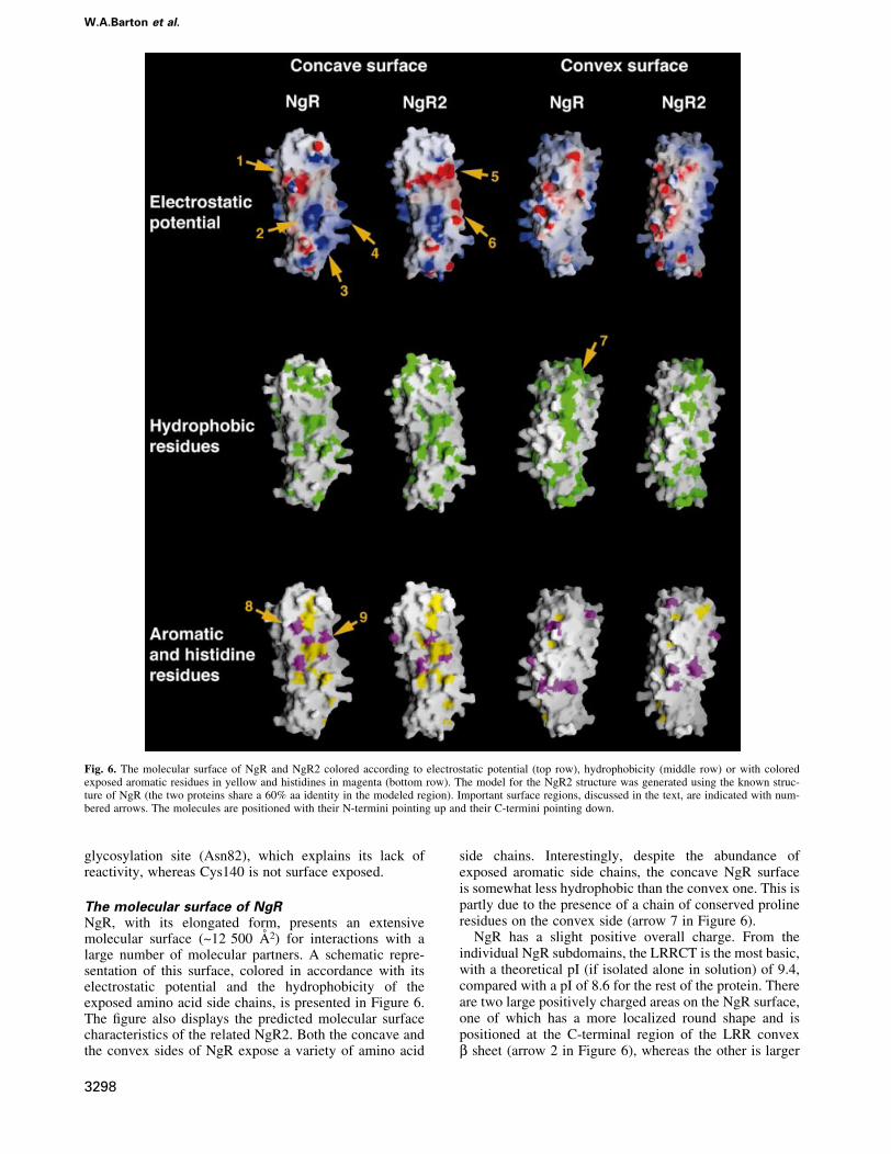
The molecular surface of NgRNgR, with its elongated form, presents an extensivemolecular surface (~12 500 AÊ 2) for interactions with alarge number of molecular partners. A schematic repre-sentation of this surface, colored in accordance with itselectrostatic potential and the hydrophobicity of theexposed amino acid side chains, is presented in Figure 6.The ®gure also displays the predicted molecular surfacecharacteristics of the related NgR2. Both the concave andthe convex sides of NgR expose a variety of amino acid
side chains. Interestingly, despite the abundance ofexposed aromatic side chains, the concave NgR surfaceis somewhat less hydrophobic than the convex one. This ispartly due to the presence of a chain of conserved prolineresidues on the convex side (arrow 7 in Figure 6).
NgR has a slight positive overall charge. From theindividual NgR subdomains, the LRRCT is the most basic,with a theoretical pI (if isolated alone in solution) of 9.4,compared with a pI of 8.6 for the rest of the protein. Thereare two large positively charged areas on the NgR surface,one of which has a more localized round shape and ispositioned at the C-terminal region of the LRR convexb sheet (arrow 2 in Figure 6), whereas the other is larger
Fig. 6. The molecular surface of NgR and NgR2 colored according to electrostatic potential (top row), hydrophobicity (middle row) or with coloredexposed aromatic residues in yellow and histidines in magenta (bottom row). The model for the NgR2 structure was generated using the known struc-ture of NgR (the two proteins share a 60% aa identity in the modeled region). Important surface regions, discussed in the text, are indicated with num-bered arrows. The molecules are positioned with their N-termini pointing up and their C-termini pointing down.
W.A.Barton et al.
3298
and more diffuse, comprising a signi®cant portion of theLRRCT subdomain and some of the loops of theC-terminal LRR repeats (arrows 3 and 4 in Figure 6). Inaddition, a large acidic region is located on the concaveside of the molecule at the N-terminal region of the centralLRR subdomain (arrow 1 in Figure 6).
Implications for the interaction of NgR withligands and co-receptorsThe major biological function of LRRs is to provide astructural framework for the formation of speci®c protein±protein interactions (Kobe and Kajava, 2001). Severalstructures of macromolecular complexes involving LRRfamily members have been reported. They share thecommon theme that the LRR-containing proteins utilizelarge areas of exposed molecular surface to recognize andbind other proteins with high af®nity. The bindinginterface usually resides within the LRR concave b sheet.In some cases, though, such as in the Iba/Willebrandfactor complex structure, most of the interactions involvethe LRRCT and LRRNT subdomains.
Our deletion analysis (Figure 1) documents that theNgR LRR, LRRCT and LRRNT subdomains are allinvolved in ligand binding. In order to gain insight into theprecise molecular regions within these subdomains, whichmight mediate speci®c interactions, we generated a modelof the structure of NgR2, a close sequence homolog ofNgR (Figure 2A), which nevertheless displays completelydistinct ligand preferences (Figure 3). Figure 6 illustratesthe molecular surface characteristics of NgR and NgR2.The surface properties of mouse NgR and of human andmouse NgR3 (Figure 2A) are very similar to those ofhuman NgR and NgR2, respectively, and are not presentedin Figure 6. The concave faces of both NgR and NgR2 are
dominated by aromatic residues such as tyrosines andphenylalanines and by histidines. This is a commonfeature of proteins belonging to the LRR family; and,although high-af®nity ligand interactions involving theseconserved residues are expected to provide the bulk of thebinding energy, their strict conservation implies that theycannot be responsible for de®ning the ligand speci®cityof NgR. However, Figure 6 shows that a number ofstrategically positioned charged residues are also availablefor interaction in this area. Several basic and acidicpatches, unique only to one of the two receptors, suggestpossible regions involved in ligand recognition andbinding. For example, as indicated in Figure 6, the NgR2surface region (arrow 5) is very acidic, whereas thecorresponding region in NgR is uncharged. In addition, thelarge basic NgR surface region, comprising part of theLRRCT subdomain and nearby LRR regions (arrows 3and 4), is mostly uncharged in NgR2 and even partiallyacidic (arrow 6). This NgR positively charged regionmight be responsible for interactions with acidic regions ofits ligands. Indeed, Nogo-66 has an overall negativecharge and, furthermore, contains two consecutive acidicresidues (Glu31 and Glu32), which are necessary for NgRbinding (GrandPre et al., 2002). Interestingly, in ourcrystal, the large acidic patch on the NgR concave face(arrow 1) is packing against the basic LRRCT subdomainof another NgR molecule. This interaction might corres-pond to lower-af®nity ligand-independent receptor/recep-tor association, which is observed experimentally at thecell surface (Fournier et al., 2002). Upon ligand approxi-mation, the electrostatic receptor/receptor interactionwould be disrupted in favor of the higher-af®nity vander Waals ligand/receptor contacts, leading to the re-orientation of receptor/co-receptor complexes and to the
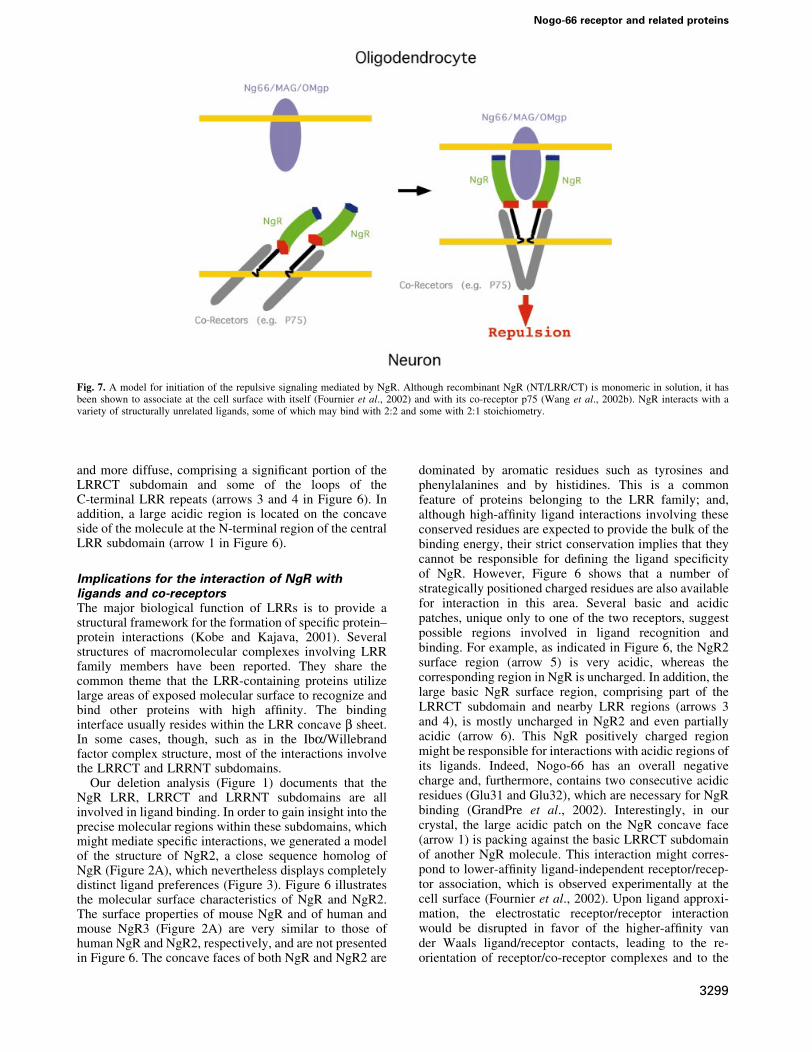
Fig. 7. A model for initiation of the repulsive signaling mediated by NgR. Although recombinant NgR (NT/LRR/CT) is monomeric in solution, it hasbeen shown to associate at the cell surface with itself (Fournier et al., 2002) and with its co-receptor p75 (Wang et al., 2002b). NgR interacts with avariety of structurally unrelated ligands, some of which may bind with 2:2 and some with 2:1 stoichiometry.
Nogo-66 receptor and related proteins
3299

transduction of the repulsive signal to the inside of the cell.Figure 7 represents such a model for the initiation of NgR-mediated signaling.
Another intriguing and unique feature of the NgRmolecular surface is the abundance of exposed histidineresidues (magenta in Figure 6). Other known LRR proteinshave a much smaller number of exposed histidines, andeven NgR2 has ®ve fewer than NgR. Comparison of thelocation of the exposed histidine residues in NgR andNgR2 (Figure 6, bottom row) identi®es a region at theinterface between the LRRNT and the LRR domains ofNgR (arrows 8 and 9) as another potentially importantligand-recognition surface. Finally, it should be noted thatNgR interacts with multiple and structurally distinctligands, and each of them is likely to utilize differentstructural features on the large NgR surface to recognizeand bind the receptor. Indeed, the three currently knownNgR ligands have very distinct molecular architectures:OMgp is a highly glycosylated LRR protein somewhatsimilar to NgR; MAG is composed of several immuno-globulin domains; and Nogo-A is a short (66 aa) domain ofunknown structure or, even more likely, unstructured in itsunbound form.
Conclusions and perspectivesThe NgR plays a central role in mediating myelin-dependent restriction of axon outgrowth by three distinctligands. We identi®ed two proteins with a high degree ofsimilarity to NgR. Despite high levels of amino acididentity with NgR in the ligand-binding LRRNT/LRR/LRRCT domain, these proteins do not bind MAG,Nogo-66, OMgp or NgR. Therefore, they do not appearto function in this pathway. The identity of the ligandsfor these proteins is yet to be determined. The lackof binding of the NgR ligands to NgR2 and NgR3emphasizes the unique nature of NgR, rendering thepossibility of redundant binding proteins for myelin-derived inhibitors much less probable. If there are anyadditional receptors for myelin-derived inhibitors of axonregeneration, they seem very unlikely to be structurallyrelated to NgR.
We report the structure of the ligand-binding LRRNT/LRR/LRRCT domain of NgR. It shares some similaritywith, but has numerous distinctions from, previouslydescribed LRR proteins. Amongst the unique features isthe presence of a proline strip on the concave face of themolecule, preventing the formation of secondary structurein this region. There are numerous large basic patches onthe surface of the NgR, especially near the LRRCT region.It is particularly tempting to speculate that the essential di-acidic motif in the region of Nogo-66 (Asp 31, Asp 32)most distant from the oligodendrocyte membrane mightinteract with this region. Comparison of NgR with closelyrelated proteins highlights those surface residues andregions most likely to participate in interaction withdifferent ligands. Such areas include two histidine residueson the convex face and a selected group of charged patchesnear the edges of the convex surface. The results presentedhere should lead to a greater understanding of themolecular basis for the inhibition of axon regenerationby CNS myelin.
Materials and methods
NgR2 and NgR3 expressionThe mouse NgR3 cDNA was ampli®ed by PCR from mouse adult braincDNA from the signal sequence to the stop codon. The ampli®ed productwas ligated into the pSecTag2 vector such that the vector encoded a signalsequence (from the vector), followed by a Myc tag and the mature NgR3sequence. The human NgR2 cDNA was derived from two human ESTclones, AW293195.1 and BE222737.1. A His6 tag was inserted betweenthe signal sequence and the mature polypeptide in a eukaryotic expressionvector. After transfection of DNA into COS-7 cells, the expression of His-NgR2 or Myc-NgR3 protein of the appropriate size was veri®ed byepitope-tag immunoblot. AP-Nogo-66, AP-MAG, AP-NgR and AP-OMgp binding studies and anti-epitope-tag immunohistology wereconducted as described previously (Fournier et al., 2001, 2002; Liuet al., 2002).
Protein expression and puri®cationNgR (aa 1±311) was cloned into the pcDNA5FRT vector and transfectedinto HEK293/FRT cells (Gibco). Hygromycin-resistant stable cell lineswere selected that express ~5±7 mg protein per liter of culture. Cells weregrown in large scale in roller bottles and supplemented with serum-freemedia. NgR was puri®ed from the media by cation exchangechromotography, followed by ammonium sulfate precipitation and gel®ltration chromotography. NgR migrates as a monomer on gel ®ltrationcolumns and is monomeric in solution as judged by analyticalultracentrifugation (data not shown). The two observed NgR bands onSDS±PAGE gels (Figure 4A) are the result of heterogeneousglycosylation and can be converted to a single band by enzymaticin vitro deglycosylation.
Crystallization and structure determinationPuri®ed NgR was concentrated to 10 mg/ml in a buffer containing250 mM NaCl and 10 mM HEPES (pH 7.2) and crystallized at roomtemperature against a reservoir containing 3.7 M NaCl and 100 mM MES(pH 6.5). The space group is P3121 with a = b = 123.96 AÊ , c = 120.17 AÊ ,and one molecule in the asymmetric unit. The ®nal solvent content, aftercrystal dehydration, was 85%. Heavy-atom soaks were performed in4.5 M NaCl at pH 6.5. All data were processed using DENZO andSCALEPACK (Otwinowski and Minor, 1997). The tungsten-cluster(W12) positions were identi®ed using the program TRAWL, a real-spacepatterson search program. A model of the W12 structure was input intoautoSHARP (C.Vonrhein, personal communication) and used to calculatea solvent ¯attened map of excellent quality.
Model building was initially performed using Arp/Warp (CCP4, 1994)and later proceeded through an iterative process of building in O andre®nement of the model in CNS (Jones et al., 1991; Brunger et al., 1998).Stereochemical analysis of the re®ned models using PROCHECK of theCCP4 package (CCP4, 1994) revealed main chain and side chainparameters better than or within the typical range of values for proteinstructures determined at corresponding resolutions. None of the NgRresidues fell in the disallowed region of the Ramachandran plot, whereas68% of the residues were in the most favored region. Molecular graphic®gures were created with MolScript (Kraulis, 1991), Raster3D (Merrittand Bacon, 1997) and GRASP (Nicholls et al., 1991).
AP-NgR binding assays and DRG axon outgrowth-inhibitionassaysFor af®nity chromatography, 20 mg of His6-tagged AP or AP-Nogo-66were immobilized on a Ni+2-containing resin (100 ml). The resin wasincubated with 100 mg of NgR in 200 ml of 50 mM NaHEPES, 200 mMNaCl, 0.1% BSA (pH 7.5). After washing, bound protein was analyzed byNgR immunoblot (Fournier et al., 2001). For AP-Nogo-66 bindingexperiments to puri®ed NgR protein, Nunc Maxisorp 96-well plates werecoated with or without 0.5 mg of NgR per well and then blocked withBSA. Solutions containing 100 nM AP-Nogo-66 or 100 nM AP in thepresence or absence of 20 mM soluble NgR were incubated in the wellsfor 1 h. After washing, bound AP was detected spectrophotometricallywith p-nitrophenol phosphate as substrate. To assess neurite outgrowth,dissociated rat P4-6 DRG neurons were plated on surfaces coated with90 ng of control GST or 90 ng of GST±Nogo-66 with the addition of450 ng of NgR protein or 450 ng of control GST protein. After 6 h, cellswere ®xed and scored for neurite outgrowth per neuron as describedpreviously (Fournier et al., 2002; Liu et al., 2002).
W.A.Barton et al.
3300

Coordinates and sequencesCoordinates and sequences have been deposited in the Protein Data Bankand GenBank (PDB accession number, 1P8T; DDBJ/EMBL/GenBankaccession Nos: BK001302 for human NgR2, BK001303 for humanNgR3, BK001304 for mouse NgR2 and BK001305 for mouse NgR3)
Acknowledgements
We thank Drs Seth Darst and Gabby Rudenko for supplying W-clustercompounds; and Dr Craig Ogata for assistance with crystallographic datacollection at APS, Chicago, IL (NE-CAT). We thank Drs Mi Sha andColleen Mullen of Biogen, Inc. (Cambridge, MA) for cDNA plasmidsencoding AP-OMgp and NgR2, respectively. This work was supported bygrants to D.B.N. from the New York State Spinal Cord Injury ResearchProgram and to S.M.S. from the NIH and the McKnight Foundation forNeuroscience. D.B.N. is a PEW fellow and a Bressler Scholar. S.M.S. isan Investigator of the Patrick and Catherine Weldon Donaghue MedicalResearch Foundation.
References
Bandtlow,C., Zachleder,T. and Schwab,M.E. (1990) Oligodendrocytesarrest neurite growth by contact inhibition. J. Neurosci., 10, 3837±3848.
Bartsch,U. et al. (1995) Lack of evidence that myelin-associatedglycoprotein is a major inhibitor of axonal regeneration in the CNS.Neuron, 15, 1375±1381.
Brunger,A.T. et al. (1998) Crystallography & NMR system: a newsoftware suite for macromolecular structure determination. ActaCrystallogr. D, 54, 905±921.
CCP4 (1994) The CCP4 suite: programs for X-ray crystallography. ActaCrystallogr. D, 50, 760±763.
David,S. and Aguayo,A.J. (1981) Axonal elongation into peripheralnervous system `bridges' after central nervous system injury in adultrats. Science, 214, 931±933.
Davies,S.J., Fitch,M.T., Memberg,S.P., Hall,A.K., Raisman,G. andSilver,J. (1997) Regeneration of adult axons in white matter tractsof the central nervous system. Nature, 390, 680±683.
Dechant,G. and Barde,Y.A. (2002) The neurotrophin receptor p75NTR:novel functions and implications for diseases of the nervous system.Nat. Neurosci., 5, 1131±1136.
Domeniconi,M. et al. (2002) Myelin-associated glycoprotein interactswith the Nogo66 receptor to inhibit neurite outgrowth. Neuron, 35,283±290.
Dou,C.L. and Levine,J.M. (1994) Inhibition of neurite growth by theNG2 chondroitin sulfate proteoglycan. J. Neurosci., 14, 7616±7628.
Evdokimov,A.G., Anderson,D.E., Routzahn,K.M. and Waugh,D.S.(2001) Unusual molecular architecture of thee Yersinia pestiscytotoxin M: a leucine rich repeat protein with the shortestrepeating unit. J. Mol. Biol., 312, 807±821.
Fournier,A.E., GrandPre,T. and Strittmatter,S.M. (2001) Identi®cation ofa receptor mediating Nogo-66 inhibition of axonal regeneration.Nature, 409, 341±346.
Fournier,A.E., Gould,G.C., Liu,B.P. and Strittmatter,S.M. (2002)Truncated soluble Nogo receptor binds Nogo-66 and blocksinhibition of axon growth by myelin. J. Neurosci., 22, 8876±8883.
Fournier,A.E., Takizawa,B.T. and Strittmatter,S.M. (2003) Rho kinaseinhibition enhances axonal regeneration in the injured CNS.J. Neurosci., 23, 1416±1423.
GrandPre,T., Nakamura,F., Vartanian,T. and Strittmatter,S.M. (2000)Identi®cation of the Nogo inhibitor of axon regeneration as aReticulon protein. Nature, 403, 439±444.
GrandPre,T., Li,S. and Strittmatter,S.M. (2002) Nogo-66 receptorantagonist peptide promotes axonal regeneration. Nature, 417, 547±551.
He,X.L., Bazan,J.F., McDermott,G., Park,J.B., Wang,K., Tessier-Lavigne,M., He,Z. and Garcia,K.C. (2003) Structure of the nogoreceptor ectodomain. A recognition module implicated in myelininhibition. Neuron, 38, 177±185.
Hendrickson,W.A. (1991) Determination of macromolecular structuresfrom anomalous diffraction of synchrotron radiation. Science, 254,51±58.
Heras,B., Edeling,M.A., Byriel,K.A., Jones,A., Raina,S. and Martin,J.L.(2003) Dehydration converts DsbG crystal diffraction from low tohigh resolution. Structure, 11, 139±145.
Holm,L. and Sander,C. (1998) Touring protein fold space with DALI/FSSP. Nucleic Acids Res., 26, 316±319.
Huber,A.B. and Schwab,M.E. (2000) Nogo-A, a potent inhibitor ofneurite outgrowth and regeneration. Biol. Chem., 381, 407±419.
Huizinga,E.G., Tsuji,S., Romijn,R.A.P., Schiphorst,M.E., de Groot,P.G.,Sixma,J.J. and Gros,P. (2002) Structures of the glycoprotein Iba andits complex with von Willebrand factor A1 domain. Science, 297,1176±1179.
Jones,T.A., Zou,J.Y., Cowan,S.W. and Kjeldgaard,M. (1991) Improvedmethods for building protein models in electron density maps and thelocation of errors in these models. Acta Crystallogr. A, 47, 110±119.
Kim,J.E., Bonilla,I.E., Qiu,D. and Strittmatter,S.M. (2003a) Nogo-C issuf®cient to delay nerve regeneration. Mol. Cell. Neurosci., in press.
Kim,J.E., Li,S., GrandPre,T., Qiu,D. and Strittmatter,S.M. (2003b) Axonregeneration in young adult mice lacking Nogo-A/B. Neuron, 38, 187±199.
Kobe,B. and Deisenhofer,J. (1993) Crystal structure of porcineribonuclease inhibitor; a protein with leucine rich repeats. Nature,366, 751±756.
Kobe,B. and Deisenhofer,J. (1995) A structural basis of the interactionsbetween leucine-rich repeats and protein ligands. Nature, 374, 183±186.
Kobe,B. and Kajava,A.V. (2001) The leucine rich repeat as a proteinrecognition motif. Curr. Opin. Struct. Biol., 11, 725±732.
Kraulis,P.J. (1991) Molscript: A program to produce both detailed andschematic plots of portein structures. J. Appl. Cryst., 24, 946±950.
Li,S. and Strittmatter,S.M. (2003) Delayed systemic nogo-66 receptorantagonist promotes recovery from spinal cord injury. J. Neurosci., 23,4219±4127.
Liu,B.P., Fournier,A., GrandPre,T. and Strittmatter,S.M. (2002) Myelin-associated glycoprotein as a functional ligand for the Nogo-66receptor. Science, 297, 1190±1193.
Marino,M., Braun,L., Cossart,P. and Ghosh,P. (1999) Structure of theInlB leucine rich repeats, a domain that triggers host cell invasion bythe pathogen L.monocytogenes. Mol. Cell, 4, 1063±1072.
McGee,A.W. and Strittmatter,S.M. (2003) The Nogo-66 receptor:focusing myelin inhibition of axon regeneration. Trends Neurosci.,26, 193±198.
McKerracher,L., David,S., Jackson,D.L., Kottis,V., Dunn,R.J. andBraun,P.E. (1994) Identi®cation of myelin-associated glycoproteinas a major myelin-derived inhibitor of neurite growth. Neuron, 13,805±811.
Merrit,E.A. and Bacon,D.J. (1997) Raster3D Version 2: Photorealisticmolecular graphics. Methods Enzymol., 277, 505±524.
Mukhopadhyay,G., Doherty,P., Walsh,F.S., Crocker,P.R. andFilbin,M.T. (1994) A novel role for myelin-associated glycoproteinas an inhibitor of axonal regeneration. Neuron, 13, 757±767.
Nicholls,A., Sharp,K. and Honig,B. (1991) Protein folding andassociation: insights from the interfacial and thermodynamicproperties of hydrocarbons. Proteins, 11, 281±296.
Otwinowski,Z. and Minor,W. (1997) Processing of X-ray diffractiondata collected in oscillation mode. Methods Enzymol., 276, 307±326.
Papageorgiou,A.C., Shapiro,R. and Acharya,K.R. (1997) Molecularrecognition of human angiogenin by placental ribonucleaseinhibitorÐan x-ray crystallographic study at 2.0 AÊ resolution.EMBO J., 16, 5162±5177.
Pignot,V. et al. (2003) Characterization of two novel proteins, NgRH1and NgRH2, structurally and biochemically homologous to theNogo-66 receptor. J. Neurochem., 85, 717±728.
Pot,C. et al. (2002) Nogo-A expressed in Schwann cells impairs axonalregeneration after peripheral nerve injury. J. Cell Biol., 159, 29±35.
Prinjha,R., Moore,S.E., Vinson,M., Blake,S., Morrow,R., Christie,G.,Michalovich,D., Simmons,D.L. and Walsh,F.S. (2000) Inhibitor ofneurite outgrowth in humans. Nature, 403, 383±384.
Raineteau,O., Z'Graggen,W.J., Thallmair,M. and Schwab,M.E. (1999)Sprouting and regeneration after pyramidotomy and blockade of themyelin-associated neurite growth inhibitors NI 35/250 in adult rats.Eur. J. Neurosci., 11, 1486±1490.
Richardson,P.M., McGuinness,U.M. and Aguayo,A.J. (1980) Axonsfrom CNS neurons regenerate into PNS grafts. Nature, 284, 264±265.
Roux,P.P. and Barker,P.A. (2002) Neurotrophin signaling through thep75 neurotrophin receptor. Prog. Neurobiol., 67, 203±233.
Savio,T. and Schwab,M.E. (1989) Rat CNS white matter, but not graymatter, is nonpermissive for neuronal cell adhesion and ®beroutgrowth. J. Neurosci., 9, 1126±1133.
Schafer,M., Fruttiger,M., Montag,D., Schachner,M. and Martini,R.(1996) Disruption of the gene for the myelin-associated glycoprotein
Nogo-66 receptor and related proteins
3301

improves axonal regrowth along myelin in C57BL/Wlds mice.Neuron, 16, 1107±1113.
Schnell,L. and Schwab,M.E. (1990) Axonal regeneration in the rat spinalcord produced by an antibody against myelin-associated neuritegrowth inhibitors. Nature, 343, 269±272.
Schwab,M.E. and Caroni,P. (1988) Oligodendrocytes and CNS myelinare nonpermissive substrates for neurite growth and ®broblastspreading in vitro. J. Neurosci., 8, 2381±2393.
Snow,D.M., Lemmon,V., Carrino,D.A., Caplan,A.I. and Silver,J. (1990)Sulfated proteoglycans in astroglial barriers inhibit neurite outgrowthin vitro. Exp. Neurol., 109, 111±130.
Thallmair,M., Metz,G.A., Z'Graggen,W.J., Raineteau,O., Kartje,G.L.and Schwab,M.E. (1998) Neurite growth inhibitors restrict plasticityand functional recovery following corticospinal tract lesions. Nat.Neurosci., 1, 124±131.
Wang,K.C., Koprivica,V., Kim,J.A., Sivasankaran,R., Guo,Y.,Neve,R.L. and He,Z. (2002a) Oligodendrocyte-myelin glycoproteinis a Nogo receptor ligand that inhibits neurite outgrowth. Nature, 417,941±944.
Wang,K.C., Kim,J.A., Sivasankaran,R., Segal,R. and He,Z. (2002b) P75interacts with the Nogo receptor as a co-receptor for Nogo, MAG andOMgp. Nature, 420, 74±78.
Wong,S.T., Henley,J.R., Kanning,K.C., Huang,K.H., Bothwell,M. andPoo,M.M. (2002) A p75NTR and Nogo receptor complex mediatesrepulsive signaling by myelin-associated glycoprotein. Nat. Neurosci.,5, 1302±1308.
Received April 16, 2003; revised and accepted May 13, 2003
Note added in proof
While this manuscript was under review, X.L.He and colleagues reportedthe structure of soluble human NgR ectodomain (He et al., 2003). Thetwo structures are essentially the same, with the exception of one surface-exposed loop (b ®nger) in the LRRNT subdomain, which in theirstructure adopts an extended conformation, whereas in ours it packsagainst the surface of the receptor and is involved in a crystal-packingcontact. The discussions about the potential location of the ligand-bindinginterface differ somewhat due to the fact that in our study we use theadditional information that NgR2 and NgR3 do not interact with theknown NgR ligands. In addition, V.Pignot and colleagues independentlyreported the identi®cation of NgR2 and NgR3, which they have namedNgRH1 and NgRH2, respectively (Pignot et al., 2003).
W.A.Barton et al.
3302