structure function and splice site analysis of the synaptogenic
TRANSCRIPT
Cellular/Molecular
Structure Function and Splice Site Analysis of theSynaptogenic Activity of the Neurexin-1� LNS Domain
Ethan R. Graf,1 Yunhee Kang,1,2,3 Anna M. Hauner,1 and Ann Marie Craig1,2
1Department of Anatomy and Neurobiology, Washington University School of Medicine, St. Louis, Missouri 63110, 2Brain Research Centre and Departmentof Psychiatry, University of British Columbia, Vancouver, British Columbia, Canada V6T 2B5, and 3Department of Anatomy and Division of Brain Korea 21Biomedical Science, Korea University College of Medicine, Seoul, South Korea
Recent findings suggest that the neurexin-neuroligin link promotes both GABAergic and glutamatergic synaptogenesis, but the mecha-nism by which neurexins influence the clustering of appropriate neuroligins and postsynaptic differentiation remains unclear. Previousstudies suggested that the presence or absence of alternatively spliced residues at splice site 4 (S4) in the neurexin LNS domain mayregulate neurexin function. We demonstrate that addition of the S4 insert selectively reduces the ability of neurexin-1� to clusterneuroligin-1/3/4 and glutamatergic postsynaptic proteins, although clustering of neuroligin-2 and GABAergic postsynaptic proteinsremain strong. Furthermore, addition of the S4 insert decreases the binding affinity of neurexin-1� to neuroligins-1 and -4 but has littleeffect on binding to neuroligins-2 and -3. Additional structure-function studies reveal the neurexin binding interface mediating synap-togenic activity to be composed primarily of residues in the �2�3, �6�7, and �10�11 loops on one rim of the LNS domain � sandwich.Mutation of two predicted Ca 2�-binding residues disrupts postsynaptic protein clustering and binding to neuroligins, consistent withprevious findings that neurexin-neuroligin binding is Ca 2� dependent. Glutamatergic postsynaptic clustering was more readily dis-rupted by the mutagenesis than GABAergic postsynaptic protein clustering. Perhaps neurexins-neuroligins, or neurexin-1� at least, ismost important for GABA synapse formation or controlling the balance of GABA and glutamate synapses. These results suggest thatdifferential neurexin-neuroligin binding affinities and splice variations may play an instructive role in postsynaptic differentiation.
Key words: synaptogenesis; neuroligin; glutamate; GABA; gephyrin; PSD-95; postsynaptic
IntroductionDuring synaptogenesis, a neuron must cluster the properpostsynaptic proteins opposite the correct presynaptic axon ter-minals. Recent work suggests that neurexins and neuroligins arecore components of both glutamatergic and GABAergic synapses(Brose, 1999; Graf et al., 2004; Prange et al., 2004; Chih et al.,2005). Presynaptic neurexins and postsynaptic neuroligins likelywork together with other synaptic cell adhesion molecules of thecadherin and ephrin families, and secreted proteins such as pen-traxins, to mediate CNS synaptogenesis (Garner et al., 2002; Liand Sheng, 2003; Biederer, 2005; Craig et al., 2006). We haveshown previously that neurexin-1� expressed in a fibroblast orattached to a bead is able to cluster both GABAergic and gluta-matergic postsynaptic receptors and scaffolding proteins in con-tacting dendrites (Graf et al., 2004). Furthermore, our data sug-gested that neurexins may mediate synaptic differentiation bybinding to the proper neuroligin isoforms, thereby causing the
clustering of appropriate postsynaptic proteins. Whereasneuroligin-1 localizes to glutamatergic synapses (Song et al.,1999), neuroligin-2 localizes primarily to GABAergic synapses(Graf et al., 2004; Varoqueaux et al., 2004). In addition, whereasdirect aggregation of neuroligins-1/3/4 coaggregates the gluta-matergic scaffolding protein postsynaptic density-95 (PSD-95)but not the GABAergic scaffolding protein gephryin, aggregationof neuroligin-2 coaggregates both PSD-95 and gephryin,gephryin to a greater extent than PSD-95 (Graf et al., 2004). Thus,the differential binding affinities of the various neurexin isoformsand splice variants to the four neuroligin isoforms may be asource of instruction for postsynaptic differentiation.
Neurexins are a family of presynaptic transmembrane pro-teins composed of six main isoforms and multiple splice variants(Ushkaryov et al., 1992; Tabuchi and Sudhof, 2002). In situ hy-bridization studies have shown that the six major neurexin iso-forms are expressed in both excitatory and inhibitory neurons(Ullrich et al., 1995), suggesting that postsynaptic differentiationmay not be dependent on the general isoform expressed by thepresynaptic axon. All neurexins are expressed in multiple splicevariants (Ushkaryov et al., 1992), and �-neurexins have onesplice site that is of particular interest, because it is located in theLNS domain (named after laminin, neurexin, sex hormone-binding protein), the only domain of neurexin-1� that is bothnecessary and sufficient for synaptogenic activity (Graf et al.,2004). Addition of the 30 aa insert at this site, called splice site 4
Received March 31, 2005; revised March 13, 2006; accepted March 14, 2006.This work was supported by National Institutes of Health Grants MH70860 and NS34448, Canadian Institutes of
Health Research and Michael Smith Foundation for Health Research establishment grants (A.M.C.), and a NationalScience Foundation predoctoral fellowship (E.R.G.). We thank Huaiyang Wu for excellent preparation of neuroncultures and Chenghua Wu and Matt Sibley for assistance with cloning and quantitation.
Correspondence should be addressed to Ann Marie Craig, Brain Research Centre, University of British ColumbiaHospital F149, University of British Columbia, 2211 Wesbrook Mall, Vancouver, British Columbia, Canada V6T 2B5.E-mail: [email protected].
DOI:10.1523/JNEUROSCI.1253-05.2006Copyright © 2006 Society for Neuroscience 0270-6474/06/264256-10$15.00/0
4256 • The Journal of Neuroscience, April 19, 2006 • 26(16):4256 – 4265
(S4), was reported previously by biochemical assays to com-pletely abolish binding of neurexin-1� to neuroligin (Ichtchenkoet al., 1995, 1996) (but see Boucard et al., 2005) and to abolishneurexin-neuroligin-mediated cell adhesion (Nguyen and Sud-hof, 1997; Dean et al., 2003).
Thus, we wished to determine whether insertion or deletion ofthe alternatively spliced residues at S4 affected the synaptogenicactivity of neurexin-1�. We show here that rather than destroy-ing the ability of neurexin to bind to the various neuroligins,addition of the S4 insert alters binding and synaptogenic activityin a more complex way. In addition, we used a structure-functionassay to determine the loops and individual residues that consti-tute the binding interface mediating synaptogenic activity of theneurexin-1� LNS domain.
Materials and MethodsCell culture. Cultures were prepared from hippocampal neurons usingmethods described previously (Goslin et al., 1998). Hippocampi weredissected from embryonic day 18 (E18) rat embryos and dissociated bytrypsinization and trituration. Neurons were plated at a final concentra-tion of 300,000 cells/dish on poly-L-lysine-coated coverslips in 60 mmculture dishes in MEM supplemented with 10% horse serum. After 2– 4h, coverslips were inverted over an astroglia feeder layer in serum-freeMEM with N2 supplements, 0.1% ovalbumin, and 1 mM pyruvate. After2 d, neuron cultures were treated with cytosine arabinoside (5 �M; Cal-biochem, La Jolla, CA) to prevent the overgrowth of glia. Cultures weremaintained in N2.1 media, feeding the cells once per week by replacingone-third of the media per dish. Neurons were treated with either 100 �M
APV (Research Biochemicals, Natick, MA) or 10 �M (�)-5-methyl-10,11-dihydro-5H-dibenzo [a,d] cyclohepten-5,10-imine maleate(Alexis Corporation, San Diego, CA) beginning on day 7. All analyseswere performed on neurons that were 8 –10 d in vitro.
COS-7 cells were maintained in DMEM supplemented with 10% fetalbovine serum and 1% penicillin/streptomycin. For protein expression inCOS cells, cells were transfected with Effectene Transfection Reagent(Qiagen, Hilden, Germany) and fixed 24 h later. For fibroblast-neuroncocultures, COS-7 cells were transfected with Effectene or Lipofectamine2000 (Invitrogen, San Diego, CA) and trypsinized 24 h later. Cells werewashed twice with FBS supplemented DMEM and plated onto neurons.Fibroblast cells were allowed to adhere and grow on neurons for 24 hbefore fixation.
DNA constructs. The expression vector for soluble neurexin-1�-Fccontaining amino acids 1–262 of mouse neurexin-1� was received fromP. Scheiffele (Columbia University, New York, NY) (Scheiffele et al.,2000). Neurexin-1�-cyan fluorescent protein (CFP) was made as de-scribed previously (Graf et al., 2004). This form of neurexin-1� lacks anyinsert at splice site 4. To add the insert at splice site 4 to both solubleneurexin-Fc and membrane bound neurexin-CFP, amino acids GNND-NERLAIARQRIPYRLGRVVDEWLLDK were inserted between residues200 and 201 by PCR and triple ligation using silent restriction sites.Mutations of the individual or three to five tandem residues to alaninewere created with the Stratagene (La Jolla, CA) Site-Directed Mutagene-sis kit. All neurexin variants were verified by sequencing. To create myc-tagged neurexins, the myc tag sequence EQKLISEEDL was added to theneurexin mature N terminus. The hemagglutinin (HA)-taggedneuroligins-1 and -2 (gift from P. Scheiffele) (Scheiffele et al., 2000) bothcontain the neuroligin-1 signal sequence followed by the HA tag (YPY-DVPDYA) and the mature N terminus of each of the neuroligins insertedinto the pNice expression vector. HA-neuroligin-3 and -4 were createdby replacing the neuroligin-2 sequence following HA with the matureneuroligin-3 and -4 sequences from human neuroligin-3 (BC051715;Open Biosystems, Huntsville, AL) and human neuroligin-4 (BC034018;Open Biosystems). These forms correspond to splice variantsneuroligin-1 A2� B�, neuroligin-2 A2�, neuroligin-3 A2�, andneuroligin-4 lacking inserts.
Antibodies. The following mouse monoclonal antibodies were used:gephyrin (mAb7a; IgG1; 1:500; Synaptic Systems, Goettingen, Germa-
ny); PSD-95 [although the term PSD-95 is used for simplicity, this anti-body recognizes PSD-95, PSD-93, SAP102, and SAP97 (Sugiyama et al.,2005); 6G6 –1C9; IgG2a; 1:500; Affinity Bioreagents, Golden, CO], neu-roligin-1/3/4 (this antibody recognizes neuroligins-1, -3, and -4 but not-2 (Song et al., 1999; Bolliger et al., 2001); 4F9; IgG2a; 1:1000; SynapticSystems); synapsin (46.1; IgG1; 1:100; Synaptic Systems); HA (IgG2b;1:500; Roche Products, Welwyn Garden City, UK); myc (9E10; IgG1;1:2000; Upstate Biotechnology, Lake Placid, NY). Rabbit antibodies wereused against synapsin (1:1000; Chemicon, Temecula, CA). A rabbit an-tibody was generated against a unique peptide of neuroligin-2 RGGGV-GADPAEALRPACP conjugated to keyhole limpet hemocyanin via thecysteine (custom generated through Zymed, San Francisco, CA). Theantibody was affinity purified against the peptide antigen coupled toSepharose. This neuroligin-2 antibody specifically recognizesneuroligin-2 expressed in COS cells and gives the same pattern of immu-noreactivity at GABA synapses of neurons as previously reported forother neuroligin-2 antibodies (Graf et al., 2004; Varoqueaux et al., 2004).Secondary antibodies were: Alexa488, Alexa568, and Alexa647 conju-gated anti-mouse IgG1, anti-mouse IgG2a, anti-mouse IgG2b, and anti-rabbit (Invitrogen); and FITC anti-human (Jackson ImmunoResearch,West Grove, PA).
Immunocytochemistry. Generally, neurons were fixed for 15 min inwarm PBS with 4% paraformaldehyde and 4% sucrose and permeabil-ized with 0.25% Triton X-100. For experiments involving immunocyto-chemistry for neuroligin-1/3/4 or -2 in the cocultures, cells were fixed in�20°C methanol for 10 min. Fixed cells were incubated in 10% BSA (30min; 37°C), appropriate primary antibody (Ab) in PBS with 3% BSA(overnight; 20°C), and secondary Ab (45 min; 37°C). Coverslips weremounted in elvanol (Tris-HCl, glycerol, and polyvinyl alcohol with 2%1,4-diazabicyclo[2,2,2]octane). Fibroblast-neuron cocultures were gen-erally incubated in mouse IgG1 anti-gephyrin, mouse IgG2a anti-PSD-95, and rabbit anti-synapsin followed by secondary antibodies conju-gated to Alexa 488, Alexa 568, and Alexa 647, respectively. Alternately,cocultures were labeled for neuroligin-1/3/4 or neuroligin-2 in the Alexa568 channel and mouse IgG1 anti-synapsin in the Alexa 647 channel. Todetermine surface localization of mutated myc-neurexin-CFP proteinsin transfected COS cells, cells were stained live with myc antibody for 15min at room temperature before being fixed and incubated with second-ary antibody. Fluorescence and phase-contrast images were capturedwith a Photometrics (Huntington Beach, CA) Sensys cooled CCD cam-era mounted on a Zeiss (Thornwood, NY)Axioskop microscope with a63� 1.4 numerical aperture oil objective using MetaMorph imaging soft-ware (Molecular Devices, Sunnyvale, CA). Images were prepared usingAdobe Photoshop 5.5 software (Adobe Systems, San Jose, CA).
Neurexin-neuroligin binding. Both splice variants (�S4 and �S4) andselected point mutant forms of soluble neurexin-Fc (human IgG) wereexpressed in COS cells. Protein was collected in serum-free media, puri-fied with protein A columns from an ImmunoPure IgG Purification Kit(Pierce, Rockford, IL), and concentrated in PBS with Centricon filters(Millipore, Molsheim, France). To determine neurexin binding to neu-roligin, neuroligin-transfected COS cells were costained live with HAantibody and 50 ng/�l soluble neurexin-Fc protein for 15 min at roomtemperature. Cells were then fixed and incubated with Alexa568-conjugated anti-mouse IgG2b to detect surface HA-neuroligins andFITC-conjugated anti-human Ig to detect bound neurexin-Fc.
Image analysis. Sets of cells used for quantification were stained simul-taneously. Images were randomized before quantification so that theexperimenter was blind to the treatment group. For the neuron-fibroblast coculture assay, transfected COS cells were chosen randomlybased on phase contrast showing significant contact of COS cells withdendrites. Images were taken of the postsynaptic proteins, the presynap-tic protein, and the transfected COS cell [neurexin-CFP or membrane-associated CFP (mCFP)] using the same exposure time for both experi-mental and control conditions. Images of the presynaptic andpostsynaptic proteins were thresholded, and the area for measuring wasdefined by the perimeter of the transfected COS cell. For each postsyn-aptic protein cluster, the area and total gray value was measured. A regionwas drawn around each cluster, and thresholded synapsin was measuredthrough these regions to determine which clusters were synaptic. Only
Graf et al. • Splice Site 4 Modulates Neurexin Function J. Neurosci., April 19, 2006 • 26(16):4256 – 4265 • 4257
values from postsynaptic protein clusters that were not apposed to syn-apsin were used in the final quantification.
To determine the binding affinity of soluble neurexin-Fc variants toneuroligins, regions were drawn around the exact perimeter of each cell,and the average gray values of neurexin-Fc and HA-neuroligin were mea-sured within the region. The average gray value of the off-cell back-ground was subtracted, and the ratio of average bound neurexin-Fc in-tensity to average surface HA-neuroligin intensity was determined.
Analysis was performed using MetaMorph, Microsoft (Seattle, WA)Excel, StatView, and Cricket-Graph. Statistical comparisons of immuno-fluorescence were made using Student’s unpaired t test. All data arereported as mean � SEM. Three-dimensional (3D) structure images ofthe neurexin LNS domain were generated using visual molecular dynam-ics (Humphrey et al., 1996).
ResultsEffect of alternative splicing at S4 on synaptogenic activityWe have shown previously that full-length neurexin-1�-CFPwithout the insert at splice site 4 (�S4) induces clustering ofgephyrin and PSD-95 in a fibroblast-neuron coculture assay(Graf et al., 2004). These studies suggested that neurexin clustersgephyrin and PSD-95 by binding to the various neuroligin iso-forms. Because it had been reported previously that addition ofthe insert at splice site 4 (�S4) abolishes neurexin-neuroliginbinding (Ichtchenko et al., 1995, 1996), we determined whetherthe S4 insert also affected clustering of gephyrin and PSD-95 inour coculture assay. Therefore, we inserted the amino acid se-
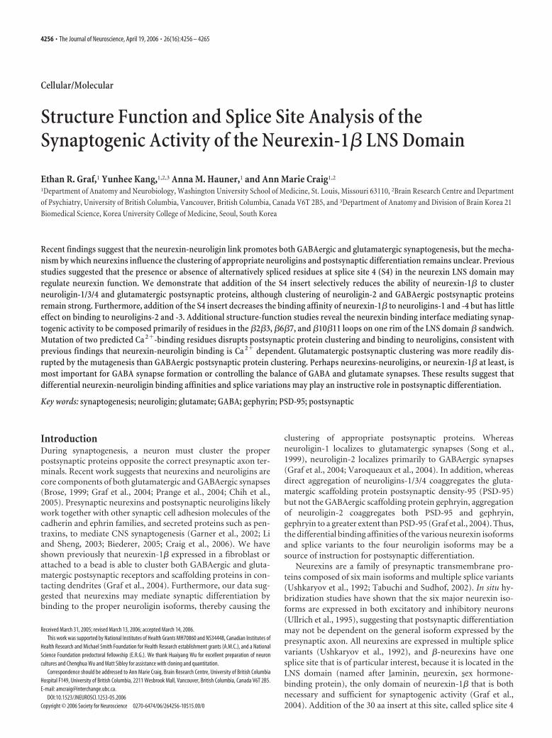
quence GNNDNERLAIARQRIPYRLGRVVDEWLLDK into theLNS domain between residues 200 and 201 of the originalneurexin-1�-CFP construct with CFP attached to the C termi-nus, creating nrx(�S4)-CFP. We then cultured COS fibroblastcells transfected with either nrx(�S4)-CFP or neurexin-CFPwith no splice site 4 insert, notated nrx(�S4)-CFP, onto 1-week-old hippocampal neuron cultures grown by the Banker method(Goslin et al., 1998). The cocultures were immunostained forgephyrin and PSD-95. As reported previously (Graf et al., 2004),nrx(�S4)-CFP induced clusters of the postsynaptic scaffoldingproteins PSD-95 and gephyrin in dendrites at contact sites withthe transfected fibroblasts (Fig. 1A). Absence of immunoreactiv-ity of the presynaptic antigen synapsin distinguished inducedclusters of gephyrin and PSD-95 from endogenous synaptic clus-ters that may happen to underlie transfected fibroblasts. Surpris-ingly, nrx(�S4)-CFP retained the ability to cluster both gephyrinand PSD-95 (Fig. 1B). For both nrx(�S4)-CFP and nrx(�S4)-CFP, clustering of gephyrin and PSD-95 was strong and oftenoccurred at the edges of the COS cells where the fibroblasts werelikely in greatest contact with their environment. Such clusteringof gephyrin and PSD-95 was never seen at dendritic contacts offibroblasts transfected with mCFP (Fig. 1C) or with N-cadherinor NgCAM (Graf et al., 2004).
To determine whether the degree of gephyrin and PSD-95clustering was altered because of addition of the S4 insert, we
Figure 1. Addition of the splice insert at S4 reduces induction of PSD-95 but not gephyrin clusters. A, B, In a fibroblast-neuron coculture assay, COS cells expressing CFP-tagged neurexin-1�without the S4 insert (A) and with the addition of the S4 insert (B) induced clustering of both gephyrin and PSD-95 in contacting dendrites. Induced clusters (arrowheads) were negative for thepresynaptic antigen synapsin unlike endogenous synapses (arrows). C, Only synapsin-positive gephyrin and PSD-95 clusters (arrows) were observed under mCFP transfected fibroblasts. D–F,Quantification of the number (D), area (E), and total integrated intensity (F ) of synapsin-negative gephyrin and PSD-95 clusters underlying transfected fibroblasts revealed reduced induction ofPSD-95 clusters by splice site 4-positive neurexin but no change in induction of gephyrin clusters compared with splice site 4-negative neurexin (*p � 0.05; n � 20 cells each). Scale bar, 10 �m.
4258 • J. Neurosci., April 19, 2006 • 26(16):4256 – 4265 Graf et al. • Splice Site 4 Modulates Neurexin Function
quantified the number, area, and total integrated intensity ofsynapsin-immunonegative gephyrin and PSD-95 clusters under-lying COS cells transfected with either nrx(�S4)-CFP or nrx(�S4)-CFP (Fig. 1D–F). The presence or absence of the S4 inserthad no effect on the number, area, or total intensity of inducedgephyrin clusters. However, nonsynaptic PSD-95 clusters weresignificantly reduced in number, area, and total intensity by 28 –44% under COS cells transfected with nrx(�S4)-CFP comparedwith cells transfected with nrx(�S4)-CFP. Thus, whereas �S4did not abolish the ability of neurexin to induce gephyrin andPSD-95 clustering, it specifically decreased PSD-95 clusteringwhile leaving gephyrin clustering unaffected. Addition of an ex-tracellular myc tag to both nrx(�S4)-CFP and nrx(�S4)-CFPand subsequent live staining with anti-myc antibody showed thatboth constructs reached the cell surface with equal efficiency,suggesting that the above results are not a result of disruptedprotein processing (supplemental Fig. 1A,B, available at www.jneurosci.org as supplemental material).
Effect of alternative splicing at S4 on recruitment and bindingto neuroliginsThe synaptogenic activity of neurexin-1� containing the S4 insertcould occur by recruitment of neuroligins or by a novel mecha-nism perhaps independent of neuroligins. We thus determinedwhether nrx(�S4)-CFP could recruit endogenous neuroligins to
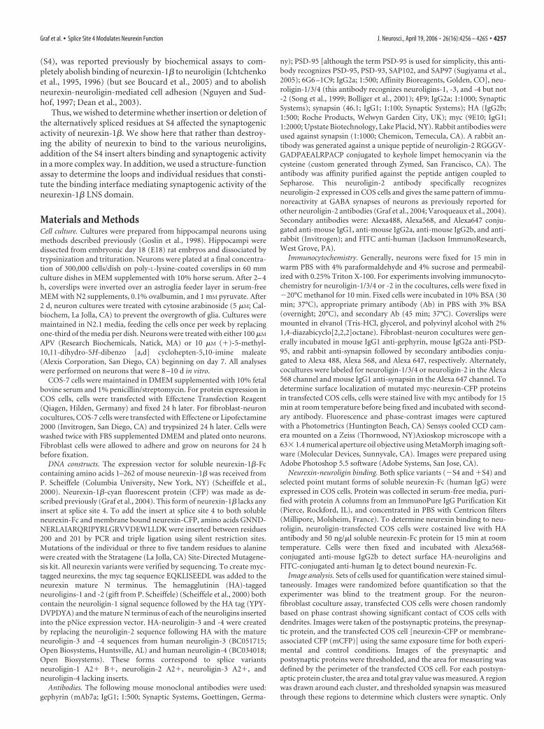
contact sites in the coculture assay, using antibodies specific ei-ther for neuroligin-2 or for neuroligins-1, -3, and -4 but not -2.Indeed, like nrx(�S4)-CFP, nrx(�S4)-CFP induced clusters ofneuroligin-1/3/4 and -2 on dendrites at sites of contact with theexpressing fibroblasts (Fig. 2A,B,D,E). Unlike the endogenoussynaptic clusters of neuroligin, the induced clusters lacked ap-posed synapsin. Such extrasynaptic clusters of neuroligin-1/3/4and -2 were not seen on dendrites in contact with cells expressingneurexin-CFP with the LNS domain deleted (Fig. 2C,F). Further-more, quantitatively, compared with the form lacking the S4 in-sert, addition of the S4 insert to neurexin-1� did not affect clus-tering of neuroligin-2 but reduced clustering of neuroligin-1/3/4by 32% (Fig. 2G–I) (*p � 0.05).
We next determined directly how addition of the S4 insertaffects binding of purified neurexin-1� to neuroligins -1, -2,-3, and -4 expressed in a heterologous system. We purifiedrecombinant soluble neurexin-Fc (the leader sequence andneurexin LNS domain fused to the human Ig constant region)either with no insert at S4, nrx(�S4)-Fc, or with the 30 aasequence inserted into S4 in the LNS domain, nrx(�S4)-Fc.We transfected COS cells with N-terminally HA-taggedneuroligin-1, -2, -3, or -4 and coincubated live with 50 ng/�lsoluble neurexin-Fc protein and with anti-HA antibody todetermine the amount of neuroligin at the surface. Unlikeprevious biochemical assays, we discovered that addition of
Figure 2. Addition of the splice insert at S4 reduces recruitment of neuroligin-1/3/4 but not neuroligin-2. A–F, In a fibroblast-neuron coculture assay, COS cells expressing CFP-taggedneurexin-1� without the S4 insert (A, D) and with the addition of the S4 insert (B, E) induced clustering of both neuroligin-1/3/4 (A, B), and neuroligin-2 (D, E) in contacting dendrites. Inducedclusters (arrowheads) were negative for the presynaptic antigen synapsin unlike endogenous synapses (arrows). C, F, Only synapsin-positive neuroligin clusters (arrows) were observed under COScells expressing CFP-tagged neurexin-1� deleted for the LNS domain. G–I, Quantification of the number (G), area (H ), and total integrated intensity (I ) of synapsin-negative neuroligin (NLG)-1, -3,-4, and -2 clusters underlying transfected fibroblasts revealed reduced induction of neuroligin-1/3/4 clusters by splice site 4-positive neurexin but no change in induction of neuroligin-2 clusterscompared with splice site 4-negative neurexin (*p � 0.05; n � 40 cells each). Scale bar, 10 �m.
Graf et al. • Splice Site 4 Modulates Neurexin Function J. Neurosci., April 19, 2006 • 26(16):4256 – 4265 • 4259
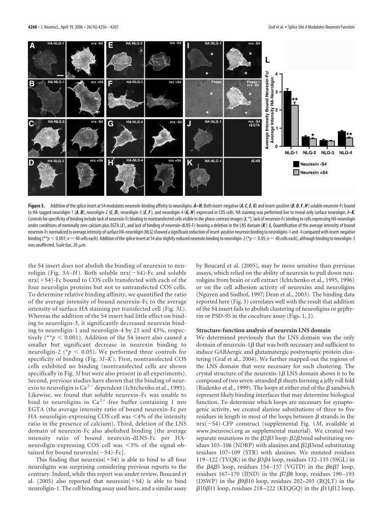
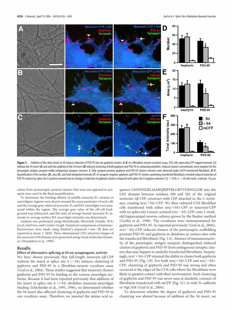
the S4 insert does not abolish the binding of neurexin to neu-roligin (Fig. 3A–H ). Both soluble nrx(�S4)-Fc and solublenrx(�S4)-Fc bound to COS cells transfected with each of thefour neuroligin proteins but not to untransfected COS cells.To determine relative binding affinity, we quantified the ratioof the average intensity of bound neurexin-Fc to the averageintensity of surface HA staining per transfected cell (Fig. 3L).Whereas the addition of the S4 insert had little effect on bind-ing to neuroligin-3, it significantly decreased neurexin bind-ing to neuroligin-1 and neuroligin-4 by 25 and 43%, respec-tively (**p � 0.001). Addition of the S4 insert also caused asmaller but significant decrease in neurexin binding toneuroligin-2 (*p � 0.05). We performed three controls forspecificity of binding (Fig. 3I–K ). First, nontransfected COScells exhibited no binding (nontransfected cells are shownspecifically in Fig. 3I but were also present in all experiments).Second, previous studies have shown that the binding of neur-exin to neuroligin is Ca 2� dependent (Ichtchenko et al., 1995).Likewise, we found that soluble neurexin-Fc was unable tobind to neuroligins in Ca 2�-free buffer containing 1 mM
EGTA (the average intensity ratio of bound neurexin-Fc perHA-neuroligin-expressing COS cell was �4% of the intensityratio in the presence of calcium). Third, deletion of the LNSdomain of neurexin-Fc also abolished binding [the averageintensity ratio of bound neurexin-dLNS-Fc per HA-neuroligin-expressing COS cell was �3% of the signal ob-tained for bound neurexin(�S4)-Fc].
This finding that neurexin(�S4) is able to bind to all fourneuroligins was surprising considering previous reports to thecontrary. Indeed, while this report was under review, Boucard etal. (2005) also reported that neurexin(�S4) is able to bindneuroligin-1. The cell binding assay used here, and a similar assay
by Boucard et al. (2005), may be more sensitive than previousassays, which relied on the ability of neurexin to pull down neu-roligins from brain or cell extract (Ichtchenko et al., 1995, 1996)or on the cell adhesion activity of neurexins and neuroligins(Nguyen and Sudhof, 1997; Dean et al., 2003). The binding datareported here (Fig. 3) correlates well with the result that additionof the S4 insert fails to abolish clustering of neuroligins or gephy-rin or PSD-95 in the coculture assay (Figs. 1, 2).
Structure-function analysis of neurexin LNS domainWe determined previously that the LNS domain was the onlydomain of neurexin-1� that was both necessary and sufficient toinduce GABAergic and glutamatergic postsynaptic protein clus-tering (Graf et al., 2004). We further mapped out the regions ofthe LNS domain that were necessary for such clustering. Thecrystal structure of the neurexin-1� LNS domain shows it to becomposed of two seven-stranded � sheets forming a jelly roll fold(Rudenko et al., 1999). The loops at either end of the � sandwichrepresent likely binding interfaces that may determine biologicalfunction. To determine which loops are necessary for synapto-genic activity, we created alanine substitutions of three to fiveresidues in length in most of the loops between � strands in thenrx(�S4)-CFP construct (supplemental Fig. 1M, available atwww.jneurosci.org as supplemental material). We created twoseparate mutations in the �2�3 loop: �2�3mid substituting res-idues 103–106 (NDRP) with alanines and �2�3end substitutingresidues 107–109 (STR) with alanines. We mutated residues119 –122 (TVQK) in the �3�4 loop, residues 132–135 (SSGL) inthe �4�5 loop, residues 154 –157 (VGTD) in the �6�7 loop,residues 167–170 (IIND) in the �7�8 loop, residues 190 –193(DSWP) in the �9�10 loop, residues 202–205 (RQLT) in the�10�11 loop, residues 218 –222 (KEQGQ) in the �11�12 loop,
Figure 3. Addition of the splice insert at S4 modulates neurexin-binding affinity to neuroligins. A–H, Both insert-negative (A, C, E, G) and insert-positive (B, D, F, H ) soluble neurexin-Fc boundto HA-tagged neuroligin-1 (A, B), neuroligin-2 (C, D), neuroligin-3 (E, F ), and neuroligin-4 (G, H ) expressed in COS cells. HA staining was performed live to reveal only surface neuroligin. I–K,Controls for specificity of binding include lack of neurexin-Fc binding to nontransfected cells visible in the phase contrast images (I, *), lack of neurexin-Fc binding to cells expressing HA-neuroliginunder conditions of nominally zero calcium plus EGTA (J ), and lack of binding of neurexin-dLNS-Fc bearing a deletion in the LNS domain (K ). L, Quantification of the average intensity of boundneurexin-Fc normalized to average intensity of surface HA-neuroligin (NLG) showed a significant reduction of insert-positive neurexin binding to neuroligins-1 and -4 compared with insert-negativebinding (**p � 0.001; n � 40 cells each). Addition of the splice insert at S4 also slightly reduced neurexin binding to neuroligin-2 (*p � 0.05; n � 40 cells each), although binding to neuroligin-3was unaffected. Scale bar, 20 �m.
4260 • J. Neurosci., April 19, 2006 • 26(16):4256 – 4265 Graf et al. • Splice Site 4 Modulates Neurexin Function
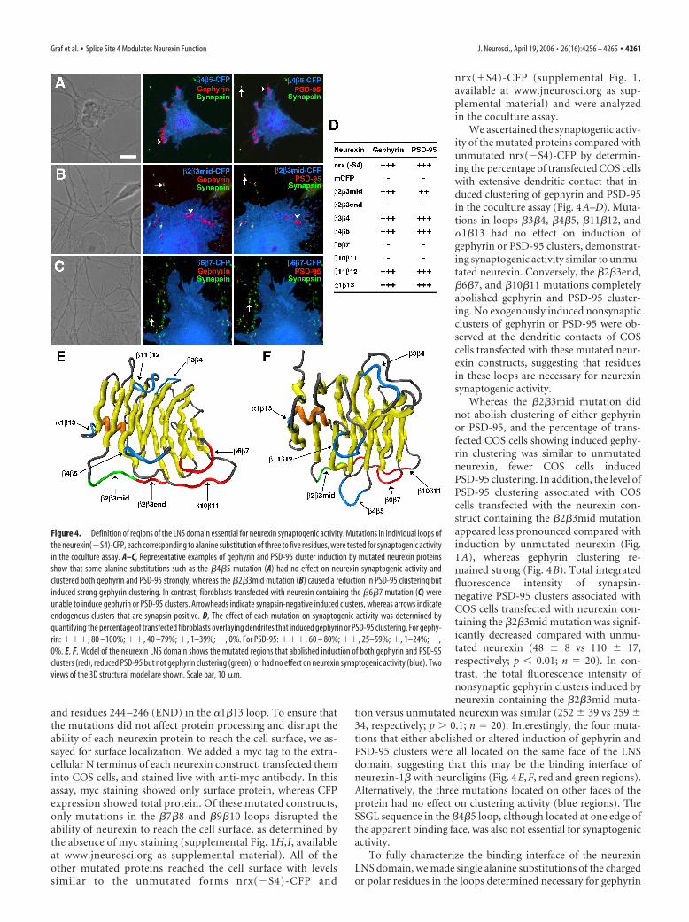
and residues 244 –246 (END) in the �1�13 loop. To ensure thatthe mutations did not affect protein processing and disrupt theability of each neurexin protein to reach the cell surface, we as-sayed for surface localization. We added a myc tag to the extra-cellular N terminus of each neurexin construct, transfected theminto COS cells, and stained live with anti-myc antibody. In thisassay, myc staining showed only surface protein, whereas CFPexpression showed total protein. Of these mutated constructs,only mutations in the �7�8 and �9�10 loops disrupted theability of neurexin to reach the cell surface, as determined bythe absence of myc staining (supplemental Fig. 1H,I, availableat www.jneurosci.org as supplemental material). All of theother mutated proteins reached the cell surface with levelssimilar to the unmutated forms nrx(�S4)-CFP and
nrx(�S4)-CFP (supplemental Fig. 1,available at www.jneurosci.org as sup-plemental material) and were analyzedin the coculture assay.
We ascertained the synaptogenic activ-ity of the mutated proteins compared withunmutated nrx(�S4)-CFP by determin-ing the percentage of transfected COS cellswith extensive dendritic contact that in-duced clustering of gephyrin and PSD-95in the coculture assay (Fig. 4A–D). Muta-tions in loops �3�4, �4�5, �11�12, and�1�13 had no effect on induction ofgephyrin or PSD-95 clusters, demonstrat-ing synaptogenic activity similar to unmu-tated neurexin. Conversely, the �2�3end,�6�7, and �10�11 mutations completelyabolished gephyrin and PSD-95 cluster-ing. No exogenously induced nonsynapticclusters of gephyrin or PSD-95 were ob-served at the dendritic contacts of COScells transfected with these mutated neur-exin constructs, suggesting that residuesin these loops are necessary for neurexinsynaptogenic activity.
Whereas the �2�3mid mutation didnot abolish clustering of either gephyrinor PSD-95, and the percentage of trans-fected COS cells showing induced gephy-rin clustering was similar to unmutatedneurexin, fewer COS cells inducedPSD-95 clustering. In addition, the level ofPSD-95 clustering associated with COScells transfected with the neurexin con-struct containing the �2�3mid mutationappeared less pronounced compared withinduction by unmutated neurexin (Fig.1A), whereas gephyrin clustering re-mained strong (Fig. 4B). Total integratedfluorescence intensity of synapsin-negative PSD-95 clusters associated withCOS cells transfected with neurexin con-taining the �2�3mid mutation was signif-icantly decreased compared with unmu-tated neurexin (48 � 8 vs 110 � 17,respectively; p � 0.01; n � 20). In con-trast, the total fluorescence intensity ofnonsynaptic gephyrin clusters induced byneurexin containing the �2�3mid muta-
tion versus unmutated neurexin was similar (252 � 39 vs 259 �34, respectively; p � 0.1; n � 20). Interestingly, the four muta-tions that either abolished or altered induction of gephyrin andPSD-95 clusters were all located on the same face of the LNSdomain, suggesting that this may be the binding interface ofneurexin-1� with neuroligins (Fig. 4E,F, red and green regions).Alternatively, the three mutations located on other faces of theprotein had no effect on clustering activity (blue regions). TheSSGL sequence in the �4�5 loop, although located at one edge ofthe apparent binding face, was also not essential for synaptogenicactivity.
To fully characterize the binding interface of the neurexinLNS domain, we made single alanine substitutions of the chargedor polar residues in the loops determined necessary for gephyrin
Figure 4. Definition of regions of the LNS domain essential for neurexin synaptogenic activity. Mutations in individual loops ofthe neurexin(�S4)-CFP, each corresponding to alanine substitution of three to five residues, were tested for synaptogenic activityin the coculture assay. A–C, Representative examples of gephyrin and PSD-95 cluster induction by mutated neurexin proteinsshow that some alanine substitutions such as the �4�5 mutation (A) had no effect on neurexin synaptogenic activity andclustered both gephyrin and PSD-95 strongly, whereas the �2�3mid mutation (B) caused a reduction in PSD-95 clustering butinduced strong gephyrin clustering. In contrast, fibroblasts transfected with neurexin containing the �6�7 mutation (C) wereunable to induce gephyrin or PSD-95 clusters. Arrowheads indicate synapsin-negative induced clusters, whereas arrows indicateendogenous clusters that are synapsin positive. D, The effect of each mutation on synaptogenic activity was determined byquantifying the percentage of transfected fibroblasts overlaying dendrites that induced gephyrin or PSD-95 clustering. For gephy-rin: ���, 80 –100%; ��, 40 –79%; �, 1–39%; �, 0%. For PSD-95: ���, 60 – 80%; ��, 25–59%; �, 1–24%; �,0%. E, F, Model of the neurexin LNS domain shows the mutated regions that abolished induction of both gephyrin and PSD-95clusters (red), reduced PSD-95 but not gephyrin clustering (green), or had no effect on neurexin synaptogenic activity (blue). Twoviews of the 3D structural model are shown. Scale bar, 10 �m.
Graf et al. • Splice Site 4 Modulates Neurexin Function J. Neurosci., April 19, 2006 • 26(16):4256 – 4265 • 4261
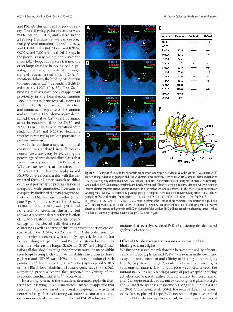
and PSD-95 clustering in the previous as-say. The following point mutations weremade: S107A, T108A, and R109A in the�2�3 loop (residues that were in the orig-inal �2�3end mutation); T156A, D157A,and D158A in the �6�7 loop; and R202A,Q203A, and T205A in the �10�11 loop. Inthe previous assay, we did not mutate thesmall �8�9 loop, but because it is near theother loops found to be necessary for syn-aptogenic activity, we mutated the singlecharged residue in that loop, N184A. Asmentioned above, the binding of neurexinto neuroligin is Ca 2� dependent (Ichtch-enko et al., 1995) (Fig. 3J). The Ca 2�-binding residues have been mapped outpreviously in the homologous lamininLNS domain (Hohenester et al., 1999; Tisiet al., 2000). By comparing the structureand amino acid sequence of the lamininand neurexin-1� LNS domains, we deter-mined the putative Ca 2�-binding aminoacids in neurexin-1� to be D137 andN208. Thus single alanine mutations weremade of D137 and N208 to determinewhether they may play a role in postsynapticprotein clustering.
As in the previous assay, each mutatedconstruct was analyzed in a fibroblast-neuron coculture assay by evaluating thepercentage of transfected fibroblasts thatinduced gephyrin and PSD-95 clusters.Whereas neurexin that contained theD157A mutation clustered gephyrin andPSD-95 at levels comparable with the un-mutated form, all other mutations eitherdecreased postsynaptic protein clusteringcompared with unmutated neurexin orcompletely abolished the synaptogenic ac-tivity of the LNS domain altogether (com-pare Figs. 5 and 1A). Mutations S107A,T108A, T156A, N184A, and Q203A hadno effect on gephyrin clustering butshowed a moderate decrease for inductionof PSD-95 clusters, both in terms of per-centage of transfected cells that causedclustering as well as degree of clustering when induction did oc-cur. Mutations D158A, R202A, and T205A disrupted synapto-genic activity more severely, moderately to greatly decreasing butnot abolishing both gephyrin and PSD-95 cluster induction. Fur-thermore, whereas the longer �2�3end, �6�7, and �10�11 mu-tations all abolished clustering, the only point mutation made withinthese loops to completely eliminate the ability of neurexin to clustergephyrin and PSD-95 was R109A. In addition, mutation of eachputative Ca2�-binding residue, D137A in the �4�5 loop and N208Ain the �10�11 loop, abolished all synaptogenic activity (Fig. 5E),supporting previous reports that suggested the activity of theneurexin-neuroligin link is Ca2� dependent.
Interestingly, none of the mutations decreased gephyrin clus-tering while leaving PSD-95 unaffected. Instead, it appeared thatmost mutations decreased the overall synaptogenic activity ofneurexin, but gephyrin clustering was more resistant to moderatedecreases in activity than was induction of PSD-95 clusters. Only
mutants that severely decreased PSD-95 clustering also decreasedgephyrin clustering.
Effect of LNS domain mutations on recruitment of andbinding to neuroliginsWe further assessed the relationship between the ability of neur-exins to induce gephyrin and PSD-95 clustering in the cocultureassay and recruitment of and affinity of binding to neuroligins(Fig. 6) (supplemental Fig. 2, available at www.jneurosci.org assupplemental material). For this purpose, we chose a subset of themutant neurexins representing a range of postsynaptic-inducingactivities and assayed relative binding affinity to neuroligins-1and -2 as representative of the major neuroligins at glutamatergicand GABAergic synapses, respectively (Song et al., 1999; Graf etal., 2004; Varoqueaux et al., 2004). For each of the mutant neur-exins chosen, plus wild-type (WT) neurexin-1� positive controland the LNS deletion negative control, we quantified the ratio of
Figure 5. Definition of single residues essential for neurexin synaptogenic activity. A–D, Although the D157A mutation (A)showed strong induction of gephyrin and PSD-95 clusters, other mutations such as T156A (B) caused moderate reduction ofPSD-95 clustering only. Other mutations such as D158A (C) caused more severe reductions in both gephyrin and PSD-95 clustering,whereas the R109A (D) mutation completely abolished gephyrin and PSD-95 clustering. Arrowheads indicate synapsin-negativeinduced clusters, whereas arrows indicate endogenous clusters that are synapsin positive. E, The effect of each mutation onsynaptogenic activity was determined by quantifying the percentage of transfected fibroblasts overlaying dendrites that inducedgephyrin or PSD-95 clustering. For gephyrin: ���, 80 –100%; ��, 40 –79%; �, 1–39%; �, 0%. For PSD-95: ���,60 – 80%; ��, 25–59%; �, 1–24%; �, 0%. Position refers to the location of the mutation or its function as a predictedCa 2�-binding residue. F, The model shows the location of residues that abolished induction of both gephyrin and PSD-95clustering (red), reduced both gephyrin and PSD-95 clustering (blue), reduced PSD-95 but not gephyrin clustering (green), or hadno effect on neurexin synaptogenic activity (purple). Scale bar, 10 �m.
4262 • J. Neurosci., April 19, 2006 • 26(16):4256 – 4265 Graf et al. • Splice Site 4 Modulates Neurexin Function
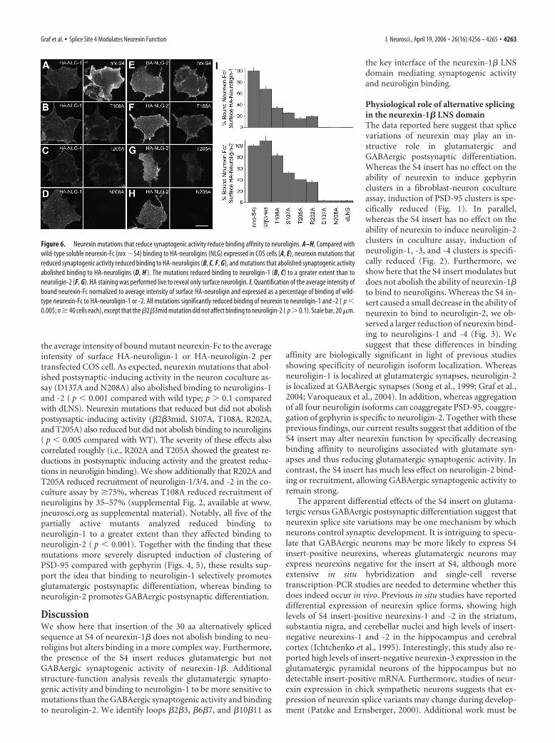
the average intensity of bound mutant neurexin-Fc to the averageintensity of surface HA-neuroligin-1 or HA-neuroligin-2 pertransfected COS cell. As expected, neurexin mutations that abol-ished postsynaptic-inducing activity in the neuron coculture as-say (D137A and N208A) also abolished binding to neuroligins-1and -2 ( p � 0.001 compared with wild type; p � 0.1 comparedwith dLNS). Neurexin mutations that reduced but did not abolishpostsynaptic-inducing activity (�2�3mid, S107A, T108A, R202A,and T205A) also reduced but did not abolish binding to neuroligins( p � 0.005 compared with WT). The severity of these effects alsocorrelated roughly (i.e., R202A and T205A showed the greatest re-ductions in postsynaptic inducing activity and the greatest reduc-tions in neuroligin binding). We show additionally that R202A andT205A reduced recruitment of neuroligin-1/3/4, and -2 in the co-culture assay by �75%, whereas T108A reduced recruitment ofneuroligins by 35–57% (supplemental Fig. 2, available at www.jneurosci.org as supplemental material). Notably, all five of thepartially active mutants analyzed reduced binding toneuroligin-1 to a greater extent than they affected binding toneuroligin-2 ( p � 0.001). Together with the finding that thesemutations more severely disrupted induction of clustering ofPSD-95 compared with gephyrin (Figs. 4, 5), these results sup-port the idea that binding to neuroligin-1 selectively promotesglutamatergic postsynaptic differentiation, whereas binding toneuroligin-2 promotes GABAergic postsynaptic differentiation.
DiscussionWe show here that insertion of the 30 aa alternatively splicedsequence at S4 of neurexin-1� does not abolish binding to neu-roligins but alters binding in a more complex way. Furthermore,the presence of the S4 insert reduces glutamatergic but notGABAergic synaptogenic activity of neurexin-1�. Additionalstructure-function analysis reveals the glutamatergic synapto-genic activity and binding to neuroligin-1 to be more sensitive tomutations than the GABAergic synaptogenic activity and bindingto neuroligin-2. We identify loops �2�3, �6�7, and �10�11 as
the key interface of the neurexin-1� LNSdomain mediating synaptogenic activityand neuroligin binding.
Physiological role of alternative splicingin the neurexin-1� LNS domainThe data reported here suggest that splicevariations of neurexin may play an in-structive role in glutamatergic andGABAergic postsynaptic differentiation.Whereas the S4 insert has no effect on theability of neurexin to induce gephyrinclusters in a fibroblast-neuron cocultureassay, induction of PSD-95 clusters is spe-cifically reduced (Fig. 1). In parallel,whereas the S4 insert has no effect on theability of neurexin to induce neuroligin-2clusters in coculture assay, induction ofneuroligin-1, -3, and -4 clusters is specifi-cally reduced (Fig. 2). Furthermore, weshow here that the S4 insert modulates butdoes not abolish the ability of neurexin-1�to bind to neuroligins. Whereas the S4 in-sert caused a small decrease in the ability ofneurexin to bind to neuroligin-2, we ob-served a larger reduction of neurexin bind-ing to neuroligins-1 and -4 (Fig. 3). Wesuggest that these differences in binding
affinity are biologically significant in light of previous studiesshowing specificity of neuroligin isoform localization. Whereasneuroligin-1 is localized at glutamatergic synapses, neuroligin-2is localized at GABAergic synapses (Song et al., 1999; Graf et al.,2004; Varoqueaux et al., 2004). In addition, whereas aggregationof all four neuroligin isoforms can coaggregate PSD-95, coaggre-gation of gephyrin is specific to neuroligin-2. Together with theseprevious findings, our current results suggest that addition of theS4 insert may alter neurexin function by specifically decreasingbinding affinity to neuroligins associated with glutamate syn-apses and thus reducing glutamatergic synaptogenic activity. Incontrast, the S4 insert has much less effect on neuroligin-2 bind-ing or recruitment, allowing GABAergic synaptogenic activity toremain strong.
The apparent differential effects of the S4 insert on glutama-tergic versus GABAergic postsynaptic differentiation suggest thatneurexin splice site variations may be one mechanism by whichneurons control synaptic development. It is intriguing to specu-late that GABAergic neurons may be more likely to express S4insert-positive neurexins, whereas glutamatergic neurons mayexpress neurexins negative for the insert at S4, although moreextensive in situ hybridization and single-cell reversetranscription-PCR studies are needed to determine whether thisdoes indeed occur in vivo. Previous in situ studies have reporteddifferential expression of neurexin splice forms, showing highlevels of S4 insert-positive neurexins-1 and -2 in the striatum,substantia nigra, and cerebellar nuclei and high levels of insert-negative neurexins-1 and -2 in the hippocampus and cerebralcortex (Ichtchenko et al., 1995). Interestingly, this study also re-ported high levels of insert-negative neurexin-3 expression in theglutamatergic pyramidal neurons of the hippocampus but nodetectable insert-positive mRNA. Furthermore, studies of neur-exin expression in chick sympathetic neurons suggests that ex-pression of neurexin splice variants may change during develop-ment (Patzke and Ernsberger, 2000). Additional work must be
Figure 6. Neurexin mutations that reduce synaptogenic activity reduce binding affinity to neuroligins. A–H, Compared withwild-type soluble neurexin-Fc (nrx �S4) binding to HA-neuroligins (NLG) expressed in COS cells (A, E), neurexin mutations thatreduced synaptogenic activity reduced binding to HA-neuroligins (B, C, F, G), and mutations that abolished synaptogenic activityabolished binding to HA-neuroligins (D, H ). The mutations reduced binding to neuroligin-1 (B, C) to a greater extent than toneuroligin-2 (F, G). HA staining was performed live to reveal only surface neuroligin. I, Quantification of the average intensity ofbound neurexin-Fc normalized to average intensity of surface HA-neuroligin and expressed as a percentage of binding of wild-type neurexin-Fc to HA-neuroligin-1 or -2. All mutations significantly reduced binding of neurexin to neuroligin-1 and -2 ( p �0.005; n � 40 cells each), except that the �2�3mid mutation did not affect binding to neuroligin-2 ( p � 0.1). Scale bar, 20 �m.
Graf et al. • Splice Site 4 Modulates Neurexin Function J. Neurosci., April 19, 2006 • 26(16):4256 – 4265 • 4263
done to specifically identify how splice variations correlate withglutamatergic versus GABAergic neurons throughout braindevelopment.
The results reported here of differential binding ofneurexin-1� to neuroligins-1, -2, -3, and -4 also highlight theimportance of considering a physiological role for each of theneuroligins. Neuroligins-1, -2, and -3 are widely expressed inbrain from postnatal day 0 (P0) to adult, and neuroligin-4 has aneven broader tissue distribution (Brose, 1999; Song et al., 1999;Bolliger et al., 2001; Varoqueaux et al., 2004). Binding affinity ofneurexin-1� to the glutamatergic synaptic neuroligins was muchgreater for neuroligin-1 than neuroligins-3 or -4. However, via-bility of the neuroligin-1 knock-out mice (Song et al., 1999) sug-gests that other neuroligins can functionally substitute in theabsence of neuroligin-1. Furthermore, linkage of mutations inhuman neuroligins-3 and -4 with autism and mental retardationindicate a specific role for these neuroligins in human cognition(Jamain et al., 2003; Laumonnier et al., 2004). Perhaps the insen-sitivity of neuroligin-3 to splice site 4 variations may be physio-logically relevant. Finally, the relatively lower affinity ofneurexin-1� for neuroligin-2 compared with neuroligin-1 is sur-prising given its more potent synaptogenic activity for GABAer-gic than glutamatergic postsynaptic proteins, suggesting that per-haps neuroligin-2 may be more abundant than the otherneuroligins in most neurons.
Neurexin-1� structure-function analysisWe have shown previously that the neurexin LNS domain is bothnecessary and sufficient for induction of postsynaptic proteinclusters (Graf et al., 2004). We report here the loops and individ-ual residues in the LNS domain that are necessary for synapto-genic activity. Alanine substitution of multiple amino acids in the�2�3, �6�7, and �10�11 loops completely abolished neurexinsynaptogenic activity, delineating the presumed binding inter-face for interaction with neuroligins. As direct confirmation, sin-gle residue substitutions in �2�3 and �10�11 reduced bindingaffinity for neuroligins (Fig. 6). These loops of the LNS domain atthe rim of the � sandwich opposite the N and C termini may forma generally conserved binding interface. The homologous surfaceof the LNS domain of sex hormone-binding globulin was directlyidentified as the steroid binding site by crystallographic analysis(Grishkovskaya et al., 2000), and indirect studies support the ideathat this surface is key to the function of agrin and laminins aswell as neurexin (Rudenko et al., 2001). It is remarkable that the30 residue S4 insert in the �10�11 loop did not completely de-stroy the neuroligin binding site; presumably, these residues foldaway from the interface, thus only partially modifying the bind-ing surface. The identification here of the �2�3 loop of neurexinas essential for synaptogenic activity is also interesting in light ofthe finding that alternative splicing at the z site in the equivalentloop of the third agrin LNS domain regulates its synaptogenicactivity (Ferns et al., 1993; Burgess et al., 1999; Rudenko et al.,1999).
Of the 12 single residue mutations tested, only three com-pletely abolished neurexin-1� synaptogenic activity. We suggestthat two of these, D137A and N208A, abolish synaptogenic activ-ity by disrupting the ability of neurexin to bind Ca 2�. Previousstudies (Ichtchenko et al., 1995), as well as our own observations(Fig. 3J), show that neurexin-neuroligin binding is Ca 2� depen-dent. Half-maximal binding of neuroligin-1 to immobilizedneurexin-1� was reported at 2 �M free Ca 2� (Nguyen and Sud-hof, 1997), suggesting that the Ca 2�-binding sites are normallysaturated in vivo. Because these two mutated residues are in an
equivalent position to the identified Ca 2�-binding aspartates inthe similar laminin LNS domain (Hohenester et al., 1999; Tisi etal., 2000), and asparagine is also capable of coordinating a cal-cium ion, it is likely that D137A and N208A are responsible forCa 2� binding in neurexin. The other essential single residue,R109A, is close to the calcium-binding residues in the 3D struc-ture (Fig. 5F) and is presumably important for binding specificneuroligin residues. Identification of the individual residues thatcontribute to synaptogenic activity of neurexin-1� as performedhere may be useful for mapping of specific interactions at theneurexin-neuroligin interface. Such analysis awaits identificationof the residues of the neuroligin acetylcholinesterase domain im-portant for neurexin binding.
It is interesting that no manipulation of the neurexin-1� LNSdomain resulted in reduced gephyrin clustering or binding toneuroligin-2 while leaving induction of PSD-95 clusters or bind-ing to neuroligin-1 unaffected. Whereas the R202A and T205Apoint mutations decreased gephyrin clustering and binding toneuroligin-2, they severely decreased PSD-95 clustering andbinding to neuroligin-1 to an even greater degree. Several otherpoint mutations as well as the larger �2�3mid mutation and theaddition of the S4 insert reduced PSD-95 clustering while leavinggephyrin clustering relatively unaffected. Like the S4 insert, the�2�3mid mutation decreased binding of neurexin toneuroligin-1 but not neuroligin-2. These results reveal that glu-tamatergic synaptogenic activity and neurexin binding to neu-roligins associated with glutamate synapses is very sensitive toneurexin modification. Conversely, all of the active neurexin mu-tants generated here, like wild-type neurexin-1�, had a morepotent inducing activity for GABAergic than glutamatergicpostsynaptic proteins. This is consistent with a recent report thatreduction of endogenous neuroligins by RNAi resulted in mor-phological and functional defects that were more severe forGABAergic than glutamatergic synapses (Chih et al., 2005). It ispossible that the neurexin-neuroligin system may be more im-portant for GABAergic synaptogenesis or for controlling the bal-ance of glutamate and GABA synapses (Graf et al., 2004; Prange etal., 2004) than for glutamatergic synaptogenesis per se. However,additional work is required to determine whether other neurexinisoforms and splice variants function in a complementary man-ner to promote glutamatergic differentiation more predomi-nantly than GABAergic differentiation. The recent findings that�-neurexins bind neuroligins, and that neurexin binding is reg-ulated by alternative splicing of neuroligin-1 (Boucard et al.,2005), raise further the potential for different physiological rolesfor different neurexin and neuroligin variants.
ReferencesBiederer T (2005) Progress from the postsynaptic side: signaling in synaptic
differentiation. Sci STKE 2005:pe9.Bolliger MF, Frei K, Winterhalter KH, Gloor SM (2001) Identification of a
novel neuroligin in humans which binds to PSD-95 and has a widespreadexpression. Biochem J 356:581–588.
Boucard AA, Chubykin AA, Comoletti D, Taylor P, Sudhof TC (2005) Asplice code for trans-synaptic cell adhesion mediated by binding of neu-roligin 1 to �- and �-neuerexins. Neuron 48:229 –236.
Brose N (1999) Synaptic cell adhesion proteins and synaptogenesis in themammalian central nervous system. Naturwissenschaften 86:516 –524.
Burgess RW, Nguyen QT, Son YJ, Lichtman JW, Sanes JR (1999) Alterna-tively spliced isoforms of nerve- and muscle-derived agrin: their roles atthe neuromuscular junction. Neuron 23:33– 44.
Chih B, Engelman H, Scheiffele P (2005) Control of excitatory and inhibi-tory synapse formation by neuroligins. Science 307:1324 –1328.
Craig AM, Graf ER, Linhoff MW (2006) How to build a central synapse:clues from cell culture. Trends Neurosci 29:8 –20.
4264 • J. Neurosci., April 19, 2006 • 26(16):4256 – 4265 Graf et al. • Splice Site 4 Modulates Neurexin Function
Dean C, Scholl FG, Choih J, DeMaria S, Berger J, Isacoff E, Scheiffele P(2003) Neurexin mediates the assembly of presynaptic terminals. NatNeurosci 6:708 –716.
Ferns MJ, Campanelli JT, Hoch W, Scheller RH, Hall Z (1993) The ability ofagrin to cluster AChRs depends on alternative splicing and on cell surfaceproteoglycans. Neuron 11:491–502.
Garner CC, Zhai RG, Gundelfinger ED, Ziv NE (2002) Molecular mecha-nisms of CNS synaptogenesis. Trends Neurosci 25:243–251.
Goslin K, Asmussen H, Banker G (1998) Rat hippocampal neurons in low-density culture. In: Culturing nerve cells, Ed 2 (Banker G, Goslin K, eds),pp 339 –370. Cambridge, MA: MIT.
Graf ER, Zhang X, Jin SX, Linhoff MW, Craig AM (2004) Neurexins inducedifferentiation of GABA and glutamate postsynaptic specializations vianeuroligins. Cell 119:1013–1026.
Grishkovskaya I, Avvakumov GV, Sklenar G, Dales D, Hammond GL, MullerYA (2000) Crystal structure of human sex hormone-binding globulin:steroid transport by a laminin G-like domain. EMBO J 19:504 –512.
Hohenester E, Tisi D, Talts JF, Timpl R (1999) The crystal structure of alaminin G-like module reveals the molecular basis of alpha-dystroglycanbinding to laminins, perlecan, and agrin. Mol Cell 4:783–792.
Humphrey W, Dalke A, Schulten K (1996) VMD: visual molecular dynam-ics. J Mol Graph 14:33–38:27–38.
Ichtchenko K, Hata Y, Nguyen T, Ullrich B, Missler M, Moomaw C, SudhofTC (1995) Neuroligin 1: a splice site-specific ligand for beta-neurexins.Cell 81:435– 443.
Ichtchenko K, Nguyen T, Sudhof TC (1996) Structures, alternative splicing,and neurexin binding of multiple neuroligins. J Biol Chem271:2676 –2682.
Jamain S, Quach H, Betancur C, Rastam M, Colineaux C, Gillberg IC, Sod-erstrom H, Giros B, Leboyer M, Gillberg C, Bourgeron T (2003) Muta-tions of the X-linked genes encoding neuroligins NLGN3 and NLGN4 areassociated with autism. Nat Genet 34:27–29.
Laumonnier F, Bonnet-Brilhault F, Gomot M, Blanc R, David A, MoizardMP, Raynaud M, Ronce N, Lemonnier E, Calvas P, Laudier B, Chelly J,Fryns JP, Ropers HH, Hamel BC, Andres C, Barthelemy C, Moraine C,Briault S (2004) X-linked mental retardation and autism are associatedwith a mutation in the NLGN4 gene, a member of the neuroligin family.Am J Hum Genet 74:552–557.
Li Z, Sheng M (2003) Some assembly required: the development of neuro-nal synapses. Nat Rev Mol Cell Biol 4:833– 841.
Nguyen T, Sudhof TC (1997) Binding properties of neuroligin 1 and neur-exin 1beta reveal function as heterophilic cell adhesion molecules. J BiolChem 272:26032–26039.
Patzke H, Ernsberger U (2000) Expression of neurexin Ialpha splice variantsin sympathetic neurons: selective changes during differentiation and inresponse to neurotrophins. Mol Cell Neurosci 15:561–572.
Prange O, Wong TP, Gerrow K, Wang YT, El-Husseini A (2004) A balancebetween excitatory and inhibitory synapses is controlled by PSD-95 andneuroligin. Proc Natl Acad Sci USA 101:13915–13920.
Rudenko G, Nguyen T, Chelliah Y, Sudhof TC, Deisenhofer J (1999) Thestructure of the ligand-binding domain of neurexin Ibeta: regulation ofLNS domain function by alternative splicing. Cell 99:93–101.
Rudenko G, Hohenester E, Muller YA (2001) LG/LNS domains: multiplefunctions– one business end? Trends Biochem Sci 26:363–368.
Scheiffele P, Fan J, Choih J, Fetter R, Serafini T (2000) Neuroligin expressedin nonneuronal cells triggers presynaptic development in contacting ax-ons. Cell 101:657– 669.
Song JY, Ichtchenko K, Sudhof TC, Brose N (1999) Neuroligin 1 is apostsynaptic cell-adhesion molecule of excitatory synapses. Proc NatlAcad Sci USA 96:1100 –1105.
Sugiyama Y, Kawabata I, Sobue K, Okabe S (2005) Determination of abso-lute protein numbers in single synapses by a GFP-based calibration tech-nique. Nat Methods 2:677– 684.
Tabuchi K, Sudhof TC (2002) Structure and evolution of neurexin genes:insight into the mechanism of alternative splicing. Genomics79:849 – 859.
Tisi D, Talts JF, Timpl R, Hohenester E (2000) Structure of the C-terminallaminin G-like domain pair of the laminin alpha2 chain harbouring bind-ing sites for alpha-dystroglycan and heparin. EMBO J 19:1432–1440.
Ullrich B, Ushkaryov YA, Sudhof TC (1995) Cartography of neurexins:more than 1000 isoforms generated by alternative splicing and expressedin distinct subsets of neurons. Neuron 14:497–507.
Ushkaryov YA, Petrenko AG, Geppert M, Sudhof TC (1992) Neurexins:synaptic cell surface proteins related to the alpha-latrotoxin receptor andlaminin. Science 257:50 –56.
Varoqueaux F, Jamain S, Brose N (2004) Neuroligin 2 is exclusively local-ized to inhibitory synapses. Eur J Cell Biol 83:449 – 456.
Graf et al. • Splice Site 4 Modulates Neurexin Function J. Neurosci., April 19, 2006 • 26(16):4256 – 4265 • 4265