structure …eisenberglab.mbi.ucla.edu/reprints/wang, structure of a nudix, acta... · substrate...
TRANSCRIPT

electronic reprint
Acta Crystallographica Section D
BiologicalCrystallography
ISSN 0907-4449
Structure of a Nudix protein from Pyrobaculum aerophilum reveals adimer with two intersubunit �-sheetsShuishu Wang, Cameron Mura, Michael R. Sawaya, Duilio Cascio and DavidEisenberg
Copyright © International Union of Crystallography
Author(s) of this paper may load this reprint on their own web site provided that this cover page is retained. Republication of this article or itsstorage in electronic databases or the like is not permitted without prior permission in writing from the IUCr.
Acta Cryst. (2002). D58, 571–578 Wang et al. � Nudix protein

Acta Cryst. (2002). D58, 571±578 Wang et al. � Nudix protein 571
research papers
Acta Crystallographica Section D
BiologicalCrystallography
ISSN 0907-4449
Structure of a Nudix protein from Pyrobaculumaerophilum reveals a dimer with two intersubunitb-sheets
Shuishu Wang, Cameron Mura,
Michael R. Sawaya, Duilio
Cascio and David Eisenberg*
UCLA-DOE Laboratory of Structure Biology,
611 Charles E. Young Drive East, 201 Boyer
Hall, Los Angeles, CA 90095-1570, USA
Correspondence e-mail: [email protected]
# 2002 International Union of Crystallography
Printed in Denmark ± all rights reserved
Nudix proteins, formerly called MutT homolog proteins, are a
large family of proteins that play an important role in reducing
the accumulation of potentially toxic compounds inside the
cell. They hydrolyze a wide variety of substrates that are
mainly composed of a nucleoside diphosphate linked to some
other moiety X and thus are called Nudix hydrolases.
Here, the crystal structure of a Nudix hydrolase from the
hyperthermophilic archaeon Pyrobaculum aerophilum is
reported. The structure was determined by the single-
wavelength anomalous scattering method with data collected
at the peak anomalous wavelength of an iridium-derivatized
crystal. It reveals an extensive dimer interface, with each
subunit contributing two strands to the �-sheet of the other
subunit. Individual subunits consist of a mixed highly twisted
and curved �-sheet of 11 �-strands and two �-helices, forming
an �-�-� sandwich. The conserved Nudix box signature motif,
which contains the essential catalytic residues, is located at the
®rst �-helix and the �-strand and loop preceding it. The
unusually short connections between secondary-structural
elements, together with the dimer form of the structure, are
likely to contribute to the thermostability of the P. aerophilum
Nudix protein.
Received 28 September 2001
Accepted 21 January 2002
PDB References: 1jrk, r1jrksf;
1k26, r1k26sf; 1k2e, 1k2esf.
1. Introduction
Nudix proteins form a large family of proteins that are found
in all organisms. They hydrolyze a wide variety of substrates
that contain a nucleoside diphosphate linked to some other
moiety, X. Therefore, these enzymes are called Nudix hydro-
lases (Bessman et al., 1996). Their functions are mainly to
reduce the level of these potentially toxic compounds and the
accumulation of metabolic intermediates (Safrany et al., 1998;
O'Handley et al., 2001). The best-studied Nudix enzyme is the
MutT protein from Escherichia coli (Shimokawa et al., 2000;
Abeygunawardana et al., 1995; Taddei et al., 1997; Wagner et
al., 1997; Bhatnagar et al., 1991; Frick et al., 1995; Porter et al.,
1996). It reduces the rate of AT to CG transversion several
thousand-fold by hydrolyzing the mutagenic nucleotide 8-oxo-
dGTP to give 8-oxo-dGMP, thus preventing it from being
incorporated into DNA (Bessman et al., 1996). Its catalytic
activity is reported to depend on the ligation of an essential
metal ion. Structural and mutagenesis studies have identi®ed
the metal ligands as part of a signature motif conserved among
the Nudix family, GX5EX7REUXEEXGU, where U is a
hydrophobic residue and X is any amino acid (Harris et al.,
2000; Shimokawa et al., 2000; Lin et al., 1996, 1997). Further-
more, the structure of this motif appears to be conserved
among three members of the Nudix family for which struc-
tures have been determined: E. coli MutT, diadenosine
electronic reprint

research papers
572 Wang et al. � Nudix protein Acta Cryst. (2002). D58, 571±578
tetraphosphate hydrolase (AP4A hydrolase) from Lupinus
angustifolius (Swarbrick et al., 2000) and an ADP-ribose
pyrophosphatase (ADPRase) from E. coli (Gabelli et al.,
2001). All three structures contain an �-�-� sandwich in which
the conserved residues in the Nudix motif are located in helix
�1 and in the loop preceding it.
Beyond the conserved catalytic core structure, Nudix
proteins demonstrate variations in peripheral structure and
oligomerization state, perhaps contributing to the wide variety
of substrate speci®cities observed in this family. For example,
the AP4A hydrolase has a structure similar to that of the E. coli
MutT, but with one extra �-strand and two more �-helices. On
a larger scale, the structures of MutT and AP4A hydrolase are
monomeric, whereas ADPRase is a dimer with its N-terminal
domain swapping between the subunits. Furthermore, in E. coli
MutT the entire adenosine moiety of the substrate analog
AMPCPP binds in the active-site cleft behind helix �1 (Lin et
al., 1997); however, in ADPRase, the terminal ribose of the
substrate binds in the cleft, while the adenine moiety binds to
the N-terminal domain.
Here, we report the crystal structure of a Nudix protein
from the hyperthermophilic archaeon P. aerophilum (PA)
determined with single-wavelength anomalous scattering data.
The PA Nudix protein was found to be a dimer, with each
subunit having a similar fold to the E. coli MutT. Unlike the
structure of ADPRase, the PA Nudix protein dimerizes
through two intersubunit �-sheets that lie in the center of the
polypeptide sequence. The active-site cleft has a distinctly
different structure from those of other Nudix hydrolases of
known structure. In this paper, the structure is analyzed in
terms of the catalytic function and the thermostability.
2. Experimental procedures
2.1. Cloning, expression and purification of the PA Nudix
The open reading frame (ORF) that encodes the putative
Nudix protein reported here was found via a BLAST search of
the PA genome with the sequence of the E. coli MutT protein.
A phosmid clone containing the PA mutT ORF was kindly
provided by the laboratory of Dr Jeffrey Miller (UCLA). The
following primers were utilized for polymerase chain reaction
(PCR) ampli®cation of the gene with Deep Vent DNA
polymerase (New England Biolabs): CATATGATCGTTAC-
CAGCGGCGTTTTA (sense) and AAGCTTTGAAA-
TTTTTCCCAGTCTATATAG (antisense). Ampli®ed DNA
was subcloned into the pCR-Blunt vector (Invitrogen) to give
an intermediary vector that was then digested with the
restriction enzymes NdeI and HindIII. The fragment
containing the mutT gene was puri®ed by gel extraction and
ligated into a bacterial expression vector [pET22b(+),
Novagen] that had been linearized by double digestion with
the same enzymes. The resulting recombinant plasmid
[designated pET22b(+)-mutT] adds a C-terminal 6�His tag to
the expressed protein. The plasmid was transformed into
chemically competent NovaBlue E. coli cells (Novagen) for
screening and the DNA sequence of the mutT gene was
con®rmed by sequencing (Davis Sequencing, Inc.) A point
mutant of PA Nudix (M16L) in which a Leu was substituted
for Met16 was prepared by overlap-extension PCR in order to
avoid heterogeneity resulting from a second translation-
initiation site at this position.
Recombinant PA Nudix was overexpressed by transfor-
mation of the pET22b(+)-mutT plasmid into chemically
competent BL21(DE3) E. coli cells. Single colonies were
used to inoculate Luria±Bertani broth supplemented with
100 mg mlÿ1 ampicillin and the cultures were grown at 310 K
to an OD600 of about 0.8. Isopropyl-�-d-thiogalactoside
(IPTG) at 1 mM was then added to induce overexpression and
cells were grown for a further 3±4 h. Cells were harvested by
centrifugation for 10 min at 8000g and stored at 253 K.
Thawed cell pellets were resuspended at room temperature
in a high ionic strength buffer containing 20 mM HEPES pH
7.8, 0.5 M NaCl, 0.5 mM PMSF, with or without 0.5%(v/v)
Triton X-100. Cells were lysed by lysozyme treatment
(�0.5 mg mlÿ1 for 30 min at room temperature) followed by
two or three passes through a French press operating at
�7.6 MPa or by sonication on ice for 5 � 1 min with 1 min
intervals. Lysed cells were maintained on ice and cell debris
was cleared by centrifugation at �37 000g for 30 min. The
supernatant of the cell lysate was either directly loaded onto
an Ni2+-charged HiTrap chelating column (Pharmacia) or
pretreated by heating to �348±353 K for 10 min and clearing
the denatured insoluble E. coli proteins by centrifugation at
37 000g for 35 min (>80% purity was achieved with this heat
treatment). The ®nal full-length protein was >99% pure as
estimated by several independent techniques (SDS±PAGE,
MALDI±TOF and electrospray mass spectrometry).
2.2. Crystallization and data collection
The puri®ed protein was exchanged by dialysis or by three
cycles of dilution and concentration into a dilute buffer
(10 mM Tris, 5 mM EDTA pH 8.0 or 5 mM HEPES, 10 mM
NaCl pH 7.5) and then concentrated to above 10.0 mg mlÿ1 at
277 K in a Centriprep ultra®ltration device. Crystallization
experiments were carried out using the hanging-drop vapor-
diffusion method. In each drop, 3±5 ml of protein solution was
mixed with an equal volume of well solution. Hampton Crystal
Screen kits I and II were used for the initial screening of
crystallization conditions. Crystals were obtained under
several conditions. The best crystals were obtained from drops
set up with well solutions containing 5±8% PEG 4000 or PEG
MME (polyethylene glycol monomethyl ether) 2000, 5%
2-propanol, 50 mM (NH4)2SO4 and 100 mM NaOAc pH 4.8
(native-1 crystals) or well solutions consisting of 100 mM MES
pH 6.2 and 15% 2-methyl-2,4-pentanediol (MPD) (native-2
crystals) at room temperature. Crystals were soaked for 5 min
in the well solution but with 30% of glycerol or with 50% of
MPD for native-2 crystals and ¯ash-frozen in a stream of N2
gas at 100 K. An iridium derivative was obtained by soaking a
crystal which was obtained under similar conditions as
native-1 crystals in the well solution containing 20 mM IrCl3
for 8 h. The crystal was then similarly ¯ash-frozen. Diffraction
electronic reprint
data from Ir-derivatized and native-1 crystals were collected at
beamline X8C, NSLS, Brookhaven National Laboratory and
those from the native-2 crystal were collected on an in-house
Rigaku FRD generator with an R-AXIS IV++ detector. For
the iridium-derivative crystal, a single set of data was collected
at the peak anomalous wavelength of iridium. The diffraction
data were indexed, integrated and scaled with the programs
DENZO and SCALEPACK (Otwinowski & Minor, 1996).
Data-processing statistics of the iridium-derivative data and
two native data sets are listed in Table 1. The iridium and
native-1 crystals are not isomorphous, despite their close
similarity in unit-cell parameters. Thus, the native data were
not used for initial phasing. After solving the structures, it
became clear that the crystals have different packing inter-
faces along the crystal z axis. The two crystals pack in the same
way in the xy plane, but the packing between layers is shifted
along the y axis by about 18.6 AÊ .
2.3. Phasing and structure determination
Initial phases were determined for the data collected at
the peak anomalous wavelength from the iridium-derivative
crystal. Heavy-atom positions were identi®ed with the
programs XTALVIEW (McRee, 1992) and SHELXD (Uson
& Sheldrick, 1999) and then re®ned with the program
MLPHARE (Otwinowski, 1991; Collaborative Computational
Project, Number 4, 1994). Density modi®cation, i.e. solvent
¯attening, histogram matching, multi-resolution modi®cation
and twofold NCS averaging, was carried out with the program
DM (Cowtan, 1994).
The automatic re®nement procedure ARP/wARP (Perrakis
et al., 1999) was then used for automatic model building. The
initial model and the electron-density maps were displayed
with the graphics program O (Jones et al., 1991) and the model
was manually rebuilt to ®x the side chains and to add residues
that have clear electron density. The model was then re®ned
using CNS (BruÈ nger et al., 1998) with NCS restraints.
�A-weighted 2Fo ÿ Fc and Fo ÿ Fc electron-
density maps were calculated from the re®ned
model and the model was manually ®xed using
the program O. This cycle was repeated until
the re®nement converged. This re®ned model
was used as a model for molecular replacement
of the native-1 data with the program AMoRe
(Navaza, 1994) or of the native-2 data with the
program EPMR (Kissinger et al., 1999) and
re®nement was similarly carried out with the
programs CNS and O.
The ®nal re®nement statistics are listed in
Table 2. Over 91% of residues in the native-1
structure and over 93% in the Ir-derivative
structure fall into the most favored regions in
the Ramachandran plot. Some glycerol or MPD
molecules from the cryogenic solution were
found in the re®ned structures (see Table 2).
Positive Fo ÿ Fc density was found near a cluster
of four His side chains, His19, His31, His89 and
His91, in the active-site cleft. A water molecule modelled into
this site was not suf®cient to account for this density. Since the
protein was puri®ed from an Ni-chelating column, an Ni2+ ion
was placed into the density. The hydroxyl group of Tyr84 side
chain is also in a good position to function as a ligand.
In the Ir-derivative structure all the Ir atoms were found to
bind in surface cavities on the edge of bulk-solvent channels.
Three of them, Ir1, Ir2 and Ir3, were found at the crystal-
packing interface, while Ir4 was found near the Nudix box
residues of subunit A. The strongest iridium peak, Ir1, was
found at a packing interface of three molecules and
surrounded by ®ve water molecules, which take ®ve positions
of an octahedral coordination. The interactions between the
Acta Cryst. (2002). D58, 571±578 Wang et al. � Nudix protein 573
research papers
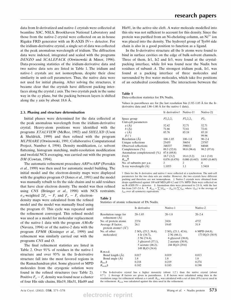
Table 1Data-collection statistics for PA Nudix.
Values in parentheses are for the last resolution bin (1.92±1.85 AÊ for the Ir-derivative data and 1.86±1.80 AÊ for the native-1 data).
Ir derivative² Native-1² Native-2³
Space group P212121 P212121 P21
Unit-cell parametersa (AÊ ) 52.42 52.71 52.71b (AÊ ) 71.66 72.61 72.61c (AÊ ) 85.76 85.18 85.18� (�) 99.96
Resolution (AÊ ) 100±1.85 100±1.8 90±2.4Unique re¯ections 25455 30654 23707Observed re¯ections 348337 398012 84048Completeness (%) 89.3 (52.0) 98.8 (96.4) 98.2 (97.0)Anomalous completeness§ (%) 87.6 (69.8)I/�(I) 18.7 (3.2) 18.4 (3.2) 14.1 (3.0)Rsym} 0.076 (0.470) 0.080 (0.443) 0.093 (0.465)No. of subunits per a.u. 2 2 4X-ray wavelength (AÊ ) 1.1053 1.1 1.5418
² Data for the Ir derivative and native-1 were collected at a synchrotron. The unit-cellparameters for the two data sets are similar. However, the two crystals have differentpacking and therefore are not isomorphous. ³ Native-2 crystals were obtained fromdrops equilibrated against 100 mM MES pH 6.2 and 15% MPD. Data were collected onan R-AXIS IV++ detector. § Anomalous data were processed to 2.0 AÊ , with the lastbin from 2.07±2.0 AÊ . } Rsym =
P j�Ihkl ÿ hIhkli�j=P
Ihkl , where hIhkli is the average ofIhkl over all symmetry equivalents.
Table 2Statistics of atomic re®nement of PA Nudix.
Ir derivative Native-1 Native-2
Resolution range forre®nement (AÊ )
20±1.85 20±1.8 20±2.4
No. of protein atoms 2374 2416 4732Average B factor,
protein atoms² (AÊ 2)26.1 19.65 31.9
No. of othermolecules³
2 SO4 (25.2, 36.6),4 Ir (34.7),2 Ni (74.4),3 glycerol (37.1),5 acetate (46.2),149 H2O (34.4)
2 SO4 (21.1, 42.6),2 Ni (66.1),6 glycerol (38.8),2 acetate (30.9),244 H2O (29.6)
6 MPD (64.8),173 H2O (30.9)
R.m.s.d.Bond length (AÊ ) 0.017 0.019 0.013Bond angle (AÊ ) 1.8 1.8 1.8
Rfree§ 0.218 0.219 0.274Rwork§ 0.182 0.183 0.190
² The Ir-derivative crystal has a higher mosaicity (about 1.2�) than the native crystal (about0.7�). ³ Average B factors are given in parentheses. § R factors were calculated using data in theresolution range for re®nement without a � cutoff. Rfree was calculated with a set of data (8%) never used inthe re®nement. Rwork was calculated against the data used in the re®nement.
electronic reprint

research papers
574 Wang et al. � Nudix protein Acta Cryst. (2002). D58, 571±578
protein atoms and Ir1 and Ir4 seem to be mediated by water.
Ir2 and Ir3 are approximately related by the twofold NCS,
each bound near a sulfate ion, as well as the side chains of
Asp65, Asp66 and Asn67 of a symmetry-related molecule.
3. Results
3.1. Protein expression and purification
The PA Nudix gene was cloned and overexpressed in E. coli
with a His tag at the C-terminus. It was found that the wild-
type protein was expressed as a mixture of the full-length and
a truncated form that lacks the ®rst 15 N-terminal residues.
Apparently, the Met16 codon acts as a second start site for
translation of this truncated protein. The DNA sequence
�10 bp upstream of the Met16 codon corresponds to a
bacterial ribosome-binding site, thus exacerbating the
problem. Similar problems have been reported with other
proteins (Matsumiya et al., 2001). In order to circumvent the
alternative start site, a point mutant (M16L) was made in
which a Leu was substituted for Met16. This mutant was
expressed as a single full-length polypeptide and was puri®ed
to >99% pure. It differs from the wild-type sequence only in
the M16L mutation and 14 amino acids appended to the
C-terminus (the 6�His tag and an eight amino-acid linker).
This M16L mutant was used for all the studies reported here.
3.2. Phasing with single-wavelength anomalous scatteringdata
The crystal structure was determined using single-
wavelength anomalous scattering data collected at a wave-
length near the peak of ¯uorescence at the LIII edge of
iridium. An anomalous difference Patterson map revealed a
single well occupied Ir site and a few much weaker potential
sites. SHELXD (Uson & Sheldrick, 1999) was used to evaluate
the heavy-atom sites and how well they correlate with the
Patterson map. The two top sites were input to MLPHARE
(Otwinowski, 1991; Collaborative Computational Project,
Number 4, 1994) for re®nement and calculating phases. The
anomalous difference Fourier map calculated from these
phases gave four unique peaks above the 5.5� contour level.
The positions of the two new peaks also matched the third and
®fth sites predicted by SHELXD. These four sites were then
input to MLPHARE for re®nement and phase calculation.
The four Ir atoms (designated Ir1, Ir2, Ir3 and Ir4) have
occupancies of 0.74, 0.50, 0.36 and 0.39 and temperature
factors of 57, 59, 64 and 67 AÊ 2, respectively, after re®nement
with MLPHARE. The atom Ir1, which has the highest occu-
pancy and lowest B factor, gave the strongest peak in the
Patterson map.
Density modi®cation with solvent ¯attening, histogram
matching and multi-resolution modi®cation using DM
(Cowtan, 1994) was carried out to improve the phases and to
identify the correct absolute con®guration. The Fourier map
after DM showed enough secondary-structure elements to
locate the twofold non-crystallographic symmetry (NCS) axis.
Twofold NCS averaging with DM signi®cantly improved the
quality of the electron-density map (Fig. 1) and the entire map
was continuous and could be traced easily. Automatic model
building with ARP/wARP (Perrakis et al., 1999) at a resolution
of 1.85 AÊ built more than 80% of the model. The map calcu-
lated at this resolution with phases from the auto-built model
showed clear side chains for more than 80% of the residues
and a complete model was built in a single round of manual
rebuilding.
3.3. Overall structure
The re®ned crystal structure of the PA Nudix protein shows
that it forms a dimer. Each subunit also contributes two
�-strands to the �-sheet of the other subunit to form an
extensive dimer interface. The dimer is held together mainly
by the two �-sheets to which both subunits contribute strands.
Hydrogen-bonding interactions occur between strand �5b of
one subunit and �6 of the other subunit (Figs. 2 and 3). There
are eight main-chain hydrogen bonds between two subunits in
the dimer. In addition, a patch of residues with hydrophobic
side chains, Ile2, Ile77, Leu94, Pro38, Pro74, Tyr61, Tyr96,
Phe92 and Met73, located at the center of the dimer interface,
also contribute to the binding energy of the dimer. The
molecular surface of each monomer, calculated with the
program GRASP (Nicholls et al., 1991), is about 7500 AÊ 2 and
the total buried surface in the dimer interface is about
2000 AÊ 2. The dimer formation in solution was con®rmed by
analytical ultracentrifugation studies (data not shown).
Each subunit (referred to as A and B) is composed of a
mixed �-sheet and two �-helices (Fig. 2). The �-sheet is highly
Figure 1A section of the initial electron-density map superimposed on the ®nalcoordinates. The map was calculated at 2.5 AÊ with phases after twofoldNCS averaging and contoured at 1.3�. The ®gure was generated with theprogram O and rendered with POV-RAY.
electronic reprint

curved and twisted and wraps around the helices. The
topology of the protein is shown in Fig. 3. Most of the strands
are connected by tight turns or short loops, with the longest
loop consisting of only six residues. The �-sheet can be divided
into a central sheet and a subsheet, which are connected
through strand �2. The N-terminal part of strand �2 forms
hydrogen bonds with the C-terminal part of �1, but then
curves and makes a subsheet with strands �3, �8 and �9.
Strands �1 and �4 to �7, together with �5a0 and �5b0 from the
other subunit, form the central �-sheet.
The two subunits are similar to each other. However,
subunit A has a long C-terminal helix that extends away from
the molecular surface, while in subunit B the C-terminal
residues turn back and form a more compact structure, making
the helix shorter (Fig. 2). The residues in the linker to the His
tag are ordered in the crystal structure, although the His tag
itself is not. In the case of subunit A, these residues extend the
helix and make contacts with nearby molecules in the crystal
packing. In one crystal form in the P21 space group (native-2)
there are four molecules per asymmetric unit, composed of a
pair of dimers, each dimer similar to native-1. Both subunits in
the dimer have long straight C-terminal helices which pack
against the C-terminal helices of the other dimer to form a
tetramer with 222 symmetry. However, the interactions
between the helices mainly involve the residues of the linker
to the His tag. Therefore, the tetramer is not physiologically
relevant.
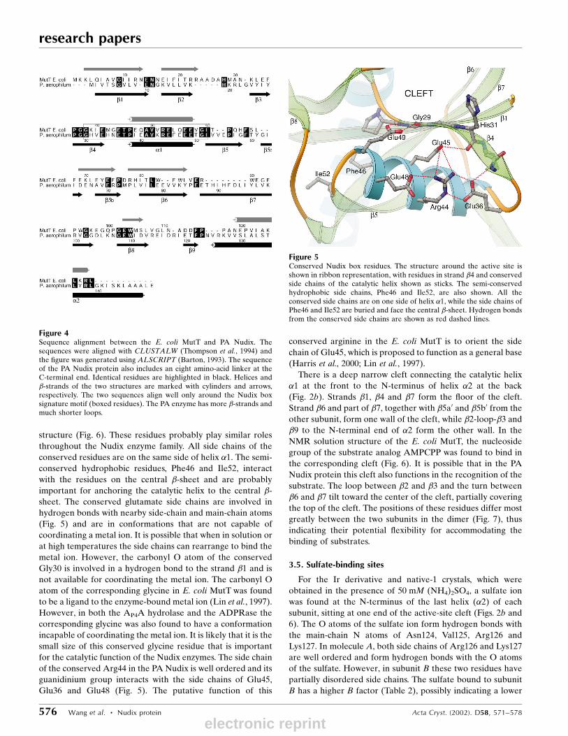
3.4. The conserved residues and the active-site cleft
The Nudix box sequence signature motif,
G30X5E36X7R44E45UXE48E49XG51U (the superscripts refer to
the residue number in the PA Nudix sequence) spans the
strand �4, helix �1 and the N-terminal part of strand �5 (Figs. 4
and 5). The conserved residues are at the same relative posi-
tions in three-dimensional space as those in the E. coli MutT
Acta Cryst. (2002). D58, 571±578 Wang et al. � Nudix protein 575
research papers
Figure 3The topology diagram of the PA Nudix structure. The strand �1 isconnected by a type II �-turn to �2, which forms hydrogen bonds with theC-terminal part of �1 and then makes a curve to form hydrogen bonds to�3, making a subsheet with strands �8 and �9. A proline makes a kink atthe end of �3, allowing �4 to form hydrogen bonds to the N-terminal halfof �1. A small loop connects �4 to helix �1. Strands �5 and �6 togetherform an almost continuous strand antiparallel to the longest strand �7.Strands �5a and �5b go to the other subunit and form part of its central�-sheet, while strands �5a0 and �5b0 are from the other subunit. The lasthelix �2 is connected to �9 by a type I �-turn. There are more �-strands inthis protein structure than in the E. coli MutT. The shaded �-strands arethe strands that are present in the E. coli MutT structure.
Figure 2Ribbon diagrams of the PA Nudix structure. (a) shows the dimer with thetwofold NCS axis perpendicular to the paper plane. Each subunit iscomposed of a long highly twisted central �-sheet and a small subsheetconnected through strand �2, sandwiched between two �-helices. Eachsubunit contributes two �-strands (�5a and �5b) to the �-sheet of theother subunit to form the dimer interface. Subunit A (on the left) has along C-terminal helix �2 with the eight amino-acid linker at theC-terminal end extending the helix, while subunit B has a shorter helix�2 and its C-terminus and the linker fold back to form a more compactstructure. (b) is a near 180� rotation of (a) along a horizontal axis. A cleftformed by the �-strands connects helix �1 at the front to the N-terminalend of helix �2 at the back. The conserved arginine and glutamate sidechains, as well as two sulfate ions found at the N-terminal end of �2 ofeach subunit, are shown as sticks. This ®gure was generated usingRIBBONS (Carson, 1997).
electronic reprint
research papers
576 Wang et al. � Nudix protein Acta Cryst. (2002). D58, 571±578
structure (Fig. 6). These residues probably play similar roles
throughout the Nudix enzyme family. All side chains of the
conserved residues are on the same side of helix �1. The semi-
conserved hydrophobic residues, Phe46 and Ile52, interact
with the residues on the central �-sheet and are probably
important for anchoring the catalytic helix to the central �-
sheet. The conserved glutamate side chains are involved in
hydrogen bonds with nearby side-chain and main-chain atoms
(Fig. 5) and are in conformations that are not capable of
coordinating a metal ion. It is possible that when in solution or
at high temperatures the side chains can rearrange to bind the
metal ion. However, the carbonyl O atom of the conserved
Gly30 is involved in a hydrogen bond to the strand �1 and is
not available for coordinating the metal ion. The carbonyl O
atom of the corresponding glycine in E. coli MutT was found
to be a ligand to the enzyme-bound metal ion (Lin et al., 1997).
However, in both the AP4A hydrolase and the ADPRase the
corresponding glycine was also found to have a conformation
incapable of coordinating the metal ion. It is likely that it is the
small size of this conserved glycine residue that is important
for the catalytic function of the Nudix enzymes. The side chain
of the conserved Arg44 in the PA Nudix is well ordered and its
guanidinium group interacts with the side chains of Glu45,
Glu36 and Glu48 (Fig. 5). The putative function of this
conserved arginine in the E. coli MutT is to orient the side
chain of Glu45, which is proposed to function as a general base
(Harris et al., 2000; Lin et al., 1997).
There is a deep narrow cleft connecting the catalytic helix
�1 at the front to the N-terminus of helix �2 at the back
(Fig. 2b). Strands �1, �4 and �7 form the ¯oor of the cleft.
Strand �6 and part of �7, together with �5a0 and �5b0 from the
other subunit, form one wall of the cleft, while �2-loop-�3 and
�9 to the N-terminal end of �2 form the other wall. In the
NMR solution structure of the E. coli MutT, the nucleoside
group of the substrate analog AMPCPP was found to bind in
the corresponding cleft (Fig. 6). It is possible that in the PA
Nudix protein this cleft also functions in the recognition of the
substrate. The loop between �2 and �3 and the turn between
�6 and �7 tilt toward the center of the cleft, partially covering
the top of the cleft. The positions of these residues differ most
greatly between the two subunits in the dimer (Fig. 7), thus
indicating their potential ¯exibility for accommodating the
binding of substrates.
3.5. Sulfate-binding sites
For the Ir derivative and native-1 crystals, which were
obtained in the presence of 50 mM (NH4)2SO4, a sulfate ion
was found at the N-terminus of the last helix (�2) of each
subunit, sitting at one end of the active-site cleft (Figs. 2b and
6). The O atoms of the sulfate ion form hydrogen bonds with
the main-chain N atoms of Asn124, Val125, Arg126 and
Lys127. In molecule A, both side chains of Arg126 and Lys127
are well ordered and form hydrogen bonds with the O atoms
of the sulfate. However, in subunit B these two residues have
partially disordered side chains. The sulfate bound to subunit
B has a higher B factor (Table 2), possibly indicating a lower
Figure 5Conserved Nudix box residues. The structure around the active site isshown in ribbon representation, with residues in strand �4 and conservedside chains of the catalytic helix shown as sticks. The semi-conservedhydrophobic side chains, Phe46 and Ile52, are also shown. All theconserved side chains are on one side of helix �1, while the side chains ofPhe46 and Ile52 are buried and face the central �-sheet. Hydrogen bondsfrom the conserved side chains are shown as red dashed lines.
Figure 4Sequence alignment between the E. coli MutT and PA Nudix. Thesequences were aligned with CLUSTALW (Thompson et al., 1994) andthe ®gure was generated using ALSCRIPT (Barton, 1993). The sequenceof the PA Nudix protein also includes an eight amino-acid linker at theC-terminal end. Identical residues are highlighted in black. Helices and�-strands of the two structures are marked with cylinders and arrows,respectively. The two sequences align well only around the Nudix boxsignature motif (boxed residues). The PA enzyme has more �-strands andmuch shorter loops.
electronic reprint

occupancy. Asn124 has main-chain dihedral angles in the left-
handed helix region of the Ramachandran plot (' ' 70� and
' 0�). This is necessary for the optimal interaction of its
backbone amide group with the sulfate ion. In one crystal
form (P21 space group with two molecules in one asymmetric
unit, data not shown), molecule B has a very low occupancy of
sulfate and the electron-density maps indicated that there are
two conformations for Asn124, with one conformation having
regular main-chain dihedral angles (' ' ÿ75� and ' ÿ25�).
We note that in E. coli MutT the amino-terminal Asn residue
of helix 2 (N119) interacts with the 6-oxo group
of dGTP (Lin et al., 1997).
4. Discussion
4.1. Comparison with other Nudix hydrolasesof known three-dimensional structure
The PA Nudix protein follows the same fold
as the E. coli MutT. However, the two struc-
tures are quite different. Fig. 6 shows the
superposition of the C� traces of E. coli MutT
onto that of the subunit A of PA Nudix. Only
�1, �1 and part of �7 can be aligned. The
r.m.s.d. over the 30 C� atoms aligned is 1.48 AÊ .
When structures are aligned based on these
secondary-structure elements, most of the long
�-strands are close in space in the two struc-
tures. However, the short �-strands and �2 are
shifted relative to each other. The �-strands of
the PA Nudix enzyme are highly curved
compared with those in the MutT and the AP4A
hydrolase. As a result, the active-site cleft is
narrower and deeper in the PA Nudix structure. This indicates
that the PA Nudix hydrolase is likely to have a different
substrate from those of MutT or AP4A hydrolase. In the crystal
structure of ADPRase complexed with ADP-ribose (Gabelli
et al., 2001), the terminal ribose group binds in the active-site
cleft, while the adenine moiety interacts with the N-terminal
domain, which is involved in dimer formation through domain
swapping. The PA Nudix does not have this N-terminal
domain and thus is unlikely to have the same ADP-sugar
hydrolase activity.
In the PA Nudix hydrolase structure, the N-terminal end of
the long helix �2 is located at the other end of the active-site
cleft opposite the catalytic helix and a sulfate ion was found
binding at the N-terminal end of �2 (Figs. 2b and 6). The
distance between the Nudix box residues to the sulfate-
binding site is about 20 AÊ . This sulfate-binding site could
potentially be a binding site for a phosphate group on the
substrate, although the distance is large. It is possible that this
Nudix enzyme hydrolyzes substrates that have a phosphate
group away from the scissile bond that can bind to the
N-terminal end of the helix �2. An attempt to identify
potential substrates for the PA Nudix protein showed that it is
inactive against 14 typical substrates for known Nudix
enzymes (ADP-ribose, ADP-mannose, ADP-glucose, GDP-
mannose, GDP-glucose, UDP-mannose, UDP-glucose, Ap2A,
Ap3A, Ap4A, NADH, deamino-NADH, NAD and FAD)
(Yang, Wang and Mura, unpublished results). These results
suggest that PA Nudix is potentially a novel Nudix enzyme.
Alternatively, PA Nudix may be inactive because of the M16L
mutation or because the active form lacks residues 1±15.
4.2. Thermostability
P. aerophilum is a hyperthermophile that grows optimally at
373 K (Volkl et al., 1996). The dimeric form of PA Nudix might
Acta Cryst. (2002). D58, 571±578 Wang et al. � Nudix protein 577
research papers
Figure 7R.m.s.d. in AÊ of the main-chain atomic positions for the superimposedsubunits of the dimer. The solid line is from the structure of the iridium-derivative crystal and the dashed line is from the native crystal. Higherr.m.s.d. was observed for residues 18±25, 79±90 and 64±69, which form thewalls of the active-site cleft, indicating their ¯exibility. The peak aroundresidue 32 is a consequence of partial disorder of the loop (higher Bfactors), while peaks around residues 110±120 and after 135 arises fromdifferences in the crystal contacts of the two subunits. A plot of ther.m.s.d. atomic positions between the dimers of the two different crystals(not shown) also gives the same peaks at residues 18±25, 79±90 and 64±69,but with a smaller scale.
Figure 6Structure superposition between the subunit A of PA Nudix and the E. coli MutT. TheE. coli MutT structure (PDB code 1tum; Lin et al., 1997) is shown in red and its boundAMPCPP shown in ball-and-stick representation. The two structures were aligned basedon �1, �1 and part of �7 of the PA structure. They have similar folds and the conservedresidues in the Nudix motif (shown as black dots) are at similar positions. However, awayfrom helix �1 the two structures are quite different. The active-site cleft is much narrowerand deeper in the PA Nudix structure. The adenosine group of the AMPCPP in the E. coliMutT structure binds in the active-site cleft. A sulfate ion was found at the N-terminus of�2 of the PA protein (shown in ball-and-stick), indicating a possible binding site for aphosphate group. The ®gure was generated with MOLSCRIPT (Kraulis, 1991).
electronic reprint
research papers
578 Wang et al. � Nudix protein Acta Cryst. (2002). D58, 571±578
contribute to the thermostability of this enzyme. Intersubunit
interactions have been proposed to be a major stabilization
mechanism for hyperthermophilic proteins (Vieille & Zeikus,
2001). In addition, ion pairs and hydrophobic interactions
between subunits make the dimer more resistant to dissocia-
tion. Two ion pairs were found between the subunits, Glu79 of
one subunit to Arg71 of the other. Another factor that may
contribute to the thermostability of this protein is that PA
Nudix has very few residues in loops. Thompson & Eisenberg
(1999) compared the sequences of about 20 complete genomes
and found that thermophilic proteins generally have loop
deletions relative to their mesophilic homologs. Sequence
alignment between E. coli MutT and PA Nudix showed a
deletion at loop 1 of the PA Nudix sequence (Fig. 4). In
addition, the loops are shortened by incorporating more
residues in the secondary-structure elements. About 75% of
residues are in �-strands and �-helices, compared with only
about 49% in the E. coli MutT protein (Fig. 4). More �-strands
are present in the PA Nudix structure and the �-strands are
mostly connected by tight �-turns.
We would like to thank Dr Hanjing Yang for lots of helpful
discussion on Nudix hydrolases and their functions.
References
Abeygunawardana, C., Weber, D. J., Gittis, A. G., Frick, D. N., Lin, J.,Miller, A.-F., Bessman, M. J. & Mildvan, A. S. (1995). Biochemistry,34, 14997±15005.
Barton, G. J. (1993). Protein Eng. 6, 37±40.Bessman, M. J., Frick, D. N. & O'Handley, S. F. (1996). J. Biol. Chem.271, 25059±25062.
Bhatnagar, S. K., Bullions, L. C. & Bessman, M. J. (1991). J. Biol.Chem. 266, 9095±9054.
BruÈ nger, A. T., Adams, P. D., Clore, G. M., DeLano, W. L., Gros, P.,Grosse-Kunstleve, R. W., Jiang, J.-S., Kuszewski, J., Nilges, N.,Pannu, N. S., Read, R. J., Rice, L. M., Simonson, T. & Warren, G. L.(1998). Acta Cryst. D54, 905±921.
Carson, M. (1997). Methods Enzymol. 277, 493±505.Collaborative Computational Project, Number 4 (1994). Acta Cryst.
D50, 760±763.Cowtan, K. (1994). Jnt CCP4/ESF±EACBM Newsl. Protein Crystal-
logr. 31, 34±38.Frick, D. N., Weber, D. J., Abeygunawardana, C., Gittis, A. G.,
Bessman, M. J. & Mildvan, A. S. (1995). Biochemistry, 34, 5577±
5586.Gabelli, S. B., Bianchet, M. A., Bessman, M. J. & Amzel, L. M. (2001).
Nature Struct. Biol. 8, 467±472.Harris, T. K., Wu, G., Nassiah, M. A. & Mildvan, A. S. (2000).
Biochemistry, 39, 1655±1674.Jones, T. A., Zou, J. Y., Cowan, S. W. & Kjeldgaard, M. (1991). Acta
Cryst. A47, 110±119.Kissinger, C., Gehlhaar, D. K. & Fogel, D. B. (1999). Acta Cryst. D55,
484±491.Kraulis, P. J. (1991). J. Appl. Cryst. 24, 946±950.Lin, J., Abeygunawardana, C., Frick, D. N., Bessman, M. J. & Mildvan,
A. S. (1996). Biochemistry, 35, 6715±6726.Lin, J., Abeygunawardana, C., Frick, D. N., Bessman, M. J. & Mildvan,
A. S. (1997). Biochemistry, 36, 1199±1211.McRee, D. E. (1992). J. Mol. Graph. 10, 44±46.Matsumiya, S., Ishino, Y. & Morikawa, K. (2001). Protein Sci. 10, 17±
23.Navaza, J. (1994). Acta Cryst. A50, 157±163.Nicholls, A., Sharp, K. & Honig, B. (1991). Proteins Struct. Funct.
Genet. 11, 281±296.O'Handley, S. F., Dunn, C. A. & Bessman, M. J. (2001). J. Biol. Chem.276, 5421±5426.
Otwinowski, Z. (1991). Proceedings of the CCP4 Study Weekend.Isomorphous Replacement and Anomalous Scattering, edited by W.Wolf, P. R. Evans & A. G. W. Leslie, p. 80. Warrington: DaresburyLaboratory.
Otwinowski, Z. & Minor, W. (1996). Methods Enzymol. 276, 307±326.Perrakis, A., Morris, R. M. & Lamzin, V. S. (1999). Nature Struct. Biol.6, 458±463.
Porter, D. W., Nelson, V. C., Fivash, M. J. & Kasprzak, K. S. (1996).Chem. Res. Toxicol. 9, 1375±1381.
Safrany, S. T., Caffrey, J. J., Yang, X., Bembenek, M., Moyer, M. B.,Burkhart, W. A. & Shears, S. B. (1998). EMBO J. 17, 6599±6607.
Shimokawa, H., Fujii, Y., Furuichi, M., Sekiguchi, M. & Nakabeppu,Y. (2000). Nucleic Acids Res. 28, 3240±3249.
Swarbrick, J. D., Bashtannyk, T., Maksel, D., Zhang, X.-R., Black-burn, G. M., Gayler, K. R. & Gooley, P. R. (2000). J. Mol. Biol. 302,1165±1177.
Taddei, F., Hayakawa, H., Bouton, M.-F., Cirinesi, A.-M., Matic, I.,Sekiguchi, M. & Radman (1997). Science, 278, 128±130.
Thompson, J. D., Higgins, D. G. & Gibson, T. J. (1994). Nucleic AcidsRes. 22, 4673±4680.
Thompson, M. J. & Eisenberg, D. (1999). J. Mol. Biol. 290, 595±604.Uson, I. & Sheldrick, G. M. (1999). Curr. Opin. Struct. Biol. 9, 643±
648.Vieille, C. & Zeikus, G. J. (2001). Microbiol. Mol. Biol. Rev. 65, 1±43.Volkl, P., Markiewicz, P., Baikalov, C., Fitz-Gibbon, S., Stetter, K. O. &
Miller, J. H. (1996). Nucleic Acids Res. 24, 4373±4378.Wagner, J., Kamiya, H. & Fuchs, R. P. P. (1997). J. Mol. Biol. 265, 302±
309.
electronic reprint