studies on transient-stage-scale … on transient-stage-scale growth on fe-22wt.% ... with a...
TRANSCRIPT
STUDIES ON TRANSIENT-STAGE-SCALE GROWTH ON Fe-22wt.% Cr
ALLOYS CONTAINING 120 PPM La + 270 PPM Ce
L. M. Fernandez Diaz1,2
, J. Zhu1,2
, G. R. Holcomb3, P. D. Jablonski
3, D. E. Alman
3, S. Sridhar
1National Energy Technology Laboratory, 626 Cochran Mill Road, Pittsburgh, PA 15236-0940,
USA 2Department of Materials Sciences and Engineering, Carnegie Mellon University, 5000 Forbes
Ave, Pittsburgh, PA 15213, USA 3National Energy Technology Laboratory, 1450 Queen Avenue SW, Albany, OR 97321-2198,
USA.
Keywords: reactive elements, high temperature oxidation, high chromium content iron alloys
Abstract
Reactive elements (RE), such as Ce, La or Y, are known to improve oxidation resistance of Fe
based alloys that form Cr2O3 scales. The current investigation aims to characterize the oxide
scale in a Fe-22 wt.% Cr alloy containing 120 ppm La and 270 ppm Ce (added during melt-stage
processing) as a function of oxidation times (at 800°C in dry air) during the transient stage of
scale formation. The surface oxidation processes were imaged in-situ through a Confocal
Scanning Laser Microscope (CSLM). The results are correlated with post-experiment
characterization through FEG-SEM and dual beam FIB-SEM. The evolution of the reactive-
elements-containing scale, its morphology and composition are determined.
Introduction
The influence of reactive elements (RE) on lowering alloy-oxidation rates has well established,1-4
but even though several mechanisms have been suggested, no accordance among them has been
reached.
Ecer and Meier studied the mechanism of Ni-Cr alloys containing 44 and 50 wt. % Cr.5 The
oxidation process is complex and it cannot be described by a single model. The growth of the
Cr2O3 oxide occurs by outwards Cr diffusion. Cr vapor is transported to the scale, resulting in
bulging and cracking and voids incorporation. Giggins and Pettit studied the effect of ThO2
dispersion in Ni-Cr alloys.1
As a result of Cr oxidation ThO2 particles get enriched in the alloy
near the interface, preventing supply of Cr atoms in the alloy to the scale. Concerns about this
proposed model are the shortage of ThO2 particles to block the Cr diffusion4. Stringer et al.
1
proposed that dispersed CeO2 and Y2O3 in Ni-Cr alloys act as nucleation sites for Cr2O3. Ecer
and Meier showed the effects of Ce additions on Ni-Cr alloysError! Bookmark not defined.
and the superficial application of CeO2 powders on Ni-Cr and Fe-Cr alloysError! Bookmark
not defined.. RE oxides are suggested to act as nucleation sites while cerium ions would
segregate to the oxide grain boundaries and decreasing the transport though them. Migration of
the solute cloud with the grain boundary would involve a reduced level of atomic movement at
the boundary. Thanneeru et al.6 studied the high-temperature oxidation kinetics of steels in the
presence of nanocrystalline ceria (NC) and La3+
doped nanocrystalline ceria (LDN) coatings.
Slower scale growth and finer grain structure with increased porosity as the La concentration was
augmented in LDN coatings were found.
In this paper state of Fe-22wt.%Cr alloy containing 120 ppm La + 270 ppm Ce oxidation after 0,
5, 15, 30 and 60 minutes in air at 800oC, which would fall within the transient stage of the scale
development (0-30 min) and once it is over (60 min)7.
313
Supplemental Proceedings: Volume 3: General Paper Selections TMS (The Minerals, Metals & Materials Society), 2009

Report Documentation Page Form ApprovedOMB No. 0704-0188
Public reporting burden for the collection of information is estimated to average 1 hour per response, including the time for reviewing instructions, searching existing data sources, gathering andmaintaining the data needed, and completing and reviewing the collection of information. Send comments regarding this burden estimate or any other aspect of this collection of information,including suggestions for reducing this burden, to Washington Headquarters Services, Directorate for Information Operations and Reports, 1215 Jefferson Davis Highway, Suite 1204, ArlingtonVA 22202-4302. Respondents should be aware that notwithstanding any other provision of law, no person shall be subject to a penalty for failing to comply with a collection of information if itdoes not display a currently valid OMB control number.
1. REPORT DATE FEB 2009 2. REPORT TYPE
3. DATES COVERED 00-00-2009 to 00-00-2009
4. TITLE AND SUBTITLE Studies on Transient-Stage-Scale Growth on Fe-22wt.% Cr AlloysContaining 120 PPM La + 270 PPM Ce
5a. CONTRACT NUMBER
5b. GRANT NUMBER
5c. PROGRAM ELEMENT NUMBER
6. AUTHOR(S) 5d. PROJECT NUMBER
5e. TASK NUMBER
5f. WORK UNIT NUMBER
7. PERFORMING ORGANIZATION NAME(S) AND ADDRESS(ES) National Energy Technology Laboratory,626 Cochran Mill Road,Pittsburgh,PA,15236-0940
8. PERFORMING ORGANIZATIONREPORT NUMBER
9. SPONSORING/MONITORING AGENCY NAME(S) AND ADDRESS(ES) 10. SPONSOR/MONITOR’S ACRONYM(S)
11. SPONSOR/MONITOR’S REPORT NUMBER(S)
12. DISTRIBUTION/AVAILABILITY STATEMENT Approved for public release; distribution unlimited
13. SUPPLEMENTARY NOTES See also ADM002300. Presented at the Minerals, Metals and Materials Annual Meeting and Exhibition(138th)(TMS 2009) Held in San Francisco, California on February 15-19, 2009. Sponsored in part by theNavy. U.S. Government or Federal Purpose Rights.
14. ABSTRACT Reactive elements (RE), such as Ce, La or Y, are known to improve oxidation resistance of Fe based alloysthat form Cr2O3 scales. The current investigation aims to characterize the oxide scale in a Fe-22 wt.% Cralloy containing 120 ppm La and 270 ppm Ce (added during melt-stage processing) as a function ofoxidation times (at 800?C in dry air) during the transient stage of scale formation. The surface oxidationprocesses were imaged in-situ through a Confocal Scanning Laser Microscope (CSLM). The results arecorrelated with post-experiment characterization through FEG-SEM and dual beam FIB-SEM. Theevolution of the reactive- elements-containing scale, its morphology and composition are determined.
15. SUBJECT TERMS
16. SECURITY CLASSIFICATION OF: 17. LIMITATION OF ABSTRACT Same as
Report (SAR)
18. NUMBEROF PAGES
8
19a. NAME OFRESPONSIBLE PERSON
a. REPORT unclassified
b. ABSTRACT unclassified
c. THIS PAGE unclassified
Standard Form 298 (Rev. 8-98) Prescribed by ANSI Std Z39-18
Experimental
An alloy (F2) with a mischmetal levels of 120 ppm La + 270 ppm Ce was prepared based on a
nominal composition of Fe-22Cr-0.5Mn-0.1Ti (weight percent alloys). Glow Discharge Mass
Spectroscopy (GDMS) was used to analyze rare earth element content of the alloys, as
summarized in Table 1 below.
Sample chemistries.
El./Mat B F Na Mg Al Si P S Cl K Ca Ti V Cr
F3 [wt%] 0.02 <0.01 0.04 0.2 330 8.3 16 46 0.12 <0.05 0.12 770 12 Matrix
El/Mat Mn Fe Co Cu Zn Y Zr Nb Mo La Ce Pr Nd C
F3 [wt%] 0.56 Matrix 23 8.4 <5 <0.5 0.91 =<30 1.7 290 610 54 19 0.019
Table 1. Composition of F2.
Samples were polished using SiC paper number 320 and subsequently numbers 800 and 1200.
Grinding was done using diamond paste suspensions of 6, 3 and 1 µm.
The oxidation experiments were carried out in the gold-image hot-stage of a Confocal Scanning
Laser Microscope (CSLM). During oxidation the sample surface was continuously scanned by
using an imaging technique for obtaining high-resolution optical images [8]. The furnace
chamber was evacuated and refilled with dry air, which was subsequently allowed to flow for 10
minutes. The flow rate was around 500 ml/s. The required heating time from RT to 800°C was
40s and the samples were maintained at this temperature for 0, 5, 15, 30 and 60 minutes after
which they were cooled to room temperature. O minutes oxidation corresponds to heat up –
immediate cool down (0 hold zone).
The samples were polished and marked with a Vickers Hardness Testing Machine. Marking were
used to allow the recognition of specific places that could be of interest. Once the marks were
made, the position of RE inclusions was easily locatable on the surface. Its evolution was
followed with CLSM and the same places were examined again after oxidation. The marked
surface was characterized before and after the experiments by a Philips XL Field Emission
scanning electron microscope (SEM). The accelerating voltage determining the energy and
wavelength of electrons in the electron beam was 10 kV. The resolution in the secondary
electron mode at 10 kV was 3.5 nm at a working distance of 10 mm.
Cross sections were obtained by milling a rectangular hole (ca. 7 x 3 µm) on the surface with a
Nova 600 DualBeam system. To avoid charge effects the surface was previously covered with
7.0 nm of Pt. The region of interest to be cross sectioned (around 20 µm) was encapsulated with
1 µm of Pt to avoid damage while milling. The surface was cross sectioned to a depth of 4-5 µm
using a 30 kv Ga+ ion beam with a current of ca. 0.5 nA. Cross-section were obtained and SEM
images of selected features in the cross-sections were taken.
Results and Discussion
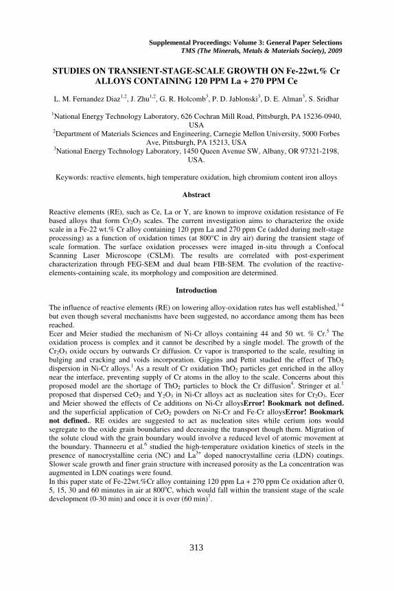
The terminology used to explain the results is summarized in figure 1. The RE sites or RE
containing sites are meant to be the locations where RE were found before the oxidation and that
after the oxidation process happened to become big particles on the surface. By “far from RE
sites” it is meant all surface spots which are not in the immediate surroundings of the RE sites
314
and includes both, nodules and ground material. On the contrary, by “sites near from RE” it is
meant the immediate locations surrounding the RE formed particles. Oxide nodules are the
nodules formed during oxidation and found in the surface afterwards. The bulk is the ground
material behind the nodules.
Figure 1. SEM picture showing the different parts analyzed in the surface of the samples.
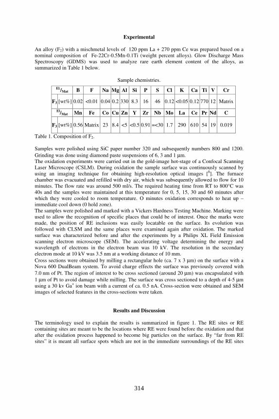
Morphology
Figure 2. SEM images of F2 samples oxidized for 5, 15, 30 and 60 min. The SEM images show
the nucleation site, area around nucleation sites, area far from the nucleation sites and the ridges
formed on the surface. (Magnification: 50000X for nucleation particle, near nucleation particle
and far from the nucleation particle and 12000X for ridges).
The oxidation of the samples was followed by CSLM. The surface starts oxidizing when the
temperature reaches 600°C, corresponding to 30 s according to the temperature profile explained
in the experimental section. The formation of ridges starts around 1:45 min oxidation. From this
point on, nothing seems to be happening in the surface but scale growing.
500 nm 2 µm
500 nm
500 nm
500 nm
500 nm
500 nm 500 nm
500 nm 500 nm
500 nm
500 nm
500 nm
500 nm
2 µm
2 µm
5 min
15 min
30 min
60 min
RE site Near RE site Far RE site Ridges
2 µm
315
The samples used in the heating up – immediate cool down acquired a very intense copper color
and their examination by SEM resulted in the obtaining of blurry images. Nevertheless, only few
small nodules and no ridges are observable in the surface.
In figure 2 SEM are shown pictures from different points of the surface: on particles, area
surrounding particles, area far from particles (50000X) and ridges (12000X); at different
oxidation times: 5, 15, 30 and 60 minutes.
The pictures show that the oxide nodule size is larger far from the RE-particles than in their
vicinity, when comparing columns 2 and 3 in figure 2.
However, the size of the oxides formed on the particles appears to be largest (column 1 in fig. 2).
With time, the increase of oxide particles size on the particles is evident. Also an increase of the
oxide particle size is observable with time in the area surrounding the particles.
The size of the oxide particles far from the nucleation sites increase from 5 to 30 minutes. After
60 minutes the grain size seems comparable to the size after 30 minutes.
Regarding ridges, after 5 minutes oxidation they are small and barely distinguishable. After 15
minutes of oxidation they are well visible. The size of the nodules constituting the ridges size
seem to be comparable at 15, 30 and 60 minutes.
Composition
The compositional evolution of the reactive elements containing sites with the oxidation time is
summarized as follows: in general it can be said that reactive elements (Ce, La, Ti) as well as the
bulk material (Fe) decrease with time at this sites while O, Cr, and Mn increase. Maybe La has a
tendency to diffuse towards the surface but when the oxidation time is long enough, Cr and Mn
oxides will bury it under an oxides layer. This indicates the formation of chromium and
manganese oxides over the reactive elements, forming a layer covering and burying them, which
supports the hypothesis of RE acting as nucleation sites for chromium and manganese oxides.
The comparison of composition evolution with time between areas surrounding RE containing
sites and areas far from them are shown yields the following results: Ti amount is very little in
the surface and its behavior is the same in both areas, far and near RE sites. Manganese is much
higher in case of sites far from the surface, increasing with the oxidation time, while RE
surrounding areas are shown to be depleted in this element. Around RE sites the amount of Mn
tends to decrease with time. Depleted Mn zones are also shown to be Cr depleted at 5 and15
minutes oxidation, while after this oxidation time the quantities of Cr are comparable far and
near RE (being a bit higher near them). The explanation for this phenomenon could be the fact
that RE are acting as nucleation sites for Cr and Mn oxides. At short oxidation times REs are
attracting all Cr around to become wrapped with Cr oxides. Longer oxidation times allow Cr to
diffuse towards RE sites and no Cr depleted area is observed any more. The explanation is not
valid for Mn due to its much lower concentration in the bulk material. Regarding Fe, RE
surrounding areas are rich in this element at low oxidation times (5 and 15 minutes; due to the
lack of Cr and Mn). At 30 minutes its value drastically decreases in these zones, because of the
higher amount of Cr. Regarding Fe is the bulk material, it can be said that at 60 minutes its value
far and near the RE sites is comparable.
Regarding the ground composition evolution with oxidation time, it can be said that the initial
steel evolves to a mixture of Fe, Cr and Mn oxides (spinel) which is getting richer with time in
Cr and Mn oxides, diminishing the quantity of Fe. Cr and Mn are diffusing towards the surface
with the increasing oxidation time. The increase/decrease of the elements diminishes its
magnitude from 30 minutes (O from 15 min) indicating that a steady state is being reached and
therefore the transient stage may be over, which is in good agreement with the literature values.9
The composition near the particles is pretty much comparable to the bulk composition at all
times, indicating that the presence of RE is avoiding the formation of chromium oxide nodules.
316

This agrees with the results shown in the previous section, where it can be seen the zones near
the particles being morphologically comparable to the bulk material.
The study of evolution of nodules composition with oxidation time shows that the first nodules
can be observed after 5 minutes oxidation. These initial oxide nodules are very rich in Fe. With
oxidation time the composition evolves to oxides richer in Cr and Mn and poorer in Fe.
As oxide nodules, ridges are found to be formed in the surface after 5 minutes of oxidation. They
are made out mainly of Fe, Cr, O, Mn and some Ti. A parallel behavior between Cr and Mn can
be observed. Oxygen seems to reach a steady state after 15 minutes, as well the amount of Ti
does not vary very much from that time.
The evolution with oxidation time of Fe, Cr, O, Mn and Ti on different parts of the surface can
be summarize as follows:
• Fe: bulk > nodules > ridges (irregular behavior) ~ particle. Decreases with time, in all
cases.
• Cr: ridges > nodules > bulk > particle (analogue behavior in ridges and nodules).
Increases with time, in all cases.
• Mn: ridges > nodules > particle > bulk (but for 30 minutes, dramatic decrease in the
rigdes, keeps tendency in the nodules). Tendency to increase in all cases.
• Ti: ridges > particle > bulk = nodules. Increases with time in the ridges but decreases in
the particles. No significant in the bulk or in the nodules.
Dual Beam FIB Cross-Sections
In figure 3 are shown the cross sections obtained for the different oxidation times: before and
after 0, 5, 15, 30 and 60 minutes (figure 3).
The cross section of a RE inclusion before oxidation showed a void which showed not to have
any surrounding layer. The dimensions of this feature are: height - 1.10 µm, width – 0.44 µm,
diagonal – 0.96 µm. A region showing different color during the cross-sectioning is found.
Images at higher magnification show intense different color forming a layer parallel to the
surface.
The cross sectioned particle in the sample corresponding to the 0-holding zone shows no oxide
layer is observed after this quick heating. Two phases seem to be differentiated in the RE
particle. In this case the dimensions of the found particle were of 0.72 µm height and 2 µm
width. The particle in this case seems to be porous.
The images obtained after cross-sectioning of sample after 5 minutes oxidation show no rides on
top of grain boundaries or even no scale are observable in the sample. The RE particle found
have the following dimensions: width – 1.14 µm, height 1.18 µm, diagonal – 1.17 µm.
After 15 minutes oxidation, the RE particle found had the following dimensions: height –
0.63 µm, width – 1.73 µm, diagonal – 0.96 µm. An average of the scale thickness in the cross-
sectioned area gave a thickness of 69 nm, nevertheless the layer is quite inhomogeneous and
nodules are often found. Their height is around 0.17 µm. There are other areas where the scale
thins and the effect is the contrary: it seems the bulk is forming a nodule surrounded by the
oxides layer. The ridges formed on the alloy grain boundaries have the following dimensions:
height – 302 nm, width – 616 nm. Also a small amount of internal oxidation could be observed.
Preliminary TEM studies were carried out in a comparable RE dual phase particle of F2 samples
oxidized for 15 minutes. According to the EDX results on the TEM sample, Ti segregated in the
lighter gray part of the particle, and the darker part segregate RE elements. if this is the case for
this FIB particle, and given the fact that Ti sometimes segregates with RE elements, we expect
the lighter gray part to be some Ti phase. But further EDS work is needed to confirm this
postulation.
According to this initial results a division of the different phases in the particle can be made as
shown in figure 3: the region marked as I compressing the lighter phase would be made out of Ti
317

oxides; region II, the darker phase, would be made out of RE oxides and a thinner layer or
chromium oxides would be the outermost layer, marked as region III.
Figure 3. Cross section of F2 samples before and after 0, 5, 15, 30 and 60 minutes oxidation.
After 30 minutes oxidation a dual phase RE particle can be again seen. The dimensions are:
height – 1.26 µm, width – 1.74 µm, diagonal – 1.73 µm. The average of scale thickness, 66 nm,
was comparable to the thickness found after 15 minutes oxidation. As in the 15 minutes case,
nodules were found, with average dimensions of height – 0.20 µm, width – 0.26 µm. Also
thinner sections in which the scale seemed to be surrounding a nodule formed by the bulk alloy
were observed. Grain boundaries were found to be about the same height as after the ones
formed after 15 minutes oxidation, but broader: height – 358 nm, width – 661nm. After this
oxidation time, a lighter layer underneath some sections of the scale could be observed.
After 60 minutes oxidation, the RE particle shows again two phases being in this case the lighter
phase predominant. The dimensions of the particle are: height – 1.62 µm, width – 1.87 µm,
diagonal – 1.72 µm. The thickness of the scale found in this case was of 109 nm (thicker than for
15 and 30 minutes oxidation). Nodules were also found in the scale: height – 0.19 µm, width –
0.21 µm. Their size is comparable with the other oxidation times. But in this case voids could be
seen in these parts of thicker scale. The opposite nodules, which a lighter core and where the
scale is thicker where shown to be in these case made out of a lighter material than the bulk. This
lighter layer can be also found in some regions underneath the scale. Regarding ridges, they were
found again on top of grain boundaries: height – 426 nm, width – 1014 nm. The ridges are in this
case a bit higher but broader.
In figure 4 the evolution in width and height of measured ridges with oxidation time is shown.
The width of ridges is superior to its height and they tend to broaden more significantly with
oxidation time while it could be said that the height slightly increases from 15 to 30 minutes and
then it keeps constant. Ridges grow on top of grain boundaries10
(figure 3) presumably because
of preferential diffusion of Mn and Cr. It is also logical to assume that formation of grain
boundaries in the scale will take place over the alloy grain boundaries. Then the fact of
preferential ridges broadening over growing in the longitudinal dimension could be indicative of
RE blocking grain boundaries in the scale, while more Cr and Mn are being provided from the
alloy and therefore these oxides have to be accommodated, resulting in broader ridges at the
interface.
318
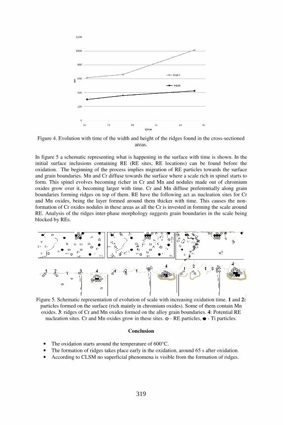
Figure 4. Evolution with time of the width and height of the ridges found in the cross-sectioned
areas.
In figure 5 a schematic representing what is happening in the surface with time is shown. In the
initial surface inclusions containing RE (RE sites, RE locations) can be found before the
oxidation. The beginning of the process implies migration of RE particles towards the surface
and grain boundaries. Mn and Cr diffuse towards the surface where a scale rich in spinel starts to
form. This spinel evolves becoming richer in Cr and Mn and nodules made out of chromium
oxides grow over it, becoming larger with time. Cr and Mn diffuse preferentially along grain
boundaries forming ridges on top of them. RE have the following act as nucleation sites for Cr
and Mn oxides, being the layer formed around them thicker with time. This causes the non-
formation of Cr oxides nodules in these areas as all the Cr is invested in forming the scale around
RE. Analysis of the ridges inter-phase morphology suggests grain boundaries in the scale being
blocked by REs.
Figure 5. Schematic representation of evolution of scale with increasing oxidation time. 1 and 2:
particles formed on the surface (rich mainly in chromium oxides). Some of them contain Mn
oxides. 3: ridges of Cr and Mn oxides formed on the alloy grain boundaries. 4: Potential RE
nucleation sites. Cr and Mn oxides grow in these sites. - RE particles, - Ti particles.
Conclusion
• The oxidation starts around the temperature of 600°C.
• The formation of ridges takes place early in the oxidation, around 65 s after oxidation.
• According to CLSM no superficial phenomena is visible from the formation of ridges.
319

• SEM results show an increase in the oxide particle size on the nucleation site, in the
surroundings of the nucleation site and far from it with time.
• Within the first 90s phenomena as formation of ridges or opening of holes in the surface
can be observed. After that all that seems to be happening in the surface is the growing of
the scale in terms of oxide particles size. The morphology of the scale at 5 min is difficult
to distinguish but after 15 minutes it is well formed (ridges and oxide particles in all
points of the surface well distinguishable).
• Ce and La are localized at specific sites in the surface (inclusions) and no significant
amount can be found forming ridges, nodules or ground. RE act as nucleation sites for Cr
and Mn oxides. They tend to be wrapped and buried by these oxides with increasing
oxidation time.
• Depletion zones around RE are remarkable at low oxidation times (5 and 15 minutes)
regarding Cr. These areas are depleted of Mn, decreasing its amount with time.
• Areas around RE sites are comparable to bulk material, both morphologically and
compositionally.
• Cr and Mn diffuse towards the surface with time.
• Transient stage of oxidation seems to be over at 30 minutes.
• Ground material evolves with time, transforming spinel into oxides richer in Cr and Mn
and poorer in Fe. The same behavior observed for oxide nodules and ridges.
• Ti is mainly present at RE sites and ridges.
• Fe is more significant in the ground material, while Cr, Mn and Ti are more significant in
ridges.
• Oxidation features (scale, nodules, ridges) are only observable after 15 minutes oxidation.
• The thickness of the scale is comparable in samples oxidized for 15 and 30 minutes, but
increases for 60 minutes oxidation.
• The RE particles found seem to segregate into dual phase particles with oxidation time.
The characterization and differentiation of these particles is future TEM work.
• Two different kind of nodules are found in the scale. The dark nodules seem to be just
scale formed while the lighter ones resemble to the lighter phase found in the RE
particles. The characterization of these nodules should also be done by TEM.
• Ridges are found in all cases above alloy grain boundaries. With increasing oxidation
time they tend to broaden but not significant increase in height is detected. This suggests
RE could be blocking Cr, Mn and Ti diffusion through the scale grain boundaries.
• Lighter layers can be found in some regions beneath the scale. It could be a possible
relation to the lighter phase found in the RE particles.
1 J. Stringer, B. A. Wilcox and R. I. Jaffee: Oxid. Met. Vol. 11(1972), p. 5.
2 G. M. Ecer and G. H. Meier: Oxid. Met. Vol. 13 (1979), p. 159.
3 M. Ecer, Singh and G. H. Meier: Oxid. Met. Vol.18 (1982), p. 55.
4 C. S. Giggins and F. S. Pettit: Met. Trans. Vol. 2 (1971), p. 1071.
5 G. M. Ecer and G. H. Meier: Oxid. Met. Vol. 13 (1979), p. 119.
6 R. Thanneeru, S. Pati., S. Deshpande and S. Seal: Act. Mat. Vol. 55 (2007), p. 3457.
7 M. Hajduga and J. Kučera: Oxid. Met. Vol. 29 (1988), p. 121.
8 H. Yin, H. Shibata, T. Emi and M. Suzuki: ISIJ Int. Vol. 37 (1997), p. 936.
9 M. Hajduga and J. Kucera: Oxid. Met. Vol. 29 (1988), p. 121.
10 L. M. Fernandez Diaz, J. Zhu, G. R. Holcomb, P. D. Jablonski, D. E. Alman, S. Sridhar,
presented at the 4th
International Conference on Diffusion in Solids and Liquids DSL-2008, 9-11
July 2008, Barcelona, Spain. (Accepted for publication).
320