subselective measurement of coronary blood flow … measurement of coronary blood flow velocity...
TRANSCRIPT

1332 lACC Vol. 8. No . 6December 1986:1332-40
Subselective Measurement of Coronary Blood Flow Velocity Using aSteerable Doppler Catheter
DAVID H. SIBLEY, MD, HUNTLY D. MILLAR, MSc, CRAIG J. HARTLEY, PHD ,
PATRICK L. WHITLOW, MD, FACC
Birmingham. Alabama
The accuracy of coronary arteriography to predict obstruction to coronary blood flow has recently been questioned. Assessment of coronary hemodynamic variablesand vasodilator reserve may provide more reliable information regarding the significance of coronary stenosis. To provide a clinically safe and reliable methodof measuring coronary blood flow velocity and coronaryflow reserve, a 3F steerable Doppler catheter capable ofsubselective placement in the coronary circulation wasdeveloped and validated in an animal model. Coronaryblood flow velocity measured with the catheter correlated with simultaneous measurements by a previouslyvalidated external cuff type Doppler probe (r = 0.97),coronary sinus flow collections (r = 0.78) and femoral
The ability to predict signific ant impairment of coron aryblood flow from visual interpretation of coronary arteriograms is open to question . The effect of a coronary stenosi son myocardial perfusion is an integrated response of ananatomic-hemodynamic system. Severity of coronary stenosis is one part of the anatomic system and percent stenosisalone does not describe the supply of blood flow to themyoc ardium. Coronary perfusion pressure , coronary vascular tone, collateral flow and stenosis configuration allaffec t myocardial perfusion. Kirkeeide et al. ( I) showedthat coronary flow reserve is a single functional measure ofcoronary stenos is severity that is determined by all of thehemodynamic variables and geometric characteristics of that
From the Division of Card iovascular Disease, Department of Medicine .Univers ity of Alabama at Birmingham . Birmingham. Alabama. This studywas supported in part by a grant from the Cardiovascular Research andTraining Ce nter at the University of Alabama at Birmingham . Birmingham .Alabama. and Grant 5T32 HL07327 from the National Heart . Lung. andBlood Inst itute. Nat ional Institutes of Health . Bethesda. Maryland .
Manuscript received March 10 , 1986; revised manuscript received June10. 1986. accepted July 7. 1986.
Address for reprints : David H. Sibley . MD. Associate in Cardiolo gy.Division of Cardiovascular Diseases. University of Alabama at Birmingham. University Station. Birmingham. Alabama 35294.
101986 by the American College of Cardiology
artery flow collections (r = 0.96). The extravascularDoppler cuff measurements of rest flow velocity andvasodilator reserve were not significantly different withor without the catheter in the artery, indicating that theDoppler catheter caused no obstruction to blood flow.
The Doppler catheter has recorded stable and reproducible signals without complications in 28 patients , including 62 separate arterial cannulations. Thus: I) the3F Doppler coronary catheter is nonobstructing, steerable and safe; 2) there is an excellent correlation of bloodflow velocity with volume collections; and 3) the catheterprovides a reliable method of determining coronary bloodflow velocity and coronary vasodilator reserve.
(J Am Coil Cardiol 1986;8:1332-40)
stenosis. White et al. (2) found that physiologic assessmentof coronary obstructive disease using coronary flow reservedoes not correlate well with percent diameter stenosi s byangiography.
Coronary blood flow velocity and vasodilator reservehave been measured during open chest surgery using electromagnetic flow meters (3) and an epicardial suction Doppler probe (4) . However, hemodynamic data can be obtainedby these method s only during surgical procedures, severelylimiting data collection . Coronary sinus thermodilutiontechn iques cannot selectively measure flow in a single coronary artery with the exception of the left anterior descendingartery, Xenon clearance techniques measure regional bloodflow, but cannot provide a cont inuous assessment of secondto second changes in selective coronary blood flow. Also,coronary flow rates greater than 200 mllmin per 100 g cannotbe accurately measured and more than two flow measurements are not possible with current clearan ce techniques(5) . Videodensitometric method s are able to assess rapidchanges in regional coronary blood flow; however , determination of coronary flow reserve by digital subtractionangiography requires precisely timed contrast injections andpatient cooperation. The system required to obtain such datamay be too complex for routine clinical use (6,7).
0735-1097/86/ $3.50

JACC Vol. H. No.6December IYX6:1332-40
SIBLEY ET AL.MEA SUREMENT OF CORONARY BLOOD FLOW
1333
Hartley and Cole (8. 9) developed the first Doppl er coronary catheter capable of on-line continuous measurementsof coronary blood flow veloc ity and coronary flow reservein humans. A 20 MHz piezoelectric crystal was placed atthe tip of a Sones catheter. Because of its size (SF). thiscatheter was too large to be placed subselectively in thecoro nary circulation. More recentl y. W ilson et al. (10 ) valida ted the ability of a 3F coro nary catheter with a sidemounted piezoelectric crystal to provide accurate continuouson-line measurements of co ronary blood flow velocity andvasodi lator reserve . However . poor signal quality was obtained in 12 of 70 patients studied with this catheter. Alimitation of this system appeared to be inability to steerthe catheter away from small branch vessels and the arterialwall . Because no catheter lumen was available for guidewire placement. it was difficult to contro l direction of thecatheter in the coronary circul ation.
To provide a consistent and clinica lly safe method ofmeasuring coro nary blood flow velocity and coronary flowreserve. we developed a 3F Doppler coronary catheter withan intern al lumen that accommodates a standard angioplastysteera ble guide wire. Th e guide wire allows precise placement and easy adjustment of the catheter in the coron arycirculation. The purpose of this study was to assess thereliabilit y of this catheter in determining coronary bloodflow veloci ty and coronary vasodi lator reserve. We reportdata to docum ent that this catheter is nonobstructing . steerable and safe and has a linear co rrela tion of blood flowvelocity with flow over a wide range of values .
MethodsCatheter design. A circul ar 20 MHz ceramic crystal
capable of both transmitting and receiving acoustic signalsis attached to the tip of a USCI Rentrop reperfusion catheter(Millar Instruments). This catheter is 4F in the main body.with a wire weave for torque control. and the distal 20 cmtip tapers to 3F ( I mm outer diameter). Two wires attachedto the crystal pass within the catheter between the outercatheter sheath and an inner lumen tubing that traverses thefull length of the catheter. The intern al lumen is 0 .015 inch(0.038 ern) and will accomm odate a 0. 014 inch (0. 0356
ern) flexible steerable guide wire (Fig. I). An electricalleakage test was zero at either polarity. using a device capable of detecting I /-LA at 600 Y.
Instrumentation. A range-gated 20 MHz pulsed Doppler velocimeter (Baylor College of Med icine) conn ected tothe proximal end of the catheter detects the Doppler shiftof the echoes from the blood cells within the adju stablesample volume which is I to 12 mm distal to the cathetertip. The Doppler frequency is related to the velocity of thereflectors (red blood ce lls) by the Doppler equation: ,M =2.F.Y/C.cos( 0). where M is the Doppl er shift frequency. Fis the transmitter frequency (20 MHz) . Y is the averagevelocity of the blood cells with in the sample volume. C isthe speed of sound in blood ( 1,500 m/s) and () is the anglebetween the sound beam and the direction of blood flow.Using an end-mounted crystal with the catheter parallel ( ± 20°)to the vessel axis, cos(0) = I ± 6%, and the relation between the Doppler shift and veloci ty is appro ximately 3.75cm/s per kHz. The 20 MHz ultrasound frequ ency and the62 .5 kHz pulse repetition frequency allow the record ing ofveloci ties up to 115 cm/s at I to 12 mm distances from thecrystal face.
The pulsed Doppl er velocimeter provides two simultaneous outputs that represent the spatially averaged veloc itywithin the sample volume. Each is a measure of the frequenc y of zero crossi ngs of the Doppl er audio signal ca librated at 0 .25 Y/kH z, but eac h has different amounts offiltr ation . The phasic output displays the pulsatile velocitysignal with a band pass from direct current to 15 Hz, andthe mean output (which elimin ates the pulsations) has a bandpass from direct current to 0 .25 Hz. Because of the uncertainties in relating the measured frequency shift within thesample volume to the average velocity across the lumen orto volume flow, the data are expressed in terms of themeasured frequency shift in kHz. The phasic and meanvelocity signals from the pulsed Doppler velocimeter wererecorded on a Hewlett-Packard strip chart recorder (model7758 0) with bioelectric ampl ifiers .
Invitro studies . The relation between the mean Dopplerfrequency shift measured by the Doppler catheter and timedvolum e collections was determ ined by placing the Doppl ercatheter in a 2 .6 mm diameter polyethylene tube system
Figure I. The 3F Doppler coronary catheter in profile.A lumen extends the entire length of the catheter andwill accommodate a 0.014 inch (0.0356 cm) guidewire.
epoxyinternal sheath ICry,stal
wire leads catheter body
1 .'~'lJnd hole
0.039" i -.J!1'" T " . .
I0.015" , 0.014 gUldewlre
1 " . 1
1334 SIBLEY ET AL.MEASUREMENT OF CORONARY BLOOD FLOW
JACC Vol. X. No.6December 19X6:1332-40
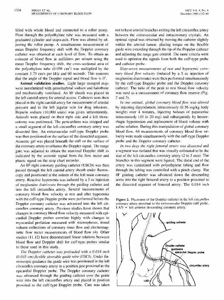
Figure 2. Placementof the Dopplercatheter in the left circumflexcoronary artery proximal to the extravascular Doppler cuff probe.LAD = left anterior descending coronary artery.
not to have arterial branches exiting the left circumflex arterybetween the extravascular and intracoronary crystals. Anoptimal signal was obtained by moving the catheter slightlywithin the arterial lumen. placing torque on the flexibleguide wire extending through the tip of the Doppler catheterand adjusting the range gate control. The same criteria wereused to optimize the signals from both the cuff-type probeand catheter probe.
Sixty-seven measurements 01" rest and hyperemic coronary blood flow velocity (induced by a 5 cc injection ofmeglumine diatrizoate) were then performed simultaneouslyby the cuff-type Doppler probe and the Doppler coronarycatheter. The ratio of the peak to rest blood flow velocitywas used as a measurement of coronary flow reserve (Fig.2 and 3).
In one animal, global coronary blood flow was alteredby injecting dipyridamole intravenously (0.56 mg/kg bodyweight) over 4 minutes. injecting boluses of papaverineintravenously (10 to 20 mg) and subsequently by hemorrhagic hypotension and replacement of blood volume withsaline solution. During this manipulation of global coronaryblood flow, 64 measurements of coronary blood flow velocity were made simultaneously with the cuff-type Dopplerprobe and the Doppler coronary catheter.
In two dogs the right femoral artery was dissected anda segment was isolated that was visually estimated to be thesize of the left circumflex coronary artery (2 to 3 mm). Thebranches in this segment were ligated. The distal end of theartery was cannulated with polyethylene tubing and flowthrough the tubing was controlled with a pinch clamp. The8F guiding catheter was advanced down the descendingaorta into the right femoral artery to a position proximal tothe dissected segment of femoral artery. The 0.014 inch
filled with whole blood and connected to a roller pump.Flow through the polyethylene tube was measured with agraduated cylinder and stopwatch. Flow was altered by adjusting the roller pump. A simultaneous measurement ofmean Doppler frequency shift with the Doppler coronarycatheter was obtained at each level of flow. To obtain anestimate of blood flow in milliliters per minute using themean Doppler frequency shift. the cross-sectional area ofthe polyethylene tube (0.056 crrr') was multiplied by theconstant 3.75 cm/s per kHz and 60 seconds. This assumesthat the angle of the Doppler signal and blood flow is 0°.
Animal validation studies. Eight large mongrel dogswere anesthetized with pentobarbital sodium and halothaneand mechanically ventilated. An 8F sheath was placed inthe left carotid artery for arterial access. Catheters were thenplaced in the right carotid artery for measurement of arterialpressure and in the left jugular vein for drug infusions.Heparin sodium (10,000 units) was given intravenously.Animals were placed on their right side and a left thoracotomy was performed. The pericardium was stripped anda small segment of the left circumflex coronary artery wasdissected free. An extravascular cuff-type Doppler probewas then positioned on the surface of the dissected segment.Acoustic gel was placed beneath the cuff on the surface ofthe coronary artery to enhance the Doppler signal. The rangegate was adjusted to obtain the maximal Doppler shift asindicated by the acoustic signal from the flow meter andphasic signal on the strip chart recorder.
An 8F right coronary guiding catheter (USCI)) was thenpassed through the left carotid artery sheath under fluoroscopy and positioned at the ostium of the left main coronaryartery. Reactive hyperemia was induced by a 5 cc injectionof meglumine diatrizoate through the guiding catheter andinto the left circumflex artery. Several measurements ofcoronary blood flow velocity at rest and after hyperemiawith the cuff-type Doppler probe were performed before theDoppler coronary catheter was advanced into the left circumflex coronary artery. Previous studies have shown thatchanges in coronary blood flow velocity measured with epicardial Doppler probes correlate highly with changes inmyocardial perfusion measured with microspheres, timedvolume collections of coronary sinus flow and electromagnetic flow meter measurements of blood flow (4). Otherstudies (11,12) have demonstrated linear relations betweenblood flow and Doppler shift for cuff-type probes similar
to those used in this study.The Doppler catheter was preloaded with a 0.014 inch
(0.035 cm) flexible steerable guide wire (USC/). Under fluoroscopic guidance the guide wire was positioned in the leftcircumflex coronary artery in a position just proximal to theepicardial Doppler probe. The Doppler coronary catheterwas advanced through the guiding catheter over the guidewire into the left circumflex artery and placed in positionproximal to the cuff-type Doppler probe. Care was taken
v
JACe Vol. X. No, 6December /986: 1332-40
SIBLEY ET AL.MEASUREMENT OF CORONARY BLOOD FLOW
1335
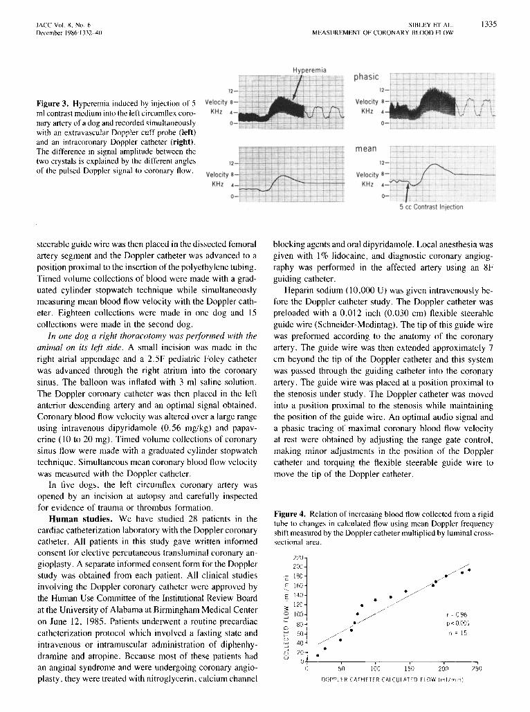
Figure 3. Hyperemia induced by injection of 5ml contrast medium into the left circumflex coronary artery of a dog and recorded simultaneouslywith an extravascular Doppler cuff probe (left)and an intracoronary Doppler catheter (right).The difference in signal amplitude between thetwo crystals is explained by the different anglesof the pulsed Doppler signal to coronary flow.
; : .I \ .
\0.
mean
12- j- ·j • • • •
Ve locity 8-.-t~ _ ~ ~- ,~,.:KHz ~-~t -' -- .. :.~
.._.1-' ... L . . .0- ' ,- . . t. .
I L- 0_5 cc Contrast Injection
steerable guide wire was then placed in the dissected femoralartery segment and the Doppler catheter was advanced to a
position proximal to the insertion of the polyethylene tubing.Timed volume collections of blood were made with a graduated cylinder stopwatch technique while simultaneouslymeasuring mean blood flow velocity with the Doppler catheter. Eighteen collections were made in one dog and 15
collections were made in the second dog.In one dog a right thoracotomy was performed with the
animal on its left side. A small incision was made in theright atrial appendage and a 2.5F pediatric Foley catheter
was advanced through the right atrium into the coronarysinus. The balloon was inflated with 3 ml saline solution.
The Doppler coronary catheter was then placed in the leftanterior descending artery and an optimal signal obtained.Coronary blood flow velocity was altered over a large rangeusing intravenous dipyridamole (0.56 mg/kg) and papaverine (10 to 20 mg). Timed volume collections of coronarysinus flow were made with a graduated cylinder stopwatchtechnique. Simultaneous mean coronary blood flow velocitywas measured with the Doppler catheter.
In five dogs, the left circumflex coronary artery wasopened by an incision at autopsy and carefully inspectedfor evidence of trauma or thrombus formation.
Human studies. We have studied 28 patients in thecardiac catheterization laboratory with the Doppler coronarycatheter. All patients in this study gave written informedconsent for elective percutaneous transluminal coronary angioplasty. A separate informed consent form for the Dopplerstudy was obtained from each patient. All clinical studiesinvolving the Doppler coronary catheter were approved bythe Human Use Committee of the Institutional Review Board
at the University of Alabama at Birmingham Medical Centeron June 12, 1985. Patients underwent a routine precardiac
catheterization protocol which involved a fasting state andintravenous or intramuscular administration of diphenhy
dramine and atropine. Because most of these patients hadan anginal syndrome and were undergoing coronary angioplasty, they were treated with nitroglycerin, calcium channel
blocking agents and oral dipyridamole. Local anesthesia wasgiven with I% lidocaine, and diagnostic coronary angiog
raphy was performed in the affected artery using an 8Fguiding catheter.
Heparin sodium (10,000 U) was given intravenously before the Doppler catheter study. The Doppler catheter waspre loaded with a 0.012 inch (0.030 em) flexible steerableguide wire (Schneider-Medintag). The tip of this guide wirewas preformed according to the anatomy of the coronaryartery. The guide wire was then extended approximately 7
ern beyond the tip of the Doppler catheter and this systemwas passed through the guiding catheter into the coronary
artery. The guide wire was placed at a position proximal tothe stenosis under study. The Doppler catheter was movedinto a position proximal to the stenosis while maintainingthe position of the guide wire. An optimal audio signal anda phasic tracing of maximal coronary blood flow velocityat rest were obtained by adjusting the range gate control,making minor adjustments in the position of the Dopplercatheter and torquing the flexible steerable guide wire tomove the tip of the Doppler catheter.
Figure 4. Relation of increasing blood flow collected from a rigidtube to changes in calculated flow using mean Doppler frequencyshift measured by the Doppler catheter multiplied by luminal crosssectional area.
220
200 /.180
// ../ •cE 160 //,/--e
<,
/E 140 •• ./
~120 • ,/.
0 100 . // r =096--' .>u;
80~ P< 00010
:;1/~w n = 15e-
/~uw--' •..-J0 •U
I I , I ,0 50 100 150 200 250
DOPPLER CATHETER CALCULATED FLOW (ml Zmin l

1336 SIBLEY ET AL.MEASUREMENT OF CORONARY BLOOD FLOW
lACC Vol. 8. No. 6December 1'186:1332-40
12
Figure 6. Relation of 64 simultaneous measures of mean coronaryblood flow velocity by a cuff-type Doppl er probe and an intracoronary Doppler catheter. Mean velocity was altered over a widerange ( 1.53 to 10.6 kHz).
I••
r = 0.97p <O.OOIn = 64
•••
••• •• •I.
•...,-....
2 3 4 5 6 7 8 9 10
CUFF TYPE DOPPLER PROBE VELOCITY (KHzl
~.
If'•
-;:;:r::.::; 10=:uod 8>a::w::. 6:r::;(u~ 4a::erZo~ 2ua::~ 0+-- -,-- -,-- ---,-- ---,-- ---,-- ---,-- -,- -,- -,- -& 1 11s
To assess the vasodilator reserve of the particular corollary artery under study. coronary blood flow velocity wasmeasured after intracoronary injection of meglumine diatrizoate. Baseline recordings were made of phasic and meancoronary blood flow velocity. Meglumine diatrizoate (5 rnl)was injected through the guiding catheter into the coronaryostium. Changes in blood flow velocity were recorded continuously until the flow velocity returned to baseline. Theextra total radiation dose from the fluoroscopic guidancewas less than 0.05 rad per case.
Statistical analysis. Pearson correlation coefficients wereused to compare the Doppler catheter mean flow velocitywith other measurements of blood fl ow. A paired t test wasused to assess differences in coronary flow velocity beforeand after left circumflex coronary cannulation with the Doppler catheter. Probability values were considered significantif less than 0.05.
Results
In Vitro Studies
Measurements of blood flow calculated from the meanvelocity and polyethylene tube cross-sectional area wereclosely correlated with timed collections of blood flow overa broad range (r = 0.96. range = 14 to 192 ml/min,slope = 10.15) (Fig. 4).
Animal Validation Studies
Femoral artery volume velocity measurements. Thefemoral artery volume collections were signifi cantly correlated with mean blood now velocity measured by theDoppler catheter in two dogs (Fig. 5). Fifteen volume collections were made from the first dog with a range of 6 to266 ml/rnin (r = 0.98. slope 23.72). Eighteen volume collections were made from the second animal with a range of19 to 282 mllmin (r = 0.96, slope 28.45).
Cuff-t ype Doppler probe versus intracoronary Doppler catheter. Sixty-four simultaneous measurements ofcoronary blood flow velocity at rest were obtained with thecuff-type Doppler probe placed immediately distal to theintracoronary Doppler catheter position (Fig. 6). Coronaryblood flow velocity was altered over a wide range of coronary now velocity and the paired measurements correlatedhighly (r = 0.97. range 1.53 to 10.6 kli z, slope = 1.03).
Sixty-seven simultaneous measurements of peak to restvelocity ratio were obtained with the cuff-type Doppler probepositioned immediately distal to the intracoronary Dopplercatheter (Fig. 7). The mean value for the external Dopplercuff was 3.15 kHz (SD = 1.50) and 2.81 (SO = 1.19) forthe Doppler catheter. The 67 peak to rest velocity ratio
r = 0.72p< 0 0001
n = 67
00
2 3 4 5 6 8
CUFF TYPE DOPPL ER PR OBE VELOCITY (KHz)
.;,... .., .· .;;set I•••
•1+--....,.------,---r--..,..---,------r----,- -
1 9
~,."7 7:r:~
~ 6Uog 5a::wt;:j 4;=;) 3a::<.oJ...J
it 2ao
Figure 7. Relat ion of 67 simultaneous measures of peak to restcoro nary flow velocity ratio by a cuff-type Doppl er probe and anintracoro nary Doppler catheter.
8
300250200150
Volume ml /rnin
10050
• Dog 1
o Dog 210
8NI 6~
~ 400Q)
2>
00
- 2J
Figure 5. Relation of pulsat ile in vivo blood flow in a fem oralartery to simultaneo us mean blood flow ve locity in that arterymeasured by a Doppler catheter in two dogs.
12

JACC Vol. 8. No.6December 1986:1332-40
SIBLEY ET ALMEASUREMENT OF CORONARY BLOOD FLOW
1337
Figure 9. Human phasicDoppler tracings fromthe leftmain(LM),left anterior descending (LAD), left circumflex (Lex) and right(RCA) coronary arteries. Note the greater amplitude of systolicflow velocity in the right coronary artery compared with the leftanterior descending artery. Dias = diastolic; Sys = systolic.
coronary artery induced by a 5 ml injection of megluminediatrizoate is seen in Figure 10. Figure II demonstrates restand peak coronary blood flow velocity measured proximalto a 90% stenosis of the middle left anterior descendingcoronary artery before and after this vessel was dilated bycoronary angioplasty.
Stable velocity recordings were obtained in all 62 coronary artery cannulations. No complications occurred duringthe procedures. There was no evidence of dissection orthrombus formation on subsequent angiograms.
Velocity 4
KHz
o .
: I·
Ve locity !4
KHz
'LAD: - .i • ..•
_J_ . , ... •
Velocity 4~' " :-:~ : .•. -=KHz ' ;. :.L. ".
2 L " " ; . -I .. .. _ .
o- . t: .t: ......, • ·. 1.
Sy. Dia.
Discussion
We have developed and validated a 3F Doppler cathetercapable of measuring blood flow velocity and vasodilatorreserve in the major arteries and branches of the coronarycirculation. The miniaturization of the Doppler crystal hasmade possible a I mm (outer diameter) catheter with a lumencapable of accommodating the same flexible steerable guidewire used in coronary angioplasty. An important advantageof this system is that it can be safely and easily passed inthe major arteries of the coronary circulation. The steerablenature of the guide wire catheter system allows precise adjustment and positioning of the Doppler crystal. This wasnot possible with previous Doppler catheters; consequently,the safety of positioning and stability of signals was notoptimal (8,10).
Comparison with previous studies. The cuff-type Doppler probe used for validation of our intracoronary catheteris very similar to the epicardial probe validated by previousinvestigators (4, II , 12). Their studies showed that changesin coronary blood flow velocity measured with the Dopplerprobe correlated highly with changes in myocardial perfusion measured with microspheres, timed volume collections
20
19 •~
E 18-<, •u17 •-"-
~ 16 •0 • •....JCL. 15Ul::> 14 • • r = 0.78zUi
13 • P = 0.003:-
"" 12 • n = 12<!z0 11""0 •u 10
45 5.0 5.5 6.0 6.5 7.0 75 8.0 8.5 9.0 9.5 10.0
LAD CORONARY FLOW VELOCITY (KHz)
Human Studies
The Doppler coronary catheter was used to measure coronary blood flow velocity in 28 patients undergoing electivepercutaneous transluminal coronary angioplasty. The procedures included 62 separate coronary artery cannulations38 in the left anterior descending, 14 in the left circumflexand lOin the right coronary artery-and I in a saphenousvein graft to the right coronary artery. Examples of thephasic tracings from the left main, right, left circumflex andleft anterior descending coronary arteries are shown in Figure 9. An example of a hyperemic response in the right
Figure 8. Relation between increases in mean bloodflow velocityin the left anteriordescending (LAD)coronary artery measured inone dog with a Dopplercatheterand simultaneous coronary sinusblood flow.
values were positively correlated (r = 0.72, range = 1.30to 8.12, slope = 0.902).
Coronary sinus volume collections. Twelve timed volume collections of coronary sinus flow were positively correlated with rest mean coronary blood flow velocity measured in the left anterior descending coronary artery(r = 0.78, range = 10.5 to 19.5 cc/min, slope = 1.15)(Fig. 8).
Evaluation of possible coronary obstruction producedby the Doppler coronary catheter. Measurements withthe epicardial cuff-type Doppler probe before the Dopplercatheter was placed in the coronary artery were averaged.The mean rest flow velocity was 3.12 kHz (SO = 1.62).The same number of epicardial Doppler measurements wereaveraged immediately after the Doppler catheter was placedin the left circumflex artery and the mean rest flow velocitywas 3.19 kHz (SO = 1.45). The mean peak to rest velocityratio was 3.04 (SO = 1.15) without and 3.18 (SO = 1.65)with the Doppler catheter in the left circumflex artery. Themeans were not significantly different using a paired t test.
Autopsy examination. In five animals a longitudinalincision was made in the left circumflex coronary arteryafter the Doppler studies. There was no gross evidence ofthrombus formation, dissection or intimal damage.

133X SIBLEY ET AL.MEASUREMENT OF CORONARY BLOOD Fl.OW
lAce Vol. X. No. f>December IYX6: 1.1.12 AO
50% Mid RCA StenosisMean
®
I I~.
Velocity 8 II
KHz 4;""--~-:-~.
oI
1 . .. !I - .j.-i· ..
veIOCitY8 f·~t · .~~ . .i.:::i 1-:,'KHz .. .. .; : .
4 .I . ,. : !I •
Of , ::: .1
o 5f 10Seconds
15
Figure 10. Contrast hyperemia (b) induced proximal to the 50% stenosis inthemid-right coronary artery (RCA) seenon the left (a).
5cc contrast injection
of coronary sinus flow. electromagnetic flow meter measurements of blood flow and timed collections of flow inperipheral arteries.
With our Doppler catheter. changes in mean rest bloodflow velocity in the left circumflex artery were comparedwith simultaneous measurements by this previously validated cuff-type probe. Over a large range of flow velocities(1.53 to 10.6 kHz) the correlation was linear (r = 0.97).There is a systematic difference of approximately I kHzbetween the Doppler catheter and cuff measurements offrequency shift. This is most likely attributed to the different
angles of the Doppler crystal in relation to blood flow.Theoretically. the Doppler cuff crystal is oriented at a 45°angle and the Doppler catheter crystal at a 0° angle in relationto blood flow.
When coronary vasodilator reserve was varied over alarge range. the values obtained from the cuff-type probeand Doppler catheter were positively and significantly correlated: however. the vasodilator reserve values were notas closely correlated as those found by Wilson et al. (10).Our measurements with the cuff-type Doppler probe beforeand after the Doppler catheter was placed in the coronary
99"10 Mid LAD Stenosis - Preangroplasty
Figure II. a, A99%stenosis of the leftanteriordescending artery ina 54 yearold man with NewYork Heart Association class III angina. b, Restandpeak coronary flow velocity measured proximal to the stenosis. c, The Doppler catheter inposition proximal to the stenosis with the guidewire extended out of the lumen. d, Afterangioplasty, the stenosis is reduced to 10%. e, Subsequent contrast hyperemia measured proximalto the stenosis.
15
°i_'1 l 1 I. t -\ i ~o I~ 20 30 40ISeconds
5cc contrast injection
10
Seconds
®
I I~ 1 ' .
r I, r ! 1li t . . _!! I,' .
Velocity 4L j' ~ -! . . t ' .,; .. I • I
KHz 2~~NlM\Or ' I i :: 1 .. ,.. . . ..
o 1 5
5cc contrast injection

lACC Vol. 8. No.6December 1986:1332-40
SIBLEY ET AL.MEASUREMENT OF CORONARY BLOOD FLOW
1339
artery showed no significant change in either rest meancoronary blood flow velocity or coronary vasodilator reserve . These data suggest that this intracoronary Dopplercatheter does not obstruct coronary flow and agree withWilson's data (10) using a similar 3F coronary catheter.
Validation studies. Data from our study substantiatethat change in mean coronary blood flow velocity (measuredin kilohertz shift) is linearly correlated with change in bloodflow. This validation was done in several ways . First wevaried blood flow through a rigid nonpulsatile tube andsimultaneously measured mean Doppler frequency shift withthe Doppler coronary catheter. The correlation coefficientwas 0.96 and agreed with previous results obtained by Marcus et al. (4) when validating an epicardial Doppler probe.Femoral artery collections of blood volume correlated wellwith simultaneous mean blood flow velocity measured withthe Doppler catheter (r = 0 .96). This is very similar to thecorrelations obtained by Cole and Hartley (8) from femoralartery collections using a larger 8F Sones Doppler catheter.The timed volume collections of coronary sinus flow weresignificantly correlated with rest mean coronary blood flowvelocity, but were less highly correlated (r = 0.78) thanother volume-velocity correlations reported in this study.This lower correlation is probably due to the difficulty inmaintaining the position of a balloon catheter in the coronarysinus over a I hour time period while flow was varied overa wide range .
These animal studies are consistent with the results ofWilson et al. (10) using a Doppler catheter of the same sizebut with a side-mounted crystal. The end-mounted crystalhas advantages over the side-mounted crystal. This is mostobvious in the Doppler equation itself (Doppler frequencyshift = 2.f.V/C x cos (0». The angle of the side-mountedcrystal in relation to blood flow is 45°. If this angle wereto vary 15° in either direction and blood flow velocity wereto remain constant, the Doppler frequency shift could varyas much as 23% from baseline. On the other hand, with anend-mounted crystal , the theoretical angle of the acousticsignal to blood flow is 0°. If the angle of the tip of theDoppler catheter were to change by 15° in either direction ,the change in the Doppler frequency shift would be a maximum of 3.5%.
Human studies. Another advantage ofan end-mountedcrystal is the presence of a lumen in the catheter that allowspassage of a flexible steerable guide wire. This guide wireprovides easy and safe access to the coronary circulationand its main branches . These features were most obviousin the first 28 patients studied with this Doppler coronarycatheter. Stable, acceptable acoustic signals were obtainedin all 28 patients . This is partly attributed to the maneuverability of the catheter tip within the arterial lumen. Whenlow frequency sounds indicative of wall motion were present, torque was applied to the guide wire and the positionof the catheter tip could be rotated into the mainstream of
blood flow. By passing the catheter over the guide wire ,we were able to selectively steer away from small branchvessels. Previous studies with a side-mounted Doppler crystal showed that measurements of phasic coronary blood flowvelocity were unobtainable in 12 of 70 patients studied because of the inability to precisely move the catheter tip ( 10).
Safety of the intracoronary Doppler catheter. Therewas no electrical leakage using a device capable of detectingI }-LA at 600 V. In five dogs, the Doppler catheter wasmanipulated for several hours in the left circumflex coronaryartery . On subsequent dissection. there was no evidence ofthrombus formation, dissection or intimal damage. Histologic evidence of endothelial erosion was not evaluated buthas been assessed in previous studies. These studies usinga Rentrop catheter with a side-mounted crystal revealed theendothelium to be intact without evidence of erosion (10) .The placement of the Doppler crystal at the tip of the catheterin this study would not be expected to change these findings .
In the 28 patients studied with this catheter , including62 cannulations of human coronary arteries, there were nocomplications or evidence of coronary spasm. On subsequent angiograms, there was no evidence of coronary dissection or thrombus formation . The presence of the Dopplercatheter in the coronary artery was never associated withangina, ventricular arrhythmia or signs and symptoms ofleft ventricular dysfunction.
Limitations of this study. One potential problem incorrelating changes in flow with mean blood flow velocityis possible change in vessel caliber as flow is varied . Ifvessel caliber were to increase after a vasodilating agentwas administered, flow could increase while mean bloodflow velocity remained constant. If the relation betweenchanges in mean blood flow velocity and flow were to remain linear over a wide range, this would indicate no significant change in the diameter of the vessel being studied .Our data confirmed a very linear correlation over a rangeof flows (6 to 282 cc/min) when collected in the femoralartery . Previous studies (4 ,10, II) also showed excellentlinear correlations between maximal flow velocity and flowover a wide range of flow rates . Studies of complianceproperties of epicardial coronary arteries in dogs providefurther evidence that the caliber of the vessel does not changewith increasing flow (13,14) . Douglas and Greenfield (14)found a 10% increase in the radius of epicardial coronaryarteries when static pressure was increased to 50 mm Hg.Only a 3.7 % change in radius occurred when the samechange in pressure was applied dynamically. Cole and Hartley (8) noted that arterial pressure returned to within 5 mmHg of baseline at the time of maximal hyperemic coronary11 ow . They reasoned that any decrease in arterial dimensionsecondary to a decrease in arterial pressure would be negligible .
The normal proximal circumflex arteries of the 25 kgdogs used in this study were approximately 2 mm in di-

1340 SIBLEY ET AL.MEASUREMENT OF CORONARY BLOOD FLOW
lAce Vol. 8. No.6December 1986:t332-40
ameter. This is smaller than the proximal segments of normal human coronary arteries. The cross-sectional area ofthe distal segment of the Rentrop catheter is less than 0.8mm". In a normal left anterior descending artery, this is lessthan 7% of the cross-sectional area of the proximal segmentand less than 22% of the area of a middle segment (15). Ithas previously been shown (16) that maximal coronary bloodflow is not affected until at least 36 ± 10% luminal areanarrowing is present. However, one must consider the possibility that this small coronary catheter could obstruct flowin arteries that are diffusely diseased.
A major limitation ofour method was the lack ofa directcorrelation of flow and mean blood flow velocity in thecoronary circulation. Although mean blood flow velocityand coronary sinus flow were positively correlated, bloodflow was not varied over a wide range and the number ofcollections was small. The cuff-type Doppler flow velocitywas varied over a wide range by several maneuvers andcorrelated strongly with velocity measured by the Dopplercatheter. Blood flow velocity measured by the cuff-typeDoppler probe has been shown previously to correlate highlywith changes in blood flow; however, this is an indirectcorrelation of blood flow with mean blood flow velocity.An experiment measuring coronary blood flow and simultaneous mean blood flow velocity would require cannulationof the distal coronary artery and ligation of its branches.This is impractical considering the subsequent myocardialischemia and probable infarction that would result. Thelength of time required to make the alterations in coronaryblood flow over a wide range would preclude this experiment.
Clinical implications. Several previous studies (17,18)measuring coronary blood flow at rest and during exerciseshowed that in normal patients coronary flow increased byapproximately 165% over rest levels. This is similar to theincreases in coronary blood flow velocity after a selectiveinjection of contrast material (8). rhus, it appears that coronary vasodilator reserve is an excellent measure of the abilityof regional coronary vasculature to increase the flow ofblood to the myocardium it supplies. As Kirkeeide et al.(I) pointed out, vasodilator reserve is a single integratedmeasure not only of stenosis severity and all of its geometriccharacteristics, but also of coronary driving pressure, collateral blood flow and vasomotor tone.
The steerable Doppler coronary catheter provides a safenew method of determining coronary vasodilator reserve inhumans. It has tremendous potential in assessing the physiologic significance of coronary disease in patients with ananginal syndrome who otherwise appear to have anatomically insignificant disease. It should be useful in assessingthe neural, humoral and pharmacologic control of coronaryblood flow. The acute and chronic efficacy of coronaryangioplasty may be better assessed with an on-line instan-
taneous measure of flow as the procedure is performed. Asmore experience is gained with the Doppler catheter, coronary hemodynamics will playa larger role in our understanding of ischemic heart disease.
ReferencesI. Kirkeeide RL, Gould KL, Parsel L. Assessment of coronary stenoses
by myocardial perfusion imaging during pharmacologic coronary vasodilation. VII. Validation of coronary flow reserve as a single integratedfunctional measure of stenosis severity reflecting all its geometricdimensions. 1 Am Coil Cardiol 1986;7:103-13.
2. White CW, Wright CB, Doty DB, et al. Does visual interpretation ofthe coronary arteriogram predict the physiologic importance of a coronary stenosis? N Engl 1 Med 1984;310:819-24.
3. Oldham HN Jr, lones R, Harris CC, Howe WR, Goodrich lK, SabistonDC lr. Intraoperative relationships between regional myocardial distribution of bypass graft flow and the coronary collateral circulation.1 Thorac Cardiovasc Surg 1979;77:32-8.
4. Marcus ML, Wright CB, Doty DB, et al. Measurements of coronaryvelocity and reactive hyperemia in the coronary circulation of humans.Circ Res 1981;49:877-91.
5. Engel Hl. Assessment of regional myocardial blood flow by the precordial 133xenon clearance technique. In: W Schaper, ed. The Pathophysiology of Myocardial Perfusion. Amsterdam: Elsevier/NorthHolland Biomedical Press, 1979:58.
6. Legrand V, Hodgson 1 MeB, Bates ER, et al. Abnormal coronaryflow reserve and abnormal radionuclide exercise test results in patientswith normal coronary angiograms. 1 Am Coil Cardiol 1985;6:1245-53.
7. Marcus ML, White CW. Coronary flow reserve in patients with normalcoronary angiograms. J Am Coli Cardiol 1985;6:1254-6.
8. Cole IS, Hartley Cl. The pulsed Doppler coronary artery catheter.Preliminary report of a new technique for measuring rapid changes incoronary artery flow velocity in man. Circulation 1977;56:18-25.
9. Hartley Cl, Cole JS. A single-crystal ultrasonic catheter-tip velocityprobe. Med Instrum 1974;8:241-3.
10. Wilson RF, Laughlin DE, Ackell PH, et al. Transiuminal subselectivemeasurement of coronary artery blood flow velocity and vasodilatorreserve in man. Circulation 1985;72:82-92.
II. Hartley CJ, Cole JS. An ultrasonic pulsed Doppler system for measuring blood flow in small vessels. 1 Appl Physiol 1974;37:626-9.
12. Ishida T, Lewis LM, Hartley Cl , Entman ML, Field JB. Comparisonof hepatic extraction of insulin and glucagon in conscious and anesthetized dogs. Endocrinology 1983;112:1098-108.
13. Patel OJ, lanicki JS. Static elastic properties of the left coronarycircumflex artery and the common carotid artery in dogs. Circ Res1970;27:149-58.
14. Douglas JE, Greenfield lC. Epicardial coronary artery compliance inthe dog. Circ Res 1970;27:921-9.
15. Vieweg WVR, Alpert JS, Hagan AD. Caliber and distribution ofnormal coronary artery anatomy. Cathet Cardiovasc Diagn 1976;2:269-80.
16. Folts JD, Gallagher K, Rowe GG. Hemodynamic effects of controlleddegrees of coronary artery stenosis in short-term and long-term studiesin dogs. 1 Thorac Cardiovasc Surg 1977;73:722-7.
17. Parker JO, West RO, Digiwsi S. The effect of nitroglycerin on coronary blood flow and the hemodynamic response to exercise in coronaryartery disease. Am J Cardiol 1971;27:59-65.
18. Holmberg S, Serzysko W, Vamauskas E. Coronary circulation duringheavy exercise in control subjects and patients with coronary heartdisease. Acta Med Scand 1971;190:465-80.