surveillance for highly pathogenic avian influenza virus ... · careful thought has been given to...
TRANSCRIPT
Christopher S. Jennelle, Michelle Carstensen, Erik C. Hildebrand, Louis Cornicelli, Paul Wolf,
Daniel A. Grear, Hon S. Ip, Kaci K. Vandalen, Larissa A. Minicucci
In 2015, a major outbreak of highly pathogenic avian influen-za virus (HPAIV) infection devastated poultry facilities in Min-nesota, USA. To understand the potential role of wild birds, we tested 3,139 waterfowl fecal samples and 104 sick and dead birds during March 9–June 4, 2015. HPAIV was isolated from a Cooper’s hawk but not from waterfowl fecal samples.
Wild birds of the orders Anseriformes (ducks, geese, and swans) and Charadriiformes (gulls and shore-
birds) are believed to be the predominant reservoir for avian influenza viruses (AIVs) (1), and most AIV subtypes are low pathogenicity (LPAIV) (2). Only subtypes H5 and H7 are commonly associated with highly pathogenic AIVs (HPAIVs), which sometimes arise from mutation after in-troduction of LPAIV in domestic poultry (3). The main transmission route of AIVs in birds is fecal-oral, with viral shedding in both feces and through the upper respiratory tract (4). Transmission involves direct or indirect contact between susceptible birds and infectious birds or fomites (5). A novel HPAIV (H5N2) strain discovered in North America in 2014, a reassortant with Eurasian (EA) and North American (AM) lineage genes (6), had been detected in domestic poultry and wild birds as far east as Kentucky, USA, through January 2016. Of 7,084 wild birds sampled by US federal and state agencies during December 2014–June 2015, a total of 98 (1.4%) tested positive for HPAIV (EA/AM H5N1, EA/AM H5N2, EA H5N8, or other EA H5); these birds were 68 dabbling ducks, 20 geese, 7 rap-tors, 2 passerines, and 1 diving duck (7).
In Minnesota, USA, HPAIV subtype H5N2 was first confirmed in a poultry facility (hereafter termed facility) in Pope County on March 4, 2015. The scope of the outbreak in Minnesota was unprecedented, and by mid-June 2015, the virus had been found in 23 counties with confirmed cas-es at 104 sites (98 turkey facilities, 5 chicken facilities, 1 backyard flock). The outbreak resulted in the depopulation of 9 million birds (8) and an economic loss of at least $650 million (9). Given that wild waterfowl are reservoirs for AIVs and that their movement could contribute to HPAIV spread, we conducted surveillance to detect HPAIV in wild waterfowl feces, selected dead birds, and live birds display-ing neurologic impairment.
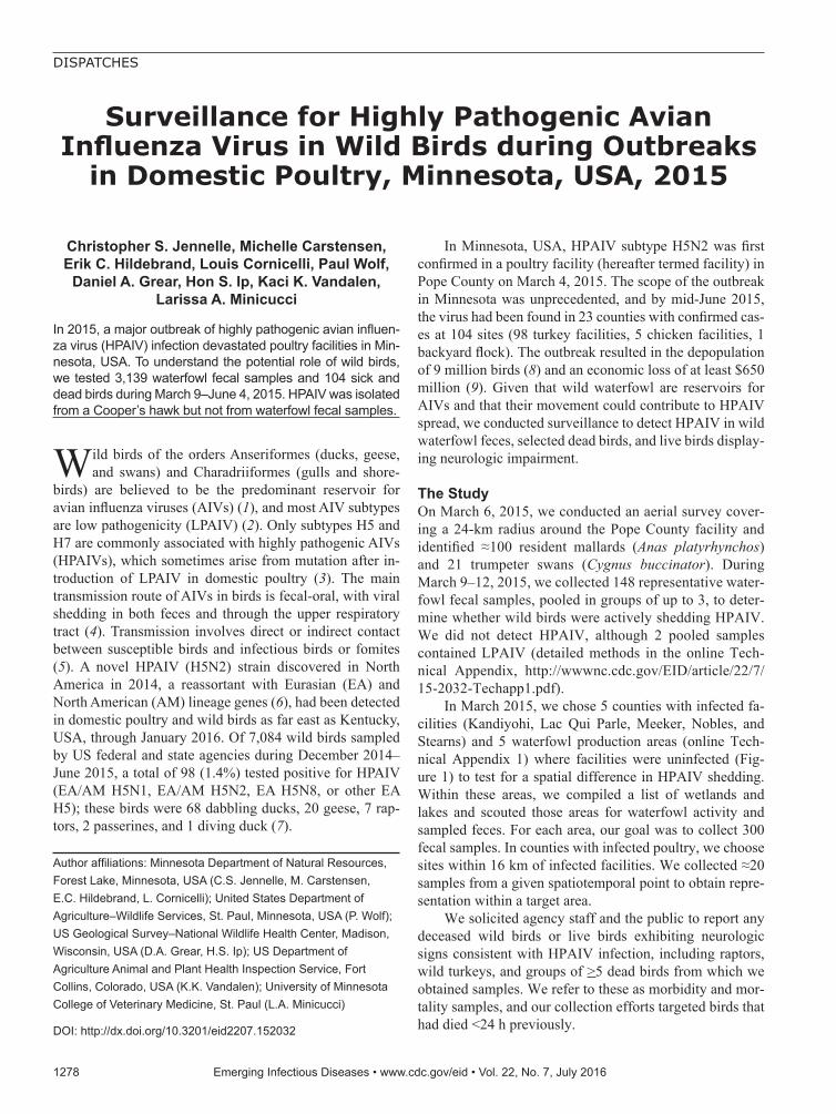
The Study On March 6, 2015, we conducted an aerial survey cover-ing a 24-km radius around the Pope County facility and identified ≈100 resident mallards (Anas platyrhynchos) and 21 trumpeter swans (Cygnus buccinator). During March 9–12, 2015, we collected 148 representative water-fowl fecal samples, pooled in groups of up to 3, to deter-mine whether wild birds were actively shedding HPAIV. We did not detect HPAIV, although 2 pooled samples contained LPAIV (detailed methods in the online Tech-nical Appendix, http://wwwnc.cdc.gov/EID/article/22/7/ 15-2032-Techapp1.pdf).
In March 2015, we chose 5 counties with infected fa-cilities (Kandiyohi, Lac Qui Parle, Meeker, Nobles, and Stearns) and 5 waterfowl production areas (online Tech-nical Appendix 1) where facilities were uninfected (Fig-ure 1) to test for a spatial difference in HPAIV shedding. Within these areas, we compiled a list of wetlands and lakes and scouted those areas for waterfowl activity and sampled feces. For each area, our goal was to collect 300 fecal samples. In counties with infected poultry, we choose sites within 16 km of infected facilities. We collected ≈20 samples from a given spatiotemporal point to obtain repre-sentation within a target area.
We solicited agency staff and the public to report any deceased wild birds or live birds exhibiting neurologic signs consistent with HPAIV infection, including raptors, wild turkeys, and groups of >5 dead birds from which we obtained samples. We refer to these as morbidity and mor-tality samples, and our collection efforts targeted birds that had died <24 h previously.
Surveillance for Highly Pathogenic Avian Influenza Virus in Wild Birds during Outbreaks
in Domestic Poultry, Minnesota, USA, 2015
1278 Emerging Infectious Diseases • www.cdc.gov/eid • Vol. 22, No. 7, July 2016
DISPATCHES
Author affiliations: Minnesota Department of Natural Resources, Forest Lake, Minnesota, USA (C.S. Jennelle, M. Carstensen, E.C. Hildebrand, L. Cornicelli); United States Department of Agriculture–Wildlife Services, St. Paul, Minnesota, USA (P. Wolf); US Geological Survey–National Wildlife Health Center, Madison, Wisconsin, USA (D.A. Grear, H.S. Ip); US Department of Agriculture Animal and Plant Health Inspection Service, Fort Collins, Colorado, USA (K.K. Vandalen); University of Minnesota College of Veterinary Medicine, St. Paul (L.A. Minicucci)
DOI: http://dx.doi.org/10.3201/eid2207.152032
Surveillance for HPAIV, Minnesota
In April 2015, which coincided with the peak rates of infection in Minnesota facilities (8), we collected 2,991 waterfowl fecal samples and pooled them into 1,027 brain-heart–infusion media vials; 1,591 samples (548 pooled) were obtained from counties with infected facilities, and 1,400 samples (479 pooled) were collected from waterfowl production areas without facilities (Figure 1). Although HPAIV was not detected in these samples, 30 pooled sam-ples (representing 85 individual birds) tested positive for LPAIV. Apparent LPAIV fecal prevalence was 0.012 (95% CI 0.007–0.018) in counties with infected poultry, 0.008 (95% CI 0.004–0.014) in counties without infection, and 0.010 (95% CI 0.007–0.014) in the combined study area. Given that HPAIV was not detected and that we could not sample every individual bird in the waterfowl population, if HPAIV were present, there was a 95% probability that
fecal prevalence was between 0 and 0.181% in areas with infection and 0 and 0.224% in areas without infection.
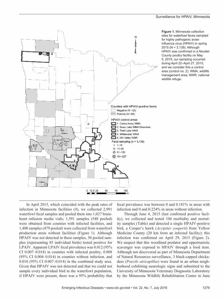
Through June 4, 2015 (last confirmed positive facil-ity), we collected and tested 104 morbidity and mortal-ity samples (Table) and detected a single HPAIV-positive bird, a Cooper’s hawk (Accipiter cooperii) from Yellow Medicine County (20 km from an infected facility); this infection was confirmed on April 29, 2015 (Figure 2). We suspect that this woodland predator and opportunistic scavenger was exposed to HPAIV through a food item. Although not discovered as part of Minnesota Department of Natural Resources surveillance, 3 black-capped chicka-dees (Poecile atricapillus) were found in an urban neigh-borhood exhibiting neurologic signs and submitted to the University of Minnesota Veterinary Diagnostic Laboratory by the Minnesota Wildlife Rehabilitation Center in June
Emerging Infectious Diseases • www.cdc.gov/eid • Vol. 22, No. 7, July 2016 1279
Figure 1. Minnesota collection sites for waterfowl feces sampled for highly pathogenic avian influenza virus (HPAIV) in spring 2015 (N = 3,139). Although HPAIV was confirmed in a Nicollet County poultry facility on May 5, 2015, our sampling occurred during April 22–April 27, 2015, and we consider this a control area (control no. 2). WMA, wildlife management area; NWR, national wildlife refuge.

DISPATCHES
2015; in 1 bird there was weak detection of Eurasian H5 RNA, but no virus was recovered and no sequence could be obtained directly from the sample (7). All 3 birds demon-strated multifocal encephalitis, which was likely the cause for the neurologic signs (A. Armien, pers. comm.).
ConclusionsMorbidity and mortality samples yielded the only HPAIV detected in our surveillance of Minnesota wild birds, de-spite the relatively small number of samples. This sample type has proven valuable for HPAIV detection in wild birds in other states; 32% of HPAIV detections nationwide and 90% of HPAIV detections within the Mississippi flyway were derived from this source during December 2014–June 2015 (7). Evolving HPAIV strains can elicit clinical signs and death in young immunologically naive ducks (10), and targeted sampling of waterfowl postbreeding areas for dead or neurologically impaired hatch-year birds might prove useful for future HPAIV surveillance (11).
Careful thought has been given to the design of sur-veillance programs for avian influenza (12). The study objectives, coupled with the methodologic limitations of available approaches, drive the sampling tool ultimately applied. Although opportunistic sampling (e.g., morbidity and mortality surveillance) is accessible to most agencies,
it is not suited for formal population-level inferences. For estimating AIV shedding prevalence, swab sampling of oropharyngeal and cloacal cavities in live birds or the trachea and cloaca in recently deceased birds is optimal because AIV replicates and sheds through the digestive tract (13) and the upper respiratory system (14). For investigating exposure history, sampling blood from live or recently dead birds for serologic testing would be more appropriate, although timing, location, and mechanism of exposure cannot be determined.
Most of our samples were obtained from waterfowl feces. The outbreak’s speed required a quickly deploy-able method to collect adequate sample sizes and imple-ment spatial design elements that would allow a meaning-ful comparison between known areas with infection and areas of the state apparently without infection. Modeling has shown that AIV maintenance in wild bird populations is mediated by environmental transmission (15), and the detection of LPAIV in waterfowl fecal samples supports that conclusion. No HPAIV was detected in waterfowl feces, although there was 95% probability of apparent fe-cal prevalence throughout the study area of 0 to 0.1%. Thus, we conclude that during the 2015 HPAIV (H5N2) outbreak in Minnesota poultry, HPAIV contamination in wild waterfowl feces was not widespread.
1280 Emerging Infectious Diseases • www.cdc.gov/eid • Vol. 22, No. 7, July 2016
Table. Wild birds collected (n = 104) for highly pathogenic avian influenza virus screening as part of MNDNR morbidity and mortality sampling efforts, Minnesota, USA, March 9–June 4 2015 Order* Family Genus and species Common name Count Anseriformes Anatidae Branta canadensis Canada goose 8 Cygnus buccinators Trumpeter swan 3 Aix sponsa Wood duck 2 Anas platyrhynchos Mallard 2 Galliformes Phasianidae Phasianus colchicus Ring-necked pheasant 8 Meleagris gallopavo Wild turkey 17 Pelicaniformes Pelicanidae Pelicanus erythrorhynchos American white pelican 1 Accipitriformes Cathartidae Cathartes aura Turkey vulture 1 Accipitridae Haliaeetus leucocephalus Bald eagle 5 Accipiter striatus Sharp-shinned hawk 8 Accipiter cooperii† Cooper’s hawk 6 Buteo platypterus Broad-winged hawk 1 Buteo jamaicensis Red-tailed hawk 3 Gruiformes Rallidae Rallus limicola Virginia rail 1 Porzana carolina Sora 1 Fulica americana American coot 9 Gruidae Grus canadensis Sandhill crane 1 Charadriiformes Laridae Larus delawarensis Ring-billed gull 1 Larus argentatus Herring gull 1 Columbiformes Columbidae Columba livia Rock pigeon 2 Zenaida macroura Mourning dove 1 Strigiformes Strigidae Bubo virginianus Great horned owl 3 Caprimulgiformes Caprimulgidae Chordeiles minor Common nighthawk 1 Passeriformes Sturnidae Sturnus vulgaris European starling 10 Parulidae Setophaga striata Blackpoll warbler 1 Setophaga palmarum Palm warbler 1 Emberizidae Melospiza lincolnii Lincoln’s sparrow 1 Icteridae Euphagus carolinus Rusty blackbird 3 Quiscalus quiscula Common grackle 1 *1 sparrow not listed was identified to order Passeriformes. †1 HPAIV-positive Cooper’s hawk confirmed on April 29, 2015.

Surveillance for HPAIV, Minnesota
AcknowledgmentsMany people were involved in the coordination of sampling, collection of samples, and logistical support for this study. We regret that we cannot name each person involved in these efforts, but we thank the participants from the Minnesota Department of Natural Resources; US Department of Agriculture–National Veterinary Services Laboratory; US Department of Agriculture–Wildlife Services; US Fish and Wildlife Service, US Geological Survey – National Wildlife Health Center; US Department of Agriculture– National Wildlife Research Center; University of Minnesota College of Veterinary Medicine, Public Health and Preventive Medicine Residents; University of Minnesota Veterinary Diagnostic Laboratory; and citizens of Minnesota who reported morbidity and mortality samples. We also thank Robert Dusek, Susan Shriner, and an anonymous reviewer for helpful comments on earlier drafts of the manuscript.
Dr. Jennelle is a research scientist in the Wildlife Health Pro-gram of the Minnesota Department of Natural Resources, Forest Lake, Minnesota, USA. He is interested in the ecology of wildlife diseases, quantitative methods for understanding wildlife disease dynamics, and conservation and management of wildlife populations.
References 1. Webster RG, Bean WJ, Gorman OT, Chambers TM, Kawaoka Y.
Evolution and ecology of influenza A viruses. Microbiol Rev. 1992;56:152–79.
2. Swayne DE, Suarez DL. Highly pathogenic avian influenza. Rev Sci Tech. 2000;19:463–82.
3. Kawaoka Y, Nestorowicz A, Alexander DJ, Webster RG. Molecular analyses of the hemagglutinin genes of H5 influenza viruses: origin of a virulent turkey strain. Virology. 1987;158:218–27. http://dx.doi.org/10.1016/0042-6822(87)90256-X
Emerging Infectious Diseases • www.cdc.gov/eid • Vol. 22, No. 7, July 2016 1281
Figure 2. Wild bird morbidity and mortality samples (n = 104) screened for highly pathogenic avian influenza virus (HPAIV) in Minnesota through June 4, 2015. A Cooper’s hawk was confirmed to be HPAIV positive in Yellow Medicine County on April 29, 2015, whereas weak titers of Eurasian H5 RNA were detected in a sampled black-capped chickadee from Ramsey County collected in June 2015.

DISPATCHES
4. Franҫa MS, Brown JD. Influenza pathobiology and pathogenesis in avian species. In: Compans RW, Oldstone MBA, editors. Influenza pathogenesis and control, vol. I. New York: Springer International Publishing; 2014. p. 221–42.
5. Stallknecht DE, Brown JD. Tenacity of avian influenza viruses. Rev Sci Tech. 2009;28:59–67.
6. World Organisation for Animal Health. Summary of immediate notifications and follow-ups—2014. Highly pathogenic avian influenza [cited 2015 Oct 14]. http://www.oie.int/wahis_2/public/wahid.php/Diseaseinformation/Immsummary
7. US Department of Agriculture. December 2014–June 2015 wild bird highly pathogenic avian influenza cases in the United States. 2015 Dec [cited 2016 Feb 15]. https://www.aphis.usda.gov/ wildlife_damage/downloads/
8. USDA Animal and Plant Health Inspection Service. Update on avian influenza findings: poultry findings confirmed by USDA’s National Veterinary Services Laboratory. 2015 [cited 2015 Oct 16]. https://www.aphis.usda.gov/wps/portal/aphis/ourfocus/ animalhealth/sa_animal_disease_information/
9. University of Minnesota Extension. Economic impact of the avian flu, updated 7/10/2015. 2015 Jul [cited 2015 Oct 16]. http://www.extension.umn.edu/community/economic-impact-analysis/reports/docs/Avian-flu-update-fact-sheet.pdf
10. Pantin-Jackwood MJ, Swayne DE. Pathobiology of Asian highly pathogenic avian influenza H5N1 virus infections in ducks. Avian Dis. 2007;51(Suppl):250–9. http://dx.doi.org/10.1637/7710-090606R.1
11. Hénaux V, Parmley J, Soos C, Samuel MD. Estimating transmission of avian influenza in wild birds from incomplete epizootic data: implications for surveillance and disease spread. J Appl Ecol. 2013;50:223–31. http://dx.doi.org/10.1111/ 1365-2664.12031
12. Hoye BJ, Munster VJ, Nishiura H, Klaassen M, Fouchier RAM. Surveillance of wild birds for avian influenza virus. Emerg Infect Dis. 2010;16:1827–34. http://dx.doi.org/10.3201/eid1612.100589
13. Webster RG, Yakhno M, Hinshaw VS, Bean WJ, Copal Murti K. Intestinal influenza: replication and characterization of influenza viruses in ducks. Virology. 1978;84:268–78. http://dx.doi.org/10.1016/0042-6822(78)90247-7
14. Sturm-Ramirez KM, Hulse-Post DJ, Govorkova EA, Humberd J, Seiler P, Puthavathana P, et al. Are ducks contributing to the endemicity of highly pathogenic H5N1 influenza virus in Asia? J Virol. 2005;79:11269–79. http://dx.doi.org/10.1128/JVI.79.17.11269-11279.2005
15. Breban R, Drake JM, Stallknecht DE, Rohani P. The role of environmental transmission in recurrent avian influenza epidemics. PLOS Comput Biol. 2009;5:e1000346. http://dx.doi.org/10.1371/journal.pcbi.1000346
Address for correspondence: Christopher S. Jennelle, Minnesota Department of Natural Resources, 5463-C W Broadway Ave, Forest Lake, MN 55025, USA; email: [email protected]
1282 Emerging Infectious Diseases • www.cdc.gov/eid • Vol. 22, No. 7, July 2016
Sources 1. Longcore JE, Pessier AP, Nichols DK.
Batrachochytrium dendrobatidis gen. et sp. nov., a chytrid pathogenic to amphibians. Mycologia. 1999;91:219–27. http://dx.doi.org/10.2307/ 3761366
2. Martel A, Spitzen-van der Sluijs A, Blooi M, Bert W, Ducatelle R, Fisher MC, et al. Batrachochytrium salamandrivorans sp. nov. causes lethal chytridiomycosis in amphibians. Proc Natl Acad Sci U S A. 2013;110:15325–9. http://dx.doi.org/10.1073/pnas.1307356110
Batrachochytrium salamandrivorans is a recently discovered fungus that kills amphib-
ians. It is related to B. dendrobatidis, which also kills amphibians (from the Greek dendron, “tree,” and bates, “one who climbs,” referring to a genus of poison dart frogs). Batrachochytrium is de-rived from the Greek words batrachos, “frog,” and chytra, “earthen pot” (describing the structure that contains unreleased zoospores); salamandriv-orans is from the Greek salamandra, “salamander,” and Latin vorans, “eating,” which refers to extensive skin destruction and rapid death in infected salamanders.
Batrachochytrium salamandrivorans [bə-trayʹ-koh-kitʺ-ri-um saʺ-la-man-dri-vo’rans]
Address for correspondence: Elizabeth Kurylo, Centers for Disease Control and Prevention, 1600 Clifton Rd NE, Mailstop E03, Atlanta, GA 30329-4027, USA; email: [email protected]
DOI: http://dx.doi.org/10.3201/eid2207.ET2207
Basal infection in skin of a fire salamander (Salamandra salamandra) characterized by extensive epidermal necrosis, high numbers of intra-epithelial colonial chytrid thalli, and loss of epithelial integrity. Photo by A. Martel and F. Pasmans, courtesy of Wikipedia.
etymologia