tartrate-resistant acid phosphatase: kaija three
TRANSCRIPT

TARTRATE-RESISTANT ACID PHOSPHATASE: THREE-DIMENSIONAL STRUCTURE AND STRUCTURE-BASED FUNCTIONAL STUDIESStudies on the enzyme using recombinant protein produced by baculovirus expression vector system in insect cells
HELENAKAIJA
Research Center for Molecular Endocrinology,
Faculty of Medicine andBiocenter Oulu,
University of Oulu
OULU 2002


HELENA KAIJA
TARTRATE-RESISTANT ACID PHOSPHATASE: THREE-DIMENSIONAL STRUCTURE AND STRUCTURE-BASED FUNCTIONAL STUDIESStudies on the enzyme using recombinant protein produced by baculovirus expression vector system in insect cells
Academic Dissertation to be presented with the assent ofthe Faculty of Medicine, University of Oulu, for publicdiscussion in the Auditorium 9 of the University Hospitalof Oulu, on September 13th, 2002, at 12 noon.
OULUN YLIOPISTO, OULU 2002

Copyright © 2002University of Oulu, 2002
Supervised byProfessor Pirkko Vihko
Reviewed byProfessor Raili MyllyläProfessor Juha Rouvinen
ISBN 951-42-6776-1 (URL: http://herkules.oulu.fi/isbn9514267761/)
ALSO AVAILABLE IN PRINTED FORMATActa Univ. Oul. D 691, 2002ISBN 951-42-6775-3ISSN 0355-3221 (URL: http://herkules.oulu.fi/issn03553221/)
OULU UNIVERSITY PRESSOULU 2002

Kaija, Helena, Tartrate-resistant acid phosphatase: three-dimensional structure andstructure-based functional studies Studies on the enzyme using recombinant proteinproduced by baculovirus expression vector system in insect cellsResearch Center for Molecular Endocrinology, Faculty of Medicine and Biocenter Oulu, Universityof Oulu, P.O.Box 5000, FIN-90014 University of Oulu, Finland Oulu, Finland2002
Abstract
Osteoporosis is a disease characterized by abnormalities in the amount and architectural arrangementof bone tissue, which leads to impaired skeletal strength and increased susceptibility to fractures.Type 5 tartrate-resistant acid phosphatase (TRACP, AcP5) has been suggested to participate directlyin bone resorption.
In this study, baculovirus expression vector system in insect cells was used to gain large amountsof recombinant type 5 acid phosphatase for structure determination, structure-based functionalstudies and production of monoclonal antibodies. Active and inactive forms of the enzyme wereseparated from each other by cation-exchange chromatography, and characterized. The enzyme wascrystallized and the three-dimensional structure was determined. Based on the three-dimensionalstructure of the active site five different enzyme variants were constructed, produced in insect cells,and purified. The wild type enzyme and the mutated forms were characterized, and their kineticparameters were determined. The importance of amino acids that were expected to be essential forthe acid phosphatase activity was confirmed. The acid phosphatase activity and reactive oxygenspecies generating activity of this dual enzyme proved to exploit different amino acids in theirreaction mechanisms.
Further studies are needed to clarify the physiological substrates of TRACP in vivo. The findingsof this study could form a base for construction of inhibitors for TRACP that could be usefultherapeutic agents for osteoporosis and related bone disorders.
Keywords: recombinant proteins, tartrate-resistant acid phosphatase, bone resorption,three-dimensional structure, monoclonal antibodies, site-directed mutagenesis, reactive ox-ygen species


Acknowledgements
This work was carried out at Biocenter Oulu and at the Research Center for Molecular Endocrinology, which also acts as a WHO Collaborating Centre for Research on Reproductive Health, Faculty of Medicine, University of Oulu.
I wish to express my sincere gratitude to my supervisor Professor Pirkko Vihko, MD, PhD, for giving me the opportunity to prepare my thesis work as a member of her research group. Her experience, support and encouragement made it possible for me to accomplish this work. I also owe gratitude to Docent Veli Isomaa, PhD, Riitta Kurkela, PhD, Katja Porvari, PhD, and Docent Hellevi Peltoketo, PhD, for their patient guidance and for sharing their wide knowledge in the field of research work.
I am grateful to Professor Raili Myllylä, PhD and Professor Juha Rouvinen, PhD, for their critical review of this work. I also wish to thank Anna Vuolteenaho, MSc, for careful revision of the language.
I owe my gratitude to all my co-authors for their irreplaceable contribution to this work. Professor H. Kalervo Väänänen, MD, PhD, Jussi Halleen, PhD, and Sari Alatalo, MSc, from the Department of Anatomy, University of Turku, Finland, are acknowledged for guiding me in the world of bone research, and in particular Jussi Halleen for his enthusiastic attitude. Professor Gunter Schneider, PhD, Professor Ylva Lindqvist, PhD, Jia Jia, PhD, and Eva Johansson, PhD, from Karolinska Institutet, Stockholm, Sweden, have done valuable work in crystallization and protein modelling. They have also given me the possibility to visit their excellent laboratory in Stockholm. I also wish to thank Dr Göran Andersson from Karolinska Institutet, Stockholm, Sweden, for the original TRACP construct. I owe my thanks to the staff of the Medical Library and the Photography Laboratory of the Medical Faculty for their pleasant and efficient services, Juhani Heikkilä and Jaakko Heikkilä for the maintenance of computers, and secretaries Anne Ollila, Anja Raatikainen and Sirpa Annanperä for holding the strings of everything in their hands.

I owe special acknowledgements to Marja-Riitta Hurnasti, Mirja Mäkeläinen, Marja-
Liisa Norrena and Pirkko Ruokojärvi for their invaluable technical assistance as well as their pleasant company during these years. I also wish to thank warmly all my colleagues and all the personnel in the Centre, present and past. Particularly I wish to thank "upstairs protein people" for all their help and for enjoyable moments in the lab, and Anne Kivinen and Sirpa Rannikko for their friendship.
I am grateful to all my friends and relatives for bringing joy and happiness into my life. With Anneli Ipatti, Päivikki Heikkilä, Seija Eidsvig, Riitta Rautio-Autere and Anitta Tauriainen I have shared "adventures in the wonderland" for more than twenty years. I wish to thank my brother Risto and his family Eila, Juha and Lauri as well as my husband's family for their support. Most of all, I wish to thank my mother.
Finally, I wish to express my deepest gratitude to my dear husband, Mauri Miihkinen. "I should see the garden far better", she said to herself, " if I could get to the top of the hill: and here's a path leading to it -- at least; no, it doesn't do that" -- (after going a few yards along the path, and turning several sharp corners), "but I suppose it will at last. But how curiously it twists! It's more like a corkscrew than a path! Well, this turn goes to the hill, I suppose -- no it doesn't! This goes straight back to the house! Well then, I'll try it the other way." Mauri has managed to do the trick beyond human comprehension, he has understood that there is logic behind it all. Not to mention that he has taught me to press "enter" and "any key", without it this would never have been possible. And cats and rats, thanks!
This study has been supported by the Research Council for Medicine of the Academy
of Finland, Sigrid Juselius Foundation, Magnus Ehrnrooth Foundation, Ida Montin Foundation, and Biocenter Oulu. Oulu, June 2002 Helena Kaija "Life what is it but a dream" Verses in italics: Lewis Carroll, Through the Looking Glass and Alice in the Wonderland

Abbreviations
AcNPV Autographa californica nuclear polyhedrosis virus AcP acid phosphatase BEVS baculovirus expression vector system bp base pair(s) EPR electron paramagnetic resonance IEF isoelectric focusing Km Michaelis-Menten constant kcat turnover number kcat/Km catalytic efficiency kDa kilodalton Mab monoclonal antibody MHC major histocompatibility complex M-CSF macrophage colony-stimulating factor NADPH nicotinamide adenine dinucleotide phosphate NF-κB transcription factor NF-κB NMR nuclear magnetic resonance Pi inorganic orthophosphate PAP prostatic acid phosphatase pNPP para-nitrophenyl phosphate PPase protein serine/threonine phosphatase PTPase protein tyrosine phosphatase PTH parathyroid hormone PU.1 transcriptor factor PU.1 RANK receptor activator of NF-κB RANKL receptor activator of NF-κB ligand ROS reactive oxygen species SDS-PAGE sodium dodecyl sulfate polyacrylamide gel electrophoresis Sf Spodoptera frugiperda TNF tumor necrosis factor TRACP tartrate-resistant acid phosphatase

TRAP tartrate-resistant acid phosphatase TR-AP tartrate-resistant acid phosphatase UV ultra violet Vmax maximal reaction velocity Å Ångström

List of original papers
This thesis is based on the following articles, which are referred to in the text by their Roman numerals:
I Halleen J, Kaija H, Stepan J, Vihko P & Väänänen HK (1998). Studies on the protein tyrosine phosphatase activity of tartrate-resistant acid phosphatase. Arch Biochem Biophys 352:97-102.
II Kaija H, Jia J, Lindqvist Y, Andersson G & Vihko P (1999). Tartrate-resistant bone
acid phosphatase: Large-scale production and purification of the recombinant enzyme, characterization, and crystallization. J Bone Miner Res 14:424-430.
III Lindqvist Y, Johansson E, Kaija H, Vihko P & Schneider G (1999). Three-
dimensional structure of a mammalian purple acid phosphatase at 2.2 Å resolution with a µ-(Hydr)oxo bridged di-iron center. J Mol Biol 291:135-147.
IV Kaija H, Alatalo S, Halleen J, Lindqvist Y, Schneider G, Väänänen HK & Vihko P
(2002). Phosphatase and oxygen radical generating activities of mammalian purple acid phosphatase are functionally independent. Biochem Biophys Res Commun 292:128-132.


Contents
Abstract Acknowledgements Abbreviations List of original papers Contents 1 Introduction................................................................................................................... 13 2 Review of the literature................................................................................................. 14
2.1 Phosphatases .......................................................................................................... 14 2.2 Purple acid phosphatases ....................................................................................... 15 2.3 TRACP................................................................................................................... 15
2.3.1 Expression and cell localization...................................................................... 15 2.3.2 TRACP gene ................................................................................................... 17 2.3.3 Structure of the active site............................................................................... 18 2.3.4 Three-dimensional structure............................................................................ 21 2.3.5 Substrate specificity, inhibitors and activators ................................................ 21 2.3.6 Proposed mechanism of hydrolysis of phosphate esters by TRACP............... 22 2.3.7 Mechanism of ROS generation by TRACP..................................................... 24 2.3.8 Proteolytic cleavage and carbohydrate content ............................................... 25 2.3.9 Serum TRACP as a marker of bone resorption ............................................... 26
2.4 Cells containing TRACP........................................................................................ 27 2.4.1 Osteoclasts and bone resorption...................................................................... 27 2.4.2 Macrophages and immune defence................................................................. 29
2.5 Biological function of TRACP............................................................................... 30 2.6 Recombinant protein production............................................................................ 31
3 Outlines of the present study......................................................................................... 33 4 Materials and methods .................................................................................................. 34
4.1 Production and purification of the recombinant enzyme (II) ................................. 34 4.2 Characterization of the recombinant enzyme (I, II) ............................................... 34
4.2.1 Acid phosphatase activity................................................................................ 34 4.2.2 Separation of the active and inactive form and stability measurements .......... 35
4.3 Crystallization (II), structure determination (III)

and site-directed mutagenesis (IV)......................................................................... 35 4.3.1 Substrate specificity ........................................................................................ 35 4.3.2 Kinetic parameters for phosphatase activity ................................................... 36 4.3.3 Radical oxygen species generating activity..................................................... 36
4.4 Monoclonal antibodies (I)...................................................................................... 36 5 Results........................................................................................................................... 37
5.1 Production and purification of recombinant TRACP (II) ....................................... 37 5.2 Characterization of wild-type TRACP, monoclonal antibodies (I, II).................... 37 5.3 Crystallization and structure determination (III).................................................... 38 5.4 Site-directed mutagenesis and amino acids important for acid phosphatase activity (IV) .......................................................................... 40
6 Discussion..................................................................................................................... 41 6.1 Baculovirus expression vector system ................................................................... 41 6.2 Characterization of TRACP ................................................................................... 42 6.3 Monoclonal antibodies........................................................................................... 43 6.4 Three-dimensional structures and reaction mechanism ......................................... 43 6.5 Classification of TRACP........................................................................................ 45 6.6 Site-directed mutagenesis ...................................................................................... 46
7 Conclusions................................................................................................................... 47 8 References..................................................................................................................... 48

1 Introduction
Mammalian tartrate-resistant acid phosphatase (TRACP), also known as type 5 acid phosphatase (AcP5, E.C. 3.1.3.2.), is a basic glycoprotein whose expression under physiological conditions is restricted to osteoclasts and activated macrophages. Together with similar enzymes isolated from animals, plants and fungi, it belongs to the group of purple acid phosphatases. The physiological functions of purple acid phosphatases in biological systems are not yet known, and no natural substrates have been unambiguously established. Mice deficient in TRACP develop mild osteopetrosis (Hayman et al. 1996), and overexpression of TRACP leads to accelerated bone turnover (Angel et al. 2000), suggesting that TRACP has an important role in bone resorption.
The catalytic site of mammalian TRACP contains a binuclear iron center, where one of the irons is redox-active, while the other is stabilized to ferric state (Antanaitis & Aisen 1983). The binuclear iron center of purple acid phosphatases has been studied extensively using various spectroscopic techniques. The enzyme may exist in two forms: an inactive purple form, where the redox-active iron is in the ferric state, and an active pink form, where it is reduced to the ferrous state (Antanaitis & Aisen 1983). The active enzyme is also capable of generating reactive oxygen species through Fenton's reaction, where the ferrous ion reacts with hydrogen peroxide to produce highly destructive hydroxyl radicals (Garret et al. 1990).
Red kidney bean acid phosphatase was the first purple acid phosphatase whose three-dimensional structure was determined (Sträter et al. 1995). Our group has long experience of studying prostatic acid phosphatase (PAP), an enzyme that is sensitive to inhibition by tartrate. We have carried out studies on PAP's enzymatic properties and described its structure both on gene and protein levels (Vihko et al. 1988, Roiko et al. 1990, Vihko et al. 1993, Schneider et al. 1993, Lindqvist et al. 1993, Lindqvist et al. 1994, Porvari et al. 1994, Virkkunen et al. 1994). The aim of this study was to determine the three-dimensional structure of a mammalian tartrate-resistant acid phosphatase and in particular its active site, to characterize amino acids predicted to be important for the acid phosphatase activity, to study the substrate specificity of the enzyme, and to produce monoclonal antibodies against TRACP. Knowledge of the active site structure is valuable for the design of enzyme inhibitors that could be useful as therapeutic agents for conditions with accelerated bone resorption.
2 Review of the literature
2.1 Phosphatases
Phosphate ester bond functions as an extremely important linkage within the living cell. It participates in storage and transfer of the genetic information, carries chemical energy and regulates the activity of enzymes and signaling molecules in the cell. Enzymes capable of acting on ester bonds (Enzyme Commission classification number EC 3.1) and catalyzing the cleavage of phosphate esters (EC 3.1.3) constitute the subclass of phosphohydrolases, i.e. phosphatases.
Phosphatases can be further classified according to several frameworks (Vincent et al. 1992). They can be divided into groups based on their substrate type. Non-specific phosphatases catalyze the hydrolysis of almost any phosphate ester, whereas protein phosphatases prefer phosphoproteins or phosphopeptides as substrates. Non-specific phosphatases can be divided into alkaline and acid phosphatases based on their optimal pH for catalysis. Non-specific alkaline and acid phosphatases recycle phosphate in metabolic reactions (Lohse et al. 1995).
Acid phosphatases (EC 3.1.3.2) form a group of four isoenzymes with differences originating at the structural level of the gene: prostatic, lysosomal, erythrocytic and macrophagic acid phosphatases. Acid phosphatases can also be distinguished based on their molecular weight into low molecular weight acid phosphatases and high molecular weight acid phosphatases, or on the basis of their resistance to inhibition by tartrate into tartrate-sensitive and tartrate-resistant acid phosphatases. Reaction catalyzed by acid phosphatases:
orthophosphoric monoester + H2O = alcohol + phosphate Protein phosphatases comprise two groups based upon their substrate specificity.
Protein-tyrosine phosphatases (PTPases, EC 3.1.3.48) prefer to remove phosphate from tyrosine residue and serine/threonine protein phosphatases (PPases, EC 3.1.3.16) from serine or threonine residue. The PTPase group includes dual-specificity phosphatases,

15
which are capable of hydrolyzing phosphorylated tyrosine, serine and threonine. Protein phosphatases regulate signal-transduction pathways (Lohse et al. 1995, Neel & Tonks 1997, Zhang 1998). Approximately 30% of intracellular proteins are subject to reversible protein phosphorylation (Barford et al. 1998).
One obvious difference among phosphatases is the presence or absence of metal ion cofactors.
2.2 Purple acid phosphatases
Purple acid phosphatases can easily be distinguished from other acid phosphatases by their purple color in solution. The color is due to the presence of a binuclear iron center (Davis & Averill 1982) and arises from tyrosine-to-iron charge transfer transition (Gaber et al. 1979, Davis & Averill 1982). The Committee of Scientific Advisors of the International Osteoporosis Foundation has endorsed the name type 5 acid phosphatase (AcP5) for mammalian purple acid phosphatases. The name is based on the relative electrophoretic motility of acid phosphatases, when these enzymes are being sorted electrophoretically under nondenaturing conditions. Type 5 acid phosphatases migrate fastest (Li et al. 1970). Purple acid phosphatases or type 5 acid phosphatases are not yet listed in Enzyme Nomenclature.
Type 5 acid phosphatases are resistant to inhibition by tartrate, and are therefore called also tartrate-resistant acid phosphatases (TRACP, other abbreviations used: TRAP, TR-AP). Resistance to inhibition by L+tartrate distinguishes type 5 acid phosphatases from acid phosphatases of lysosomal (von Figura & Weber 1978) or prostatic (Vihko et al. 1978) origin. Terms related to the enzyme's origin, osteoclastic acid phosphatase and macrophagic acid phosphatase, are also used. The abbreviation PAP is frequently used as reference to type 5 acid phosphatases referring to the name purple acid phosphatases. However, PAP is commonly known as the abbreviation for prostatic acid phosphatase, and it is therefore misleading.
In the present work, the name tartrate-resistant acid phosphatase (TRACP) is used for type 5 acid phosphatases and the name purple acid phosphatases for corresponding non-mammalian enzymes.
2.3 TRACP
2.3.1 Expression and cell localization
TRACP is expressed by bone-resorbing osteoclasts and activated macrophages (Minkin 1982, Efstradiadis & Moss 1985, Bevilacqua et al. 1991). It was first purified from

16
bovine spleen in 1954 (Sundararajan & Sarma 1954). In 1970 it was identified in hairy cells of patients with leukemic reticuloendotheliosis (hairy cell leukemia) (Li et al. 1970), and in 1973 a similar enzyme was isolated from porcine uterine fluid (Chen et al. 1973). In 1973 it was discovered to contain iron (Campbell & Zerner 1973), but the presence of two iron atoms per molecule of enzyme was not confirmed until 1978 (Campbell et al. 1978). It has since been isolated from various mammalian tissues including human (Allen et al. 1989), bovine (Lau et al. 1987a), and rat (Andersson et al. 1984) bone, human (Ketcham et al. 1985) and rat (Hara et al. 1984) spleen, human lungs (Efstratiadis & Moss 1985) and placenta (Ketcham et al. 1989).
TRACP mRNA expression has been detected in neonatal rats in skeletal tissues and with lower levels also in spleen, thymus, liver, skin, kidney, lung, heart, and brain (Ek-Rylander et al. 1991a) and in adult rats in cells of central and peripheral nervous system (Lång et al. 2001). Using murine tissues TRACP expression and activity has been demonstrated to be localized in tissues that possess cells originating from the osteoclast/macrophage lineage (Hayman et al. 2000a,b). The expression of TRACP is increased in some pathological conditions: in Gaucher´s disease (Robinson & Glew 1980, Lam et al. 1981), which is a lysosomal storage disease, in leukemic reticuloendotheliosis (Yam 1974, Lam et al. 1976, 1981, Ketcham et al. 1985), in HIV-induced encephalopathy (Schindelmeiser et al. 1989) and in osteoporosis and metabolic bone diseases (Delmas 1992, Cheung et al. 1995, Halleen et al.1996).
Purple acid phosphatases have been described from plants: red kidney bean (Klabunde et al. 1994), sweet potato (Durmus et al. 1999), Arabidopsis thaliana (del Pozo et al. 1999), and soybean (LeBansky et al. 1992, Hegeman & Grabau 2001), microbial organisms and fungi (Schenk et al. 2000a,b). Mammalian and plant enzymes are enzymatically and spectroscopically very similar, despite the fact that instead of two iron atoms plant enzymes contain one iron and one zinc or manganese atom (Merkx and Averill 1999, Schenk et al. 1999, 2001), and differ by their amino acid composition and molecular weight (Klabunde et al. 1995, Schenk et al. 2000b). Sequence homology is low and the only totally conserved residues are the seven invariant metal coordinating amino acids (Klabunde et al. 1995).
Histochemical, cytochemical and immunocytochemical studies reveal that TRACPs are localized in lysosomes and lysosome-like organelles or intracellular transcytotic vesicles in osteoclasts, and in antigen presentation route in activated macrophages (Katayama et al. 1972, Lam et al. 1976, Andersson et al. 1986, Clark et al. 1989, Schindelmeiser et al. 1987, Reinholt et al. 1990a, Halleen et al. 1999a, Hayman et al. 2000b, Räisänen et al. 2001). No obvious hydrophobic membrane-spanning domain has been identified.
The major secretory product of the porcine uterus under the influence of progesterin is a tartrate-resistant acid phosphatase called uteroferrin. Uteroferrin and other TRACPs are products of the same gene and share high sequence identity (Ling & Roberts 1993). Uteroferrin differs from intracellular TRACPs in the fact that it is secreted by pig uterine glandular epitelium cells and transported to fetal blood circulation (Buhi et al. 1982, Baumbach et al. 1986).

17
2.3.2 TRACP gene
Tartrate-resistant acid phosphatases constitute a group of related metalloenzymes. One single gene encodes mammalian TRACPs (Ling & Roberts 1993). The TRACP gene has been localized to human chromosome 19 (Lord et al. 1990, Leach et al. 1994) and to mouse chromosome 9 (Grimes et al. 1993), being a member of syntenic genes localized to these human and mouse chromosomes.
The genes encoding pig (Simmen et al. 1989, Vallet & Fahrenkrug 2000), rat (Ek-Rylander et al. 1991a), human (Cassady et al. 1993, Reddy et al. 1995a, Fleckenstein et al. 1996), and mouse (Cassady et al. 1993, Reddy et al. 1993) TRACP have been cloned and characterized. The sequences are highly conserved. Human and porcine enzymes have 85% homology (Ketcham et al. 1989) and porcine and bovine enzymes 90% homology (Hunt et al. 1987). Rat bone TRACP is 94% identical with human placental enzyme (Ek-Rylander et al. 1991a) and 89% identical with uteroferrin (Ek-Rylander et al. 1991a). The overall sequence similarity of rat osteoclastic acid phosphatase to rat lysosomal acid phosphatase is only 41%, and to prostatic acid phosphatase 44% (Himeno et al. 1989, Roiko et al. 1990, Ek-Rylander et al. 1991a). Studies of human and murine TRACP genes have revealed that both of these genes contain five exons with the initiation code lying in exon 2, exon 1 remaining noncoding (Fleckenstein & Drexler 1997). Exon/intron organization of the 5´-flanking region of murine, human and porcine uteroferrin gene is similar, but they differ in respect to the size of the intron (Reddy et al. 1995a, Vallet & Fahrenkrug 2000). Figure 1 indicates box diagrams comparing sizes of the exons for porcine uteroferrin, human TRACP and mouse TRACP gene.
Fig. 1. Comparing sizes of the exons for porcine uteroferrin gene, human TRACP gene and mouse TRACP gene (Figure modified from Vallet & Fahrenkrug 2000).

18
Studies on the 5´-flanking region of the TRACP gene have indicated that transcriptional regulation of this enzyme is rather complex (Lamp & Drexler 2000) and not yet fully described. Two different promotors have been identified locating in human gene within the sequence from -1903 bp to -1bp (Reddy et al. 1995a,b). A putative repressor for one of the promotors and a putative enhancer for both promotors have been described (Reddy et al. 1995b). Iron and hemin have been found to regulate one of these promoters transcriptionally (Alcantara et al. 1994, Reddy et al. 1995b, 1996, 1998, Fleckenstein et al. 2000). The most important region for iron control lies between sequences -1846 bp and -1240 bp in mouse gene (Reddy et al. 1996). TRACP gene expression is enhanced by iron and inhibited by hemin.
Estrogen has been shown to prevent bone loss by reducing the mRNA levels of osteoclastic bone resorbing enzymes TRACP and carbonic anhydrase II (Zheng et al. 1995). Porcine uteroferrin gene contains progesterone and estrogen response elements (Simmen et al. 1989). They have, however, not been found in other TRACPs, and it has therefore been suggested that cells other than osteoclasts mediate estrogen effects in the bone environment (Collier et al. 1998).
TRACP is translated as a single polypeptide (Hayman et al. 1991). Ketcham and his co-workers have reported an open reading frame of 969 bp corresponding to a protein of 323 amino acids for human placental TRACP (Ketcham et al. 1989). According to Lord and his co-workers, the open reading frame of human macrophage sequence is 975 bp encoding 325 amino acids (Lord et al.1990). Both groups have reported a putative signal sequence of 19 amino acids. Some of the isolated enzymes are single polypeptides while others are isolated as two-subunit proteins. It has been suggested that TRACPs are synthesized as latent proenzymes and activated by cleavage into subunits (Orlando et al. 1993, Ljusberg et al. 1999).
2.3.3 Structure of the active site
A binuclear metal center is a common feature for proteins in the class of metallohydrolases. These proteins have, however, many different kinds of structures, functional groups and chemical mechanisms to accelerate hydrolysis reactions (reviewed by Wilcox 1996, Sträter et al. 1996, Kimura 2000). Sequence alignments have revealed a signature motif DXH-(X)≈25-GDXXD-(X) ≈25-GNHD/E that is common for a wide variety of phosphoesterases cleaving phosphoesterbonds in many different substrates. It has been identified in tartrate-resistant/purple acid phosphatases, Serine/Threonine phosphatases, diadenosine tetraphosphatases, exonucleases, 5´-nucleasidases and sphingomyelin phosphodiesterases. (Koonin 1994, Zhuo et al. 1994, Lohse et al. 1995, Aravind & Koonin 1998, Oddie et al. 2000, Knöfel & Sträter 2001). All those metallophosphatases which contain the first sequence motif seem also to posses a second common motif GH-(X)≈50-GHXH (Klabunde et al. 1996). Sequences of proteins belonging to these groups contain five conserved blocks of residues that surround the seven invariant metal binding residues (Lohse et al. 1995). Alignment of some TRACP-like sequences from different sources in table 1 (Oddie et al. 2000).

19

20
All isolated tartrate-resistant acid phosphatases have similar physical and enzymatic properties. They are all monomeric iron-containing glycoproteins with Mr 30-40 kDa. TRACPs have basic isoelectric point (Vincent & Averill 1990a, Ek-Rylander et al. 1991b) and optimal enzymatic activity at acidic pH. (Vincent & Averill 1990a).
Many research groups have studied tartrate-resistant/purple acid phosphatases and their binuclear metal center using various spectroscopic methods (reviewed by Doi et al. 1988). The distinctive purple color of TRACP was identified by resonance Raman spectra to be due to tyrosine phenolate-to-metal charge transfer transition (Gaber et al. 1979, Antanaitis et al. 1982). Final evidence for a spin-coupled binuclear iron center at the active site of TRACP was gained from EPR (electro paramagnetic resonance) spectra and magnetic susceptibility data (Davis & Averill 1982, Antanaitis et al. 1983). Each molecule of TRACP binds two iron atoms in a binuclear cluster (Campbell et al. 1978, Davis & Averill 1982). The presence of two types of iron, a chromophoric tyrosine coordinated species that remains ferric and a colorless species that cycles between ferric and ferrous states depending on the reduction-oxidation state has been demonstrated (Antanaitis & Aisen 1983).
The enzyme can exist in two different forms: an enzymatically active pink form where the binuclear iron unit is in the ferrous-ferric state, and enzymatically inactive purple diferric state (Davis & Averill 1982). Mild reductive agents can change the inactive form into active form (Davis & Averill 1982). The reduction does not disrupt the tyrosine-iron coordination (Gaber et al. 1979, Antanaitis et al. 1982, Lambert et al. 1997). The diferric-state is EPR silent (Antanaitis et al. 1983, Day et al. 1988) and ferrous-ferric state elicits an intense EPR signal, distinctive to this class of enzymes (Davis & Averill 1982, Antanaitis et al. 1983, Day et al. 1988). Resonance Raman spectra show vibration, which suggests that the strong antiferromagnetic coupling between the irons results from an µ-oxo-bridge (Sjöberg et al. 1982).
The visible spectrum of TRACP was first published in 1960 (Glomset & Porath 1960) and after that in numerous studies (Campbell & Zerner 1973, Schlosnagle et al. 1974, Keough et al. 1980, Davis et al. 1981, Antanaitis & Aisen 1983, Antanaitis et al. 1983, Vincent et al. 1991a, and references therein). The purple form of the enzyme has absorption maximum between 550-570 nm and a near-UV shoulder between 315-320 nm. The pink form has absorption maximum between 505-510 nm and a near-UV shoulder at 310 nm. Preservation of the near-UV shoulder at the wavelength that is distinct to disulfide bond suggests that such potential bonds remain unaffected by reduction (Doi et al. 1988).

21
2.3.4 Three-dimensional structure
The three-dimensional structure of kidney bean purple acid phosphatase was determined in 1995 by Sträter and his co-workers at 2.9 Å resolution (Sträter et al. 1995). Kidney bean purple acid phosphatase is a metalloenzyme with Fe(III)-Zn(II) dinuclear active site. Kidney bean enzyme is a homodimeric protein with Mr 110 kDa containing two domains in each subunit. It has the shape of a twisted heart and overall dimensions 40 Å by 60 Å by 75 Å. The active site is at the bottom of the pocket formed by the monomers. The function of the smaller NH2-terminal domain of each subunit is unclear. The COOH-terminal domain contains the active site. The larger domain forms an α/β-type structure. It contains five α helices and two mixed β sheets with 6 and 7 β strands. The β sheets form a sandwich-like structure. The α helices connect the parallel strands in the β sheets. The two sandwich-like structures are connected with one α helix and two β strands. The iron is coordinated by Tyr167, His325 and Asp135, and the zinc is coordinated by His286, His 323 and Asn201. The metal ions are bridged by Asp164. (Sträter et al. 1995).
The primary structure of mammalian enzymes from different sources is conserved with over 80% homology. Except for the blocks of sequence homology in the metal coordinating region there is low amino acid similarity and very low sequence homology between mammalian and plant enzymes. NMR studies of uteroferrin and bovine spleen enzymes reveal the ligation of one tyrosine and one histidine to the Fe(III) and the ligation of one histidine to the Fe(II) (Wang et al. 1992, 1996). These residues are also conserved in kidney bean enzyme (Sträter et al. 1995). When the zinc ion of kidney bean enzyme is replaced with Fe(II), the enzyme's spectroscopic and kinetic properties show close similarity to mammalian enzymes (Battistuzzi et al. 1997).
The active site of mammalian TRACPs could be expected to be similar to that of kidney bean purple acid phosphatase and an average secondary structure prediction suggested that mammalian TRACPs belong to the βαβαβ-type structures, as does the kidney bean enzyme (Sträter et al. 1995, Denu et al. 1996). In 1999 three independent structures of mammalian tartrate-resistant acid phosphatase were reported almost simultaneously: pig TRACP at 1.65 Å resolution (Guddat et al. 1999), the rat bone enzyme first at 2.7 Å resolution (Uppenberg et al. 1999) and a month later at 2.2 Å resolution in this study.
2.3.5 Substrate specificity, inhibitors and activators
TRACPs prefer as substrates phosphoric esters of aromatic alcohols such as pNPP and phosphotyrosine, and compounds containing a phosphoanhydride linkage such as nucleotide di- and triphosphates (Hara et al. 1984, Ketcham et al. 1985, Anderson & Toverud 1986, Kato et al. 1986, Lau et al. 1987a, Hayman et al. 1989, Allen et al. 1989, Andersson et al. 1989, Janckila et al. 1992a, Cheung et al. 1995). Phosphoric esters of aliphatic alcohols, such as nucleotide monophosphates, phosphoserine and phosphothreonine are not hydrolyzed (Anderson & Toveryd 1986, Lau et al. 1987a, Andersson et al. 1989, Cheung et al. 1995).

22
Acid phosphatase activity of TRACP is inhibited by phosphate, molybdate, zink, copper, fluoride, vanadate, arsenate, tungstate, and ascorbate (Robinson & Glew 1980, Keough et al. 1982, Antanaitis & Aisen 1985, Ketcham et al. 1985, Anderson & Toverud 1986, Hayman et al. 1989, Allen et al. 1989, Crans et al. 1992, Janckila et al. 1992a, Cheung et al. 1995, Andersson & Ek-Rylander 1995, Halleen et al. 1996). Oxidation by e.g. hydrogen peroxide causes inhibition of the enzyme activity (Ketcham et al. 1985, Anderson & Toverud 1986, Hayman et al. 1989, Janckila et al. 1992a, Halleen et al. 1996, Beck et al. 1999). Reaction product phosphate and its analogs vanadate, arsenate and molybdate cause competitive inhibition (Keough et al. 1982, Antanaitis & Aisen 1985, Janckila et al. 1992a). Fluoride, tungstate, copper and zinc cause noncompetitive inhibition (Hayman et al. 1989, Crans et al. 1992, Halleen et al. 1996).
The enzyme is activated by mild reducing agents, such as β-mercaptoethanol (Ketcham et al. 1985, Hayman et al. 1989, Janckila et al. 1992a, Halleen et al. 1996). Strong reducing agents, such as dithionite, lead to removal of iron and thus cause inhibition of the activity (Keough et al. 1980, Ketcham et al. 1985, Hayman et al. 1989). Ascorbate together with iron activates TRACP (Hayman et al. 1989, Halleen et al. 1996). Manganese and magnesium are reported to have an activating effect on TRACP (Hayman et al. 1989).
2.3.6 Proposed mechanism of hydrolysis of phosphate esters by TRACP
Phosphoryl-transfer reactions have been intensively studied because of their primary significance in biological events. Hydrolysis of phosphate esters proceeds either through dissociative or associative mechanism (Vincent et al. 1992). The main question is whether or not there is a covalent phosphoenzyme intermediate in the reaction. (Reviewed by Sträter et al. 1996). A study on bovine tartrate-resistant acid phosphatase mechanism reported evidence for a phosphoenzyme intermediate (Vincent et al. 1991b). More resent studies have, however, not found such intermediate (Wynne et al. 1995).
Based on spectroscopic studies and on the crystal structure of plant purple acid phosphatase (Sträter et al. 1995), the following mechanism of hydrolysis shown in Figure 2 has been suggested by Klabunde and his co-workers (Klabunde et al. 1996). The phosphate group of the substrate binds to the divalent metal ion, which together with the histidine ligands preorientates the phosphate group and enhances the electrophilicity of the phosphorus center. After that a nucleophilic attack occurs by the hydroxide ligand bound to the Fe3+ ion, and the configuration at the phosphorus center is inverted through a pentacoordinate transition-state in which the leaving group and the attacking nucleophile are in apical position. In addition to the metal ions the protonated side chains of histidine residues reduce the energy of the pentacovalent transition-state through the formation of hydrogen bonds. An additional histidine may facilitate by a small movement the protonation of the leaving alcohol group.

23
Fig. 2. Proposed mechanism of hydrolysis of phosphate group by TRACP (Figure modified from Klabunde et al. 1996).
Tartrate-resistant/purple acid phosphatases have been studied in order to clarify the
role of metal ions in the catalytic mechanisms of metalloenzymes. Three possible mechanisms have been proposed, presented in Figure 3 as reviewed by Kimura (Kimura 2000). In mechanism a), the dianionic phosphate monoester substrate binds to Me2+ (Me= Fe or Zn) and after that phosphate is attacked by the Fe3+ -OH. In mechanism b), phosphate ester forms a bridging between the Fe3+ and Me2+ ions. The bridged phosphate ester is attacked by the µ-OH bridge, which acts as a nucleophile. In mechanism c), the inert Fe3+ -OH acts as a general base and deprotonates a second water molecule, which then attacks the Me2+ bound phosphate ester.

24
Fig. 3. Role of metal ions in the catalytic mechanism of metalloenzymes (Figure modified from Kimura et al. 2000).
2.3.7 Mechanism of ROS generation by TRACP
In osteoclasts and macrophages, superoxide is generated by the action of NADPH-oxidase on molecular oxygen (Garret et al. 1990, Steinbeck et al. 1992, 1994, Key et al. 1994, Silverton 1994, Silverton et al. 1995, Hall et al. 1995, Darden et al. 1996). TRACPs, like other metalloproteins containing a redox-active iron, are able to catalyze the generation of reactive oxygen species from superoxide by the mechanism known as Haber-Weiss-Fenton chemistry (Sibille et al. 1987, Hayman & Cox 1994, Halleen et al. 1999a. Räisänen et al. 2001). Reactive oxygen species (ROS) include hydroxyl radicals, hydrogen peroxide, and singlet oxygen. Superoxide itself may not be directly injurious. Highly reactive hydroxyl radicals formed through Fenton's reaction can break peptide bonds and cause disruption of RNA, DNA and protein structures (Greenwald et al. 1989,

25
Ries et al. 1992, Boldt 1999). Figure 4 describes reactions important for the ROS generation by TRACP.
O2 → (NADPH-oxidase) O2
−⋅→ (superoxide dismutase) H2O2 → (catalase) H2O + O2
TRACP-Fe3+ (purple) + O2
−⋅ → TRACP-Fe2+ (pink) + O2
H2O2 + TRACP-Fe2+ (pink) → OH· + OH- + TRACP-Fe3+ (purple)
Fig. 4. Reactions important for ROS generation.
2.3.8 Proteolytic cleavage and carbohydrate content
Some TRACPs appear as one single polypeptide chain after purification, while others are cleaved into two fragments by proteolytic cleavage (Ketcham et al. 1985, Hayman et al. 1991, Halleen et al. 1996). TRACP purified from bone tissue exists primarily as two smaller fragments (Hayman et al. 1989, Halleen et al. 1996, Ek-Rylander et al. 1997, Ljusberg et al. 1999). The size of the smaller fragment varies between Mr 15-16 kDa and that of the larger fragment between Mr 18-23 kDa. Purified recombinant bone-derived enzyme exists as one polypeptide chain form (Hayman & Cox 1994, Ek-Rylander et al. 1997). A small portion of the recombinant enzyme appears after disulfide reduction as two smaller fragments with Mr 16 kDa and 20 kDa (Ek-Rylander et al. 1997). The recombinant enzymes can be turned into two-fragment form by cleavage with serine proteinase trypsin or chymotrypsin or cystein proteinase papain (Orlando et al. 1993, Ljusberg et al. 1999).
The cleavage occurs within an exposed loop in the residue 155-161 region (Ljusberg et al. 1999). This highly antigenic loop is a general feature of the structures of TRACPs (Orlando et al. 1993, Ljusberg et al. 1999). Cleavage of the protein into fragments enhances the acid phosphatase activity (Ljusberg et al. 1999). Activation is accompanied by a shift in pH optimum to more basic values (Ljusberg et al. 1999, Funhoff et al. 2001a). Proteolytic cleavage changes also the EPR spectrum into one distinct to two-fragment TRACPs (Funhoff et al. 2001b). The reason for the cleavage of the enzyme into smaller fragments is not clear. It has been suggested that the enzyme is synthesized as a latent proenzyme which is activated by proteolytic cleavage (Ljusberg et al. 1999).

26
The carbohydrate content of tartrate-resistant acid phosphatases has been most thoroughly studied using porcine uteroferrin (Chen et al. 1973, Saunders et al. 1985, Roberts et al. 1986, Baumbach et al. 1991). The uteroferrin polypeptide chain has two potential N-glycosylation sites, Asn97 and Asn128 (Baumbach et al. 1991). Both of these glycosylation sites are conserved in human (Ketcham et al. 1989, Lord et al. 1990) and rat (Ek-Rylander et al. 1997) enzymes. Both sites are located in the N-terminal larger fragment, residues Asn97 and Asn128 in human placental TRACP (Ketcham et al. 1989) and Asn118 and Asn149 in rat bone TRACP (Ek-Rylander et al. 1997). Bovine spleen enzyme has been reported to have only one attachment site for N-linked oligosaccharides (Ketcham et al. 1989). In uteroferrin only one of the sites, Asn 97 seems to carry carbohydrate chain (Ketcham et al. 1989, Baumbach et al. 1991). There is significant charge heterogeneity among TRACPs, and this might reflect varying degrees of glycosylation (Lau et al. 1987a). Human bone TRACP has been demonstrated to contain only high-mannose type carbohydrates (Halleen et al. 1996). The small molecular mass difference in bone-derived TRACP and recombinant osteoclastic TRACP seems not to be due to differences in N-glycosylation, since the difference in motility is still present after N-glycase treatment (Ek-Rylander et al. 1997).
2.3.9 Serum TRACP as a marker of bone resorption
Type 5 acid phosphatases present in serum have been named 5a and 5b (Stepan et al. 1989). They differ in pH optimum, which is 5.0 for 5a and 5.5-6.0 for 5b (Alatalo et al. 2000), and in carbohydrate content, 5a containing sialic acid while 5b does not (Janckila et al. 1992a,b, Halleen et al. 2000). Isoform 5b is derived from osteoclasts and therefore useful as a marker for osteoclast activity (Alatalo et al. 2000, Janckila et al. 2001a,b). The source of isoform 5a is so far unestablished.
TRACP expression and activity is associated with bone resorption (Lam et al. 1982, Anderson & Toverud 1986, Lau et al. 1987a, Zaidi et al.1989, Kraenzlin et al. 1990, Moss 1992, Rico & Villa 1993, Chamberlain et al. 1995, Moss et al. 1995, Halleen et al. 1996, 2000, 2001, Hannon et al. 1998, Nakasato et al. 1999, Seregni et al. 2001). Increased bone resorption can be found in physiological bone remodeling and growth in children (Lam et al. 1978) and in various pathological osteolytic processes. Elevated TRACP levels have been measured in the serum of patients with osteoporosis (Rico and Villa 1993, Chamberlain et al. 1995, Cheung et al. 1995, Halleen et al. 1996), bone metastases in cancer (Lam et al. 1982, Lau et al. 1987b, Wada et al. 1999), hyperparathyroid bone disease (Cheung et al. 1995, Lau et al. 1987b, Kraenzlin et al. 1990) and Paget's disease (Lau et al. 1987b, Kraenzlin et al. 1990, Kehely & Moss 1992, Rico & Villa 1993).
TRACP has been known for decades to be a marker for hairy cell leukemia (Hoyer et al. 1997, Lamp & Drexler 2000). Increasing proof of its participation in bone metabolism has made it a promising marker for osteoporosis. Instability of the enzyme activity causes a problem in assays based on measuring the activity of the enzyme. In serum TRACP has also been demonstrated to form complexes with α2-macroglobulin (Brehme et al. 1999).

27
Specimens with high bilirubin content can cause nonspecificity of the analytical reaction (Alvarez et al. 1999). Several immunological tests for TRACP have been developed for research purposes and also for clinical use (Lam et al. 1982, Echetebu et al. 1987, Kraenzlin et al. 1990, Chamberlain et al. 1995, Cheung et al. 1995, Yaziji et al. 1995, Janckila et al. 1996, 1997, Halleen et al. 1996, 1998, 1999b).
2.4 Cells containing TRACP
2.4.1 Osteoclasts and bone resorption
The skeleton is a complex organ system that is under a constant state of flux. It serves mechanical, protective and metabolic functions, most importantly calsium homeostasis (Mundy & Guise 1999). The cellular process of bone activity is referred to as bone remodeling, the balance between bone formation and bone resorption. There is an interdependency of bone resorbing osteoclastic and bone forming osteoblastic activities. Osteoclasts are recruited to a particular site on the bone surface, and when their task is completed, they signal the osteoblasts to attend the same site (Edelson & Kleerekoper 1996). Osteoblasts have been shown to produce collagen that activates osteoclasts (Holliday et al. 1997).
Imbalance in the interrelationship between osteoblasts and osteoclasts can lead to osteoporosis. Osteoporosis is characterized by decreased bone mass and increased susceptibility to fracture in an aging individual or as a consequence of metabolic bone disease of unknown (Paget's disease) or known (hyperparathyroid bone disease) etiology. (Edelson & Kleerekoper 1996). Sufficient dietary calsium intake and serum vitamin D levels have beneficial effects on bone volume (Mezquita-Raya et al. 2001). Both bone mass and density is influenced by genetic factors as well (Brown et al. 2001). It has recently been appreciated that, as well as cell proliferation and activity, bone cell apoptosis may be a significant determinant of bone shape and size (Boyce et al. 1999, Plotkin et al. 1999, Manolagas 2000, Weinstein & Manolagas 2000, da Paz et al. 2001).
Osteoclasts are multinucleated bone-resorbing cells that originate from precursors of the monocyte-macrophage lineage of hemopoietic stem cells in the bone marrow (Walker 1975, Quinn et al. 1996, 1998). The precursors are released as monocytes to the bloodstream and collect at sites of bone resorption (Scheven et al. 1986). Osteoclasts are formed by fusion of post-mitotic monocytes by influence of bone-derived stromal cell elements (Quinn et al. 1996).
The role of stromal cells in the control of osteoclast differentiation and activity seems to be important but not yet fully understood (Matayoshi et al. 1996, Yasuda et al. 1998, Udagawa et al. 1990, Suda et al. 1999, Migliaccio et al. 2000, Kondo et al. 2001). Osteotropic factors such as parathyroid hormone (PTH), interleukin 11 (IL-11) and 1,25-dihydroxyvitamin D3 enhance osteoclast formation (Suda et al. 1992, Takeda et al. 1999) and activity (Martin & Ugadawa 1998). The mechanism of function of these regulators

28
can be both direct and indirect (Martin & Ugadawa 1998, Oursler et al. 1991, Oursler et al. 1993). Target cells for these factors are most likely osteoblasts/stromal cells, in which they induce expression of elements that control osteoclast formation and differentiation (Yasuda et al. 1998, Suda et al. 1999). These elements probably include NF-κB, TNF, RANK, RANKL (Yasuda et al. 1998, Abu-Amer et al. 1998, Blair et al. 2000, Shevde 2000, Zhang et al. 2001, Mancino et al. 2001, Hofbauer & Heyfelder 2001), hemopoietic transcription factor PU.1 (Tondravi et al. 1997) and M-CSF (Takeshita et al. 2000, Toyosaki-Maeda et al. 2001, Mancino et al. 2001, Nishino 2001). Studies of the sequence of events during differentiation have revealed that TRACP and proteinase cathepsin K activity appear simultaneously at 3-5 days, whereas major increases in expression of proteinases and acid secretion occurs later, at 10-14 days (Blair et al. 2000).
Osteoclasts are characteristically TRACP positive (Minkin 1982, Price et al. 1995) and have the ability to form resorption pits (Howship's lacunae) on bone surface (Suda et al. 1992, Teitelbaum 2000). Resorbing osteoclasts reveal four functionally and structurally different membrane domains: sealing zone (clear zone), ruffled border, basolateral membrane and functional secretory domain (Salo et al. 1997). Figure 5 shows an illustration of a bone resorbing osteoclast.
Fig. 5. Schematic illustration of a bone resorbing osteoclast (Figure modified from Väänänen & Horton 1995).

29
During the resorption osteoclasts are tightly sealed to the surface of bone through sealing zone membrane domain (Silver et al. 1988, Väänänen & Horton 1995). Osteoclasts attach via cell-surface reseptors to the bone (Flores et al. 1992, Duong & Rodan 1999, Ihara et al. 2001). The αvβ3 integrin is the major receptor on the osteoclast that can interact with ligands such as osteopontin and vitronectin in the bone matrix (Medhora et al. 1993, Mimura et al. 1994, Ruoslahti 1996). Osteopontin and vitronectin contain an RGD amino acid sequence that interacts with integrins, and their attachmant can be inhibited by RGD-containing peptides (Flores et al. 1992). It has been demonstrated that besides osteoblasts also osteoclasts are able to synthetize osteopontin, thus being able to facilitate their own attachment to the bone surface (Merry et al. 1993, Dodds et al. 1995) and initiation of the resorption process (Arai et al. 1993).
Osteoclasts dissolve bone minerals and degrade organic matrix by secretion of acid and proteases through ruffled border (Miller 1985, Blair et al. 1986, 1989, Silver et al. 1988, Baron et al. 1986, Moonga et al. 1990, Väänänen et al. 1990, Inoue et al. 1999, Reinholt et al. 1999). Targeted disruption of the proton pump has been shown to cause osteopetrosis (Li et al. 1999). Inhibition of the acidification affects osteoclast differentiation (Laitala-Leinonen et al. 1999) and activity (Lees et al. 2001).
The degraded bone material is endocytosed via ruffled border membrane and transported through the osteoclast in membranous vesicles to the functional secretory domain (Isaki & Hanaoka 1995, Salo et al. 1997, Nesbit & Horton 1997). TRACP has been found in these transcytotic vesicles, suggesting that TRACP participates in the fragmentation of endocytosed material (Halleen et al. 1999a).
Sex steroids regulate bone mineral density both on women and men (Greendale et al. 1997, Riggs 2000, Paglia et al. 2001). Estrogen has an inhibitory effect on osteoclast function (Oursler et al. 1993, 1994, Gruber et al. 2001), possibly by promoting osteoclast apoptosis (Hughes et al. 1996, Boyce et al. 1999), or by modulating osteoclast differentiation (Huang et al. 1998, Qu et al. 1999). The anabolic effects of high-dose estrogen are caused by increased osteoblast activity (Bord et al. 2001). Osteoclasts have been shown to posses functional androgen reseptors (Pederson et al. 1999), and androgen is therefore a potentional regulator of bone resorption activity of osteoclasts (Pederson et al. 1999). The powerful inhibitory effect of calcitonin on osteoclast activity is mediated by calcitonin reseptors on osteoclasts (Cornish et al. 2001).
Oxygen-derived free radicals have been suggested to be involved in the events in formation, differentiation and activation of osteoclasts (Garret et al. 1990, Hall et al. 1995).
2.4.2 Macrophages and immune defence
Macrophages are mononuclear cells capable of endocytosing material destined to degradation. This material may be either cells/cell components or injurious and infectious foreign agents and micro-organisms. Macrophages together with polymorphonuclear leukocytes are responsible for non-specific tissue defense by phagocytosing and thus for removing foreign material and tissue debris caused by tissue damage. Macrophages are

30
also involved in the highly specific immune defense system. Macrophages act as antigen presenting cells by taking up foreign material and breaking it down into fragments, which can be recognized by T-cells, leading to the activation of the immune response. (Burkitt et al. 1993). Macrophages originate from the same lineage of progenitor cells in the bone marrow as osteoclasts. Activated macrophages are known to posses elevated levels of TRACP. It has been suggested that TRACP has an important role in immune defense, and an analogous route to osteoclast transcytosis might function in macrophages in the antigen presentation (Hayman et al. 2000b, Räisänen et al. 2001).
2.5 Biological function of TRACP
The physiological function(s) of TRACP are not known. Diverse functions for mammalian TRACPs have been proposed, including acid phosphatase activity, reactive oxygen species generating activity and iron transport. These activities may have different roles in different cells and cell compartments.
Targeted disruption of TRACP gene results in mild osteopetrosis in mice (Hayman et al. 1996) whereas overexpression of TRACP has been shown to cause increased rate of bone turnover leading to osteoporosis (Angel et al. 2000). These findings suggest an important role for TRACP in bone metabolism. It has been reported that TRACP is able to dephosphorylate two bone-matrix proteins, osteopontin and bone sialoprotein, thus regulating the attachment of osteoclasts to the bone surface (Ek-Rylander et al. 1994, 1997). These studies refer to the role of acid phosphatase activity of the enzyme in the resorption lacuna, which has an acidic pH. In bone resorbing osteoclasts, TRACP has been found in vesicles transporting bone matrix degradation products through the cell (Clark et al. 1989, Salo et al. 1997, Halleen et al. 1999a). In osteoclasts the physiological function of TRACP has therefore been hypothesized to be the destruction of endocytosed matrix degradation products by highly destructive reactive oxygen species (Halleen et al. 1999a).
Mice with disrupted TRACP gene have disordered inflammatory responses and reduced population of phagocytosing macrophages (Bune et al. 2001). This would imply an important role for TRACP in the defense mechanisms of macrophages. The observation that TRACP is found in bone marrow derived dendritic cells and macrophages that function as antigen-presenting cells suggests that it participates in processing of macromolecular antigens (Hayman et al. 2001). This hypothesis is supported by co-localization of TRACP with MHC II molecules and phagocytosed bacteria in the same late endosomal/lysosomal vesicles (Räisänen et al. 2001). Phagocytosing cells containing TRACP activity have been found associated with aged and deformed erythrocytes in bovine spleen (Schindelmeiser et al. 1987). TRACP may participate in degradation of cell membrane phosphoproteins in phagocytosed red blood cells (Schindelmeiser et al. 1987) and in iron metabolism (Antanaitis & Aisen 1983).
The major function of porcine uteroferrin appears to be iron transport from the mother to the developing fetus (Buhi et al. 1982, Roberts et al. 1986).

31
2.6 Recombinant protein production
Recombinant protein production technology provides means for obtaining large quantities of protein for function and structure analysis, drug design, diagnostics, therapy, and vaccines. Mammalian protein production is no longer restricted to the cells that generate the protein naturally. The baculovirus expression vector system (BEVS) in insect cells was first used by Smith and his coworkers in 1983 (Smith et al. 1983). This method has since become widely used in the production of complicated recombinant proteins, giving biologically active products on a short time scale with high yields (Summers & Smith 1987, Vihko et al. 1993). The baculovirus-based system is an eukaryotic expression system and it is therefore able to produce overexpressed recombinant proteins with proper folding, disulfide bond formation and oligomerization. This system is also capable of performing many post-translational modifications including O-linked and N-linked glycosylation, phosphorylation, acylation, amidation, carboxymethylation and cleavage of certain proteins to their active forms. (Luckow 1991).
Baculoviruses are double-stranded DNA viruses that infect different species of insects as their natural hosts. Baculovirus are not known to infect vertebrate hosts (Carbonell et al. 1985, Carbonell & Miller 1987), thus providing a safe method for protein production. The baculovirus DNA used in most of the baculovirus expression vector systems is the Autographa californica nuclear polyhedrosis virus (AcNPV) DNA.
The most frequently used insect cells that are susceptible to AcNPV infections are cell lines Sf9 and Sf21. Both of these lines are originally established from ovarian tissues of Spodoptera frugiperda larvae. These cell lines may be grown in suspension and can therefore be used in bioreactor. Transfer vector containing the foreign DNA fragment is cotransfected with linearized AcNPV virus DNA into the insect cells. This allows recombination between homologous sites, transferring the foreign gene from the vector to the AcNPV DNA. The desired recombinant protein can be produced, harvested and purified.
A schematic description of large-scale production of recombinant proteins with Sf9 or Sf21 insect cells in 30-liter bioreactor is presented in Figure 6 (Picture kindly provided by Riitta Kurkela & Katja Porvari.)

32
Fig. 6. Recombinant protein production by BEVS.

3 Outlines of the present study
The present work was aimed at clarifying the substrate specificity and function of tartrate-resistant acid phosphatase based on the three-dimensional structure. The knowledge of the active site structure would give valuable information for the design of TRACP inhibitors, which could be useful therapeutic agents for osteoporosis and related bone disorders. Specific aims of the study were:
− to produce and purify sufficient amounts of recombinant tartrate-resistant acid phosphatase, to crystallize the protein and determine its three-dimensional structure
− to characterize amino acids predicted to be important for the acid phosphatase activity of the enzyme
− to produce monoclonal antibodies

4 Materials and methods
Detailed description of the materials and methods is to be found in the original papers I-IV.
4.1 Production and purification of the recombinant enzyme (II)
Transfer vector pVL1392 containing the rat bone TRACP DNA fragment was cotransfected with linearized Autographa californica nuclear polyhedrosis virus DNA into Spodoptera frugiperda Sf9 cells (Vihko et al. 1993). The construct was prepared in Dr Anderssons laboratory at Huddinge University Hospital, Sweden (Ek-Rylander et al. 1997). Five days postinfection the medium was collected and concentrated, and the enzyme was purified using cation exchange and hydrophobic interaction chromatography steps, including Streamline SP, S-Sepharose 26/10, and Phenyl Superose HR 5/5 columns. The purity of the enzyme was evaluated by SDS-PAGE runs under nonreduced and reduced conditions (Wyckoff et al. 1977). Protein concentrations were measured according to the Bradford method (Bradford 1976). Proper cleavage of the recombinant protein was confirmed by N-terminal sequencing (Kalkkinen & Tilgmann 1988).
4.2 Characterization of the recombinant enzyme (I, II)
4.2.1 Acid phosphatase activity
Acid phosphatase activities were determined by monitoring the formation of pNP from pNPP. Activity measurements were done at +37°C, pH 4.9 or 5.8 (Ek-Rylander et al. 1997) in the presence of L(+) tartrate. Induced activity measurements were done after

35
reduction by 1 mM ascorbic acid and 0.1 mM FeCl3 (Ek-Rylander et al. 1997). One unit of enzyme releases one µmol of pNP per minute per mg enzyme at +37 °C.
4.2.2 Separation of the active and inactive form and stability
measurements
The different forms of purified TRACP were separated by cation exchange chromatography step using Mono-S column PC 1.6/5 connected to a SMART micropurification system (Pharmacia, Sweden). To determine whether the reduction of the enzyme is associated with color change, the reduction and incubation at +37°C were performed using 600 µg of purified rat bone TRACP in a total volume of 100 µl. Absorption spectra were determined using a Shimadzu UV 160-A spectrophotometer (Kyoto, Japan).
4.3 Crystallization (II), structure determination (III) and site directed
mutagenesis (IV)
Crystallization and structure determination was done at the Department of Medical Biochemistry & Biophysics, Karolinska Institutet, Stockholm, Sweden. The methods used are described in detail in papers II and III. Site-directed mutagenesis was achieved by using QuikChangeTM Site-directed mutagenesis Kit (Stratagene, TX, USA). This method allows site-directed mutagenesis using double stranded DNA templates (Bremer et al. 1996). The mutation products were sequenced using ABI PRISM BigDyeTM Terminator Cycle Sequencing Ready Reaction Kit (Perkin-Elmer, USA) to verify the mutation and the integrity of the rest of the DNA.
4.3.1 Substrate specificity
Substrate specificity was studied by measuring phosphatase activity by the method published by Black & Jones (Black & Jones 1983). pNPP, β-glycerophosphate, o-phospho-L-serine and o-phospho-L-tyrosine were used as substrates. One unit of enzyme releases one µmol of Pi per minute per mg enzyme at +37 °C.

36
4.3.2 Kinetic parameters for phosphatase activity
In the kinetic measurements of phosphatase activity pNPP was used as the substrate. The steady state constants for pNPP were derived from the analysis of rate versus substrate concentration. Km and Vmax values were calculated using the GraphPadPrism software. For pH profiles of the enzymes, the activity measurements were performed at pH values 3.5, 4.0, 4.5, 5.0, 5.5, 6.0 and 6.5.
4.3.3 Radical oxygen species generating activity
The formation of ROS was determined by monitoring the formation of malondialdehyde acetal from degradation products of deoxyribose (Halliwell et al. 1987). ROS generation activity studies were performed by Dr Jussi Halleen and MSc Sari Alatalo.
4.4 Monoclonal antibodies (I)
Monoclonal antibodies were raised using purified recombinant TRACP as antigen (Höyhtyä et al. 1987). Mabs were produced at Diabor Ltd (Oulu, Finland).

5 Results
5.1 Production and purification of recombinant TRACP (II)
The baculovirus expression vector system was used to overproduce TRACP in insect cells. The amount of TRACP activity in the culture medium reached the maximum level 5-6 days after the infection. The medium was harvested at day 5, when only a few viable cells could be detected, and the contents of the cells had been released into the culture medium. The recombinant protein was purified from the medium to homogeneity by two-step cation exchange chromatography followed by a hydrophobic interaction chromatography step, resulting in 0.5-1.0 mg of pure TRACP/l of harvested medium. N-terminal sequencing confirmed that the recombinant protein was properly cleaved.
The mutant proteins were produced and purified the same way as the wild-type enzyme.
5.2 Characterization of wild-type TRACP and
monoclonal antibodies (I, II)
The purified protein was analyzed by SDS-PAGE. Under nonreducing conditions, a 34 kDa protein was identified. Isoelectric focusing showed the isoelectric point of recombinant TRACP to be at ∼9, corresponding to the calculated pI of rat bone TRACP (Ek-Rylander 1991a).
The specific acid phosphatase activity of the purified wild-type recombinant protein varied in different purification batches. The average value for specific activity after the final purification step was ∼ 120 U, 1 unit corresponding to 1 µmol of p-nitrophenol formed/minute/mg enzyme at +37°C. When the purified protein was mildly reduced, the activities measured were 2- to 6-fold higher. Incubation at +37°C activated the protein to the same level as attained by the addition of β-mercaptoethanol. Prolonged incubation at

38
+37°C dropped the activity near the original value. At this stage the enzyme could not be activated by β-mercaptoethanol. The activation and inhibition of TRACP were associated with color changes. The non-reduced purple form had its absorption maximum at 540 nm, the reduced pink form at 505 nm, and the further reduced yellowish form at 450 nm.
The specific activity of the enzyme toward phosphotyrosine was comparable to the activity with p-nitrophenyl phosphate. Phosphoserine and β-glycerophosphate were not hydrolyzed by the enzyme.
Mono-S cation exchange chromatography of the purified wild-type recombinant TRACP resulted in separation of the purple and pink forms of the protein. The less active purple form was significantly more abundant and it eluted before the pink form from the column. The specific activity of the purple form was 54.2 U/mg and of the pink form 349.2 U/mg.
Monoclonal antibodies were raised in mice. Three Mab-producing clones - U1, Z1, and V1 - were selected. U1 detected TRACP in Western blotting, while Z1 and V1 were able to bind the active enzyme. Monoclonal antibody Z1 was used in developing a new bone-specific immunoassay for rat TRACP 5b activity (manuscript in press).
5.3 Crystallization and structure determination (III)
In the final conditions for crystallization, the reservoir solution contained 0.1 M HEPES, pH 7.5, 2.24 M ammonium sulphate, 10 % polyethylene glycol 400, and 0.05 M zinc sulphate. The formation of crystals took two months to two years. The crystals formed were small with a typical diameter of 30 µm. Removal of glycosyl units did not speed the process. The crystals were orthorhombic, space group P212121, with cell dimensions a = 57.2 Å, b = 69.5 Å, and c = 87.2 Å. The data collection on the few available crystals succeeded with some difficulties at the second attempt. Structure determination with molecular replacement proceeded without major obstacles.
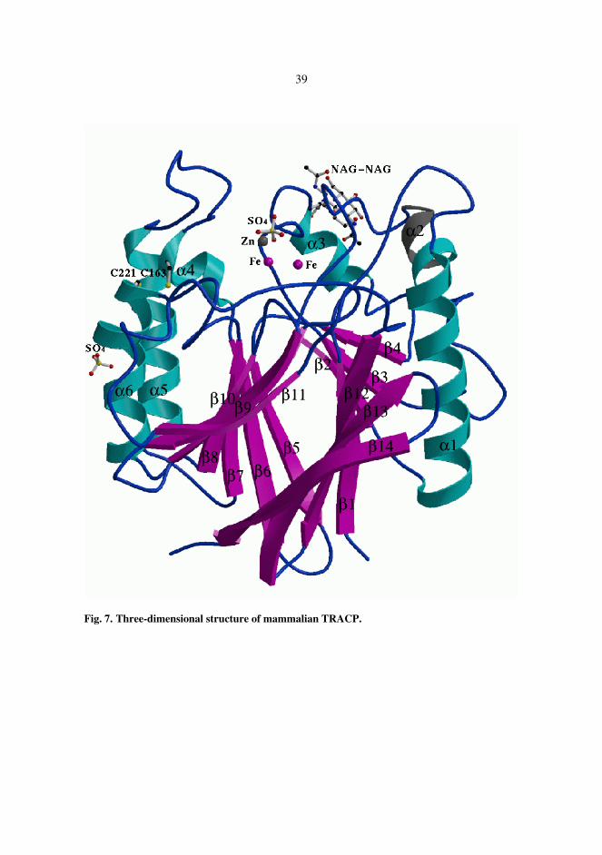
The three-dimensional structure of rat bone TRACP was determined at 2.2 Å resolution, Figure 7. The mammalian enzyme is a monomeric enzyme comprising one domain. The core of the enzyme was formed by two seven-stranded β-sheets which packed together, making up a β-sandwich. The β-sandwich was surrounded by three solvent-exposed α-helices on both sides. The di-iron center was located on the other edge of the β-sandwich. Two N-acetylglucosamine (NAG) molecules linked to the side chain of Asn118 were found in well-defined electron density. One zinc and one sulphate ion were found in the active site and one further zinc and sulphate ion were involved in crystal contacts. One of the iron ions was ligated to the side chains of Asp35, Tyr76, and His244 and the other iron ion was ligated to Asn112, His207, and His242. The latter iron formed a coordination bond to one of the oxygen atoms of a bound sulphate ion. This oxygen atom formed also a hydrogen bond to the side chain of Asn112. Another oxygen of the sulphate ion interacted with the side chain of His113 and the third oxygen coordinated the bound Zn ion. The metal ions were bridged through the coordination to one of the carboxylate oxygen atoms of Asp73. The distance of the metal ions was 3.5 Å. The electron density map indicated that a µ-(hydr)oxo bridge is found in the metal center.
39
Fig. 7. Three-dimensional structure of mammalian TRACP.

40
5.4 Site-directed mutagenesis and amino acids important for acid
phosphatase activity (IV)
The substrate specificity of TRACP was not affected by the mutations. The Km values for pNPP were of the same order of magnitude for all mutants, with the largest decrease (about five-fold) occurring in the His113Gln and His216Ala mutants compared to the wild-type enzyme. Significant decreases in the kcat values were seen for the mutants at the positions His113 and His216, where substitutions led to a drop in rate by up to two orders of magnitude. In terms of catalytic efficiency, the mutations at position 113 were most deleterious, with a drop in kcat /Km by up to 75-fold (His113Ala).
The kcat/Km versus pH profiles of wild-type TRACP and the Asp267Ala mutant were very similar, and the catalytic efficiency of this mutant was not significantly impaired over the whole pH range measured. The His216Ala mutant showed a shift in pH dependency and recovered to reach similar kcat/Km values as wild-type TRACP at very low pH values. Substitutions at the position His113 led to a mutant enzyme with very low catalytic efficiency over the whole pH range.
The rate versus pH curves for the AcP and ROS-generating activities of wild-type recombinant rat TRACP were different from each other, with the pH optimum for phosphoester hydrolysis close to pH 4.5 and the pH optimum for ROS-generating activity at pH 6.5. In order to determine further whether there are any differences between the AcP and the ROS-generating activities of recombinant rat TRACP, we studied if the mutants at the positions His113 and His216, which were almost inactive as acid phosphatase, were able to generate ROS. We found out that the ROS-generating capacity was only slightly impaired in the four mutants. For both histidine residues, the mutation to alanine inactivated the ROS-generating activity more than the mutation to glutamine.
The absorption maximum of the wild-type enzyme in the visible region of the spectrum was at 514 nm. The Asp267Ala mutant was slightly more oxidized, as indicated by the shift in the absorption maximum to 534 nm. The absorption maximum of all the other variants was at 507 nm – 514 nm.

6 Discussion
6.1 Baculovirus expression vector system
Application of the baculovirus expression vector system (BEVS) provides an easy access to large quantities of recombinant protein, which is biologically active and in most cases properly processed post-translationally. This kind of recombinant protein can be used for both enzymatical and structural characterization. Bone tissue is a particularly challenging material to be used in protein purification. Gaining sufficient amounts of bone tissue may be difficult or cause ethical problems, and the yields are usually not very high (Allen et al. 1989, Halleen et al. 1996). We were able to purify recombinant rat TRACP 0.5-1.0 mg to homogeneity per each liter of culture medium. In the production we used either 0.5-liter spinner flasks or produced the protein as a batch culture in a 30-liter bioreactor. Large-scale production and purification systems provided us with adequate quantities of protein for crystallization experiments (Vihko et al. 1993, Kurkela et al. 1995). Purification yields of up to 8 mg/liter culture medium for TRACP have been reported (Hayman & Cox 1994, Ek-Rylander et al. 1997). The baculovirus expression vector system is becoming an increasingly popular method for recombinant protein production due to its ability to produce functional proteins. Accelerated protein expression rate in cells may lead to a situation where glycosylation and amidation fail to "keep pace" with the protein production (Luckow 1991). In the case of glycoproteins this may result in lower glycosylation of the recombinant protein compared to the native form and to heterogeneity of the protein produced. This is considered as the only weak point of BEVS. In structure-based studies heterogeneity of the protein may cause difficulties in the crystallization process.

42
6.2 Characterization of TRACP
Recombinant rat tartrate-resistant acid phosphatase was produced as a mixture of the active, mixed valenced and the inactive, diferric forms of the protein. The specific activity of the purified preparation varied from one purification batch to another, depending on the relative amount of both forms, the diferric form being, however, more dominating in each batch. Large amounts of the protein allowed us to purify and separate these forms in sufficient amounts for their characterization. These forms should not be mixed up with the two differentially glycosylated forms of tartrate-resistant acid phosphatases 5a and 5b (Stepan et al. 1989) existing in serum and originating from different sources.
The inactive, diferric purple form could be transformed by reductive agents to the active, mixed valenced pink form and further to the extensively reduced yellowish form. The purple form was also spontaneously activated to the pink form upon incubation in +37°C. TRACP isolated from blood had significantly lower acid phosphatase activity compared to the bone-derived protein. These results led us to the hypothesis of a redox regulation of osteoclastic TRACP activity: TRACP is synthesized as an inactive oxidized form and then reduced to its functionally active form. Once it has completed its biological function in the cell, it is secreted into the circulation and inactivated by further reduction. This was further supported by our observation that the yellowish form brought about by reduction could no longer be activated. The yellowish form could thus represent the form described earlier inactivated by loss of its iron ions (Doi et al. 1988). In the blood circulation the enzyme would be destroyed and its iron content could be recycled. TRACP would thus have one possible function in iron metabolism.
In our studies, and shown also by other groups, proteolytic cleavage of the enzyme to its two-subunit form activates the enzyme. Ljusberg and her co-workers have suggested that the cysteine proteinase family might have a regulatory role in TRACP activation by converting the newly synthesized single polypeptide chain to its enzymatically active form (Ljusberg et al. 1999). This form is also more redox-sensitive (Ljusberg et al. 1999). According to their studies the cleavage shifts the pH optimum of the acid phosphatase activity to more basic values (Ljusberg et al. 1999, Funhoff et al. 2001b). This is consistent with our observation of the recombinant protein's more acidic pH optimum compared to the protein isolated from tissues. This group has postulated that the proteolytic cleavage results in the loss of a three-residue segment, thus changing interactions between residues and leading to an increase in Lewis acidity of the divalent metal with a concomitant increase in the electrophilicity of the enzyme-bound substrate (Funhoff et al. 2001a,b).

43
6.3 Monoclonal antibodies
Recombinant protein production using BEVS is a suitable method to gain antigen for raising monoclonal antibodies. Two of our antibodies raised in mouse using purified recombinant TRACP were able to bind the active rat recombinant enzyme, and one of the antibodies recognized the denatured form of the protein and could be used in Western blotting. None of the antibodies produced by us recognized native or denatured human bone derived TRACP (unpublished observation). Janckila and his co-workers studied the species specificity of monoclonal antibodies to human TRACP, and obtained both a species-specific and nonspecific antibody, demonstrating that in spite of the highly conserved sequences of mammalian TRACPs species-specific determinants exist and should be recognized when using experimental animals (Janckila et al. 1998). Several urinary and serum markers for studying human skeletal disorders have been developed, but use of biochemical markers in animal studies is less examined. Monoclonal antibodies produced in this study were used to developed a specific immunoassay that can be used to monitor the effects of different drugs in vivo in an experimental model, where osteoporosis has been induced by orchidectomy or ovariectomy in rats (manuscript in press).
6.4 Three-dimensional structures and reaction mechanism
Crystallization of the enzyme required thousands of experiments and took from several months up to two years. The crystals were very small with a typical diameter of 30µm. Heterogeneity of the glycosylation of the recombinant protein was suspected to cause the delay in crystal formation. The reason for this may also have been a mere technical detail of incompletely sealed crystallization wells allowing evaporation of the liquid. The crystals did, however, diffract well, and the data of the few available crystals were collected and processed. The structure was determined using the coordinates of the only structure of purple acid phosphatases existing so far, a plant enzyme isolated from red kidney bean.
Plant purple acid phosphatase is a dimer and has an additional domain of unknown function at the N-terminus which was not included in the model used in molecular replacement. The sequence identity between kidney bean and rat enzyme is only 18%. The three-dimensional structures of kidney bean enzyme and mammalian TRACP in this study and in two of the almost simultaneously published studies are nevertheless very similar (Sträter et al. 1995, Guddat et al. 1999, Uppenberg et al. 1999). Especially the core of the enzyme subunit is well preserved, while many of the loops differ in size and conformation. Figure 8 presents superposition of the structure of mammalian TRACP and purple acid phosphatase from red kidney bean.

44
Fig. 8. Superposition of TRACP (pink) and red kidney bean (purple) purple acid phosphatase.
The loop folding over the active site of mammalian TRACP does not exist in the plant
enzyme. This loop is also missing in the structure of rat bone enzyme by Uppenberg and his co-workers, presumably due to proteolytic cleavage (Uppenberg et al. 1999). The electron density map confirmed the presence of a µ-(hydr)oxo bridge in the metal center in both our study and in the study by Guddat and his coworkers (Guddat et al. 1999), while this could not be localized either in plant enzyme or in the rat enzyme structure by Uppenberg and co-workers (Uppenberg et al. 1999). The reason for this is probably the low resolution of these structures (kidney bean 2.9Å, rat 2.7Å).
Based on the data in this study and previous spectroscopic studies of tartrate-resistant/purple acid phosphatatases we have proposed the following reaction mechanism, which is closely consistent with the mechanism proposed for red kidney bean enzyme (Klabunde et al. 1996). In this mechanism the substrate binds to the enzyme with a coordination to the Fe(II). This places the hydroxyl ion in the ligand sphere of the Fe(III) ion in a position suitable for in-line attack on the phosphorus ion. Because the attack is delivered from the side opposite the alcohol group of the substrate, it will result in inversion of the configuration of the phosphorus ion. The inversion of the configuration argues against the formation of a covalent phosphorylated intermediate.

45
6.5 Classification of TRACP
As suggested by earlier secondary structure predictions, the mammalian TRACP proved to posses a βαβαβ-motif, which incorporates it to the large group of metallohydrolases with positioning of two metal ions by a central βαβαβ-scaffold. In some cases the metal appears to be essential either for the structure or for the enzymatic mechanism, and in other cases the role of the metal ions is more obscure (Zhuo et al. 1994, Nordlund & Eklund 1995). Serine/Threonine protein phosphatases (PPases) belong to this same group of phosphoesterases. PPases and mammalian tartrate-resistant acid phosphatases share fragments of amino acid sequences of significant homology. Among the conserved residues are those important in metal-binding, i.e. tyrosine, histidine, glutamic acid, and aspartic acid (Vincent & Averill 1990b). The active site structures are similar, but they do have some differences (Egloff et al. 1995, Kissinger et al. 1995, Knöfel & Sträter 2001, reviewed by Wilcox 1996, Barford et al. 1998, Kimura 2000). Metallohydrolases have in common that they hydrolyze phosphoproteins phosphorylated at their serine or threonine residues and the artificial substrate p-nitrophenyl phosphate (Vincent & Averill 1990b). We and others have reported that TRACP does not hydrolyze free or peptide-bound serine or threonine. TRACP has, however, been reported to hydrolyze protein-bound serine and threonine such as in osteopontin (Ek-Rylander et al. 1994, 1997), which is consistent with this classification. Specific phosphorylation of osteopontin has furthemore been shown to be important for its interactions with osteoclasts (Katayama et al. 1998).
Protein tyrosine phosphatases, PTPases, form another group of protein phosphatases. TRACP has been shown to hydrolyze phosphotyrosine, and we have suggested that TRACP might function as a PTPase in vivo. In spite of substrate specificity, TRACP has not turned out to have similarities with PTPases on structural level. Enzymes belonging to the group of protein tyrosine phosphatases require no metal ions or cofactors (Zhang 1997). PTPases do not share much sequence similarity, but contain an active site motif of one cysteine and one arginine residue, CX5R. This motif is missing in the structures of TRACPs (Fauman & Saper 1996). Acid phosphatases tend to show rather broad substrate specificity in general. Prostatic acid phosphatase (PAP) hydrolyzes phosphotyrosine in vitro (Nguyen et al. 1990, Porvari et al. 1994) without having structural motifs distinct to PTPases (Schneider et al. 1993). PAP has a readily accessible active site, and this has been speculated to give rise to the broad range of substrates hydrolyzed by the enzyme (Schneider et al. 1993). In mammalian TRACPs, a long insertion in the loop between β6 and α5 folds over the active site, resulting in a less open substrate-binding pocket compared to the plant enzyme (Figure 8) with similar substrate specificity. Fragments of conserved amino acid sequences existing in purple acid phosphatases and PPases are not to be found in PTPases (Zhang 1997). Ljusberg and her co-workers have argued TRACP's function as a PTPase in vivo with its cellular localization (Ljusberg et al. 1999). TRACP has been shown to be localized in lysosome-like vesicular compartments, whereas tyrosine phosphorylation occurs is cytosomal matrix of cells. The substrate specificity and physiological substrate of TRACP in biological conditions needs further investigation.

46
6.6 Site-directed mutagenesis
Our results from analyses of mutated forms of TRACP were consistent with most of the conclusions that were made by us and the other groups based on the crystallographic studies (Sträter et al. 1995, Guddat et al. 1999, Uppenberg et al. 1999). The active site structure revealed hydrogen bond interactions of the conserved residues His113 and His216 with oxygen atoms of bound phosphate. Site-directed mutagenesis confirmed these residues to be important in phosphoester hydrolysis. Based on the active site structure we proposed that Asp267, which is also conserved in plant enzyme, acts as a general acid that protonates the leaving group. Our suggestion was argued for by the fact that this residue is not present in PPases and it is too far from the substrate's phosphate group (8Å) to be able to participate directly in the reaction (Knöfel & Sträter 2001). Our mutagenesis studies did indeed reveal that the role of Asp267 is less likely than we had suggested.
The di-iron center is essential for both phosphate hydrolysis and reactive oxygen species generation of TRACP, and the same active site is thus employed in both mechanisms. For site-directed mutagenesis we chose those amino acid residues we expected to be important for the acid phosphatase activity. Mutation of these residues did not impair ROS generating activity as it did acid phosphatase activity, which led us to suggest that these activities are independent of each other. The fact that these two activities have different pH optima supports their independent function and possibly different cell compartment localization. One possibility would also have been to mutate one or more of the metal ion ligand binding residues, and to study its consequences on both of the activities (Zhuo 1994). The three-dimensional structure of an acid phosphatase-inactive form might be useful in clarifying the issue of TRACP's substrate specificity and the identification of natural substrate(s).

7 Conclusions
Here we have used the baculovirus expression vector system to produce recombinant rat tartrate-resistant acid phosphatase for characterization and structural analysis and as method to produce antigen for antibody production. The three-dimensional structure of the active site determined was used to predict the amino acids important for acid phosphatase activity. The role of these amino acids was studied using site-directed mutagenesis. Conserved amino acid residues His113 and His216 proved to have a critical role in acid phosphatase activity, while radical oxygen species generating activity seems to employ other so far unidentified residues. The substrate specificity of the enzyme was not altered by the mutations and kinetic parameters for the wild type enzyme and mutated forms were determined. The three-dimensional structure provided evidence for the µ-(hydr)oxo bridged di-iron center. Hypothesis of the redox regulation of osteoclastic TRACP activity was proposed as well as of the mechanism for phosphate hydrolysis. The di-iron metal center with conserved amino acid residues and the βαβαβ core-motif link type 5 tartrate-resistant acid phosphatase to the diverse group of metallohydrolases with different functions and substrate specificities. This study gives information for further investigation on the physiological substrate(s) of the enzyme and for construction of spesific inhibitors as well as for studying the role of the enzymes dual activity in vivo.
8 References
Abu-Amer Y, Ross FP, McHugh KP, Livolsi A, Peyron JF & Teitelbaum SL (1998). Tumor necrosis factor-alpha activation of nuclear transcription factor-kappaB in marrow macrophages is mediated by c-Src tyrosine phosphorylation of Ikappa Balpha. J Biol Chem 273:29417-29423.
Alatalo SL, Halleen JM, Hentunen TA, Mönkkönen J & Väänänen HK (2000). Rapid screening method for osteoclast differentiation in vitro that measures tartrate-resistant acid phosphatase 5b activity secreted into the culture medium. Clin Chem 46:1751-1754.
Alcantara O, Reddy SV, Roodman GD & Boldt DH (1994). Transcriptional regulation of the tartrate-resistant acid phosphatase (TRAP) gene by iron. Biochem J 298 ( Pt 2):421-425.
Allen SH, Nuttleman PR, Ketcham CM & Roberts RM (1989). Purification and characterization of human bone tartrate-resistant acid phosphatase. J Bone Miner Res 4:47-55.
Alvarez L, Peris P, Bedini JL, Pares A, Monegal A, Guanabens N, Mas E, Aibar C & Ballesta AM (1999). High bilirubin levels interfere with serum tartrate-resistant acid phosphatase determination: relevance as a marker of bone resorption in jaundiced patients. Calcif Tissue Int 64:301-303.
Anderson TR & Toverud SU (1986). Purification and characterization of purple acid phosphatase from developing rat bone. Arch Biochem Biophys 247:131-139.
Andersson G, Ek-Rylander B & Hammarström LE (1984). Purification and characterization of a vanadate-sensitive nucleotide tri- and diphosphatase with acid pH optimum from rat bone. Arch Biochem Biophys 228:431-438.
Andersson GN, Ek-Rylander B, Hammarström LE, Lindskog S & Toverud SU (1986). Immunocytochemical localization of a tartrate-resistant and vanadate-sensitive acid nucleotide tri- and diphosphatase. J Histochem Cytochem 34:293-298.
Andersson G, Lindunger A & Ek-Rylander B (1989). Isolation and characterization of skeletal acid ATPase--a new osteoclast marker? Connect Tissue Res 20:151-158.
Andersson G & Ek-Rylander B (1995). The tartrate-resistant purple acid phosphatase of bone osteoclasts--a protein phosphatase with multivalent substrate specificity and regulation. Acta Orthop Scand Suppl 266:189-194.
Angel NZ, Walsh N, Forwood MR, Ostrowski MC, Cassady AI & Hume DA (2000). Transgenic mice overexpressing tartrate-resistant acid phosphatase exhibit an increased rate of bone turnover. J Bone Miner Res 15:103-110.
Antanaitis BC, Strekas T & Aisen P (1982). Characterization of pink and purple uteroferrin by resonance Raman and CD spectroscopy. J Biol Chem 257:3766-3770.
Antanaitis BC & Aisen P (1983). Uteroferrin and the purple acid phosphatases. Adv Inorg Biochem 5:111-136.
Antanaitis BC, Aisen P & Lilienthal HR (1983). Physical characterization of two-iron uteroferrin. Evidence for a spin-coupled binuclear iron cluster. J Biol Chem 258:3166-3172.

49
Antanaitis BC & Aisen P (1985). Effects of perturbants on the pink (reduced) active form of uteroferrin. Phosphate-induced anaerobic oxidation. J Biol Chem 260:751-756.
Arai N, Ohya K & Ogura H (1993). Osteopontin mRNA expression during bone resorption: an in situ hybridization study of induced ectopic bone in the rat. Bone Miner 22:129-145.
Aravind L & Koonin EV (1998). The HD domain defines a new superfamily of metal-dependent phosphohydrolases. Trends Biochem Sci 23:469-472.
Barford D, Das AK & Egloff MP (1998). The structure and mechanism of protein phosphatases: insights into catalysis and regulation. Annu Rev Biophys Biomol Struct 27:133-164.
Baron R, Neff L, Roy C, Boisvert A & Caplan M (1986) Evidence for a high and specific concentration of (Na+,K+)ATPase in the plasma membrane of the osteoclast. Cell 46:311-320.
Battistuzzi G, Dietrich M, Locke R & Witzel H (1997). Evidence for a conserved binding motif of the dinuclear metal site in mammalian and plant purple acid phosphatases: 1H NMR studies of the di-iron derivative of the Fe(III)Zn(II) enzyme from kidney bean. Biochem J 323 ( Pt 3):593-596.
Baumbach GA, Ketcham CM, Richardson DE, Bazer FW & Roberts RM (1986). Isolation and characterization of a high molecular weight stable pink form of uteroferrin from uterine secretions and allantoic fluid of pigs. J Biol Chem 261:12869-12878.
Baumbach GA, Saunders PT, Ketcham CM, Bazer FW & Roberts RM (1991). Uteroferrin contains complex and high mannose-type oligosaccharides when synthesized in vitro. Mol Cell Biochem 105:107-117.
Beck JL, Durack MC, Hamilton SE & De Jersey J (1999). Irreversible inactivation of purple acid phosphatase by hydrogen peroxide and ascorbate. J Inorg Biochem 73:245-252.
Bevilacqua MA, Lord DK, Cross NC, Whitaker KB, Moss DW & Cox TM (1991). Regulation and expression of type V (tartrate-resistant) acid phosphatase in human mononuclear phagocytes. Mol Biol Med 8:135-140.
Black MJ & Jones ME (1983). Inorganic phosphate determination in the presence of a labile organic phosphate: Assay for carbamyl phosphate phosphatase activity. Biochemistry 135:233-238.
Blair HC, Kahn AJ, Crouch EC, Jeffrey JJ & Teitelbaum SL (1986). Isolated osteoclasts resorb the organic and inorganic components of bone. J Cell Biol 102:1164-1172.
Blair HC, Teitelbaum SL, Ghiselli R & Gluck S (1989). Osteoclastic bone resorption by a polarized vacuolar proton pump. Science 245:855-857.
Blair HC, Sidonio RF, Friedberg RC, Khan NN & Dong SS (2000). Proteinase expression during differentiation of human osteoclasts in vitro. J Cell Biochem 78:627-637.
Boldt DH (1999). New perspectives on iron: an introduction. Am J Med Sci 318:207-212. Bord S, Beavan S, Ireland D, Horner A & Compston JE (2001). Mechanisms by which high-dose
estrogen therapy produces anabolic skeletal effects in postmenopausal women: role of locally produced growth factors. Bone 29:216-222.
Boyce BF, Hughes DE, Wright KR, Xing L & Dai A (1999). Recent advances in bone biology provide insight into the pathogenesis of bone diseases. Lab Invest 79:83-94.
Bradford MM (1976). A rapid and sensitive method for the quantitation of microgram quantities of protein utilizing the principle of protein-dye binding. Anal Biochem 72:248-254.
Brehme CS, Roman S, Shaffer J & Wolfert R (1999). Tartrate-resistant acid phosphatase forms complexes with alpha2-macroglobulin in serum. J Bone Miner Res 14:311-318.
Brown MA, Haughton MA, Grant SF, Gunnell AS, Henderson NK & Eisman JA (2001). Genetic control of bone density and turnover: role of the collagen 1alpha1, estrogen receptor, and vitamin D receptor genes. J Bone Miner Res 16:758-764.
Buhi WC, Ducsay CA, Bazer FW & Roberts RM (1982). Iron transfer between the purple phosphatase uteroferrin and transferrin and its possible role in iron metabolism of the fetal pig. J Biol Chem 257:1712-1723.
Bune AJ, Hayman AR, Evans MJ & Cox TM (2001). Mice lacking tartrate-resistant acid phosphatase (Acp 5) have disordered macrophage inflammatory responses and reduced clearance of the pathogen, Staphylococcus aureus. Immunology 102:103-113.
Burkitt HC,Young B & Heath JW. Wheater's Functional Histology. A Text and Colour Atlas (1993) Hong Kong:Churchill Livingstone, p. 192-199.

50
Campbell HD & Zerner B (1973). A low-molecular-weight acid phosphatase which contains iron. Biochem Biophys Res Commun 54:1498-1503.
Campbell HD, Dionysius DA, Keough DT, Wilson BE, De Jersey J, and Zerner B (1978). Iron-containing acid phosphatases: comparison of the enzymes from beef spleen and pig allantoic fluid. Biochem Biophys Res Commun 82:615-620.
Carbonell LF, Klowden MJ & Miller LK (1985). Baculovirus-mediated expression of bacterial genes in dipteran and mammalian cells. J Virol 56:153-160.
Carbonell LF & Miller LK (1987). Baculovirus interaction with nontarget organisms: a virus-borne reporter gene is not expressed in two mammalian cell lines. Appl Environ Microbiol 53:1412-1417.
Cassady AI, King AG, Cross NC & Hume DA (1993). Isolation and characterization of the genes encoding mouse and human type-5 acid phosphatase. Gene 130:201-207.
Chamberlain P, Compston J, Cox TM, Hayman AR, Imrie RC, Reynolds K & Holmes SD (1995). Generation and characterization of monoclonal antibodies to human type-5 tartrate-resistant acid phosphatase: development of a specific immunoassay of the isoenzyme in serum. Clin Chem 41:1495-1499.
Chen TT, Bazer FW, Cetorelli JJ, Pollard WE & Roberts RM (1973). Purification and properties of a progesterone-induced basic glycoprotein from the uterine fluids of pigs. J Biol Chem 248:8560-8566.
Cheung CK, Panesar NS, Haines C, Masarei J & Swaminathan R (1995). Immunoassay of a tartrate-resistant acid phosphatase in serum. Clin Chem 41:679-686.
Clark SA, Ambrose WW, Anderson TR, Terrell RS & Toverud SU (1989). Ultrastructural localization of tartrate-resistant, purple acid phosphatase in rat osteoclasts by histochemistry and immunocytochemistry. J Bone Miner Res 4:399-405.
Collier FM, Huang WH, Holloway WR, Hodge JM, Gillespie MT, Daniels LL, Zheng MH & Nicholson GC (1998). Osteoclasts from human giant cell tumors of bone lack estrogen receptors. Endocrinology 139:1258-1267.
Cornish J, Callon KE, Bava U, Kamona SA, Cooper GJ & Reid IR (2001). Effects of calcitonin, amylin, and calcitonin gene-related peptide on osteoclast development. Bone 29:162-168.
Crans DC, Simone CM, Holz RC & Que L, Jr. (1992). Interaction of porcine uterine fluid purple acid phosphatase with vanadate and vanadyl cation. Biochemistry 31:11731-11739.
da Paz LH, de F, V, Teng NC, dos Reis LM, Pereira RM & Jorgetti V (2001). Effect of 17beta-estradiol or alendronate on the bone densitometry, bone histomorphometry and bone metabolism of ovariectomized rats. Braz J Med Biol Res 34:1015-1022.
Darden AG, Ries WL, Wolf WC, Rodriguiz RM & Key LL, Jr. (1996). Osteoclastic superoxide production and bone resorption: stimulation and inhibition by modulators of NADPH oxidase. J Bone Miner Res 11:671-675.
Davis JC, Lin SS & Averill BA (1981). Kinetic and optical spectroscopic studies on the purple acid phosphatase from beef spleen. Biochemistry 20:4062-4067.
Davis JC & Averill BA (1982). Evidence for a spin-coupled binuclear iron unit at the active site of the purple acid phosphatase from beef spleen. Proc Natl Acad Sci U S A 79:4623-4627.
Day EP, David SS, Peterson J, Dunham WR, Bonvoisin JJ, Sands RH & Que L, Jr. (1988). Magnetization and electron paramagnetic resonance studies of reduced uteroferrin and its "EPR-silent" phosphate complex. J Biol Chem 263:15561-15567.
del Pozo JC, Allona I, Rubio V, Leyva A, de la PA, Aragoncillo C & Paz-Ares J (1999). A type 5 acid phosphatase gene from Arabidopsis thaliana is induced by phosphate starvation and by some other types of phosphate mobilising/oxidative stress conditions. Plant J 19:579-589.
Delmas PD (1992). Clinical use of biochemical markers of bone remodeling in osteoporosis. Bone 13 Suppl 1:S17-S21.
Denu JM, Stuckey JA, Saper MA & Dixon JE (1996). Form and function in protein dephosphorylation. Cell 87:361-364.
Dodds RA, Connor JR, James IE, Rykaczewski EL, Appelbaum E, Dul E & Gowen M (1995). Human osteoclasts, not osteoblasts, deposit osteopontin onto resorption surfaces: an in vitro and ex vivo study of remodeling bone. J Bone Miner Res 10:1666-1680.

51
Doi K, Antanaitis BC & Aisen P (1988). The binuclear iron centers of uteroferrin and the purple acid phosphatase. Structure and bonding 70:1-26.
Duong LT & Rodan GA (1999). The role of integrins in osteoclast function. J Bone Miner Metab 17:1-6.
Durmus A, Eicken C, Sift BH, Kratel A, Kappl R, Huttermann J & Krebs B (1999). The active site of purple acid phosphatase from sweet potatoes (Ipomoea batatas) metal content and spectroscopic characterization. Eur J Biochem 260:709-716.
Echetebu ZO, Cox TM & Moss DW (1987). Antibodies to porcine uteroferrin used in measurement of human tartrate-resistant acid phosphatase. Clin Chem 33:1832-1836.
Edelson GW & Kleerekoper M (1996). Pathophysiology of osteoporosis. In: Sartoris DJ, editor, New York: Marcel Dekker, Inc, 1-18.
Efstratiadis T & Moss DW (1985). Tartrate-resistant acid phosphatase of human lung: apparent identity with osteoclastic acid phosphatase. Enzyme 33:34-40.
Egloff MP, Cohen PT,Reinemer P & Barford D (1995) Crystal structure of the catalytic subunit of human protein phosphatase 1 and its complex with tungstate. J Mol Biol 254:942-959.
Ek-Rylander B, Bill P, Norgard M, Nilsson S & Andersson G (1991a). Cloning, sequence, and developmental expression of a type 5, tartrate-resistant, acid phosphatase of rat bone. J Biol Chem 266:24684-24689.
Ek-Rylander B, Bergman T & Andersson G (1991b). Characterization of a tartrate-resistant acid phosphatase (ATPase) from rat bone: hydrodynamic properties and N-terminal amino acid sequence. J Bone Miner Res 6:365-373.
Ek-Rylander B, Flores M, Wendel M, Heinegard D & Andersson G (1994). Dephosphorylation of osteopontin and bone sialoprotein by osteoclastic tartrate-resistant acid phosphatase. Modulation of osteoclast adhesion in vitro. J Biol Chem 269:14853-14856.
Ek-Rylander B, Barkhem T, Ljusberg J, Öhman L, Andersson KK & Andersson G (1997). Comparative studies of rat recombinant purple acid phosphatase and bone tartrate-resistant acid phosphatase. Biochem J 321 ( Pt 2):305-311.
Fauman EB & Saper MA (1996). Structure and function of the protein tyrosine phosphatases. Trends Biochem Sci 21:413-417.
von Figura K & Weber E (1978) An alternative hypothesis of cellular transport of lysosomal enzymes in fibroblasts. Effect of inhibitors of lysosomal enzyme endocytosis on intra- and extra-cellular lysosomal enzyme activities. Biochem J 176:943-950.
Fleckenstein E, Dirks W, Dehmel U & Drexler HG (1996). Cloning and characterization of the human tartrate-resistant acid phosphatase (TRAP) gene. Leukemia 10:637-643.
Fleckenstein E & Drexler HG (1997). Tartrate-resistant acid phosphatase: gene structure and function. Leukemia 11:10-13.
Fleckenstein EC, Dirks WG & Drexler HG (2000). The human tartrate-resistant acid phosphatase (TRAP): involvement of the hemin responsive elements (HRE) in transcriptional regulation. Leuk Lymphoma 36:603-612.
Flores ME, Norgård M, Heinegård D, Reinholt FP & Andersson G (1992). RGD-directed attachment of isolated rat osteoclasts to osteopontin, bone sialoprotein, and fibronectin. Exp Cell Res 201:526-530.
Funhoff EG, Klaassen CHW, Samyn B, Van Beeumen J & Averill BA (2001a). The highly exposed loop region in mammalian purple acid phosphatase controls the catalytic activity. Chembiochem 2:355-363.
Funhoff EG, Ljusberg J, Wang Y, Andersson G & Averill BA (2001b). Mutational analysis of the interaction between active site residues and the loop region in mammalian purple acid phosphatases. Biochemistry 40:11614-11622.
Gaber BP, Sheridan JP, Bazer FW & Roberts RM (1979). Resonance Raman scattering from uteroferrin, the purple glycoprotein of the porcine uterus. J Biol Chem 254:8340-8342.
Garrett IR, Boyce BF, Oreffo RO, Bonewald L, Poser J & Mundy GR (1990). Oxygen-derived free radicals stimulate osteoclastic bone resorption in rodent bone in vitro and in vivo. J Clin Invest 85:632-639.
Glomset J & Porath J (1960). Some properties of a phophatase from bovine spleen. Biochim Biophys Acta 39:1-8.

52
Greendale GA, Edelstein S & Barrett-Connor E (1997). Endogenous sex steroids and bone mineral density in older women and men: the Rancho Bernardo Study. J Bone Miner Res 12:1833-1843.
Greenwald RA, Rush SW, Moak SA & Weitz Z (1989). Conversion of superoxide generated by polymorphonuclear leukocytes to hydroxyl radical: a direct spectrophotometric detection system based on degradation of deoxyribose. Free Radic Biol Med 6:385-392.
Grimes R, Reddy SV, Leach RJ, Scarcez T, Roodman GD, Sakaguchi AY, Lalley PA & Windle JJ (1993). Assignment of the mouse tartrate-resistant acid phosphatase gene (Acp5) to chromosome 9. Genomics 15:421-422.
Gruber HE, Puzanov IJ, Bennett M, Kumar V, V & Gordon B (2001). Alterations in osteoclast morphology following long-term 17beta-estradiol administration in the mouse. BMC Cell Biol 2:3.
Guddat LW, McAlpine AS, Hume D, Hamilton S, De Jersey J & Martin JL (1999). Crystal structure of mammalian purple acid phosphatase. Structure Fold Des 7:757-767.
Hall TJ, Schaeublin M, Jeker H, Fuller K & Chambers TJ (1995). The role of reactive oxygen intermediates in osteoclastic bone resorption. Biochem Biophys Res Commun 207:280-287.
Halleen J, Hentunen TA, Hellman J & Väänänen HK (1996). Tartrate-resistant acid phosphatase from human bone: purification and development of an immunoassay. J Bone Miner Res 11:1444-1452.
Halleen JM, Hentunen TA, Karp M, Käkönen SM, Pettersson K & Väänänen HK (1998). Characterization of serum tartrate-resistant acid phosphatase and development of a direct two-site immunoassay. J Bone Miner Res 13:683-687.
Halleen JM, Räisanen S, Salo JJ, Reddy SV, Roodman GD, Hentunen TA, Lehenkari PP, Kaija H, Vihko P & Väänänen HK (1999a). Intracellular fragmentation of bone resorption products by reactive oxygen species generated by osteoclastic tartrate-resistant acid phosphatase. J Biol Chem 274:22907-22910.
Halleen JM, Karp M, Viloma S, Laaksonen P, Hellman J, Käkönen SM, Stepan JJ, Holmes S, Väänänen H & Pettersson K (1999b). Two-site immunoassays for osteoclastic tartrate-resistant acid phosphatase based on characterization of six monoclonal antibodies. J Bone Miner Res 14:464-469.
Halleen JM, Alatalo SL, Suominen H, Cheng S, Janckila AJ & Väänänen HK (2000). Tartrate-resistant acid phosphatase 5b: a novel serum marker of bone resorption. J Bone Miner Res 15:1337-1345.
Halleen JM & Ranta R (2001). Tartrate-resistant acid phosphatase as a serum marker of bone resorption. Am Clin Lab 20:29-30.
Halliwell B, Gutteridge JM & Aruoma OI (1987). The deoxyribose method: a simple "test-tube" assay for determination of rate constants for reactions of hydroxyl radicals. Anal Biochem 165:215-219.
Hannon R, Blumsohn A, Naylor K & Eastell R (1998). Response of biochemical markers of bone turnover to hormone replacement therapy: impact of biological variability. J Bone Miner Res 13:1124-1133.
Hara A, Sawada H, Kato T, Nakayama T, Yamamoto H & Matsumoto Y (1984). Purification and characterization of a purple acid phosphatase from rat spleen. J Biochem (Tokyo) 95:67-74.
Hayman AR, Warburton MJ, Pringle JA, Coles B & Chambers TJ (1989). Purification and characterization of a tartrate-resistant acid phosphatase from human osteoclastomas. Biochem J 261:601-609.
Hayman AR, Dryden AJ, Chambers TJ & Warburton MJ (1991). Tartrate-resistant acid phosphatase from human osteoclastomas is translated as a single polypeptide. Biochem J 277 ( Pt 3):631-634.
Hayman AR & Cox TM (1994). Purple acid phosphatase of the human macrophage and osteoclast. Characterization, molecular properties, and crystallization of the recombinant di-iron-oxo protein secreted by baculovirus-infected insect cells. J Biol Chem 269:1294-1300.
Hayman AR, Jones SJ, Boyde A, Foster D, Colledge WH, Carlton MB, Evans MJ & Cox TM (1996). Mice lacking tartrate-resistant acid phosphatase (Acp 5) have disrupted endochondral ossification and mild osteopetrosis. Development 122:3151-3162.

53
Hayman AR, Bune AJ & Cox TM (2000a). Widespread expression of tartrate-resistant acid phosphatase (Acp 5) in the mouse embryo. J Anat 196 ( Pt 3):433-441.
Hayman AR, Bune AJ, Bradley JR, Rashbass J & Cox TM (2000b). Osteoclastic tartrate-resistant acid phosphatase (Acp 5): its localization to dendritic cells and diverse murine tissues. J Histochem Cytochem 48:219-228.
Hegeman CE & Grabau EA (2001). A novel phytase with sequence similarity to purple acid phosphatases is expressed in cotyledons of germinating soybean seedlings. Plant Physiol 126:1598-1608.
Himeno M, Fujita H, Noguchi Y, Kono A & Kato K (1989). Isolation and sequencing of a cDNA clone encoding acid phosphatase in rat liver lysosomes. Biochem Biophys Res Commun 162:1044-1053.
Hofbauer LC & Heufelder AE (2001). Role of receptor activator of nuclear factor-kappaB ligand and osteoprotegerin in bone cell biology. J Mol Med 79:243-253.
Holliday LS, Welgus HG, Fliszar CJ, Veith GM, Jeffrey JJ & Gluck SL (1997). Initiation of osteoclast bone resorption by interstitial collagenase. J Biol Chem 272:22053-22058.
Hoyer JD, Li CY, Yam LT, Hanson CA & Kurtin PJ (1997). Immunohistochemical demonstration of acid phosphatase isoenzyme 5 (tartrate-resistant) in paraffin sections of hairy cell leukemia and other hematologic disorders. Am J Clin Pathol 108:308-315.
Hughes DE, Dai A, Tiffee JC, Li HH, Mundy GR & Boyce BF (1996). Estrogen promotes apoptosis of murine osteoclasts mediated by TGF-beta. Nat Med 2:1132-1136.
Hunt DF, Yates JR, III, Shabanowitz J, Zhu NZ, Zirino T, Averill BA, Daurat-Larroque ST, Shewale JG, Roberts RM & Brew K (1987). Sequence homology in the metalloproteins; purple acid phosphatase from beef spleen and uteroferrin from porcine uterus. Biochem Biophys Res Commun 144:1154-1160.
Höyhtyä M, Vihko P, Vuolas L, Tryggvason K & Vihko R (1987). High-affinity monoclonal antibodies specific for human prostatic acid phosphatase. Clin Chem 33:103-107.
Igarashi Y, Lee MY & Matsuzaki S (2001). Heparin column analysis of serum type 5 tartrate-resistant acid phosphatase isoforms. J Chromatogr B Biomed Sci Appl 757:269-276.
Ihara H, Denhardt DT, Furuya K, Yamashita T, Muguruma Y, Tsuji K, Hruska KA, Higashio K, Enomoto S, Nifuji A, Rittling SR & Noda M (2001). Parathyroid hormone-induced bone resorption does not occur in the absence of osteopontin. J Biol Chem 276:13065-13071.
Inoue M, Yoshida H & Akisaka T (1999). Visualization of acidic compartments in cultured osteoclasts by use of an acidotrophic amine as a marker for low pH. Cell Tissue Res 298:527-537.
Isaki H & Hanaoka H (1995). The effects of high dose of parathyroid hormone on fetal osteoclasts and their precursors in vivo: an ultrastructural-cytochemical study. Anat Rec 243:421-429.
Janckila AJ, Woodford TA, Lam KW, Li CY & Yam LT (1992a). Protein-tyrosine phosphatase activity of hairy cell tartrate-resistant acid phosphatase. Leukemia 6:199-203.
Janckila AJ, Latham MD, Lam KW, Chow KC, Li CY & Yam LT (1992b). Heterogeneity of hairy cell tartrate-resistant acid phosphatase. Clin Biochem 25:437-443.
Janckila AJ, Lear SC, Martin AW & Yam LT (1996). Epitope enhancement for immunohistochemical demonstration of tartrate-resistant acid phosphatase. J Histochem Cytochem 44:235-244.
Janckila AJ, Cardwell EM & Yam LT (1997). Characterization of monoclonal antibodies specific to human tartrate-resistant acid phosphatase. Hybridoma 16:175-182.
Janckila AJ, Walton SP & Yam LT (1998). Species specificity of monoclonal antibodies to human tartrate-resistant acid phosphatase. Biotech Histochem 73:316-324.
Janckila AJ, Takahashi K, Sun SZ & Yam LT (2001a). Naphthol-ASBI phosphate as a preferred substrate for tartrate-resistant acid phosphatase isoform 5b. J Bone Miner Res 16:788-793.
Janckila AJ, Takahashi K, Sun SZ & Yam LT (2001b). Tartrate-resistant acid phosphatase isoform 5b as serum marker for osteoclastic activity. Clin Chem 47:74-80.
Kalkkinen N & Tilgman C (1988). A gas-pulsed liquid-phase sequencer contructed from a Beckman 890D instrument by using Applied Biosystems delivery and cartridge blocks. J Protein Chem 7:242-243.

54
Katayama I, Li CY& Yam LT (1972). Ultrastructural cytochemical demonstration of tartrate-resistant acid phosphatase isoenzyme activity in "hairy cells" of leukemic reticuloendotheliosis. Am J Pathol 69:471-482.
Kato T, Hara A, Nakayama T, Sawada H, Hamatake M & Matsumoto Y (1986) Purification and characterization of purple acid phosphatase from rat bone. Comp Biochem Physiol B 83:813-817.
Kehely A & Moss DW (1992). Circulating levels of tartrate-resistant acid phosphatase in macrophage-activated lung disease. Ann Clin Biochem 29 ( Pt 2):172-175.
Keough DT, Dionysius DA, De Jersey J & Zerner B (1980). Iron-containing acid phosphatases: characterization of the metal-ion binding site of the enzyme from pig allantoic fluid. Biochem Biophys Res Commun 94:600-605.
Keough DT, Beck JL, De Jersey J & Zerner B (1982). Iron-containing acid phosphatases: interaction of phosphate with the enzyme from pig allantoic fluid. Biochem Biophys Res Commun 108:1643-1648.
Ketcham CM, Baumbach GA, Bazer FW & Roberts RM (1985). The type 5, acid phosphatase from spleen of humans with hairy cell leukemia. Purification, properties, immunological characterization, and comparison with porcine uteroferrin. J Biol Chem 260:5768-5776.
Ketcham CM, Roberts RM, Simmen RC & Nick HS (1989). Molecular cloning of the type 5, iron-containing, tartrate-resistant acid phosphatase from human placenta. J Biol Chem 264:557-563.
Key LL, Jr., Wolf WC, Gundberg CM & Ries WL (1994). Superoxide and bone resorption. Bone 15:431-436.
Kimura E (2000). Dimetallic hydrolases and their models. Curr Opin Chem Biol 4:207-213. Kissinger CR, Parge HE, Knighton DR, Lewis CT, Pelletier LA, Tempczyk A, Kalish VJ, Tucker
KD, Showalter RE, Moomaw EW & (1995) Crystal structures of human calcineurin and the human FKBP12-FK506-calcineurin complex. Nature 378:641-644.
Klabunde T, Stahl B, Suerbaum H, Hahner S, Karas M, Hillenkamp F, Krebs B & Witzel H (1994). The amino acid sequence of the red kidney bean Fe(III)-Zn(II) purple acid phosphatase. Determination of the amino acid sequence by a combination of matrix-assisted laser desorption/ionization mass spectrometry and automated Edman sequencing. Eur J Biochem 226:369-375.
Klabunde T, Sträter N, Krebs B & Witzel H (1995). Structural relationship between the mammalian Fe(III)-Fe(II) and the Fe(III)-Zn(II) plant purple acid phosphatases. FEBS Lett 367:56-60.
Klabunde T, Sträter N, Fröhlich R, Witzel H & Krebs B (1996). Mechanism of Fe(III)-Zn(II) purple acid phosphatase based on crystal structures. J Mol Biol 259:737-748.
Knöfel T & Sträter N (2001). Mechanism of hydrolysis of phosphate esters by the dimetal center of 5'-nucleotidase based on crystal structures. J Mol Biol 309:239-254.
Kondo Y, Irie K, Ikegame M, Ejiri S, Hanada K & Ozawa H (2001). Role of stromal cells in osteoclast differentiation in bone marrow. J Bone Miner Metab 19:352-358.
Koonin EV (1994). Conserved sequence pattern in a wide variety of phosphoesterases. Protein Sci 3:356-358.
Kraenzlin ME, Lau KH, Liang L, Freeman TK, Singer FR, Stepan J & Baylink DJ (1990). Development of an immunoassay for human serum osteoclastic tartrate-resistant acid phosphatase. J Clin Endocrinol Metab 71:442-451.
Kurkela R, Herrala A, Henttu P, Nai H & Vihko P (1995). Expression of active, secreted human prostate-specific antigen by recombinant baculovirus-infected insect cells on a pilot-scale. Biotechnology (N Y ) 13:1230-1234.
Laitala-Leinonen T, Lowik C, Papapoulos S & Väänänen HK (1999). Inhibition of intravacuolar acidification by antisense RNA decreases osteoclast differentiation and bone resorption in vitro. J Cell Sci 112 ( Pt 21):3657-3666.
Lam KW, Dekker PT, Castleman J & Yam LT (1976). Studies of intracellular distribution of acid phosphatase 5 in the spleen cells of leukemic reticuloendotheliosis by isopycnic gradient centrifugation. Clin Chim Acta 70:289-295.
Lam WK, Eastlund DT, Li CY & Yam LT (1978). Biochemical properties of tartrate-resistant acid phosphatase in serum of adults and children. Clin Chem 24:1105-1108.

55
Lam KW, Li CY, Yam LT & Desnick RJ (1981). Comparison of the tartrate-resistant acid phosphatase in Gaucher's disease and leukemic reticuloendotheliosis. Clin Biochem 14:177-181.
Lam KW, Siemens M, Sun T, Li CY & Yam LT (1982). Enzyme immunoassay for tartrate-resistant acid phosphatase. Clin Chem 28:467-470.
Lambert E, Chabut B, Chardon-Noblat S, Deronzier A, Chottard G, Bousseksou A, Tuchagues J-P, Laugier J, Bardet M & Latour J-M (1997). Synthesis, structural, magnetic, and redox properties of asymmetric diiron complex with a single terminally bound phenolate ligand. Relevance to the purple acid phosphatase enzymes. J Am Chem Soc 119:9424-9437.
Lamp EC & Drexler HG (2000). Biology of tartrate-resistant acid phosphatase. Leuk Lymphoma 39:477-484.
Lau KH, Freeman TK & Baylink DJ (1987a). Purification and characterization of an acid phosphatase that displays phosphotyrosyl-protein phosphatase activity from bovine cortical bone matrix. J Biol Chem 262:1389-1397.
Lau KH, Onishi T, Wergedal JE, Singer FR & Baylink DJ (1987b). Characterization and assay of tartrate-resistant acid phosphatase activity in serum: potential use to assess bone resorption. Clin Chem 33:458-462.
Leach RJ, Reus BE, Hundley JE, Johnson-Pais TL & Windle JJ (1994). Confirmation of the assignment of the human tartrate-resistant acid phosphatase gene (ACP5) to chromosome 19. Genomics 19:180-181.
LeBansky BR, McKnight TD & Griffing LR (1992). Purification and characterization of a secreted purple acid phosphatase from soybean suspension cultures. Plant Physiol 99:391-395.
Lees RL, Sabharwal VK & Heersche JN (2001). Resorptive state and cell size influence intracellular pH regulation in rabbit osteoclasts cultured on collagen-hydroxyapatite films. Bone 28:187-194.
Li CY, Yam LT & Lam KW (1970). Studies of acid phosphatase isoenzymes in human leukocytes demonstration of isoenzyme cell specificity. J Histochem Cytochem 18:901-910.
Li YP, Chen W, Liang Y, Li E & Stashenko P (1999). Atp6i-deficient mice exhibit severe osteopetrosis due to loss of osteoclast-mediated extracellular acidification. Nat Genet 23:447-451.
Lindqvist Y, Schneider G & Vihko P (1993). Three-dimensional structure of rat acid phosphatase in complex with L(+)-tartrate. J Biol Chem 268:20744-20746.
Lindqvist Y, Schneider G & Vihko P (1994). Crystal structures of rat acid phosphatase complexed with the transition-state analogs vanadate and molybdate. Implications for the reaction mechanism. Eur J Biochem 221:139-142.
Ling P & Roberts RM (1993). Uteroferrin and intracellular tartrate-resistant acid phosphatases are the products of the same gene. J Biol Chem 268:6896-6902.
Ljusberg J, Ek-Rylander B & Andersson G (1999). Tartrate-resistant purple acid phosphatase is synthesized as a latent proenzyme and activated by cysteine proteinases. Biochem J 343 Pt 1:63-69.
Lohse DL, Denu JM & Dixon JE (1995). Insights derived from the structures of the Ser/Thr phosphatases calcineurin and protein phosphatase 1. Structure 3:987-990.
Lord DK, Cross NC, Bevilacqua MA, Rider SH, Gorman PA, Groves AV, Moss DW, Sheer D & Cox TM (1990). Type 5 acid phosphatase. Sequence, expression and chromosomal localization of a differentiation-associated protein of the human macrophage. Eur J Biochem 189:287-293.
Luckow VA (1991). Cloning and expression of heterologous genes in insect cells with baculovirus vectors. In: Prokop A, Bajpai RK, and Ho CS, editors, New York: McGraw-Hill, 97-152.
Lång P, Schultzberg M & Andersson G (2001). Expression and distribution of tartrate-resistant purple acid phosphatase in the rat nervous system. J Histochem Cytochem 49:379-396.
Mancino AT, Klimberg VS, Yamamoto M, Manolagas SC & Abe E (2001). Breast cancer increases osteoclastogenesis by secreting M-CSF and upregulating RANKL in stromal cells. J Surg Res 100:18-24.
Manolagas SC (2000). Birth and death of bone cells: basic regulatory mechanisms and implications for the pathogenesis and treatment of osteoporosis. Endocr Rev 21:115-137.
Martin TJ & Udagawa N (1998). Hormonal regulation of osteoclast function. TEM 9:6-12.

56
Matayoshi A, Brown C, DiPersio JF, Haug J, Abu-Amer Y, Liapis H, Kuestner R & Pacifici R (1996). Human blood-mobilized hematopoietic precursors differentiate into osteoclasts in the absence of stromal cells. Proc Natl Acad Sci U S A 93:10785-10790.
Medhora MM, Teitelbaum S, Chappel J, Alvarez J, Mimura H, Ross FP & Hruska K (1993). 1 alpha,25-dihydroxyvitamin D3 up-regulates expression of the osteoclast integrin alpha v beta 3. J Biol Chem 268:1456-1461.
Merkx M & Averill BA (1999). Probing the role of the trivalent metal in phosphate ester hydrolysis: preparation and characterization of purple acid phosphatases containing Al(III)Zn(II) and In(III)Zn(II) active sites, including the first example of an active aluminium enzyme. J Am Chem Soc 121:6683-6689.
Merry K, Dodds R, Littlewood A & Gowen M (1993). Expression of osteopontin mRNA by osteoclasts and osteoblasts in modelling adult human bone. J Cell Sci 104 ( Pt 4):1013-1020.
Mezquita-Raya P, Munoz-Torres M, Luna JD, Luna V, Lopez-Rodriguez F, Torres-Vela E & Escobar-Jimenez F (2001). Relation between vitamin D insufficiency, bone density, and bone metabolism in healthy postmenopausal women. J Bone Miner Res 16:1408-1415.
Migliaccio S, Newbold RR, Teti A, Jefferson WJ, Toverud SU, Taranta A, Bullock BC, Suggs CA, Spera G & Korach KS (2000). Transient estrogen exposure of female mice during early development permanently affects osteoclastogenesis in adulthood. Bone 27:47-52.
Miller SC (1985). The rapid appearance of acid phosphatase activity at the developing ruffled border of parathyroid hormone activated medullary bone osteoclasts. Calcif Tissue Int 37:526-529.
Mimura H, Cao X, Ross FP, Chiba M &Teitelbaum SL (1994). 1,25-Dihydroxyvitamin D3 transcriptionally activates the beta 3-integrin subunit gene in avian osteoclast precursors. Endocrinology 134:1061-1066.
Minkin C (1982). Bone acid phosphatase: tartrate-resistant acid phosphatase as a marker of osteoclast function. Calcif Tissue Int 34:285-290.
Moonga BS, Moss DW, Patchell A & Zaidi M (1990). Intracellular regulation of enzyme secretion from rat osteoclasts and evidence for a functional role in bone resorption. J Physiol 429:29-45.
Moss DW (1992). Changes in enzyme expression related to differentiation and regulatory factors: the acid phosphatase of osteoclasts and other macrophages. Clin Chim Acta 209:131-138.
Moss DW, Raymond FD & Wile DB (1995). Clinical and biological aspects of acid phosphatase. Crit Rev Clin Lab Sci 32:431-467.
Mueller EG, Crowder MW, Averill BA & Knowles JR (1993). Purple acid phosphatase catalyzes the direct transfer of a phospho group from substrate to water. J Am Chem Soc 115:2974-2975.
Mundy GR & Guise TA (1999). Hormonal control of calcium homeostasis. Clin Chem 45:1347-1352.
Nakasato YR, Janckila AJ, Halleen JM, Väänänen HK, Walton SP & Yam LT (1999). Clinical significance of immunoassays for type-5 tartrate-resistant acid phosphatase. Clin Chem 45:2150-2157.
Neel BG & Tonks NK (1997). Protein tyrosine phosphatases in signal transduction. Curr Opin Cell Biol 9:193-204.
Nesbitt SA & Horton MA (1997). Trafficking of matrix collagens through bone-resorbing osteoclasts. Science 276:266-269.
Nguyen L, Chapdelaine A & Chevalier S (1990). Prostatic acid phosphatase in serum of patients with prostatic cancer is a specific phosphotyrosine acid phosphatase. Clin Chem 36:1450-1455.
Nishino I, Amizuka N & Ozawa H (2001). Histochemical examination of osteoblastic activity in op/op mice with or without injection of recombinant M-CSF. J Bone Miner Metab 19:267-276.
Nordlund P & Eklund H (1995). Di-iron-carboxylate proteins. Curr Opin Struct Biol 5:758-766. Oddie GW, Schenk G, Angel NZ, Walsh N, Guddat LW, De Jersey J, Cassady AI, Hamilton SE &
Hume DA (2000). Structure, function, and regulation of tartrate-resistant acid phosphatase. Bone 27:575-584.
Orlando JL, Zirino T, Quirk BJ & Averill BA (1993). Purification and properties of the native form of the purple acid phosphatase from bovine spleen. Biochemistry 32:8120-8129.
Oursler MJ, Osdoby P, Pyfferoen J, Riggs BL & Spelsberg TC (1991). Avian osteoclasts as estrogen target cells. Proc Natl Acad Sci U S A 88:6613-6617.
57
Oursler MJ, Landers JP, Riggs BL & Spelsberg TC (1993). Oestrogen effects on osteoblasts and osteoclasts. Ann Med 25:361-371.
Oursler MJ, Pederson L, Fitzpatrick L, Riggs BL & Spelsberg T (1994). Human giant cell tumors of the bone (osteoclastomas) are estrogen target cells. Proc Natl Acad Sci U S A 91:5227-5231.
Paglia F, Dionisi S, De Geronimo S, Rosso R, Romagnoli E, Raejentroph N, Ragno A, Celi M, Pepe J, D'Erasmo E & Minisola S (2001). Biomarkers of bone turnover after a short period of steroid therapy in elderly men. Clin Chem 47:1314-1316.
Pederson L, Kremer M, Judd J, Pascoe D, Spelsberg TC, Riggs BL & Oursler MJ (1999). Androgens regulate bone resorption activity of isolated osteoclasts in vitro. Proc Natl Acad Sci U S A 96:505-510.
Plotkin LI, Weinstein RS, Parfitt AM, Roberson PK, Manolagas SC & Bellido T (1999). Prevention of osteocyte and osteoblast apoptosis by bisphosphonates and calcitonin. J Clin Invest 104:1363-1374.
Porvari KS, Herrala AM, Kurkela RM, Taavitsainen PA, Lindqvist Y, Schneider G & Vihko PT (1994). Site-directed mutagenesis of prostatic acid phosphatase. Catalytically important aspartic acid 258, substrate specificity, and oligomerization. J Biol Chem 269:22642-22646.
Price CP, Kirwan A & Vader C (1995). Tartrate-resistant acid phosphatase as a marker of bone resorption. Clin Chem 41:641-643.
Quinn JM, Sabokbar A & Athanasou NA (1996). Cells of the mononuclear phagocyte series differentiate into osteoclastic lacunar bone resorbing cells. J Pathol 179:106-111.
Quinn JMW, Neale S, Fujikawa Y, McGee JOD & Athanasou NA (1998). Human osteoclast formation from blood monocytes, peritoneal macrophages, and bone marrow cells. Calsif Tissue Int 62:527-531.
Reddy SV, Scarcez T, Windle JJ, Leach RJ, Hundley JE, Chirgwin JM, Chou JY & Roodman GD (1993). Cloning and characterization of the 5'-flanking region of the mouse tartrate-resistant acid phosphatase gene. J Bone Miner Res 8:1263-1270.
Reddy SV, Kuzhandaivelu N, Acosta LG & Roodman GD (1995a). Characterization of the 5'-flanking region of the human tartrate-resistant acid phosphatase (TRAP) gene. Bone 16:587-593.
Reddy SV, Hundley JE, Windle JJ, Alcantara O, Linn R, Leach RJ, Boldt DH & Roodman GD (1995b). Characterization of the mouse tartrate-resistant acid phosphatase (TRAP) gene promoter. J Bone Miner Res 10:601-606.
Reddy SV, Alcantara O, Roodman GD & Boldt DH (1996). Inhibition of tartrate-resistant acid phosphatase gene expression by hemin and protoporphyrin IX. Identification of a hemin-responsive inhibitor of transcription. Blood 88:2288-2297.
Reddy SV, Alcantara O & Boldt DH (1998). Analysis of DNA binding proteins associated with hemin-induced transcriptional inhibition. The hemin response element binding protein is a heterogeneous complex that includes the Ku protein. Blood 91:1793-1801.
Reinholt FP, Widholm SM, Ek-Rylander B & Andersson G (1990a). Ultrastructural localization of a tartrate-resistant acid ATPase in bone. J Bone Miner Res 5:1055-1061.
Reinholt FP, Hultenby K, Oldberg A & Heinegård D (1990b). Osteopontin--a possible anchor of osteoclasts to bone. Proc Natl Acad Sci U S A 87:4473-4475.
Reinholt FP, Hultenby K, Heinegård D, Marks SC, Jr., Norgård M & Anderson G (1999). Extensive clear zone and defective ruffled border formation in osteoclasts of osteopetrotic (ia/ia) rats: implications for secretory function. Exp Cell Res 251:477-491.
Rico H & Villa LF (1993). Serum tartrate-resistant acid phosphatase (TRAP) as a biochemical marker of bone remodeling. Calcif Tissue Int 52:149-150.
Ries WL, Key LL, Jr. & Rodriguiz RM (1992). Nitroblue tetrazolium reduction and bone resorption by osteoclasts in vitro inhibited by a manganese-based superoxide dismutase mimic. J Bone Miner Res 7:931-939.
Riggs BL (2000). The mechanisms of estrogen regulation of bone resorption. J Clin Invest 106:1203-1204.
Roberts RM, Baumbach GA, Saunders PT, Raub TJ, Renegar RH & Bazer FW (1986). Possible function of carbohydrate on glycoproteins secreted by the pig uterus during pregnancy. Mol Cell Biochem 72:67-79.

58
Robinson DB & Glew RH (1980). A tartrate-resistant acid phosphatase from Gaucher spleen. Purification and properties. J Biol Chem 255:5864-5870.
Roiko K, Jänne OA & Vihko P (1990). Primary structure of rat secretory acid phosphatase and comparison to other acid phosphatases. Gene 89:223-229.
Ruoslahti E (1996). RGD and other recognition sequences for integrins. Annu Rev Cell Dev Biol 12:697-715.
Räisänen SR, Halleen J, Parikka V & Väänänen HK (2001). Tartrate-Resistant Acid Phosphatase Facilitates Hydroxyl Radical Formation and Colocalizes with Phagocytosed Staphylococcus aureus in Alveolar Macrophages. Biochem Biophys Res Commun 288:142-150.
Salo J, Lehenkari P, Mulari M, Metsikkö K & Väänänen HK (1997). Removal of osteoclast bone resorption products by transcytosis. Science 276:270-273.
Saunders PT, Renegar RH, Raub TJ, Baumbach GA, Atkinson PH, Bazer FW & Roberts RM (1985). The carbohydrate structure of porcine uteroferrin and the role of the high mannose chains in promoting uptake by the reticuloendothelial cells of the fetal liver. J Biol Chem 260:3658-3665.
Schenk G, Ge Y, Carrington LE, Wynne CJ, Searle IR, Carroll BJ, Hamilton S & De Jersey J (1999). Binuclear metal centers in plant purple acid phosphatases: Fe-Mn in sweet potato and Fe-Zn in soybean. Arch Biochem Biophys 370:183-189.
Schenk G, Korsinczky ML, Hume DA, Hamilton S & DeJersey J (2000a). Purple acid phosphatases from bacteria: similarities to mammalian and plant enzymes. Gene 255:419-424.
Schenk G, Guddat LW, Ge Y, Carrington LE, Hume DA, Hamilton S & De Jersey J (2000b). Identification of mammalian-like purple acid phosphatases in a wide range of plants. Gene 250:117-125.
Schenk G, Boutchard CL, Carrington LE, Noble CJ, Moubaraki B, Murray KS, De Jersey J, Hanson GR & Hamilton S (2001). A purple acid phosphatase from sweet potato contains an antiferromagnetically coupled binuclear Fe-Mn center. J Biol Chem 276:19084-19088.
Scheven BA, Visser JW & Nijweide PJ (1986). In vitro osteoclast generation from different bone marrow fractions, including a highly enriched haematopoietic stem cell population. Nature 321:79-81.
Schindelmeiser J, Munstermann D & Witzel H (1987). Histochemical investigations on the localization of the purple acid phosphatase in the bovine spleen. Histochemistry 87:13-19.
Schindelmeiser J, Gullotta F & Munstermann D (1989) Purple acid phosphatase of human brain macrophages in AIDS encephalopathy. Pathol Res Pract 185:184-186.
Schlosnagle DC, Bazer FW, Tsibris JC & Roberts RM (1974). An iron-containing phosphatase induced by progesterone in the uterine fluids of pigs. J Biol Chem 249:7574-7579.
Schneider G, Lindqvist Y & Vihko P (1993). Three-dimensional structure of rat acid phosphatase. EMBO J 12:2609-2615.
Seregni E, Martinetti A, Ferrari L & Bombardieri E (2001). Clinical utility of biochemical marker of bone remodelling in patients with bone metastases of solid tumors. Q J Nucl Med 45:7-17.
Shevde NK, Bendixen AC, Dienger KM & Pike JW (2000). Estrogens suppress RANK ligand-induced osteoclast differentiation via a stromal cell independent mechanism involving c-Jun repression. Proc Natl Acad Sci U S A 97:7829-7834.
Sibille JC, Doi K & Aisen P (1987). Hydroxyl radical formation and iron-binding proteins. Stimulation by the purple acid phosphatases. J Biol Chem 262:59-62.
Silver IA, Murrills RJ & Etherington DJ (1988). Microelectrode studies on the acid microenvironment beneath adherent macrophages and osteoclasts. Exp Cell Res 175:266-276.
Silverton S (1994). Osteoclast radicals. J Cell Biochem 56:367-373. Silverton SF, Mesaros S, Markham GD & Malinski T (1995). Osteoclast radical interactions:
NADPH causes pulsatile release of NO and stimulates superoxide production. Endocrinology 136:5244-5247.
Simmen RC, Srinivas V & Roberts RM (1989). cDNA sequence, gene organization, and progesterone induction of mRNA for uteroferrin, a porcine uterine iron transport protein. DNA 8:543-554.
Sjoberg BM, Loehr TM & Sanders-Loehr J (1982). Raman spectral evidence for a mu-oxo bridge in the binuclear iron center of ribonucleotide reductase. Biochemistry 21:96-102.

59
Smith GE, Summers MD & Fraser MJ (1983). Production of human beta interferon in insect cells infected with a baculovirus expression vector. Mol Cell Biol 3:2156-2165.
Steinbeck MJ, Khan AU & Karnovsky MJ (1992). Intracellular singlet oxygen generation by phagocytosing neutrophils in response to particles coated with a chemical trap. J Biol Chem 267:13425-13433.
Steinbeck MJ, Appel WH, Jr., Verhoeven AJ & Karnovsky MJ (1994). NADPH-oxidase expression and in situ production of superoxide by osteoclasts actively resorbing bone. J Cell Biol 126:765-772.
Stepan JJ, Lau KH, Mohan S, Kraenzlin M & Baylink DJ (1989). Purification and N-terminal sequence of two tartrate-resistant acid phosphatases type-5 from the hairy cell leukemia spleen. Biochem Biophys Res Commun 165:1027-1034.
Sträter N, Klabunde T, Tucker P, Witzel H & Krebs B (1995). Crystal structure of a purple acid phosphatase containing a dinuclear Fe(III)-Zn(II) active site. Science 268:1489-1492.
Sträter N, Lipscomb WN, Klabunde T & Krebs B (1996). Two-metal ion catalysis in enzymatic acyl- and phosphoryl-transfer reactions. Angew Chem Int Ed Engl 35:2024-2055.
Suda N, Morita I, Kuroda T & Murota S (1993). Participation of oxidative stress in the process of osteoclast differentiation. Biochim Biophys Acta 1157:318-323.
Suda T, Takahashi N & Martin TJ (1992). Modulation of osteoclast differentiation. Endocr Rev 13:66-80.
Suda T, Takahashi N, Udagawa N, Jimi E, Gillespie MT & Martin TJ (1999). Modulation of osteoclast differentiation and function by the new members of the tumor necrosis factor receptor and ligand families. Endocr Rev 20:345-357.
Summers MD & Smith GE (1987). A manual of methods for baculovirus vectors and insect cell culture procedures. Tex Agric Exp Stn Bull 1555:1-57.
Sundararajan TA & Sarma PS (1954). Purification and properties of phosphoprotein phosphatase from ox spleen. Biochem J 56:125-130.
Takeda S, Yoshizawa T, Nagai Y, Yamato H, Fukumoto S, Sekine K, Kato S, Matsumoto T & Fujita T (1999). Stimulation of osteoclast formation by 1,25-dihydroxyvitamin D requires its binding to vitamin D receptor (VDR) in osteoblastic cells: studies using VDR knockout mice. Endocrinology 140:1005-1008.
Takeshita S, Kaji K & Kudo A (2000). Identification and characterization of the new osteoclast progenitor with macrophage phenotypes being able to differentiate into mature osteoclasts. J Bone Miner Res 15:1477-1488.
Teitelbaum SL (2000). Bone resorption by osteoclasts. Science 289:1504-1508. Tondravi MM, McKercher SR, Anderson K, Erdmann JM, Quiroz M, Maki R & Teitelbaum SL
(1997). Osteopetrosis in mice lacking haematopoietic transcription factor PU.1. Nature 386:81-84.
Toyosaki-Maeda T, Takano H, Tomita T, Tsuruta Y, Maeda-Tanimura M, Shimaoka Y, Takahashi T, Itoh T, Suzuki R & Ochi T (2001). Differentiation of monocytes into multinucleated giant bone-resorbing cells: two-step differentiation induced by nurse-like cells and cytokines. Arthritis Res 3:306-310.
Udagawa N, Takahashi N, Akatsu T, Tanaka H, Sasaki T, Nishihara T, Koga T, Martin TJ & Suda T (1990). Origin of osteoclasts: mature monocytes and macrophages are capable of differentiating into osteoclasts under a suitable microenvironment prepared by bone marrow-derived stromal cells. Proc Natl Acad Sci U S A 87:7260-7264.
Uppenberg J, Lindqvist F, Svensson C, Ek-Rylander B & Andersson G (1999). Crystal structure of a mammalian purple acid phosphatase. J Mol Biol 290:201-211.
Vallet JL & Fahrenkrug SC (2000). Structure of the gene for uteroferrin. DNA Cell Biol 19:689-696.
Vihko P, Kontturi M & Korhonen LK (1978) Purification of human prostatic acid phosphatase by affinity chromatography and isoelectric focusing. Part I. Clin Chem 24:466-470.
Vihko P, Virkkunen P, Henttu P, Roiko K, Solin T, and Huhtala ML (1988). Molecular cloning and sequence analysis of cDNA encoding human prostatic acid phosphatase. FEBS Lett 236:275-281.

60
Vihko P, Kurkela R, Porvari K, Herrala A, Lindfors A, Lindqvist Y & Schneider G (1993). Rat acid phosphatase: overexpression of active, secreted enzyme by recombinant baculovirus-infected insect cells, molecular properties, and crystallization. Proc Natl Acad Sci U S A 90:799-803.
Vincent JB & Averill BA (1990a). An enzyme with a double identity: purple acid phosphatase and tartrate-resistant acid phosphatase. FASEB J 4:3009-3014.
Vincent JB & Averill BA (1990b). Sequence homology between purple acid phosphatases and phosphoprotein phosphatases. Are phosphoprotein phosphatases metalloproteins containing oxide-bridged dinuclear metal centers? FEBS Lett 263:265-268.
Vincent JB, Crowder MW & Averill BA (1991a). Spectroscopic and kinetics studies of a high-salt-stabilized form of the purple acid phosphatase from bovine spleen. Biochemistry 30:3025-3034.
Vincent JB, Crowder MW & Averill BA (1991b). Evidence for a phosphoryl-enzyme intermediate in phosphate ester hydrolysis by purple acid phosphatase from bovine spleen. J Biol Chem 266:17737-17740.
Vincent JB, Crowder MW & Averill BA (1992). Hydrolysis of phosphate monoesters: a biological problem with multiple chemical solutions. Trends Biochem Sci 17:105-110.
Virkkunen P, Hedberg P, Palvimo JJ, Birr E, Porvari K, Ruokonen M, Taavitsainen P, Jänne OA & Vihko P (1994). Structural comparison of human and rat prostate-specific acid phosphatase genes and their promoters: identification of putative androgen response elements. Biochem Biophys Res Commun 202:49-57.
Väänänen HK, Karhukorpi EK, Sundquist K, Wallmark B, Roininen I, Hentunen T, Tuukkanen J & Lakkakorpi P (1990). Evidence for the presence of a proton pump of the vacuolar H(+)-ATPase type in the ruffled borders of osteoclasts. J Cell Biol 111:1305-1311.
Väänänen HK & Horton M (1995). The osteoclast clear zone is a specialized cell-extracellular matrix adhesion structure. J Cell Sci 108 ( Pt 8):2729-2732.
Wada N, Ishii S, Ikeda T, Enomoto K & Kitajima M (1999). Serum tartrate resistant acid phosphatase as a potential marker of bone metastasis from breast cancer. Anticancer Res 19:4515-4521.
Walker DG (1975). Control of bone resorption by hematopoietic tissue. The induction and reversal of congenital osteopetrosis in mice through use of bone marrow and splenic transplants. J Exp Med 142:651-663.
Wang Z, Ming LJ, Que L, Jr., Vincent JB, Crowder MW & Averill BA (1992). 1H NMR and NOE studies of the purple acid phosphatases from porcine uterus and bovine spleen. Biochemistry 31:5263-5268.
Wang X, Randall CR, True AE & Que L, Jr. (1996). X-ray absorption spectroscopic studies of the FeZn derivative of uteroferrin. Biochemistry 35:13946-13954.
Weinstein RS & Manolagas SC (2000). Apoptosis and osteoporosis. Am J Med 108:153-164. Wilcox DE (1996). Binuclear metallohydrolases. Chem Rev 96:2435-2458. Wyckoff M, Rodbard D & Chrambach A (1977). Polyacrylamide gel electrophoresis in sodium
dodecyl sulfate-containing buffers using multiphasic buffer systems: properties of the stack, valid Rf- measurement, and optimized procedure. Anal Biochem 78:459-482.
Wynne CJ, Hamilton SE, Dionysius DA, Beck JL & De Jersey J (1995). Studies on the catalytic mechanism of pig purple acid phosphatase. Arch Biochem Biophys 319:133-141.
Yam LT (1974). Clinical significance of the human acid phosphatases: a review. Am J Med 56:604-616.
Yasuda H, Shima N, Nakagawa N, Yamaguchi K, Kinosaki M, Mochizuki S, Tomoyasu A, Yano K, Goto M, Murakami A, Tsuda E, Morinaga T, Higashio K, Udagawa N, Takahashi N & Suda T (1998). Osteoclast differentiation factor is a ligand for osteoprotegerin/osteoclastogenesis-inhibitory factor and is identical to TRANCE/RANKL. Proc Natl Acad Sci U S A 95:3597-3602.
Yaziji H, Janckila AJ, Lear SC, Martin AW & Yam LT (1995). Immunohistochemical detection of tartrate-resistant acid phosphatase in non-hematopoietic human tissues. Am J Clin Pathol 104:397-402.
Zaidi M, Moonga B, Moss DW & MacIntyre I (1989). Inhibition of osteoclastic acid phosphatase abolishes bone resorption. Biochem Biophys Res Commun 159:68-71.

61
Zhang ZY (1997). Structure, mechanism, and specificity of protein-tyrosine phosphatases. Curr Top Cell Regul 35:21-68.
Zhang ZY (1998). Protein-tyrosine phosphatases: biological function, structural characteristics, and mechanism of catalysis. Crit Rev Biochem Mol Biol 33:1-52.
Zhang YH, Heulsmann A, Tondravi MM, Mukherjee A & Abu-Amer Y (2001). Tumor necrosis factor-alpha (TNF) stimulates RANKL-induced osteoclastogenesis via coupling of TNF type 1 receptor and RANK signaling pathways. J Biol Chem 276:563-568.
Zheng MH, Lau TT, Prince R, Criddle A, Wysocki S, Beilharz M, Papadimitriou JM & Wood DJ (1995). 17 beta-estradiol suppresses gene expression of tartrate-resistant acid phosphatase and carbonic anhydrase II in ovariectomized rats. Calcif Tissue Int 56:166-169.
Zhuo S, Clemens JC, Stone RL & Dixon JE (1994). Mutational analysis of a Ser/Thr phosphatase. Identification of residues important in phosphoesterase substrate binding and catalysis. J Biol Chem 269:26234-262.