the activation loop of pip5k functions as a membrane...
TRANSCRIPT
SC I ENCE ADVANCES | R E S EARCH ART I C L E
B IOCHEM ISTRY
1Department of Biochemistry and Molecular Biology, Michigan State University,East Lansing, MI 48824, USA. 2Department of Pharmacology, Yale School of Med-icine, New Haven, CT 06511, USA. 3Department of Chemistry, Michigan State Uni-versity, East Lansing, MI 48824, USA.*Corresponding author. Email: [email protected]
Liu et al. Sci. Adv. 2016;2 : e1600925 18 November 2016
2016 © The Authors,
some rights reserved;
exclusive licensee
American Association
for the Advancement
of Science. Distributed
under a Creative
Commons Attribution
NonCommercial
License 4.0 (CC BY-NC).
Dow
nloaded fr
The activation loop of PIP5K functions as a membranesensor essential for lipid substrate processingAizhuo Liu,1 Dexin Sui,1 Dianqing Wu,2 Jian Hu1,3*
Phosphatidylinositol 4-phosphate 5-kinase (PIP5K), a representative member of the phosphatidylinositolphosphate kinase (PIPK) family, is a major enzyme that biosynthesizes the signaling molecule PI(4,5)P2 (phos-phatidylinositol 4,5-bisphosphate) in eukaryotic cells. The stringent specificity toward lipid substrates and thehigh sensitivity to the membrane environment strongly suggest a membrane-sensing mechanism, but theunderlying structural basis is still largely unknown. We present a nuclear magnetic resonance (NMR) studyon a peptide commensurate with a PIP5K’s activation loop, which has been reported to be a determinant oflipid substrate specificity and subcellular localization of PIP5K. Although the activation loop is severelydisordered in the crystal structure of PIP5K, the NMR experiments showed that the largely unstructured peptidefolded into an amphipathic helix upon its association with the 1,2-dihexanoyl-sn-glycero-3-phosphocholine(DHPC) micellar surface. Systematic mutagenesis and functional assays further demonstrated the crucial rolesof the amphipathic helix and its hydrophobic surface in kinase activity and membrane-sensing function,supporting a working model in which the activation loop is a critical structural module conferring a membrane-sensing mechanism on PIP5K. The activation loop, surprisingly functioning as a membrane sensor, represents anew paradigm of kinase regulation by the activation loop through protein-membrane interaction, which also laysa foundation on the regulation of PIP5K (and other PIPKs) by membrane lipids for future studies.
hom
on June 21, 2018ttp://advances.sciencemag.org/
INTRODUCTIONUsing phosphatidylinositol phosphates (PIPs) as substrates, phos-phatidylinositol phosphate kinases (PIPKs) exclusively biosynthesizephosphatidylinositol 4,5-bisphosphate [PI(4,5)P2] and phosphatidylinositol3,5-bisphosphate [PI(3,5)P2], two lipid signalingmolecules crucial formanybiological processes (1).On thebasis of substrate specificity, thePIPKfamilyis further divided into three subfamilies: phosphatidylinositol 4-phosphate5-kinase (PIP5K, type I PIPK) phosphorylates phosphatidylinositol 4-phosphate [PI(4)P] into PI(4,5)P2, phosphatidylinositol 5-phosphate4-kinase (PIP4K, type II PIPK) produces the same product but withphosphatidylinositol 5-phosphate [PI(5)P] as the preferred substrate,and FYVE finger-containing phosphoinositide kinase (PIKfyve, typeIII PIPK) converts phosphatidylinositol 3-phosphate [PI(3)P] intoPI(3,5)P2, a very-low-abundance lipid signaling molecule that is es-sential in intracellular vesicle trafficking. Because the enzymes exclu-sively produce PI(4,5)P2 andPI(3,5)P2, PIPKs are involved in numerousphysiological and pathological processes (2–4) and have been proposedto be drug targets for cancer, chronic pain, diabetes, and inflamma-tion (5–10).
PIP5K is a stringent lipid kinase that processes only lipid substrates,because it cannot catalyze phosphate-transferring reaction toward anyknown soluble compound commensurate with the head group of in-dividual lipid substrates. The activity of PIP5K can be significantlymodulated by the membrane lipid composition, and the phosphatidicacid is a known activator of PIP5K (11). These facts strongly suggestthe presence of a membrane-sensing mechanism for PIP5K. The high-ly superimposable crystal structures of the kinase domains of PIP5Kand PIP4Ks revealed a canonical protein kinase fold with a large andflattened surface, which was proposed to associate with membrane
surface (12, 13). This unique membrane-binding model has been fur-ther supported by mutagenesis and functional studies (14). Accordingto this model, the catalytic site faces toward the membrane surface,and a short segment, called activation loop, in the proximity of the cat-alytic site would directly associate with the membrane (Scheme 1A).Previous biochemical studies have discovered that swapping the acti-vation loops between PIP4K and PIP5K led to not only swapped sub-strate specificity but also altered subcellular distribution (15, 16),demonstrating the pivotal role of the activation loop of PIP5K inmembrane interaction. Because it is exchangeable among differentPIPKs, the activation loop is likely to be a structurally independentmodule with certain a-helical content as indicated by secondary struc-ture prediction (Scheme 1B). Unfortunately, in all the available crystalstructures of PIPKs (12, 13), this functionally critical segment is se-verely disordered, suggestive of either the presence of multiple con-formations or being intrinsically unstructured. We reason that theactivation loop is not structured and is thus not solved in crystalstructures, unless it associates with the membrane surface. We fur-ther hypothesize that the activation loop is a critical module re-sponsible for membrane sensing through conformational changesinduced by membrane association.
To test this hypothesis, in this work, we conducted nuclearmagnetic resonance (NMR) studies on an isolated peptide com-mensurate with the activation loop region of a PIP5K from zebra-fish (zPIP5Ka). Here, we show that the largely unstructured peptideunderwent a marked conformational change upon association withthe 1,2-dihexanoyl-sn-glycero-3-phosphocholine (DHPC) micellarsurface and folded into an amphipathic helix. Further mutagenesisand activity assays revealed the crucial roles of the hydrophobic sur-face of the amphipathic helix in the kinase activity and membrane-sensing function of PIP5K. On the basis of these findings, we proposethat the activation loop acts as a membrane sensor conferring PIP5Kabsolute specificity toward the lipid substrates and high sensitivityto the membrane environment, providing a new perspective on themechanism of PIP5K activation and regulation by membrane lipids.
1 of 9

Scheme 1. The activation loop in the PIPK family. (A) The membrane-binding model of PIP5K. Note that the activation loop (red dotted line) is severely disordered inthe crystal structure of zPIP5Ka [Protein Data Bank (PDB) code: 4TZ7]. The green oval indicates the adenosine triphosphate (ATP) binding site, and the red star represents the catalytic aspartate residue. The membrane is depicted by a blue rectangle. According to this model, the activation loop would face and directly associatewith the membrane. (B) Sequence alignment of the activation loop of the PIPKs. h, Homo sapiens; z, Danio rerio; d, Drosophila melanogaster; c, Caenorhabditis elegans; yyeast; p, Arabidopsis thaliana. The highly conserved residues in each type of PIPK are highlighted in different colors. The red bars and black lines on top represent thepredicted a helices and random coils, respectively, according to secondary structure prediction by PSIPRED.
S C I ENCE ADVANCES | R E S EARCH ART I C L E
on June 21, 2018http://advances.sciencem
ag.org/D
ownloaded from
RESULTSThe isolated activation loop peptide can be prepared forNMR studiesA 21-residue peptide (AL peptide) corresponding to the activationloop of zPIP5Ka was expressed as an MBP (maltose-binding protein)fusion protein in Escherichia coli. The high yield of the fusion protein(up to 100 mg of protein per liter of culture) enabled us to prepareenough purified AL peptide for structural studies. The AL peptide re-leased from the fusion protein has an additional three residues at theN terminus derived from the tobacco etch virus (TEV) cleavage site(fig. S1A). The 15N, 13C–double-labeled peptide was prepared for NMRcharacterization. Similar to human PIP5K isoforms (11), zPIP5Kaexhibits clear preference toward PI(4)P with unsaturated acyl chains(fig. S1B). Accordingly, acyl chain sensitivity appears to be a commonfeature among PIP5Ks from different species, and our study on the ALpeptide from zPIP5Ka would likely reveal a general mechanism forPIP5K activation and regulation by membrane lipids.
The AL peptide is intrinsically unstructured in the absence ofmembrane mimicsThe AL peptide in solution renders clean NMR spectra in which theresonances of backbone amides are well dispersed, as shown in the1H–15N heteronuclear single-quantum coherence (HSQC) spectrum(Fig. 1A). The assignment of backbone resonances is achieved throughstandard triple-resonance NMR experiments, and the presence ofresidual structures in the peptide is revealed by chemical shift indices(CSIs) (17) and medium-range nuclear Overhauser effect (NOE)patterns. Although the 13Ca CSIs are rather small, indicating that,overall, the AL peptide is largely unstructured, the continuous positivevalues of 13Ca CSI and the concomitant negative values of 1Ha CSIsfor the S392-A395 segment suggest a helical conformation in thisregion (Fig. 1C). The presence of helical conformation is further con-firmed by the medium-range NOE pattern (fig. S2A). Together, theactivation loop intrinsically adopts two rapidly exchanged conforma-tions: an unstructured conformation and a partially folded conforma-tion with a helical turn at S392-A395. According to 13Ca CSIs, thepopulation of the unstructured conformation is much larger than thatof the partially folded one, which may be less than 20% (18).
Liu et al. Sci. Adv. 2016;2 : e1600925 18 November 2016 2 of 9
-
,
The AL peptide folds into an amphipathic helix in thepresence of membrane mimicsTo mimic the membrane environment, we mixed DHPC, a short-chain phospholipid commonly used in the studies of membraneproteins, with the AL peptide. When the concentration of DHPC(1%, w/v) is just about its critical micelle concentration (CMC)(0.7%, w/v), no significant conformational changes of the AL pep-tide were observed, as monitored by 1H–15N HSQC spectral com-parison (fig. S3). However, in the presence of 7.5% DHPC with asignificant amount of DHPC micelles being formed, the 1H–15NHSQC spectrum of the AL peptide changed completely (Fig. 1B).To our surprise, it turned out that two major conformations, notedas conformations I and II hereafter, coexist with roughly equal pop-ulations because there are two sets of strong resonances with sim-ilar intensities for individual residues (Fig. 1B). For conformationI, in comparison to that without DHPC, significant changes in am-ide resonances are among residues E390 and W393-H398 withaveraged differential chemical shifts of amides (19), Dav(HN),larger than 0.1 parts per million (Fig. 1E). In addition, althoughthe general CSI patterns are essentially the same as those withoutdetergent, the 13Ca CSIs for W393-L396 are three to four times largeron average (Fig. 1D). A very similar medium-range NOE pattern re-mains in this region and is absent in the rest of the region (fig. S2B).Together, both CSIs and NOEs strongly suggest that the partiallyfolded conformation in the absence of DHPC is stabilized by DHPCmicelles, likely because of a direct association with the micellar surfacethrough the a-helical segment (W393-L396), as indicated by the largeDav(HN) values in this region but negligibly small values in the rest.In contrast, conformation II appears very different from that withoutDHPC, as indicated by the large Dav(HN) values throughout the pep-tide (Fig. 1E). Both 13Ca and 1Ha CSIs strongly indicate an extendedhelical conformation starting from the N terminus all the way to L397,which is reinforced by medium-range NOEs all over this region (fig.S2C). S392 clearly lacks the corresponding medium-range NOEs withK394 and A395, implying that the extended helix may not be straightbut kinked at S392. The helix wheel analysis reveals that the formedhelix in conformation II is an amphipathic helix with a hydrophobicsurface on one side and a highly positively charged surface on the
SC I ENCE ADVANCES | R E S EARCH ART I C L E
on June 21, 2018http://advances.sciencem
ag.org/D
ownloaded from
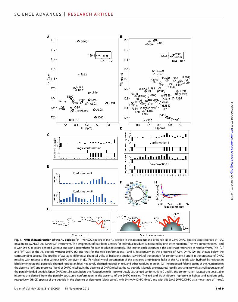
Fig. 1. NMR characterization of the AL peptide. 1H–15N HSQC spectra of the AL peptide in the absence (A) and presence (B) of 7.5% DHPC. Spectra were recorded at 10°Con a Bruker AVANCE 900-MHz NMR instrument. The assignment of backbone amides for individual residues is indicated by one-letter notations. The two conformations, I andII, with DHPC in (B) are denoted without and with a parenthesis for each residue, respectively. The inset in each spectrum is the side-chain resonance of residue W393. The 13Ca
and 1Ha CSIs of the AL peptide without DHPC (C) and that for the two conformations, I and II, respectively, in the presence of 7.5% DHPC (D) are shown below thecorresponding spectra. The profiles of averaged differential chemical shifts of backbone amides, Dav(NH), of the peptide for conformations I and II in the presence of DHPCmicelles with respect to that without DHPC are given in (E). (F) Helical wheel presentation of the predicted amphipathic helix of the AL peptide with hydrophilic residues inblack letter notations, positively charged residues in blue, negatively charged residues in red, and other residues in green. (G) The proposed folding status of the AL peptide inthe absence (left) and presence (right) of DHPC micelles. In the absence of DHPC micelles, the AL peptide is largely unstructured, rapidly exchanging with a small population ofthe partially folded peptide. Upon DHPC micelle association, the AL peptide folds into two slowly exchanged conformations (I and II), and conformation I appears to be a stableintermediate derived from the partially structured conformation in the absence of the DHPC micelles. The red and black ribbons represent a helices and random coils,respectively. (H) CD spectra of the peptide in the absence of detergent (black curve), with 5% (w/v) DHPC (blue), and with 5% (w/v) DMPC/DHPC at a molar ratio of 1 (red).
Liu et al. Sci. Adv. 2016;2 : e1600925 18 November 2016 3 of 9

SC I ENCE ADVANCES | R E S EARCH ART I C L E
on June 21, 2018http://advances.sciencem
ag.org/D
ownloaded from
opposite side, with E390 and S392 being at the boundary (Fig. 1F).Accordingly, the AL peptide in conformation II likely associateswith the surface of the DHPC micelles through hydrophobic inter-actions while exposing the positively charged surface to the bulkysolvent. This model is further supported by linewidth analysis (ta-ble S1): For conformation II, the resonances corresponding to theV386-H398 segment, but not to H391 and S392, have clear broaderlinewidths compared to those in the absence of DHPC micelles,strongly suggesting that the helical region of the AL peptide directlyassociates with, though weakly, the DHPC micelles that have amuch larger molecular weight (~20,000) and thus slower tumblingrates; in contrast, for conformation I, the residues with broaderlinewidths are restricted to the region of S392-H398, supportingan association with the DHPC micelles through the limited helicalregion (W393-L396), as revealed by CSIs (Fig. 1D) and the Dav(HN) (Fig. 1E) data. Because there are no species correspondingto conformation II in the absence of the DHPC micelles, we con-clude that micelle-associated conformation I is a stable foldingintermediate that is slowly converted into conformation II, whichassociates with the micellar surface through a much larger hydro-phobic interface (Fig. 1G). Consistent with the results derived fromthe NMR experiments, the helical content of the AL peptideincreased markedly when 5% DHPC was added, as demonstratedby the negative peak at 222 nm in circular dichroism (CD) spectra(Fig. 1H). Furthermore, we observed an even higher helical content inthe presence of DMPC (1,2-dimyristoyl-sn-glycero-3-phosphocho-line)/DHPC mixed micelles (5%, w/v; DMPC/DHPC molar ratioof q = 1), indicating that the AL peptide undergoes further confor-mational changes when the membrane-mimic environment is moreanalogous to the authentic cell membrane.
The hydrophobic surface of the amphipathic helix isessential for lipid substrate processingThe amphipathic helix shown in conformation II is induced byDHPC micelles, raising a hypothesis that the helical structure ofthe activation loop represents a functional conformation that is es-sential for PIP5K activity. To test this hypothesis, we performedsystematic mutagenesis on the hydrophobic residues in the activa-tion loop because these residues constitute the hydrophobic surfaceand are thus supposed to interact with membrane per se (Fig. 2A).The activity assays were conducted in 5% DMPC/DHPC bicelles (q =1), which support a strong activity of zPIP5Ka. Proline and glycineresidues are known helix breakers; the former introduces a kink inthe helix, whereas the latter generally destabilizes the helical structure.Proline scanning on the hydrophobic residues revealed that introdu-cing a kink at L389 or W393 markedly reduced activities toward bothlipid substrates, PI(4)P and PI(5)P, whereas the mutations at L396 andL397 had much greater detrimental effects on the activity toward PI(5)P,an unfavorable substrate of PIP5K (Fig. 2B). Glycine mutations hadno significant effects on PI(4)P processing but affected PI(5)P proces-sing differently: L398G markedly attenuated the activity, whereas theactivities of L385G, V386G, and W393G increased significantly. Ac-cordingly, the activation loop may adopt distinct conformations to ac-commodate different substrates. Together, it appears that L389 andW393, which are right in the middle of the amphipathic helix, playimportant roles in both substrate processing and recognition. To fur-ther explore the function of the hydrophobic surface of the amphi-pathic helix, L389 and W393 were mutated into glutamate andarginine, respectively. Because of the salt bridge between E389 and
Liu et al. Sci. Adv. 2016;2 : e1600925 18 November 2016
R393, the helical structure of the L389E/W393R double mutant shouldbe preserved according to secondary structure prediction by PSIPRED(Psi-blast–based secondary structure prediction) (20). However, thedouble mutation did convert the hydrophobic surface into a more hy-drophilic one. As expected, the activity of the double mutant decreasedmarkedly, and a more radical mutant with four hydrophobic residuesreplaced by charged residues completely abolished the activity ofPIP5K. Collectively, these data indicate that the structure of the am-phipathic helix within the activation loop and its hydrophobic surfaceare crucial for PIP5K function.
Disrupting the hydrophobic surface of the amphipathichelix within the activation loop affects themembrane-sensing function of PIP5KIt has been reported that the activity of PIP5K can be markedlymodulated by fatty acid composition of the lipid substrates andphosphatidic acid (11). Our study reveals that PIP5K activity is alsosensitive to the size/curvature of the membrane mimics. In the effortof cocrystallization of PIP5K with lipid substrates dissolved in deter-gents (including Triton X-100, n-dodecyl-b-D-maltoside, decyl b-D-maltopyranoside, octyl b-D-glucopyranoside, Fos-Choline-12, andDHPC), we found that the activity of PIP5K was almost completely
Fig. 2. Mutagenesis and activity measurement of the PIP5K mutants in 5%DMPC/DHPC bicelles (q = 1). (A) A model structure of the AL peptide based onthe results of the NMR experiments. The R384-L396 segment is in a helical con-formation, representing conformation II in the presence of DHPC micelles. Thehydrophobic residues of the amphipathic helix are highlighted in stick mode,and the positively charged residues are colored blue. (B) Mutagenesis of the res-idues on the hydrophobic surface of the amphipathic helix in the AL peptide andactivity assays. The mutated residues are highlighted in red. Normalized activitiesrelative to that of the wild-type (WT) enzyme toward a specific substrate [PI(4)P orPI(5)P] are shown in the table. The significant decreased and increased activitiesare highlighted in red and blue, respectively. N.D., no detectable activity; “-,” not de-tected. The results represent the average (±SD) of three independent experiments.
4 of 9
SC I ENCE ADVANCES | R E S EARCH ART I C L E
on June 21, 2018http://advances.sciencem
ag.org/D
ownloaded from
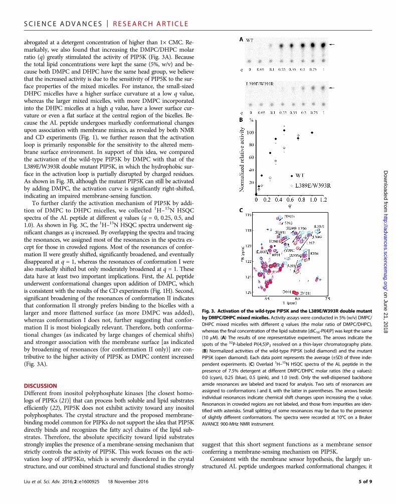
abrogated at a detergent concentration of higher than 1× CMC. Re-markably, we also found that increasing the DMPC/DHPC molarratio (q) greatly stimulated the activity of PIP5K (Fig. 3A). Becausethe total lipid concentrations were kept the same (5%, w/v) and be-cause both DMPC and DHPC have the same head group, we believethat the increased activity is due to the sensitivity of PIP5K to the sur-face properties of the mixed micelles. For instance, the small-sizedDHPC micelles have a higher surface curvature at a low q value,whereas the larger mixed micelles, with more DMPC incorporatedinto the DHPC micelles at a high q value, have a lower surface cur-vature or even a flat surface at the central region of the bicelles. Be-cause the AL peptide undergoes markedly conformational changesupon association with membrane mimics, as revealed by both NMRand CD experiments (Fig. 1), we further reason that the activationloop is primarily responsible for the sensitivity to the altered mem-brane surface environment. In support of this idea, we comparedthe activation of the wild-type PIP5K by DMPC with that of theL389E/W393R double mutant PIP5K, in which the hydrophobic sur-face in the activation loop is partially disrupted by charged residues.As shown in Fig. 3B, although the mutant PIP5K can still be activatedby adding DMPC, the activation curve is significantly right-shifted,indicating an impaired membrane-sensing function.
To further clarify the activation mechanism of PIP5K by addi-tion of DMPC to DHPC micelles, we collected 1H–15N HSQCspectra of the AL peptide at different q values (q = 0, 0.25, 0.5, and1.0). As shown in Fig. 3C, the 1H–15N HSQC spectra underwent sig-nificant changes as q increased. By overlapping the spectra and tracingthe resonances, we assigned most of the resonances in the spectra ex-cept for those in crowded regions. Most of the resonances of confor-mation II were greatly shifted, significantly broadened, and eventuallydisappeared at q = 1, whereas the resonances of conformation I werealso markedly shifted but only moderately broadened at q = 1. Thesedata have at least two important implications. First, the AL peptideunderwent conformational changes upon addition of DMPC, whichis consistent with the results of the CD experiments (Fig. 1H). Second,significant broadening of the resonances of conformation II indicatesthat conformation II strongly prefers binding to the bicelles with alarger and more flattened surface (as more DMPC was added),whereas conformation I does not, further suggesting that confor-mation II is most biologically relevant. Therefore, both conforma-tional changes (as indicated by large changes of chemical shifts)and stronger association with the membrane surface [as indicatedby broadening of resonances (for conformation II only)] are con-tributive to the higher activity of PIP5K as DMPC content increased(Fig. 3A).
DISCUSSIONDifferent from inositol polyphosphate kinases [the closest homo-logs of PIPKs (21)] that can process both soluble and lipid substratesefficiently (22), PIP5K does not exhibit activity toward any inositolpolyphosphates. The crystal structure and the proposed membrane-binding model common for PIPKs do not support the idea that PIP5Kdirectly binds and recognizes the fatty acyl chains of the lipid sub-strates. Therefore, the absolute specificity toward lipid substratesstrongly implies the presence of a membrane-sensing mechanism thatstrictly controls the activity of PIP5K. This work focuses on the acti-vation loop of zPIP5Ka, which is severely disordered in the crystalstructure, and our combined structural and functional studies strongly
Liu et al. Sci. Adv. 2016;2 : e1600925 18 November 2016
suggest that this short segment functions as a membrane sensorconferring a membrane-sensing mechanism on PIP5K.
Consistent with the membrane sensor hypothesis, the largely un-structured AL peptide undergoes marked conformational changes; it
Fig. 3. Activation of the wild-type PIP5K and the L389E/W393R double mutantby DMPC/DHPC mixedmicelles. Activity assays were conducted in 5% (w/v) DMPC/DHPC mixed micelles with different q values (the molar ratio of DMPC/DHPC),whereas the final concentration of the lipid substrate [diC16-PI(4)P] was kept the same(10 mM). (A) The results of one representative experiment. The arrows indicate thespots of the 32P-labeled PI(4,5)P2 resolved on a thin-layer chromatography plate.(B) Normalized activities of the wild-type PIP5K (solid diamond) and the mutantPIP5K (open diamond). Each data point represents the average (±SD) of three inde-pendent experiments. (C) Overlaid 1H–15N HSQC spectra of the AL peptide in thepresence of 7.5% detergent at different DMPC/DHPC molar ratios (the q values):0.0 (cyan), 0.25 (blue), 0.5 (pink), and 1.0 (red). Only the well-dispersed backboneamide resonances are labeled and traced for analysis. Two sets of resonances areassigned to conformations I and II, with the latter in parentheses. The arrows besideindividual resonances indicate chemical shift changes upon increasing the q value.Resonances in crowded regions are not labeled, and those from impurities are iden-tified with asterisks. Small splitting of some resonances may be due to the presenceof slightly different conformations. The spectra were recorded at 10°C on a BrukerAVANCE 900-MHz NMR instrument.
5 of 9
SC I ENCE ADVANCES | R E S EARCH ART I C L E
on June 21http://advances.sciencem
ag.org/D
ownloaded from
folds into two roughly equally populated conformations upon additionof DHPC micelles. Both conformations represent membrane-boundforms: Conformation II contains an extended amphipathic helix,and both Dav(NH) differential chemical shifts and NMR linewidthanalysis suggest that the helix associates with the surface of the DHPCmicelles through hydrophobic interactions; conformation I, on theother hand, is most likely derived from the minor species in the ab-sence of DHPC micelles and is stabilized through the association ofthe preexisting helical turn in the W393-L396 segment with the mi-cellar surface, as suggested by the Dav(NH) data and linewidthanalysis. Replacing the conserved hydrophobic residues on the am-phipathic helix with helix-destabilizing amino acid residues (pro-line or glycine) revealed that L389 and W393 are critical in kinaseactivity and substrate recognition (Fig. 2). Because they are right inthe middle of the amphipathic helix in conformation II, mutationson these two residues may lead to the greatest structural changes. No-tably, the unstructured L389 in conformation I is not involved inmembrane association according to the analysis of Dav(NH) data(Fig. 1E), and therefore, the critical role of L389 in kinase activity sug-gests that conformation II with the extended amphipathic helix ismore likely to be biologically relevant than conformation I. The lattermay represent a stable intermediate during the conformationalchanges necessary for kinase activation (Fig. 1G).
It is interesting that the activity of PIP5K varies markedly uponchanging the DMPC/DHPC molar ratio (the q value) in the mixedmicelles (Fig. 3). The largely suppressed activity in the presence ofpure DHPC micelles can be greatly restored when the q value in-creases up to 1. Although both NMR and CD data with the isolatedpeptide showed that it folds into helical structures in the presenceof detergent micelles, they may not necessarily represent the exactproductive conformation required for kinase activity. For example,the amphipathic helix has an apparent kink at S392 (Fig. 1, E andG, and fig. S2C), which is likely to be an artifact due to the asso-ciation of the AL peptide on the highly curved DHPC micellar sur-face. When the curvature of the mixed micellar surface is reducedalong with the increased q value, the activity of PIP5K is restoredgradually, accompanied by further conformational changes andbetter membrane association of the AL peptide (Fig. 3C). Finally,
Liu et al. Sci. Adv. 2016;2 : e1600925 18 November 2016
the L389E/W393R double mutant exhibited significantly attenuatedsensitivity to the changes in the lipid composition, indicating thatthe activation loop is at least, if not the only, a major structural ele-ment conferring high membrane sensitivity.
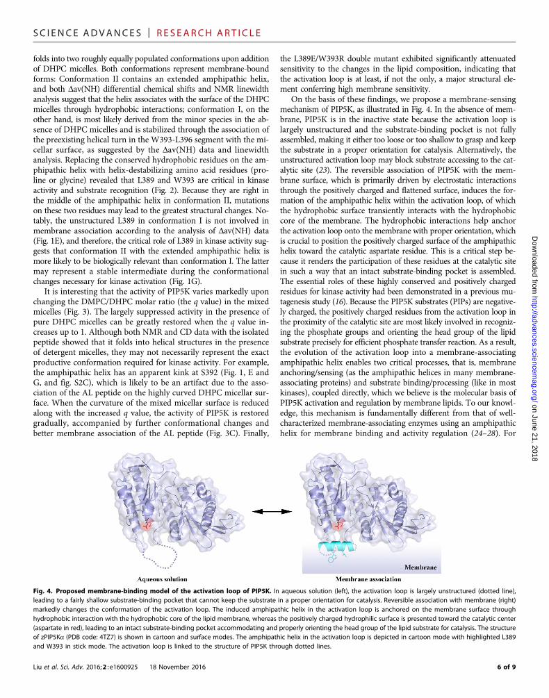
On the basis of these findings, we propose a membrane-sensingmechanism of PIP5K, as illustrated in Fig. 4. In the absence of mem-brane, PIP5K is in the inactive state because the activation loop islargely unstructured and the substrate-binding pocket is not fullyassembled, making it either too loose or too shallow to grasp and keepthe substrate in a proper orientation for catalysis. Alternatively, theunstructured activation loop may block substrate accessing to the cat-alytic site (23). The reversible association of PIP5K with the mem-brane surface, which is primarily driven by electrostatic interactionsthrough the positively charged and flattened surface, induces the for-mation of the amphipathic helix within the activation loop, of whichthe hydrophobic surface transiently interacts with the hydrophobiccore of the membrane. The hydrophobic interactions help anchorthe activation loop onto the membrane with proper orientation, whichis crucial to position the positively charged surface of the amphipathichelix toward the catalytic aspartate residue. This is a critical step be-cause it renders the participation of these residues at the catalytic sitein such a way that an intact substrate-binding pocket is assembled.The essential roles of these highly conserved and positively chargedresidues for kinase activity had been demonstrated in a previous mu-tagenesis study (16). Because the PIP5K substrates (PIPs) are negative-ly charged, the positively charged residues from the activation loop inthe proximity of the catalytic site are most likely involved in recogniz-ing the phosphate groups and orienting the head group of the lipidsubstrate precisely for efficient phosphate transfer reaction. As a result,the evolution of the activation loop into a membrane-associatingamphipathic helix enables two critical processes, that is, membraneanchoring/sensing (as the amphipathic helices in many membrane-associating proteins) and substrate binding/processing (like in mostkinases), coupled directly, which we believe is the molecular basis ofPIP5K activation and regulation by membrane lipids. To our knowl-edge, this mechanism is fundamentally different from that of well-characterized membrane-associating enzymes using an amphipathichelix for membrane binding and activity regulation (24–28). For
, 2018
Fig. 4. Proposed membrane-binding model of the activation loop of PIP5K. In aqueous solution (left), the activation loop is largely unstructured (dotted line),leading to a fairly shallow substrate-binding pocket that cannot keep the substrate in a proper orientation for catalysis. Reversible association with membrane (right)markedly changes the conformation of the activation loop. The induced amphipathic helix in the activation loop is anchored on the membrane surface throughhydrophobic interaction with the hydrophobic core of the lipid membrane, whereas the positively charged hydrophilic surface is presented toward the catalytic center(aspartate in red), leading to an intact substrate-binding pocket accommodating and properly orienting the head group of the lipid substrate for catalysis. The structureof zPIP5Ka (PDB code: 4TZ7) is shown in cartoon and surface modes. The amphipathic helix in the activation loop is depicted in cartoon mode with highlighted L389and W393 in stick mode. The activation loop is linked to the structure of PIP5K through dotted lines.
6 of 9
SC I ENCE ADVANCES | R E S EARCH ART I C L E
http://advaD
ownloaded from
example, an amphipathic helix within the M domain of cytidinetriphosphate:phosphocholine cytidylyltransferase plays an auto-inhibitory role in the membrane-unbound state, whereas it activatesthe enzyme in a membrane-bound state most likely through a long-range allosteric effect (29). In addition, somehow similar to the anti-microbial peptides that fold into an amphipathic helix and disintegratethe membrane (30), the activation loop of PIP5K may also disturb themembrane integrity and facilitate binding of the head group of thelipid substrate. Consistent with this model, disrupting the hydropho-bic surface of the amphipathic helix by mutagenesis not only markedlyreduced kinase activity (Fig. 2) but also diminished membrane-sensingfunction (Fig. 3).
In summary, because the formation of the amphipathic helix ab-solutely depends on the presence of membrane and the orientationof the critical substrate-coordinating residues is conceivably affectedby membrane properties, PIP5K exhibits a stringent specificity towardlipid substrates and is highly sensitive to the membrane environment.Because the amphipathic helix is predicted to be a common structuralfeature for all the PIPKs (Scheme 1), we therefore propose that thismembrane-sensing mechanism is likely shared among other lipid kinasesin the PIPK family. It is well known that the activation loop is a criticalsite for kinase activity regulation primarily through phosphorylation/dephosphorylation (23). The surprising function of the PIP5K activationloop as a membrane sensor represents a new paradigm on kinase reg-ulation through the unprecedented activation loop–membrane interac-tion, which lays a foundation on the regulation of PIPKs by membranelipids for future studies.
on June 21, 2018nces.sciencem
ag.org/
MATERIALS AND METHODSPreparation of the AL peptideTo overexpress the AL peptide of zPIP5Ka, BL21 (DE3) plysS com-petent cells were transformed with a plasmid construct that carries theactivation loop region (GHM384RLVKKLEHSWKALLHDGD401, inwhich the N-terminal three residues, GHM, were introduced afterTEV digestion of the His-tag) with its N terminus fused to the C ter-minus of MBP and a His-tag plus a TEV cleavage site engineered be-tween MBP and the peptide. The unlabeled peptide was produced inLB medium, whereas 15N- and 15N/13C-labeled samples in M9 mini-mal defined medium with 15N-ammonium chloride and U-13C–glucoseserved as sole sources for nitrogen and carbon atoms, respectively. Toproduce the unlabeled peptide, cell colonies from the agar culture platecontaining ampicillin and chloramphenicol after incubating overnightat 37°C were collected and pooled into 1 liter of LB supplementedwith the antibiotics. The LB rotary flask underwent vigorous shaking(220 rpm) at 37°C, and the culture was paused when optical density at600 nm (OD600) reached ~0.65. The flask was then placed in a coldroom (4°C) for half an hour, and the expression was induced with0.1 mM isopropyl b-D-1-thiogalactopyranoside (IPTG) overnight at18°C. The cells were harvested by centrifugation, and the pellet wassubjected to immediate lysis or stored at −80°C for subsequent puri-fication. To produce 15N- or 15N/13C-labeled peptides, cell coloniesfrom the agar plate were added into 1 liter of M9 instead of LB me-dium for overnight culturing at 37°C. Depending on the cell density,appropriate amounts of cells were collected by gentle centrifugation,and the cell pellet was resuspended in 1 liter of NMR isotope–enrichedM9 medium, in which the natural-abundance ammonium chlorideand glucose were substituted by 15N- and 13C-enriched chemicals, re-spectively, so that the starting reading of OD600 was ~0.4. The isotope-
Liu et al. Sci. Adv. 2016;2 : e1600925 18 November 2016
enriched cell culture was continued until the OD600 reading reached~0.65 for IPTG induction, followed by the same procedure for theLB culture.
The MBP-fused peptide was purified by a one-step Ni-columnprocedure. First, the cell pellet was resuspended in 10 ml of trisbuffer (25 mM tris and 300 mM NaCl at pH 8.0) per gram of cells,with the addition of 1 mM phenylmethylsulfonyl fluoride. The dis-solved cells were lysed through sonication, and the lysate wassubjected to centrifugation to remove cell debris. The supernatantwas then loaded into a Ni-column prebalanced with tris buffer,thoroughly mixed with 15 to 20 ml of resin, and incubated on aroller for 2 hours at 4°C. After flow-through, 50 ml of tris bufferwas added, and the Ni-column was incubated for 10 min on theroller for another flow-through. This washing step was repeatedusing the tris buffer supplemented with increasing amounts of im-idazole (10, 15, and 20 mM). The protein was eluted two timesusing 25 ml of tris buffer containing 250 mM imidazole per elutionand 10 min of column incubation in between. The elution was con-centrated using an Amicon centrifugal filter device [30,000 molecularweight cutoff (MWCO)] until the protein concentration reached100 mg/ml or higher, as measured by the Bradford method. The typ-ical yield was ~150 mg/liter of LB or ~100 mg/liter of M9 culture.
To get rid of imidazole, the buffer was changed to phosphate(150 mM NaCl and 20 mM phosphate buffer at pH 7.0) throughrepeated centrifugations with an Amicon centrifugal filter device(30,000 MWCO). The concentrated solution was transferred intoa few 1.5-ml tubes, each containing 0.5-ml solution with the pro-tein concentration at ~100 mM. Appropriate amounts of TEV pro-tease, 0.5 mM EDTA, and 1 mM dithiothreitol (DTT) were added,and the volume of the final solution in each tube was 0.7 to 0.8 ml.The tubes were incubated on a roller at room temperature for 1 to2 hours for TEV digestion. After the addition of 1 mM DTT, incuba-tion continued overnight at 4°C. The TEV-digested peptide was elutedby centrifugation for 25 min using an Amicon centrifugal filter device(10,000 MWCO) and then subjected to another elution using a newAmicon centrifugal unit to eliminate residual MBP contamination.The peptide concentration was measured on a NanoDrop. Thepeptide solution was further concentrated through evaporation un-der vacuum, and the substitution for the NMR buffer was achievedthrough dialysis using small tubes with a 1000-MWCO membrane(Sigma).
NMR measurementThe NMR samples contained 0.2 to 0.4 mM peptide solution in100 mM NaCl and 20 mM phosphate buffer at pH 7.0 with or with-out detergent. The volume of each NMR sample was ~300 ml in aShigemi tube, containing 5% D2O, 150 mM NaN3, and 50 mM 4,4-dimethyl-4-silapentane-1-sulfonic acid as inner NMR reference.
All NMR data were collected on a Bruker AVANCE 900-MHzinstrument equipped with a TCI cryoprobe. The optimum tempera-ture for NMR measurements was found to be 10°C after recording aseries of 1H–15N HSQC spectra at 5°, 10°, 15°, 20°, 25°, 30°, and 35°C,based on the appearance of resonance and the linewidth. For theNMR sample in the absence of detergent, the backbone resonance as-signment was accomplished through the analysis of three-dimensional(3D) HNCACB and 15N-dispersed [1H–1H]-NOESY (nuclear Over-hauser effect spectroscopy) spectra. For the peptide in the presenceof 7.5% DHPC, two additional 3D data sets, HNCA and HNCOCA,were recorded to facilitate NMR assignment. The mixing time for the
7 of 9

SC I ENCE ADVANCES | R E S EARCH ART I C L E
on June 21, 2018http://advances.sciencem
ag.org/D
ownloaded from
NOESY spectra was set to 200 ms. Data were processed using theprogram NMRPipe (31) and were analyzed using the program NMRView(32). More details of the NMR experiments are described in the Sup-plementary Materials.
CD measurementsThe far-ultraviolet CD spectra were recorded on a Chirascan spec-tropolarimeter (Applied Photophysics) using a 1-mm path-lengthquartz cuvette at 20°C over 200 to 260 nm with a bandwidth of1 nm. The scan speed was set with a response time of 3 s and a stepresolution of 0.3 nm. The results represent the average of three scans.All spectra were background-corrected by subtracting correspondingblank spectra without the peptide. The peptide (final concentration,~0.1 mM) was dissolved in a buffer containing 20 mM sodium phos-phate (pH 7.0) and 100 mM NaCl.
PIP5K preparation and kinase activity assayzPIP5Ka and the mutant proteins were prepared as previously re-ported (13). In brief, the expression vector (pET41b) harboring thegene of zPIP5Ka (kinase domain, residues 49 to 431, synthesized inGenScript Inc. with an optimized codon for E. coli expression) wastransformed into Rosetta 2 (DE3) Competent Cells (Novagen). Whenthe OD600 of the culture in LB medium reached 0.4 at 37°C, theculture was cooled down on ice for half an hour, and overexpressionwas induced by 0.1 mM IPTG at room temperature for an additional16 hours. The cells were harvested and suspended in a lysis buffercontaining 50 mM sodium phosphate (pH 7.3), 300 mM NaCl, 5%glycerol, 0.5% Triton X-100 (American Bioanalytical), and EDTA-freeprotease inhibitor cocktail (Roche). The cells were lysed by sonicationon ice and subjected to centrifugation (10,000g at 4°C for 30 min). Theprotein in the supernatant was purified using Co2+-resin (Talon, Clon-tech) and then applied to a size-exclusion column equilibrated withthe gel filtration buffer containing 10 mM Hepes (pH 7.3), 300mM NaCl, 5% glycerol, and 0.03% Triton X-100. The peak fractionwas used for activity assay, and the protein concentration wasmeasured using the Bradford method (Bio-Rad).
The kinase activity assay was modified from a previously re-ported approach (13). The following components were included in onereaction (50 ml): 100 ng of purified zPIP5Ka, 100 mM tris-HCl (pH 7.4),50 mM EGTA, 100 mM MgCl2, 20 mM ATP with 1 mCi [g-32P]ATP(PerkinElmer), and 10 mM diC16-PI(4)P (Echelon Biosciences Inc.).The reaction was performed at room temperature for 1 hour andwas stopped by the addition of lipid extraction solution containingchloroform, methanol, and HCl with a volume ratio of 3.3:3.7:0.1,as well as bovine follicular fluid (10 mg/ml). After vortexing for 20 s,the sample was centrifuged at 6000 rpm for 1 min, and the lowerorganic phase was collected, loaded, and separated on a thin-layerplate. The product of the reaction was quantified by a Storm 820PhosphorImager (GE). To measure the activity of PIP5K in DMPC/DHPC bicelles, diC16-PI(4)P (final concentration, 10 mM) was dis-solved in 5% (w/v) DMPC/DHPC (q = 1) mixture by incubation for30 min at room temperature followed by three cycles of freeze-thawtreatment as described above. Before adding ATP to initiate the re-action, the purified PIP5K was incubated with the lipid substratedissolved in DMPC/DHPC mixed micelles for 10 min at room tem-perature. The other processes were the same as described above. Theresults obtained in diC16-PI(4)P vesicles are shown in table S2 andthe activity measured in DMPC/DHPC bicelles (q = 1) is shown inFig. 2B.
Liu et al. Sci. Adv. 2016;2 : e1600925 18 November 2016
To prepare DMPC/DHPC (both from Avanti Polar Lipids) mixedmicelles, a bicelle sample [q = 3 (15%, w/v)] was produced accordingto previously reported approaches (33, 34). In brief, DMPC powderwas dissolved in DHPC solution with at least five cycles of freeze-thawtreatment until DMPC was completely dissolved and the solution wasclear and homogeneous at low temperatures. In each cycle, thesamples were vigorously vortexed for 1 min at room temperatureand subjected to incubation at 42°C for 10 min, followed by freezingat −80°C for 10 min and then thawing on ice. By mixing the bicellesample (q = 3; 15%, w/v) and DHPC (q = 0; 15%, w/v) with differentratios, the DMPC/DHPC mixed micelles were prepared, and the stocksolutions were stored at −80°C. The lipids were all dissolved in water.
SUPPLEMENTARY MATERIALSSupplementary material for this article is available at http://advances.sciencemag.org/cgi/content/full/2/11/e1600925/DC1fig. S1. Preparation of the AL peptide and activity assays of zebrafish PIP5Ka using differentlipid substrates.fig. S2. Two-dimensional [1H–1H] NOE strips derived from 3D 15N-dispersed [1H–1H]-NOESYspectra for the activation loop peptide.fig. S3. Overlay of 1H–15N HSQC spectra of the activation loop peptide in the absence (blue)and presence (red) of 1% DHPC detergent.table S1. Amide proton linewidth of the PIP5K-loop peptide.table S2. Mutagenesis and activity measurement of the PIP5K mutants in diC16-PI(4)P vesicles.Details of NMR experiments
REFERENCES AND NOTES1. G. Di Paolo, P. De Camilli, Phosphoinositides in cell regulation and membrane dynamics.
Nature 443, 651–657 (2006).2. M. Schramp, A. Hedman, W. Li, X. Tan, R. Anderson, PIP kinases from the cell membrane
to the nucleus. Subcell. Biochem. 58, 25–59 (2012).3. J. R. Brown, K. R. Auger, Phylogenomics of phosphoinositide lipid kinases: Perspectives
on the evolution of second messenger signaling and drug discovery. BMC Evol. Biol.11, 4 (2011).
4. R. A. Anderson, I. V. Boronenkov, S. D. Doughman, J. Kunz, J. C. Loijens,Phosphatidylinositol phosphate kinases, a multifaceted family of signaling enzymes.J. Biol. Chem. 274, 9907–9910 (1999).
5. B. D. Wright, L. Loo, S. E. Street, A. Ma, B. Taylor-Blake, M. A. Stashko, J. Jin, W. P. Janzen,S. V. Frye, M. J. Zylka, The lipid kinase PIP5K1C regulates pain signaling and sensitization.Neuron 82, 836–847 (2014).
6. J. Semenas, A. Hedblom, R. R. Miftakhova, M. Sarwar, R. Larsson, L. Shcherbina,M. E. Johansson, P. Härkönen, O. Sterner, J. L. Persson, The role of PI3K/AKT-relatedPIP5K1a and the discovery of its selective inhibitor for treatment of advanced prostatecancer. Proc. Natl. Acad. Sci. U.S.A. 111, E3689–E3698 (2014).
7. B. M. Emerling, J. B. Hurov, G. Poulogiannis, K. S. Tsukazawa, R. Choo-Wing, G. M. Wulf,E. L. Bell, H.-S. Shim, K. A. Lamia, L. E. Rameh, G. Bellinger, A. T. Sasaki, J. M. Asara,X. Yuan, A. Bullock, G. M. DeNicola, J. Song, V. Brown, S. Signoretti, L. C. Cantley,Depletion of a putatively druggable class of phosphatidylinositol kinases inhibits growth ofp53-null tumors. Cell 155, 844–857 (2013).
8. Y. Sun, D. A. Turbin, K. Ling, N. Thapa, S. Leung, D. G. Huntsman, R. A. Anderson, Type Igamma phosphatidylinositol phosphate kinase modulates invasion and proliferationand its expression correlates with poor prognosis in breast cancer. Breast Cancer Res. 12,R6 (2010).
9. M. D. Voss, W. Czechtizky, Z. Li, C. Rudolph, S. Petry, H. Brummerhop, T. Langer, A. Schiffer,H.-L. Schaefer, Discovery and pharmacological characterization of a novel small moleculeinhibitor of phosphatidylinositol-5-phosphate 4-kinase, type II, beta. Biochem. Biophys. Res.Commun. 449, 327–331 (2014).
10. N. Hayakawa, M. Noguchi, S. Takeshita, A. Eviryanti, Y. Seki, H. Nishio, R. Yokoyama,M. Noguchi, M. Shuto, Y. Shima, K. Kuribayashi, S. Kageyama, H. Eda, M. Suzuki, T. Hatta,S.-i. Iemura, T. Natsume, I. Tanabe, R. Nakagawa, M. Shiozaki, K. Sakurai, M. Shoji,A. Andou, T. Yamamoto, Structure–activity relationship study, target identification, andpharmacological characterization of a small molecular IL-12/23 inhibitor, APY0201.Bioorg. Med. Chem. 22, 3021–3029 (2014).
11. Y. V. Shulga, R. A. Anderson, M. K. Topham, R. M. Epand, Phosphatidylinositol-4-phosphate 5-kinase isoforms exhibit acyl chain selectivity for both substrate and lipidactivator. J. Biol. Chem. 287, 35953–35963 (2012).
8 of 9

SC I ENCE ADVANCES | R E S EARCH ART I C L E
http://advances.sciencem
ag.org/D
ownloaded from
12. V. D. Rao, S. Misra, I. V. Boronenkov, R. A. Anderson, J. H. Hurley, Structure of type IIbphosphatidylinositol phosphate kinase: A protein kinase fold flattened for interfacialphosphorylation. Cell 94, 829–839 (1998).
13. J. Hu, Q. Yuan, X. Kang, Y. Qin, L. Li, Y. Ha, D. Wu, Resolution of structure of PIP5K1Areveals molecular mechanism for its regulation by dimerization and dishevelled.Nat. Commun. 6, 8205 (2015).
14. L. M. Burden, V. D. Rao, D. Murray, R. Ghirlando, S. D. Doughman, R. A. Anderson,J. H. Hurley, The flattened face of type IIb phosphatidylinositol phosphate kinase bindsacidic phospholipid membranes. Biochemistry 38, 15141–15149 (1999).
15. J. Kunz, M. P. Wilson, M. Kisseleva, J. H. Hurley, P. W. Majerus, R. A. Anderson, Theactivation loop of phosphatidylinositol phosphate kinases determines signalingspecificity. Mol. Cell 5, 1–11 (2000).
16. J. Kunz, A. Fuelling, L. Kolbe, R. A. Anderson, Stereo-specific substrate recognition byphosphatidylinositol phosphate kinases is swapped by changing a single amino acidresidue. J. Biol. Chem. 277, 5611–5619 (2002).
17. D. S. Wishart, B. D. Sykes, F. M. Richards, The chemical shift index: A fast and simplemethod for the assignment of protein secondary structure through NMR spectroscopy.Biochemistry 31, 1647–1651 (1992).
18. A. Liu, R. Riek, R. Zahn, S. Hornemann, R. Glockshuber, K. Wüthrich, Peptides and proteinsin neurodegenerative disease: Helix propensity of a polypeptide containing helix 1 ofthe mouse prion protein studied by NMR and CD spectroscopy. Biopolymers 51,145–152 (1999).
19. S. Grzesiek, S. J. Stahl, P. T. Wingfield, A. Bax, The CD4 determinant for downregulation byHIV-1 Nef directly binds to Nef. Mapping of the Nef binding surface by NMR. Biochemistry35, 10256–10261 (1996).
20. D. W. A. Buchan, F. Minneci, T. C. O. Nugent, K. Bryson, D. T. Jones, Scalable webservices for the PSIPRED Protein Analysis Workbench. Nucleic Acids Res. 41,W349–W357 (2013).
21. B. González, M. J. Schell, A. J. Letcher, D. B. Veprintsev, R. F. Irvine, R. L. Williams, Structureof a human inositol 1,4,5-trisphosphate 3-kinase: Substrate binding reveals why it isnot a phosphoinositide 3-kinase. Mol. Cell 15, 689–701 (2004).
22. A. C. Resnick, A. M. Snowman, B. N. Kang, K. J. Hurt, S. H. Snyder, A. Saiardi, Inositolpolyphosphate multikinase is a nuclear PI3-kinase with transcriptional regulatory activity.Proc. Natl. Acad. Sci. U.S.A. 102, 12783–12788 (2005).
23. J. A. Adams, Activation loop phosphorylation and catalysis in protein kinases: Is therefunctional evidence for the autoinhibitor model? Biochemistry 42, 601–607 (2003).
24. R. B. Cornell, N. D. Ridgway, CTP:phosphocholine cytidylyltransferase: Function,regulation, and structure of an amphitropic enzyme required for membrane biogenesis.Prog. Lipid Res. 59, 147–171 (2015).
25. L. Danne, M. Aktas, J. Gleichenhagen, N. Grund, D. Wagner, H. Schwalbe, B. Hoffknecht,N. Metzler-Nolte, F. Narberhaus, Membrane-binding mechanism of a bacterialphospholipid N-methyltransferase. Mol. Microbiol. 95, 313–331 (2015).
Liu et al. Sci. Adv. 2016;2 : e1600925 18 November 2016
26. E. Karanasios, G.-S. Han, Z. Xu, G. M. Carman, S. Siniossoglou, A phosphorylation-regulatedamphipathic helix controls the membrane translocation and function of the yeastphosphatidate phosphatase. Proc. Natl. Acad. Sci. U.S.A. 107, 17539–17544 (2010).
27. J. Lind, T. Rämö, M. L. R. Klement, E. Bárány-Wallje, R. M. Epand, R. F. Epand, L. Mäler,Å. Wieslander, High cationic charge and bilayer interface-binding helices in a regulatorylipid glycosyltransferase. Biochemistry 46, 5664–5677 (2007).
28. B. Antonny, S. Beraud-Dufour, P. Chardin, M. Chabre, N-terminal hydrophobic residues ofthe G-protein ADP-ribosylation factor-1 insert into membrane phospholipids uponGDP to GTP exchange. Biochemistry 36, 4675–4684 (1997).
29. R. B. Cornell, Membrane lipid compositional sensing by the inducible amphipathic helixof CCT. Biochim. Biophys. Acta 1861, 847–861 (2016).
30. B. Bechinger, The structure, dynamics and orientation of antimicrobial peptides inmembranes by multidimensional solid-state NMR spectroscopy. Biochim. Biophys. Acta1462, 157–183 (1999).
31. F. Delaglio, S. Grzesiek, G. W. Vuister, G. Zhu, J. Pfeifer, A. Bax, NMRPipe: Amultidimensional spectral processing system based on UNIX pipes. J. Biomol. NMR 6,277–293 (1995).
32. B. A. Johnson, R. A. Blevins, NMR View: A computer program for the visualization andanalysis of NMR data. J. Biomol. NMR 4, 603–614 (1994).
33. L. van Dam, G. Karlsson, K. Edwards, Direct observation and characterization of DMPC/DHPC aggregates under conditions relevant for biological solution NMR. Biochim.Biophys. Acta 1664, 241–256 (2004).
34. A. A. De Angelis, S. J. Opella, Bicelle samples for solid-state NMR of membrane proteins.Nat. Protoc. 2, 2332–2338 (2007).
Acknowledgments: We thank H. Yan (Michigan State University) and Y. Ha (Yale University)for their help in this work. We also thank the staff at Max T. Rogers NMR Facility at MichiganState University for assistance in NMR experiments. Funding: This work was supportedby a Michigan State University Start-up Fund (to J.H., 2013.8-2017.7). Author contributions:J.H. formulated the project. A.L., J.H., and D.S. conducted the experiments. A.L., J.H., andD.W. wrote the manuscript. Competing interests: The authors declare that they have nocompeting interests. Data and materials accessibility: All data needed to evaluate theconclusions in the paper are present in the paper and/or the Supplementary Materials.Additional data related to this paper may be requested from the authors.
Submitted 28 April 2016Accepted 20 October 2016Published 18 November 201610.1126/sciadv.1600925
Citation: A. Liu, D. Sui, D. Wu, J. Hu, The activation loop of PIP5K functions as a membranesensor essential for lipid substrate processing. Sci. Adv. 2, e1600925 (2016).
on
9 of 9
June 21, 2018
processingThe activation loop of PIP5K functions as a membrane sensor essential for lipid substrate
Aizhuo Liu, Dexin Sui, Dianqing Wu and Jian Hu
DOI: 10.1126/sciadv.1600925 (11), e1600925.2Sci Adv
ARTICLE TOOLS http://advances.sciencemag.org/content/2/11/e1600925
MATERIALSSUPPLEMENTARY http://advances.sciencemag.org/content/suppl/2016/11/14/2.11.e1600925.DC1
REFERENCES
http://advances.sciencemag.org/content/2/11/e1600925#BIBLThis article cites 34 articles, 6 of which you can access for free
PERMISSIONS http://www.sciencemag.org/help/reprints-and-permissions
Terms of ServiceUse of this article is subject to the
registered trademark of AAAS.is aScience Advances Association for the Advancement of Science. No claim to original U.S. Government Works. The title
York Avenue NW, Washington, DC 20005. 2017 © The Authors, some rights reserved; exclusive licensee American (ISSN 2375-2548) is published by the American Association for the Advancement of Science, 1200 NewScience Advances
on June 21, 2018http://advances.sciencem
ag.org/D
ownloaded from