the active site of the p99 f-lactamase - orbi: home. j. (1984) 223, 271-274 271 printed in great...
TRANSCRIPT

Biochem. J. (1984) 223, 271-274 271Printed in Great Britain
The active site of the P99 f-lactamase from Enterobacter cloacae
Bernard JORIS,* Jean DUSART,* Jean-Marie FRERE,*II Jozef VAN BEEUMENJ Ezard L.EMANUEL,: Sigthor PETURSSON,$ Jean GAGNON§ and Stephen G. WALEYt
*Universite de Liege, Service de Microbiologie, Institut de Chimie, B6, B-4000 Sart Tilman, Liege, Belgium,t Rijksuniversiteit-Gent, Laboratorium voor Microbiologie en microbiele Genetica, K.L. Ledeganckstraat,
35, B-9000 Gent, Belgium, I Sir William Dunn School of Pathology, University of Oxford, South Parks Road,Oxford OX] 3RE, U.K., and § Department of Biochemistry, University of Oxford, South Parks Road,
Oxford OX] 3QU, U.K.
(Received 12 June 1984/Accepted 24 July 1984)
Labelling the f,-lactamase of Enterobacter cloacae P99 with a poor substrate or amechanism-based inactivator points to an active-site serine residue in a sequenceclosely resembling that of the ampC ,B-lactamase. These results establish the P99enzyme as a class-C ,B-lactamase, and the concurrence of the two approaches helps toconfirm the reliability of determining active-site sequences with the aid ofmechanism-based inactivators.
,B-Lactamases are clinically important enzymesnotable for their efficiency and diversity. That thisdiversity may not be extreme as it appears issuggested by the division of P-lactamases into threeclasses (Ambler, 1980; Jaurin & Grundstrom,1981). It is interesting that although members ofboth classes A and C are 'serine enzymes' (Knott-Hunziker et al., 1979, 1980, 1982a,b; Cohen &Pratt, 1980; Fisher et al., 1980, 1981) theirstructures differ so much that they are regarded asevolutionarily distinct (Jaurin & Grundstrom,1981). Many Gram-negative bacteria producechromosomally encoded f,-lactamases, and therecent increase in infections due to Enterobactercloacae (Neu, 1983) has focused attention on the f,-lactamase (called P99) that certain strains produceabundantly, and which has been crystallized(Charlier et al., 1983). The active-site residues of ,B-lactamases have been identified with fl-lactamsbehaving either as mechanism-based inhibitors orsubstrates that turn over slowly. In the workreported here, both methods have been used.
Materials and methodsEnterobacter cloacae, strain P99, was grown, and
the fi-lactamase purified, as described by Ross(1975) and Cartwright & Waley (1984). Chymo-
Abbreviations used: dansyl, 5-dimethylaminonaptha-lene-l-sulphonyl; h.p.l.c., high-pressure liquid chroma-tography; SP-Sephadex,. sulphopropyl-Sephadex.
H To whom correspondence and requests for reprintsshould be sent.
trypsin and tosylphenylalanylchloromethane('TPCK')-treated trypsin were from Millipore(Freehold, NJ, U.S.A.). 6fl-Iodopenicillanate waskindly given by Dr. Kemp (Pfizer Research,Sandwich, Kent, U.K.) and [3H]cloxacillin (sp.radioactivity 4.1 pCi/pmol) was the sample pre-pared previously (Knott-Hunziker et al., 1982a).Automatic sequencing was performed with an
Applied Biosystem gas-phase sequenator for thepeptides labelled with inhibitor (Hewick et al.,1981), or was done as described by Campbell et al.(1981) for the peptides labelled with substrate.Manual sequencing was carried out by the dansyl-Edman procedure (Bruton & Hartley, 1970).
ResultsPreparation of labelled enzymeThe enzyme (4.5mg, 120nmol) in 0.5ml of
50mM-phosphate, pH7, was labelled with in-hibitor by treatment with 6f,-iodopenicillanate(1.5mol/mol of enzyme). The appearance of acharacteristic u.v. absorbance at 325 nm (£12000w1 cm7l) was observed. This maximumshifted to 315 nm on denaturation with urea.Labelling with cloxacillin was possible because thekcat. for the hydrolysis of this substrate was very low(about 0.01 min- I at pH 7 and 30° C). The enzyme(0.5mM) was incubated with [3H]cloxacillin in50mM-sodium phosphate (pH7)/0.5M-NaCl, for1 min at 0° C; the reaction was stopped by adding 1vol. of acetic acid. Gel filtration was then carriedout on Sephadex G-25 in 30% (v/v) acetic acid to
Vol. 223
B. Joris and others
isolate labelled enzyme. The extent of labelling was0.8-0.9mol of 3H/mol of enzyme.
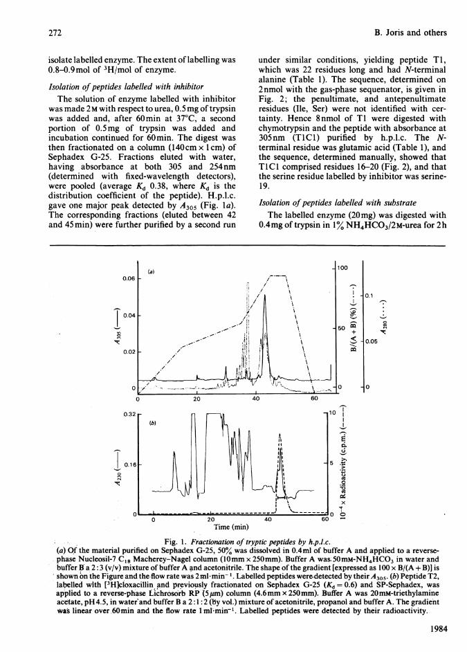
Isolation ofpeptides labelled with inhibitorThe solution of enzyme labelled with inhibitor
was made 2M with respect to urea, 0.5mg of trypsinwas added and, after 60min at 37° C, a secondportion of 0.5mg of trypsin was added andincubation continued for 60min. The digest wasthen fractionated on a column (140cm x 1 cm) ofSephadex G-25. Fractions eluted with water,having absorbance at both 305 and 254nm(determined with fixed-wavelength detectors),were pooled (average Kd 0.38, where Kd is thedistribution coefficient of the peptide). H.p.l.c.gave one major peak detected by A305 (Fig. la).The corresponding fractions (eluted between 42and 45 min) were further purified by a second run
under similar conditions, yielding peptide TI,which was 22 residues long and had N-terminalalanine (Table 1). The sequence, determined on2nmol with the gas-phase sequenator, is given inFig. 2; the penultimate, and antepenultimateresidues (Ile, Ser) were not identified with cer-tainty. Hence 8nmol of Ti were digested withchymotrypsin and the peptide with absorbance at305nm (TICI) purified by h.p.l.c. The N-terminal residue was glutamic acid (Table 1), andthe sequence, determined manually, showed thatTICI comprised residues 16-20 (Fig. 2), and thatthe serine residue labelled by inhibitor was serine-19.
Isolation ofpeptides labelled with substrateThe labelled enzyme (20mg) was digested with
0.4mg of trypsin in 1% NH4HCO3/2M-urea for 2h
10I
I_cd
._o~
-
Time (min)
Fig. 1. Fractionation of tryptic peptides by h.p.l.c.(a) Qf the material purified on Sephadex G-25, 50% was dissolved in 0.4ml of buffer A and applied to a reverse-phase Nucleosil-7 C18 Macherey-Nagel column (0mm x 250mm). Buffer A was; 50mM-NH4HCO3 in water andbuffer B a 2: 3 (v/v) mixture of buffer A and acetonitrile. The shape of the gradient [expressed as 100 x B/(A + B)] isshown on the Figure and the-flow rate was 2ml*min'1. Labelled peptides were detected by their A305. (b) Peptide T2,labelled with [3H]cloxacillin and previously fractionated on Sephadex G-25 (Kd = 0.6) and SP-Sephadex, wasapplied to a reverse-phase Lichrosorb RP (5pm) column (4.6mm x 250mm). Buffer A was 20mM-triethylamineacetate, pH4.5, in water and buffer B a 2:1: 2 (by vol.) mixture of acetonitrile, propanol and buffer A. The gradientwas linear over 60min and the flow rate I ml'min-'. Labelled peptides were detected by their radioactivity.
1984
272
,-I
x
1-1
-n0In
'lt.
1-10InC4
'11:.

The active site of P99 P-lactamasePseudomonasaeruginosa Val-Thr-Pro-Glu-Thr-Leu-Phe-Glu-Ile-Gly-Ser-Val-Ser-Lys
62 70 80ampC Ala-Asp-Ile-Ala-Lys-Lys-Gln-Pro-Val-Thr-Gln-Gln-Thr-Leu-Phe-Glu-Leu-Gly-Ser-Val-Ser-Lys-Thr
5 10 15 * 20P99 Ala-Asp-Ile-Ala-Ala-Asn-Lys-Pro-Val-Thr-Pro-Gln-Thr-Leu-Phe-Glu-Leu-Gly-Ser-Ile-Ser-Lys-Thr
TiT2 -
-Z TlClCP1
P2
Fig. 2. Active-site sequences of class-C /-lactamasesThe sequence that is deduced for the 23-residue fragment of the P99 P-lactamase is shown; the asterisk marks theserine residue that is labelled by substrate and inhibitor. Peptides obtained by the action of trypsin and pepsin aredenoted by 'T' and 'P', respectively; further digestion of tryptic peptides Ti and T2 with chymotrypsin orthermolysin gave peptides TlCl and T2H1 respectively. The sequence 1-19 of Ti was determined with the gas-phase sequenator. All the other peptides were sequenced except the last one. Above the continuous sequence areshown the sequences of ampC ,B-lactamase (middle line) (Jaurin & Grundstr6m, 1981) and Pseudomonas aeruginosa,B-lactamase (top line) (Knott-Hunziker et al., 1982a). The numbering of the ampC ,B-lactamase is derived from thecomplete sequence of the gene, including the signal peptide.
Table 1. Composition of labelled peptidesThe Table gives residues/molecule from amino acid analysis; the values in parentheses refer to residues not found bythe Edman sequence analysis. The integral values in columns (3), (5), (7), (11) and (13) are from the sequenceanalysis, and in column (9) are calculated for residues 17-19. The electrophoretic mobility (m) at pH6.5 wascalculated relative to Asp = -1 (Offord, 1977). Columns (1) and (6) refer to peptide labelled with 6f,-iodopenicillan-ate, and columns (2), (4), (8), (10) and (12) refer to radioactive peptides labelled with [3H]cloxacillin. N-Terminalresidues were identified by the dansyl procedure (Bruton & Hartley, 1970). T, P, TICI and T2Hl are explained inthe legend to Fig. 2.
Composition (residues/molecule)A
Peptide ... T1
Column ... (1)2.12.02.02.01.91.42.81.21.82.00.91.8
3H* ......... 0.9m ... -0.3N-Terminus ... Ala AlaNo. of
residues ... 22* Mol of labelled substrate bound/mol of peptide.
T2 TICI T2H1
(2) (3) (4) (5) (6) (7)1.9 2 (0.4) (0.3)1.5 21.6 2 1.4 2 1.3 12.1 2 0.4 1 1.2 11.9 21.7 1 1.5 1 1.0 13.1 3 (1.3)1.3 11.9 2 1.0 1 0.5 12.0 2 1.7 1 1.1 10.9 12.0 2 0.9 1
0.8-0.27Glu
(8) (9)
0.9 1(0.4).1.1 I
0.9-0.44
Glu Leu
7 5
P1
(10) (1 1)
1.0 1 1.02.2 2 2.00.9 1,
1.2 1 1.2 1
0.9 11.0 1 1.0 1
1.0 10.8 11.0
-0.31Phe
.93
at 37° C and the digest fractionated on a column(150cm x O.9cm) of Sephadex G-25 (superfinegrade) in 0.1 M-acetic acid at 4° C. The radioactivefraction (Kd 0.34) contained peptide TI, and asecdnd radioactive fraction (Kd 0.6) contained
peptide T2 (Table 1); further tryptic hydrolysisconverted peptide TI into peptide T2. The labelledenzyme was also digested with 1% pepsin in 1 mM-HCl/3M-guanidinium chloride for 60min at 37° C.The tryptic or peptic digests, after fractionation on
Vol. 223
Amino acidAspThrSerGluProGlyAlaValIleLeuPheLys
P2
(12) (13)
12
1.0 11.1 1
0.8 11.0
-0.15Leu
7
273
r-

274 B. Joris and others
Sephadex G-25, were further fractionated on SP-Sephadex 50 and by h.p.l.c.; the peptic digestyielded two peptides, P1 and P2 (Table 1). Theh.p.l.c. fractionation of the Sephadex fractionscontaining peptide T2 is shown on Fig. l(b).The amino acid sequence of peptide T2 showed
that it comprised residues 16-22 (Fig. 2). Thesequences of peptides P1 and P2 showed that theycomprised residues 15-23 and 17-23 respectively.Finally, peptide T2 was digested with thermolysinand the digest fractionated by h.p.l.c. The radioac-tive tripeptide (T2H1) (Table 1) contained thelabelled serine in the sequence Leu-Gly-Ser andcomprised residues 17-19 (Fig. 2). Since serine isthe only amino acid in this tripeptide with areactive side chain, it is serine-19 that is labelled bysubstrate.
DiscussionThe results on the P99 P-lactamase in Fig. 2
establish that the same serine residue is labelled by6fi-iodopenicillanate, a 'branched-pathway' f3-lactamase inactivator, and by cloxacillin, an 'inhibi-tory substrate' (Cartwright & Waley, 1983). Thesequence of the 18 residues before, and the fourresidues after, serine-19 is firmly based on thestructures of six peptides. The sequence of peptidescontaining the active-site serine residues of the ,B-lactamases of Pseudomonas aeruginosa and Escheri-chia coli K 12 (ampC gene) has been previouslyestablished (Knott-Hunziker et al., 1982a). Thecorresponding sequence obtained in the presentwork is closely similar to that of the ampC ,B-lactamase: 18 out of 23 residues are identical.Similarly, 11 out of 14 residues are the same in theP99 and Pseudamonas aeruginosa P-lactamases. Infact, among the 14 residues corresponding topositions 70-83 in the ampC,-lactamase, ten areidentical in these three fi-lactamases (Fig. 2). Ourresults clearly establish the E. cloacae P99 /3-lactamase as a member of class C, and the serineresidue labelled as the counterpart of serine-80 inthe ampC ,B-lactamase (the position of peptide Tiin the sequence still requires determination).
It was observed with various penicillin-sensitiveenzymes that the homology was much morepronounced in the immediate surroundings of thepenicillin-binding serine residue (Frere & Joris,1984). Although these homologies were not asstrong as those observed in the present study, onemay wonder whether, in the case of the class C /3-lactamases, the homology extends further away
from the active serine residue. This problemrequires further investigation.The isolation of an acyl-enzyme from cloxacillin
and the P99 P-lactamase suggests that this covalentintermediate is important in catalysis. Kineticstudies are necessary to decide whether thisintermediate is on the main reaction pathway.
The support of the Medical Research Council (U.K.)and of the National Fund for Scientific Research(Belgium), the Fonds de la Recherche ScientifiqueMedicale, Belgium (contract n° 3. 4507.83) and of theBelgian State (Action concertee n° 79/84-I 1) is gratefullyacknowledged. We thapk Mr. N. Gascoyne for carryingout the amino acid analyses and Glaxo Group ResearchLtd., Pfizer Research and Beecham Pharmaceuticals forgifts of various samples. B.J. and J.D. are respectively'Charge de Recherches' and 'Chercheur Qualifie' of theNational Fund for Scientific Research.
ReferencesAmbler, R. P. (1980) Philos. Trans. R. Soc. London Ser. B
289, 321-331Bruton, C. J. & Hartley, B. S. (1970) J. Mol. Biol. 52, 165-
178Campbell, D. G., Gagnon, J., Reid, K. B. M. &
Williams, A. F. (1981) Biochem. J. 195, 15-30Cartwright, S. J. & Waley, S. G. (1983) Med. Res. Rev. 3,
341-382Cartwright, S. J. & Waley, S. G. (1984) Biochem. J. 221,
505-511Charlier, P., Dideberg, O., Frere, J. M., Moews, P. C. &Knox, J. R. (1983) J. Mol. Biol. 171, 237-238
Cohen, S. A. & Pratt, R. F. (1980) Biochemistry 19, 3996-4003
Fisher, J., Belasco, J. G., Khosla, S. & Knowles, J. R.(1980) Biochemistry 19, 2895-2901
Fisher, J., Charnas, R. L., Bradley, S M. & Knowles,J. R. (1981) Biochemistry 20, 2726-2731
Frere, J. M. & Joris, B. (1984) CRC Crit. Rev. in the pressHewick, R. M., Hunkapiller, M. W., Hood, L. E. &
Dreyer, W. J. (1981) J. Biol. Chem. 256, 7990-7997Jaurin, B. & Grundstrom, T. (1981) Proc. Nat!. Acad. Sci.
U.S.A. 78, 4897-4901Knott-Hunziker, V., Orlek, B. S., Sammes, P. G. &
Waley, S. G. (1979) Biochem. J. 177, 365-367Knott-Hunziker, V., Orlek, B. S., Sammes, P. G. &
Waley, S. G. (1980) Biochem. J. 187, 797-802Knott-Hunziker, V., Petursson, S., Jayatilake, G. S.,
Waley, S. G., Jaurin, B. & Grundstrom, T. (1982a)Biochem. J. 201, 621-627
Knott-Hunziker, V., Petursson, S., Waley, S. G., Jaurin,B. & Grundstrom, T. (1982b) Biochem. J. 207, 315-322
Neu, H. C. (1983) Infection 11, Suppi. 2, 74-80Offord, R. E. (1977) Methods Enzymol. 47, 51-69Ross, G. W. (1975) Methods Enzymol. 43, 678-687
1984