the allosteric role of the ca2+ switch in adhesion and ...sotomayo/private/mainsr.pdf · marcos...
TRANSCRIPT
The Allosteric Role of the Ca2+ Switch in Adhesionand Elasticity of C-Cadherin
Marcos Sotomayor§ and Klaus Schulten§∗
October 3, 2007
§ Department of Physics, University of Illinois at Urbana-Champaign, and BeckmanInstitute for Advanced Science and Technology. E-mail: [email protected]∗ To whom correspondence should be addressed.
1

Abstract
Modular proteins such as titin, fibronectin, and cadherin are ubiquitouscomponents of living cells. Often involved in signaling and mechanical pro-cesses, their architecture is characterized by domains containing a variablenumber of heterogeneous “repeats” arranged in series, with either flexible orrigid linker regions that determine their elasticity. Cadherin repeats arrangedin series are unique in that linker regions also feature calcium binding mo-tifs. While it is well known that the extracellular repeats of cadherin proteinsmediate cell-cell adhesion in a calcium-dependent manner, the molecular mech-anisms behind the influence of calcium in adhesion dynamics and cadherin’smechanical response are not well understood. Here we show, using moleculardynamics simulations, how calcium ions control the structural integrity of cad-herin’s linker regions, thereby affecting cadherin’s equilibrium dynamics, theavailability of key residues involved in cell-cell adhesion, and cadherin’s me-chanical response. The all-atom, multi-nanosecond molecular dynamics simu-lations involved the entire C-cadherin extracellular domain solvated in water (a345,000 atom system). Equilibrium simulations show that the extracellular do-main maintains its crystal conformation (elongated and slightly curved) whencalcium ions are present. In the absence of calcium ions, however, it assumesa disordered conformation. The conserved residue Trp2, which is thought toinsert itself into a hydrophobic pocket of another cadherin molecule (therebyproviding the basis for cell-cell adhesion) switches conformation from exposedto intermittently buried upon removal of calcium ions. Furthermore, the over-all mechanical response of C-cadherin’s extracellular domain is characterized atlow force by changes in shape (tertiary structure elasticity), and at high forceby unraveling of secondary structure elements (secondary structure elasticity).This mechanical response is modulated by calcium ions at both low and highforce, switching from a stiff, rod-like to a soft, entropic-like behavior upon re-moval of ions. The simulations provide an unprecedented molecular view ofcalcium mediated allostery in cadherins, also illustrating the general principlesof linker mediated elasticity of modular proteins relevant for cell-cell adhesionand sound transduction, but also muscle elasticity.
2

Introduction
Development of complex multicellular organs and tissues relies on selective and robustadhesion between cells (1–4). Cadherin proteins are responsible for calcium-mediatedcell-cell adhesion and have been implicated in various biologically relevant processesrelated to tissue morphogenesis and maintenance of tissue integrity, such us neuronalconnectivity or prevention of tumor cell propagation (3–8). Members of the cadherinfamily of proteins have also been suggested to form part of the mechanotransduc-tion apparatus of the inner ear (9–12). Classical cadherins feature a cytoplasmicdomain, a single transmembrane segment, and a long extracellular domain made offive, tandemly arranged, heterogeneous cadherin repeats (1, 13–15). The repeatsare labeled EC1 to EC5, with EC1 being the most distant from the membrane (seeFig. 1 A).
Selective adhesion is achieved through trans interactions between cadherin extra-cellular domains coming from adjacent cells (8, 15, 16). In addition, cis interactionsarising from dimerization of cadherin molecules that belong to the same cell havebeen suggested to be necessary for formation of trans-bonds (16–22). The inter-action between cadherin extracellular domains likely involves one or more repeatsfrom each cadherin molecule (8, 23–29). Multiple studies using different experimen-tal techniques such as mutagenesis, electron microscopy, force measurements, NMR,and X-ray crystallography have been used to postulate and probe different modelsof trans- and cis- interactions (see (8) for a recent review). In all cases it has beenshown that Ca2+ and the EC1 repeat are both essential for at least the initial stagesof trans-bond formation.
Calcium ions seem to stabilize and rigidify extracellular domains. Indeed, electronmicroscopy (EM) and other experiments have shown that the E-cadherin extracellulardomain forms an elongated, semi-curved rod in the presence of Ca2+, while it col-lapses upon Ca2+ removal (17, 30–34). The crystal structure of a complete C-cadherinextracellular domain (35) depicts in atomic detail the elongated rod observed for E-cadherin with EM and also reveals all Ca2+ binding spots found in linker regionsbetween repeats (Fig. 1 A). Biochemical assays, mutagenesis, and quantitative forceand bead aggregation measurements have shown that disruption of some Ca2+ bind-ing spots cooperatively influence large regions of the protein structure, often abolishadhesion, and affect the stability of individual repeats (36–38). Furthermore, molec-ular dynamics simulations of single C-cadherin repeat mechanical unfolding and ofEC1-EC2 E-cadherin equilibrium dynamics have confirmed the relevant role of Ca2+
in the stability of cadherin repeats (39–41).While calcium ions provide rigidity, the EC1 repeat provides an anchor residue
(Trp2) that may intramolecularly dock into a hydrophobic pocket of EC1, or insertitself into the same hydrophobic pocket but of a neighboring EC1 coming from anadjacent cell, thereby facilitating cell-cell adhesion (17, 33, 35, 42–44). Antibodybinding and mutagenesis combined with force measurements suggest that Ca2+ bind-
3
ing allosterically modulates the availability of Trp2 (38, 45). However, a molecularand dynamic view of how calcium ions control Trp2 availability and the flexibility ofcadherin repeats is missing. Here we present molecular dynamics simulations thatreveal how Ca2+ control the flexibility and adhesive property of C-cadherin switch-ing the cadherin ectodomains from a stiff, rod-like to a soft, entropic-like regime.Moreover, the simulations depict the Ca2+ modulation of Trp2 availability.
Results and Discussion
Molecular dynamics simulations were carried out using the complete extracellulardomain of C-cadherin. The corresponding crystal structure (35), solved at 3.1 Aresolution (pdb code 1L3W), features five cadherin repeats, each made of about 110amino acids and folded in a “Greek key” motif characterized by seven β-strands form-ing two β-sheets (13). Four sets of simulations of this structure were performed. Thefirst set (labeled SimCaX) utilized a system containing the protein, water molecules,and 12 crystallographically resolved Ca2+ ions (three ions located at each linker re-gion). The second set (labeled SimApoX) was performed without Ca2+ ions. Thelast two sets, labeled SimKX and SimNaX, involved systems in which crystallograph-ically resolved Ca2+ ions were replaced by potassium (K+) and sodium (Na+) ions,respectively (Fig. 2). Further details of the systems and simulations can be found inMethods and Supplementary Material.
Influence of Ions on C-Cadherin Equilibrium Dynamics
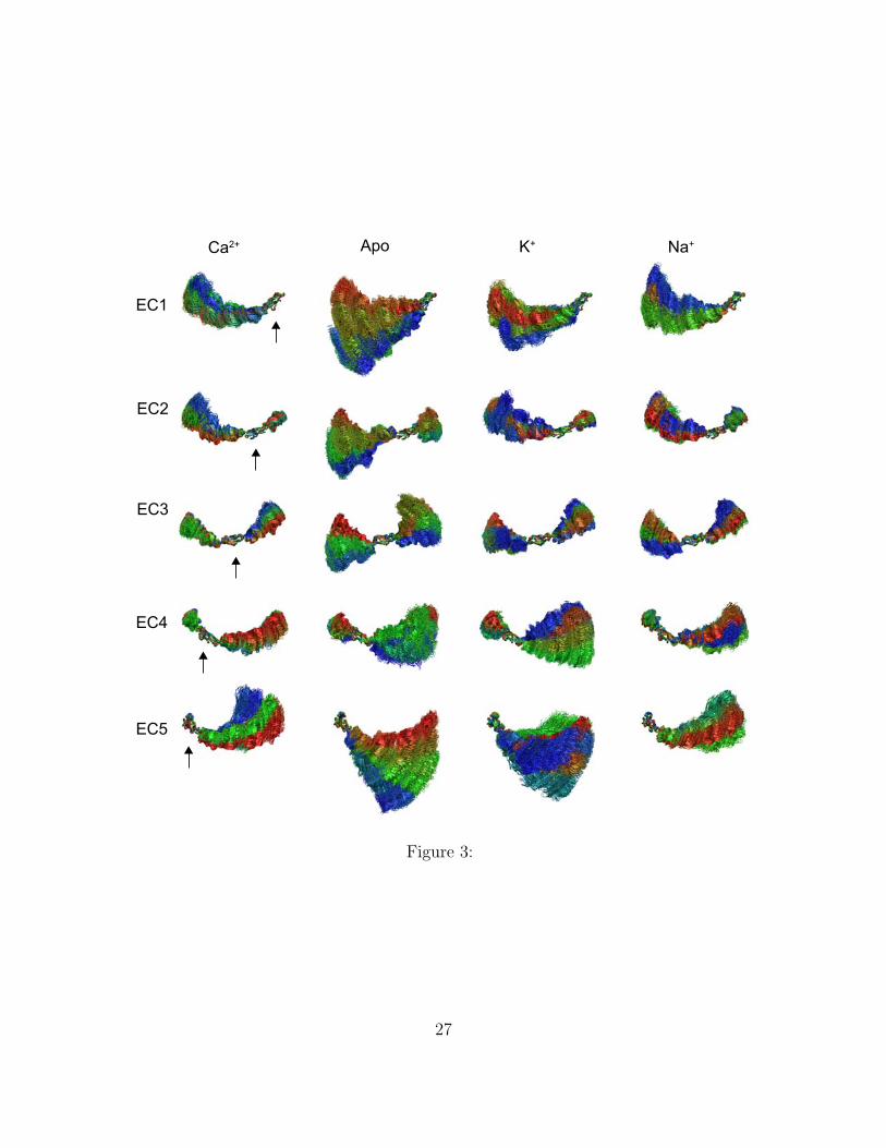
Equilibrium simulations showed that the overall curved shape of the protein was main-tained throughout 10 ns of dynamics in which Ca2+ ions were present in the model(SimCa1, Fig. 1 C), while the cadherin extracellular domain adopted a disorderedshape when calcium ions were removed (SimApo1, Fig. 1 D). Indeed, the absenceof Ca2+ resulted in strong repulsion between negatively charged residues (previouslycoordinating ions) and partial disruption of linker regions (Fig. 2 B). Complete dis-ruption and unfolding of linker regions was prevented by performing an equilibrationstep in which 1000 steps of minimization and 100 ps of dynamics were carried out withbackbone atoms harmonically restrained to their crystal conformation (as in all ourinitial equilibrations, see Methods and Table 1). The linker disruption, most clearlyseen at the linker between EC3 and EC4, favored independent motion of individualrepeats leading to the observed disordered shape (Fig. 3). The same behavior was re-produced in simulations that used different temperature control protocols (SimCa4,SimApo4, and SimApo5; Supplementary Material’s Figs. 8 and 9). Whether pro-tonation states of charged amino acids change upon Ca2+ removal remains to beelucidated. However, our results are in agreement with experiments showing thatlinkers without calcium can destabilize cadherin repeats through electrostatic inter-actions (37). Additional simulations were performed in which Ca2+ ions were replaced
4
by K+ or Na+ ions (SimK1 and SimNa1, respectively). The replacement of divalentions by monovalent ions likely reduced artifacts introduced by simple elimination ofCa2+. These control simulations showed that although the extracellular domain ofC-cadherin partially retains its curved conformation on a 10 ns time scale (Fig. 1 Eand F), inter-repeat motion was clearly enhanced and in some cases similar to thatobserved in the absence of Ca2+ (Fig. 3). Neither K+ nor Na+ ions seemed to main-tain the rigidity of the C-cadherin extracellular domain. Furthermore, unbinding ofboth types of ions was observed throughout the simulations.
Root mean square deviations (RMSD), computed for individual repeats duringSimCa1, SimApo1, SimK1, and SimNa1 (see Supplementary Material’s Fig. 10),reached stable values below 0.3 nm in all cases except one, indicating that indi-vidual repeats maintained their secondary structure on a nanosecond time-scale evenin the absence of Ca2+. Repeat EC5 exhibited large RMSD values (> 0.3 nm) duringa simulation in which Ca2+ was replaced by K+ (SimK1), likely reflecting deforma-tions induced by unbinding of this large monovalent ion. RMSD values for repeatsEC1 and EC2 were found to be consistently smaller than those for other repeats insimulations with Ca2+, K+, and Na+. The slight increase in RMSD observed whencalcium was removed (SimApo1) suggests that EC1 and EC2 are the most sensitiveto Ca2+ binding.
The outcome of our equilibrium simulations are in line with those presented byCaillez and Lavery (40, 41) (using a different force-field and simulation engine) fortwo repeats of E-cadherin and agree well with previous experimental results indi-cating that Ca2+ rigidifies the extracellular cadherin domain (30–34). Although oursimulations span a short time-scale which precludes observation of a full collapse ofcadherin repeats (as seen in electron microscopy and AFM images (30, 46)) or pos-sible unfolding, the simulations permitted us to clearly see inter-repeat motion thatcould lead to a collapsed conformation in the absence of Ca2+ (Fig. 3, SupplementaryMaterial’s Figs. 8 and 9).
Tertiary Structure Elasticity of C-cadherin
We further probed cadherin stability by using conformations from equilibrium simu-lations as the starting points for constant-velocity steered molecular dynamics simu-lations (47–51). The SMD simulations were performed on systems with and withoutCa2+ ions and on a system in which Na+ replaced the crystallographically resolvedCa2+. Our SMD setup was similar to that used in (52) as we attached both ends ofthe protein (C1
α and C540α ) to virtual springs (ks =1 kcal/mol/ A2). The free ends of
the springs moved in opposite directions at a constant velocity along the axis definedby the vector joining the protein termini. Different temperature control protocols andstretching velocities were utilized.
The SMD simulations revealed that upon stretching the complete C-cadherin ex-tracellular domain becomes straight by rearranging the relative orientation of indi-
5
vidual repeats with respect to each other (see Figs. 4 B and E). This so-called tertiarystructure elasticity (TSE) demonstrated earlier in (39, 51, 53) was observed in SMDsimulations with and without Ca2+, and with Na+ replacing Ca2+. While the apoSMD simulation shows that the linkers between repeats extend fairly easily in thiscase, the simulation with Ca2+ reveals that these ions act as molecular bearings, suchthat linker regions behave as stiff hinges and the extracellular domain responds asone unit.
We ask then whether cadherin’s TSE is reversible. We turned off the applied forcesat the end of simulations SimCa2 and SimApo2, and continued with equilibrium dy-namics in two simulations labeled SimCa3 and SimApo3 lasting 10 ns and 5 ns,respectively. In both cases the protein relaxed and the end-to-end distance partiallyrecovered the value observed before stretching (see Figs. 4 C, F, and SupplementaryMaterial’s Fig. 11 A). However, while C-cadherin partially recovered curvature in thepresence of Ca2+ (SimCa3, see Fig. 4 C), in the absence of Ca2+ (SimApo3) it relaxedinto a conformation even more disordered than the one observed before stretching (dis-order meaning that relative orientation of individual repeats was not uniform as seenin Fig. 4 F and Supplementary Material movies mI, mII, and Fig. 12). Reversibilityfor the Apo simulation arises only in regard to end-to-end distance, as confirmedby the RMSD of the complete structure computed during stretching and relaxation(Supplementary Material’s Fig. 11 B). The latter result suggests that TSE has dif-ferent molecular origins in both cases. Interestingly, RMSD of individual repeatsremained below 0.3 nm throughout the whole stretching and relaxation trajectoriesin the presence and the absence of calcium, confirming that linkers are responsiblefor the observed shape changes. Furthermore, partial recovery of C-cadherin’s shapein the presence of divalent ions suggests that its curvature is not an artifact causedby crystallographic packing.
Local deformations of the C-cadherin structure were also monitored by comput-ing average strain per residue (see Methods and (54)) during equilibration, shortstretching, and relaxation simulations in the presence and absence of Ca2+ (see Sup-plementary Material movies mIII and mIV). In the presence of Ca2+ the largest strainswere observed in loops of repeats EC3, EC4, and EC5 and the corresponding linkerregions. In the absence of Ca2+ the largest strains were observed in all linker regionsand loops, confirming once again the role of Ca2+ ions in inter-repeat motion andTSE.
Tertiary structure elasticity was first predicted through simulations of the proteinAnkyrin-R (9, 39), and subsequently confirmed through AFM experiments involvingAnkyrin-B (55). Both Ankyrin proteins feature 24 repeat units but, unlike cadherin,ankyrin repeats interact with each other through extensive hydrophobic surfaces,the parallel stack of repeats forming a curved super-helical arrangement (56). Thereversible, non-entropic, TSE observed for ankyrin resembles better the one observedhere for cadherin with calcium ions, while the TSE observed for cadherin withoutions seems to be rather entropic in origin, although some residual interactions at the
6

linkers may remain. Whether such difference in the origin of the observed TSE canbe distinguished through experimental force spectroscopy or if it is relevant at all foradhesion are questions that remain to be answered. However, a simple calculationcan set some limits for the elasticity in both cases. A freely jointed chain with nsegments of length b each would exhibit an entropic spring constant of k = 3kBT
nb2∼
0.1 mN/m (lower limit of k for cadherin without Ca2+ at low force using n = 5 andb = 5 nm), while a straight rigid rod 23 nm (L) long and 2 nm (2r) wide would exhibita longitudinal stiffness of k = Eπr2
L∼ 270 mN/m (upper limit of k for cadherin with
Ca2+ using a Young’s modulus for collagen of E = 2 GPa).A rough estimate of the elastic constant of C-cadherin with and without Ca2+
was obtained using a novel SMD protocol termed ‘length-clamp’ (SimCa13/SimCa17and SimApo9; see Methods and Supplementary Material’s Fig. 13). The estimatedspring constants from elastic forces (57), kCa2+ ∼ 50 mN/m (400/8 pN/nm) andkApo ∼ 25 mN/m (200/8 pN/nm) are within the boundaries computed above. Thesesimulations also reveal that the complete C-cadherin extracellular domain exhibits aviscoelastic behavior.
Secondary Structure Elasticity of C-cadherin
The simulations described above depict the extracellular domain of cadherin switch-ing from a rod-like to a flexible-chain like behavior upon removal of Ca2+ at lowforce regimes. While unfolding of individual domains may not play a physiologicalrole in adhesion (44), we explored the effect of calcium and sodium on the stabil-ity of individual domains when subject to large forces inducing mechanical unfolding.Two SMD simulations (SimCa2 and SimApo2) were further continued until stretchingforces unfolded one repeat (SimCa2E and SimApo2E) and eight new SMD simulations(SimCa5, SimCa6, SimCa7, SimCa8, SimCa9, SimApo6, SimApo7, and SimApo8)were performed using different steering velocities and thermodynamic ensemble pro-tocols.
The SMD simulations showed what is perhaps the most dramatic difference be-tween the elastic response of the protein with or without Ca2+. While the EC1repeat unfolded fairly easily in the absence of Ca2+ (see Fig. 5 A), unfolding of EC1in the presence of Ca2+ required a considerably larger force (1664 pN compared to858 pN at v = 10 nm/ns) that breaks a bond between residue Glu11 and a Ca2+ ion(Fig. 5 B-E and Supplementary Material’s Figs. 14, 15, and movies mV, mVI andmVII). Peak forces arising in a SMD simulation unfolding cadherin in which Ca2+
ions were replaced by Na+ (SimNa) were only slightly larger than those observed forthe simulations performed in the absence of Ca2+, reinforcing the relevance of Ca2+
for the stability of C-cadherin (see Supplementary Material’s Fig. 16).Additional simulations performed at different stretching speeds (SimCa7, SimCa8,
SimCa9, and SimApo8 in Table 1) confirmed a similar unfolding scenario and a reduc-tion in unfolding force peaks upon decrease of v, as expected (58, 59) (Supplementary
7

Material’s Fig. 17). It remains to be elucidated whether the difference observed inpeak forces for C-cadherin between simulations with and without Ca2+ is as dramaticat slow pulling speeds as it is at the speeds used in this study. Even the slowest stretch-ing velocity used in our SMD simulations is large compared to those used in AFMor other experimental techniques (1 nm/ns vs 10−5 nm/ns), since all-atom molec-ular dynamics simulations can presently achieve only a submicrosecond time-scale.Despite this limitation, multiple SMD studies, all similarly exceeding experimentalpulling speeds, have provided qualitative and quantitative predictions confirmed byexperiments (39, 51, 60–66).
The SMD simulations mentioned above were performed in the NVE ensemble.Further simulations performed in the NpT ensemble (SimCa5, SimCa6, SimApo6,SimApo7) confirmed the Ca2+ dependent mechanical stability of the C-cadherin do-main. However, even when using a small damping coefficient, Langevin dynamics(NpT) resulted in an artificial increase of unfolding peak forces and, in the worstcase, complete decoupling of forces measured at opposite ends of the protein (Sup-plementary Material’s Fig. 18).
Unfolding forces for EC1 in all cases are considerably smaller than the forces re-quired to unfold a single EC2 domain simulated under similar conditions but isolatedfrom the rest of the structure (reported in our earlier SMD simulations (39)). Theresults are consistent with temperature and denaturant induced unfolding experi-ments of E-cadherin showing that Ca2+ stabilizes cadherin repeats and that EC1 isweaker than EC2 (36). However, the mechanical unfolding of the EC2 repeat withinthe entire C-cadherin extracellular domain may differ from that of the isolated re-peat. We therefore probed the mechanical response of repeats EC2 and EC4 bysteering the center of mass of repeats EC3/EC1 and repeats EC5/EC3 in oppositedirections (effectively stretching repeats EC2 and EC4, Supplementary Material’sFig. 19 A). The simulations (SimCa10 and SimCa11) revealed that the EC4-EC5linker is weaker than linker EC1-EC2, indicating a possible mechanical hierarchy forcadherin repeats/linkers. Interestingly, the unfolding force for repeat EC2 within thecomplete domain is comparable to that of the isolated repeat (Supplementary Mate-rial’s Fig. 19 B), and both unfolding pathways exhibit an intermediate state in whichthe repeat is partially unfolded due to rupture of interactions between conservedresidues and Ca2+ ions. Such a state may be particularly relevant under physiolog-ical conditions, as it provides a safety mechanism in which cadherin can extend inresponse to sustained mechanical stimuli and easily refold when the external force isturned off.
Previous SMD simulations identified two types of mechanical unfolding processesfor immunoglobulin-like domains (67) in which water mediates rupture of hydrogenbonds concertedly (sheering mode) or one-by-one (zipper mode). In both types ofunfolding, rupture of hydrogen bonds between β-strands (concertedly or one-by-one)gives rise to the unfolding force-peak. Based on the results presented here and in (39),we add a third mechanical unfolding category in which a bridge between charged
8

amino-acids and divalent ions produce the unfolding force-peak. Thus, Ca2+ is notonly acting as a molecular bearing that facilitates cooperative motion of cadherinrepeats, but also as a staple that maintains the stability of the structure providingfurther resistance to mechanical unfolding.
Unfolding as observed with the SMD methodology employed here permitted us toalso estimate a characteristic time for stress propagation through the protein. Thepresent example shows how an unfolding event that occurred at one end of a pro-tein is perceived at the other end (by the second steering spring considered now asa sensor) after some time. The force peaks measured through spring extension atboth ends of the protein are not synchronous (see Supplementary Material Fig. 14).In fact, in SimCa2E, a delay between the appearance of force peaks at both ends ofthe protein can be used to compute a velocity v ∼ 285 A/38 ps ∼ 747 m/s at thecorresponding extension of the protein. This velocity may depend on multiple factorsincluding the thermodynamic ensemble used in the simulation, stretching velocity,spring constants used for stretching springs, and criteria used to determine the char-acteristic time. Indeed, our simulations show that damping artificially introducedby temperature control algorithms may decouple different zones of the protein fromeach other (Supplementary Material’s Fig. 18). Although protein motions are largelyover-damped (68), it has been hypothesized that stress signals could propagate overlarge distances in the cell (69). The two-end stretching approach outlined here mayserve to test this hypothesis experimentally and theoretically.
Ca2+ Allosteric Control of Residues Involved in Cell Adhesion
The dynamics of the extracellular domain, controlled by Ca2+, determines the avail-ability of repeat EC1 to form adhesive contacts with proteins coming from neighboringcells. There are also subtle changes in C-cadherin dynamics controlled by Ca2+ thatwill affect its adhesive properties. For instance, we monitored the dynamics of theconserved residue Trp2 throughout equilibrium simulations in presence and absenceof Ca2+, as well as with K+ and Na+ replacing Ca2+. As mentioned above, Trp2 isthought to mediate trans-interactions of cadherin by inserting itself in a hydrophobicpocket of another EC1 repeat coming from either the same cell or an adjacent cell (cisversus trans). The conformation of Trp2 in the C-cadherin structure is that of an ex-posed residue (see first snapshot in Fig. 6), since the crystallographic arrangement ofthe cadherin molecule leads to a proposed trans interaction between molecules of dif-ferent crystallographic unit cells. During the simulations in which calcium ions werepresent (SimCa1, SimCa3, and SimCa4), the side chain of Trp2 remained partially ex-posed at all times (Fig. 6 A and Supplementary Material’s Fig. 20 A). However, whencalcium ions were removed or replaced by monovalent ions, the EC1 domain was moremobile (see root mean square fluctuations in Supplementary Material’s Fig. 21 A) andthe sidechain of Trp2 fluctuated between two states: exposed, and partially buried(see Fig. 7, snapshots in Fig. 6 and Supplementary Material’s Figs. 20 B&C and
9
movies mVIII, mIX, mX, and mXI). In the latter state the sidechain is hidden in ahydrophobic pocket (intramolecular docking), close to residues 24 and 25. We termedthis state “partially” buried since interactions with E90 (D90 in E-cadherin) seen inother structures and NMR experiments with E-cadherin are not observed here, per-haps due to the short time scale of the simulation. The solvent accessible surfacearea for Trp2 computed throughout different equilibrium simulations also confirm thegreater availability of this side chain when Ca2+ is bound to C-cadherin (Supple-mentary Material’s Fig. 22). Our results strongly support experimental work usingantibodies and independent mutagenesis experiments combined with force measure-ments suggesting that Trp2 is allosterically modulated by calcium (38, 45).
Conclusions
The results presented here qualitatively validate molecular dynamics simulations asa tool to study the mechanical function of cadherin. The simulations, using standardand widely used parameters, account for experimentally known facts such as the roleof Ca2+ in shaping the extracellular cadherin domain, the putative allosteric effect ofCa2+ on the key binding residue Trp2, and the Ca2+ dependent stability of individualrepeats probed here through mechanical unfolding. At the same time, the simula-tions provide a unique atomistic view of cadherin dynamics. Such a detailed view isa necessary complement to experiments investigating cadherin function. Moreover,the reversible TSE observed here for C-cadherin provides support for the multipleconformations observed in desmosomal cadherins through electron tomography (70)and also suggest that the extracellular domains of cadherin molecules should not beconsidered as completely rigid units, even in the presence of calcium.
The present study serves as a step towards future molecular dynamics studies ofcadherin that should address (i) the relevance of cadherin elasticity and mechanicalintermediates for adhesion, (ii) the robustness of the intrinsic curvature of C-cadherinin response to multiple stretching and compressing cycles, (iii) the mechanical stabilityand complete unfolding of all repeats, (iv) the selectivity of adhesion molecules (71,72) in the complete adhesion complex and, eventually, (iv) the mechanical or al-losteric role of mutations affecting calcium binding sites in cadherin-23 (CDH23) andprotocadherin-15 (PCDH15) resulting in hereditary deafness (12, 73, 74). Our sim-ulations strongly suggest that deafness may arise through mutations that affect theCDH23/PCDH15 hair-cell tip link in two ways: abolishing tip-link formation by di-rectly or allosterically interfering with residues involved in adhesion or dimerization,or modifying the mechanical strength of the tip link by favoring unfolding or preclud-ing mechanical intermediates by breaking bridges between protein atoms and Ca2+
ions at linker regions.The results presented here have implications for other proteins featuring tandemly-
arranged repeats, the archetypical examples being spectrin and titin. Linkers havebeen shown to be important in spectrin elasticity (54, 75, 76), while inter-domain
10

motion and divalent ions may likely play a role in titin elasticity (77). Cadherinmay serve as an exceptional example of the design principles behind linker mediatedelasticity.
Methods
Systems
The psfgen VMD (78) plugin was utilized to build four systems containing the entirecrystal structure of C-cadherin (Protein Data Bank code 1L3W) solvated in waterwith the VMD solvate plugin. The first system with a total of 345,467 atoms in-cluded 12 crystallographicaly resolved and protein bound Ca2+ ions, 8 bulk Na+ ionsrandomly placed for cell neutralization with the autoionize VMD plugin. The secondsystem encompassing 345,407 atoms did not include Ca2+ ions, but contained 32 bulkNa+ ions. The last two systems included 12 K+ or Na+ ions replacing crystallograph-ically resolved Ca2+ ions, respectively. In addition, these two systems included 20bulk Na+ ions and encompassed 345,443 atoms. In all systems, 39 crystallographicwater molecules were kept as part of the model while N-acetylglucosamine residueswere excluded. Disulfide bonds for cysteines 448-532 and 530-539 were explicitly mod-eled for all systems. Residues Asp, Glu, Lys, and Arg were assumed to be chargedthroughout the protein, while protonation states of His residues were chosen favor-ing the formation of evident hydrogen bonds. Prior to solvation, the C-cadherinmolecule was spatially aligned such that the vector joining the Cα atoms of the ter-minal residues was oriented along the x axis. The size of the resulting systems wasabout 36.4× 10.5× 9.4 nm3.
Molecular Dynamics Simulations
All molecular dynamics simulations were performed using NAMD 2.6 (79), the CHARMM22force field for proteins with the CMAP correction (80–82) (see validation using lysozyme (83,84) in Supplementary Materials, in particular Fig. 23), and the TIP3P model for wa-ter (85). The standard set of CHARMM parameters for ions was utilized in allsimulations. Parameters for Ca2+ correspond to those obtained to reproduce theirexperimental free energy of hydration (86). Parameters for K+ and Na+ did notinclude the NBFIX correction, used in simulation of potassium channels to describeion-backbone interactions more accurately, as C-cadherin Ca2+ binding cites involveion-backbone as well as ion-side chain interactions. A summary of all simulationscarried out in our study totaling more than 150 ns is presented in Table 1.
11

Label tsim (ns) Type Ensemble γ (ps−1) Velocity (nm/ns) StartSimCa1 10.00 EQ NpT/NVEa – – –SimCa2 0.67 PCV NV – 5× 2 SimCa1 (5.0 ns)SimCa2E 1.07 PCV NV – 5× 2 SimCa2SimCa3 10.00 REL NVE – – SimCa2SimCa4 4.04 EQ NpT 5.0 – SimCa1 (1.1 ns)SimCa5 1.70 PCV NpT 5.0 5× 2 SimCa4SimCa6 2.15 PCV NpT 0.1 5× 2 SimCa4SimCa7 0.50 PCV NV – 50× 2 SimCa1 (5.0 ns)SimCa8 0.50 PCV NV – 25× 2 SimCa1 (5.0 ns)SimCa9 14.96 PCV NV – 0.5× 2 SimCa1 (5.0 ns)SimCa10 1.30 PCVb NV – 5× 2 SimCa1 (5.0 ns)SimCa11 1.30 PCVb NV – 5× 2 SimCa1 (5.0 ns)SimCa12 5.00 PCL NV – 10/13 nm/C SimCa1 (5.0 ns)SimCa13 5.00 PCL NV – 10/8 nm/C SimCa1 (5.0 ns)SimCa14 3.60 PCL NV – 10/15 nm/C SimCa1 (5.0 ns)SimCa15 2.90 PCL NV – 10/13 nm/N SimCa1 (5.0 ns)SimCa16 2.30 PCL NV – 10/14 nm/N SimCa1 (5.0 ns)SimCa17 2.30 PCL NV – 10/8 nm/N SimCa1 (5.0 ns)SimApo1 10.00 EQ NpT/NVEa – – –SimApo2 0.65 PCV NV – 5× 2 SimApo1 (5.0 ns)SimApo2E 1.05 PCV NV – 5× 2 SimApo2SimApo3 5.00 REL NVE – – SimApo2SimApo4 9.34 EQ NpT 5.0 – SimApo1 (1.1 ns)SimApo5 14.61 EQ NpT 0.1 – SimApo1 (1.1 ns)SimApo6 2.00 PCV NpT 5.0 5× 2 SimApo1 (5.0 ns)SimApo7 2.10 PCV NpT 0.1 5× 2 SimApo1 (5.0 ns)SimApo8 12.10 PCV NV – 0.5× 2 SimApo1 (5.0 ns)SimApo9 2.30 PCL NV – 10/8 nm/C SimApo1 (5.0 ns)SimK1 10.00 EQ NpT/NVEa – – –SimK2 5.00 EQ NpT/NVEa – – –SimNa1 10.00 EQ NpT/NVEa – – –SimNa2 1.77 PCV NV – 5× 2 SimNa1 (5.0 ns)
aThese simulations consisted of 1000 steps of minimization, 100 ps of dynamics with the backboneof the protein restrained (k = 1 Kcal/mol/A2), and the remaining time as free dynamics in the NpT(1 ns with γ=5 ps−1) and NVE ensembles.
bThese SMD simulations were performed by pulling repeats one and three (SimCa10) or threeand five (SimCa11) in opposite directions (see Methods).
Table 1: Summary of simulations. Labels indicate the presence (Ca) or absence (Apo) of crystal-lographic Ca2+ ions in the system. Replacement of Ca2+ by Na+ or K+ is indicated by labels Naand K, respectively. EQ denotes equilibrium simulations, PCV denotes constant velocity SMD sim-ulations, and REL denotes free dynamics simulations in the corresponding ensemble. PCL denotesconstant velocity SMD simulations in which one end of the protein is held fixed while the other endof the protein (N or C terminus) is pulled until a predefined elongation has being achieved. Then,the steering atom is held in space and the protein is allowed to relax in a so-called ‘length-clamp’steering protocol (see Methods). Initial coordinates and velocities were obtained from the last frameof the simulations mentioned in the Start column. All SMD simulations were performed by attachingsteering springs to Cα atoms of residues 1 and 540, unless otherwise stated.
A uniform integration time step of 1 fs was assumed for all types of interactionsthroughout all simulations. In all cases a cutoff of 12 A (switching function startingat 10 A) for van der Waals interactions was assumed, and the particle-mesh-Ewald(PME) method was used to compute long-range electrostatic forces without cut-off (87). The density of grid points for PME was at least 1/A3. Periodic boundaryconditions were assumed in all cases.
Langevin dynamics was utilized to maintain a constant temperature of T = 300 Kwhen indicated, with the damping coefficient set to 5 or 0.1 ps−1 for all heavy atoms(see Table 1). Constant-pressure simulations at 1 atm were conducted using a hybridNose-Hoover-Langevin piston method with a decay period of 200 fs and a dampingtimescale of 50 fs.
Constant velocity stretching simulations were performed using the steered molec-ular dynamics method (SMD) and the NAMD Tcl Forces interface. The stretchingdirection was set along the x-axis, which matched the vector connecting the Cα atomsof the N- and C-terminal residues when the systems were built. The SMD simulationswere performed by attaching the C1
α and C540α atoms of the N- and C-terminal residues
to virtual (independent) springs of stiffness ks = 1 (kcal/mol)/A2 each. The free endsof the mentioned virtual springs were then moved away of the protein at a constantvelocity of v each in opposite directions. Different velocities were used in simulations(see Table 1). The force applied at each end was computed using the extension of thevirtual springs.
Two additional SMD protocols were used. In the first protocol, the center ofmass of all Cα atoms of repeats EC1 and EC3 (SimCa10) or repeats EC3 and EC5(SimCa11) were each attached to virtual springs of stiffness ks = 1 (kcal/mol)/A2
each. The free ends of the springs were moved as in the standard SMD protocoldescribed above, effectively stretching repeats EC2 and EC4 (see Supplementary Ma-terial’s Fig. 19)
The second SMD protocol (‘length clamp’) consisted of two phases. First, astandard constant velocity SMD simulation is performed in which one end of theprotein (Cα atom of C- or N- terminus) is held fixed while the other end of the protein(Cα atom of N- or C-terminus) is pulled by a virtual spring as described above. Thesecond phase begins when a predetermined elongation has been achieved. Then, thefree end of the virtual spring is held fixed in space and the protein is allowed to relaxwhile the force applied on the spring by the protein is obtained by monitoring theextension of the virtual spring.
Analysis Tools
Coordinates of all atoms of the system were saved every picosecond of simulation forlater analysis. Overall structural deformation of the protein was monitored by com-puting root mean-square deviations (RMSD) over entire trajectories using VMD. Thecrystallographic structure served as the reference point, and positions of protein Cα
13
atoms were compared. End-to-end distances were computed as the distance betweenthe Cα atoms of the N- and C- terminal residues. Root mean-square fluctuations(RMSF) of Cα atom positions were computed using VMD and trajectories in whicheach cadherin repeat (EC1-EC5) was individually aligned to its crystal conformation.Alignment of structures for visualization purposes and for computation of RMSD orRMSF were performed by comparing positions of Cα atoms to their original positionsin the crystal using the VMD Tcl interface. The solvent accessible surface area forTrp2 was computed using VMD and assuming a probe radius of 0.14 nm.
The total energy ET of the system (reported as TOTAL3 in the NAMD output)was computed using the total potential energy U , the total kinetic energy Kc obtainedfrom step-centered velocities (v2
ci = [(xi+1−xi−1)/2∆t]2), and the total kinetic energyKh obtained from half-step velocities averaged (v2
hi = ([(xi − xi−1)/∆t]2 + [(xi+1 −xi)/∆t]2)/2). The final form of ET used was:
ET = U +2
3Kc +
1
3Kh −
4
3(Kh −Kc) +
4
3〈Kh −Kc〉 (1)
where the first three terms correspond to a total energy that is numerically well be-haved in long time-scales, while the last two terms correct short-timescale fluctuationswith a weighted exponential average (〈X〉new = 0.9375 〈X〉old + 0.0625 X).
Local deformations or strain (see Supplementary Material movies mIII and mIV)were computed following a method similar to that described in (54). First, for eachCi
α atom of the protein a list of neighboring Cjα atoms (within 1 nm) was created using
positions of the crystal conformation. Then, the local strain ui(t) was computed everytime frame (t) for every Ci
α atom using
ui(t) =1
Ni
Ni∑j
|~rj(t)− ~ri(t)||~rj(0)− ~ri(0)|
(2)
where ~ri(t) is the position of the Cα atom i at time t and Ni is the total number ofneighboring Cj
α atoms (determined as described above) for Cα atom i. The strainvalue ui(t) was then used to color each Cα atom.
14

References
[1] Takeichi, M. 1990. Cadherins: A molecular family important in selective cell-celladhesion. Annu. Rev. Biochem. 59:237–252.
[2] Hynes, R. O. 1999. Cell adhesion: old and new questions. Trends in Cell Biology.9:M33–M37.
[3] Takeichi, M. 2005. Looking for cell-cell adhesion molecules: a cadherin story.Proc. Japan Aca. 81:321–328.
[4] Gumbiner, B. M. 2005. Regulation of cadherin-mediated adhesion in morpho-genesis. Nature Reviews Molecular Cell Biology. 6:622–634.
[5] Takeichi, M. 1991. Cadherin cell adhesion receptors as a morphogenetic regulator.Science. 251:1451–1455.
[6] Takeichi, M. 1995. Morphogenetic roles of classic cadherins. Current Opinion inCell Biology. 7:619–627.
[7] Takeichi, M. 2007. The cadherin superfamily in neuronal connections and inter-actions. Nature Reviews Neuroscience. 8:11–20.
[8] Leckband, D. and A. Prakasam. 2006. Mechanism and dynamics of cadherinadhesion. Annual Review of Biomedical Engineering. 8:259–287.
[9] Corey, D. P. and M. Sotomayor. 2004. Tightrope act. Nature. 428:901–902.
[10] Sollner, C., G.-J. Rauch, J. Siemens, R. Geisler, S. C. Schuster, the Tubingen2000 Screen Consortium, U. Muller, and T. Nicolson. 2004. Mutations in cadherin23 affect tip links in zebrafish sensory hair cells. Nature. 428:955–958.
[11] Siemens, J., C. Lillo, R. A. Dumont, A. Reynolds, D. S. Williams, P. G. Gillespie,and U. Muller. 2004. Cadherin 23 is a component of the tip link in hair-cellstereocilia. Nature. 428:950–955.
[12] Kazmierczak, P., H. Sakaguchi, J. Tokita, E. M. Wilson-Kubalek, R. A. Milligan,U. Muller, and B. Kachar. 2007. Cadherin 23 and protocadherin 15 interact toform tip-link filaments in sensory hair cells. Nature. 449:87–91.
[13] Shapiro, L., P. D. Kwong, A. M. Fannon, D. R. Colman, and W. A. Hendrickson.1995. Consideration on the folding topology and evolutionary origin of cadherindomains. Proceedings of the National Academy of Sciences, USA. 92:6793–6797.
[14] Nollet, F., P. Kools, and F. van Roy. 2000. Phylogenetic analysis of the cadherinsuperfamily allows identification of six major subfamilies besides several solitarymembers. Journal of Molecular Biology. 299:551–572.
15

[15] Patel, S. D., C. P. Chen, F. Bahna, B. Honig, and L. Shapiro. 2003. Cadherin-mediated cell-cell adhesion: sticking together as a family. Current Opinion inStructural Biology. 13:690–698.
[16] Koch, A. W., D. Bozi, O. Pertz, and J. Engel. 1999. Homophilic adhesion bycadherins. Current Opinion in Structural Biology. 9:275–281.
[17] Nagar, B., M. Overdulin, M. Ikura, and J. Rini. 1996. Structural basis of calcium-induced E-cadherin rigidification and dimerization. Nature. 380:360–364.
[18] Tomschy, A., C. Fauser, R. Landwehr, and J. Engel. 1996. Homophilic adhe-sion of E-cadherin occurs by a co-operative two-step interaction of N-terminaldomains. EMBO Journal. 15:3507–3514.
[19] Yap, A. S., W. M. Brieher, M. Pruschy, and B. M. Gumbiner. 1997. Lateralclustering of the adhesive ectodomain: a fundamental determinant of cadherinfunction. Current Biology. 7:308–315.
[20] Takeda, H., Y. Shimoyama, A. Nagafuchi, and S. Hirohashi. 1999. E-cadherinfunctions as a cis-dimer at the cell-cell adhesive interface in vivo. Nature Struc-tural Biology. 6:310–312.
[21] Klingelhofer, J., O. Y. Laur, R. B. Troyanovsky, and T. S. M. 2002. Dynamicinterplay between adhesive and lateral E-cadherin dimers. Molecular and CellularBiology. 22:7449–7458.
[22] Ahrens, T., M. Lambert, O. Pertz, T. Sasaki, T. Schulthess, R.-M. Mege,R. Timpl, and J. Engel. 2003. Homoassociation of VE-cadherin follows a mech-anism common to “classical” cadherins. Journal of Molecular Biology. 325:733–742.
[23] Sivasankar, S., W. Brieher, N. Lavrik, B. Gumbiner, and D. Leckband. 1999.Direct molecular force measurements of multiple adhesive interactions betweencadherin ectodomains. Proceedings of the National Academy of Sciences, USA.96:11820–11824.
[24] Sivasankar, S., B. Gumbiner, and D. Leckband. 2001. Direct measurementsof multiple adhesive alignments and unbinding trajectories between cadherinextracellular domains. Biophysical Journal. 80:1758–1768.
[25] Chappuis-Flament, S., E. Wong, L. D. Hicks, C. M. Kay, and B. M. Gumbiner.2001. Multiple cadherin extracellular repeats mediate homophilic binding andadhesion. Journal of Cell Biology. 154:231–243.
[26] Zhu, B., S. Chappuis-Flament, E. Wong, I. E. Jensen, B. M. Gumbiner, andD. Leckband. 2003. Functional analysis of the structural basis of homophiliccadherin adhesion. Biophysical Journal. 84:4033–4042.
16

[27] Perret, E., A. Leung, and E. Evans. 2004. Trans-bonded pairs of E-cadherin ex-hibit a remarkable hierarchy of mechanical strengths. Proceedings of the NationalAcademy of Sciences, USA. 101:16472–16477.
[28] Bayas, M. V., A. Leung, E. Evans, and D. Leckband. 2006. Lifetime measure-ments reveal kinetic differences between homophilic cadherin bonds. BiophysicalJournal. 90:1385–1395.
[29] Tsukasaki, Y., K. Kitamura, K. Shimizu, A. H. Iwane, Y. Takai, and T. Yanagida.2007. Role of multiple bonds between the single cell adhesion molecules, nectinand cadherin, revealed by high sensitive force measurements. Journal of Molec-ular Biology. 367:996–1006.
[30] Pokutta, K., S. Herrenknecht, R. Kemler, and J. Engel. 1994. Conformationalchanges of the recombinant extracellular domain of E-cadherin upon calciumbinding. European Journal of Biochemistry. 223:1019–1026.
[31] Alattia, J.-R., J. B. Ames, T. Porumb, K. I. Tong, Y. M. Heng, P. Ottensmeyer,C. M. Kay, and M. Ikura. 1997. Lateral self-assembly of E-cadherin directed bycooperative calcium binding. FEBS Letters. 417:405–408.
[32] Koch, A. W., S. Pokutta, A. Lustig, and J. Engel. 1997. Calcium binding andhomoassociation of E-cadherin domains. Biochemistry. 36:7696–7705.
[33] Pertz, O., D. Bozic, A. W. Koch, C. Fauser, A. Brancaccio, and J. Engel. 1999. Anew crystal structure, Ca2+ dependence and mutational analysis reveal moleculardetails of E-cadherin homoassociation. EMBO Journal. 18:1738–1747.
[34] Haussinger, D., T. Ahrens, H.-J. Sass, O. Pertz, J. Engel, and S. Grzesiek.2002. Calcium-dependent homoassociation of E-cadherin by NMR spectroscopy:Changes in mobility, conformation and mapping of contact regions. Journal ofMolecular Biology. 324:823–839.
[35] Boggon, T. J., J. Murray, S. Chappuis-Flament, E. Wong, B. M. Gumbiner,and L. Shapiro. 2002. C-cadherin ectodomain structure and implications for celladhesion mechanisms. Science. 296:1308–1313.
[36] Prasad, A. and S. Pedigo. 2005. Calcium-dependent stability studies of domains1 and 2 of epithelial cadherin. Biochemistry. 44:13692–13701.
[37] Prasad, A., H. Zhao, J. M. Rutherford, H. N., C. Nichols, and S. Pedigo. 2006.Effect of linker segments on the stability of epithelial cadherin domain 2. PRO-TEINS: Structure, Function, and Bioinformatics. 62:111–121.
[38] Prakasam, A., Y.-H. Chien, V. Maruthamuthu, and D. E. Leckband. 2006. Cal-cium site mutations in cadherin: Impact on adhesion and evidence of coopera-tivity. Biochemistry. 45:6930–6939.
17

[39] Sotomayor, M., D. P. Corey, and K. Schulten. 2005. In search of the hair-cellgating spring: Elastic properties of ankyrin and cadherin repeats. Structure.13:669–682.
[40] Cailliez, F. and R. Lavery. 2005. Cadherin mechanics and complexation: theimportance of calcium binding. Biophysical Journal. 89:3895–3903.
[41] Cailliez, F. and R. Lavery. 2006. Dynamics and stability of E-cadherin dimers.Biophysical Journal. 91:3964–3971.
[42] Shapiro, L., A. M. Fannon, P. D. Kwong, A. Thompson, M. S. Lehmann,G. Grubel, J. F. Legrand, J. Als-Nielsen, D. R. Colman, and W. A. Hendrickson.1995. Structural basis of cell-cell adhesion by cadherins. Nature. 374:327–336.
[43] Haussinger, D., T. Ahrens, T. Aberle, J. Engel, J. Stetefeld, and S. Grzesiek.2004. Proteolytic E-cadherin activation followed by solution NMR and X-raycrystallography. EMBO Journal. 23:1699–1708.
[44] Bayas, M. V., K. Schulten, and D. Leckband. 2004. Forced dissociation of thestrand dimer interface between C-cadherin ectodomains. Mechanics and Chem-istry of Biosystems. 1:101–111.
[45] Harrison, O. J., E. M. Corps, T. Berge, and P. J. Kilshaw. 2005. The mechanismof cell adhesion by classical cadherins: the role of domain 1. Journal Cell Science.118:711–721.
[46] Baumgartner, W., P. Hinterdorfer, W. Ness, A. Raab, D. Vestweber,H. Schindler, and D. Drenckhahn. 2000. Cadherin interaction probed by atomicforce microscopy. Proceedings of the National Academy of Sciences, USA.97:4005–4010.
[47] Izrailev, S., S. Stepaniants, B. Isralewitz, D. Kosztin, H. Lu, F. Molnar, W. Wrig-gers, and K. Schulten. 1998. Steered molecular dynamics. In P. Deuflhard, J. Her-mans, B. Leimkuhler, A. E. Mark, S. Reich, and R. D. Skeel, editors, Compu-tational Molecular Dynamics: Challenges, Methods, Ideas, volume 4 of LectureNotes in Computational Science and Engineering. Springer-Verlag, Berlin, pages39–65.
[48] Isralewitz, B., J. Baudry, J. Gullingsrud, D. Kosztin, and K. Schulten. 2001.Steered molecular dynamics investigations of protein function. Journal of Molec-ular Graphics and Modeling. 19:13–25. Also in Protein Flexibility and Folding,L. A. Kuhn and M. F. Thorpe, editors, Biological Modeling Series (Elsevier).
[49] Isralewitz, B., M. Gao, and K. Schulten. 2001. Steered molecular dynamicsand mechanical functions of proteins. Current Opinion in Structural Biology.11:224–230.
18

[50] Gao, M., M. Sotomayor, E. Villa, E. Lee, and K. Schulten. 2006. Molecular mech-anisms of cellular mechanics. Physical Chemistry - Chemical Physics. 8:3692–3706.
[51] Sotomayor, M. and K. Schulten. 2007. Single-molecule experiments in vitro andin silico. Science. 316:1144–1148.
[52] Grater, F., J. Shen, H. Jiang, M. Gautel, and H. Grubmuller. 2005. Mechani-cally induced titin kinase activation studied by force-probe molecular dynamicssimulations. Biophysical Journal. 88:790–804.
[53] Lee, E. H., J. Hsin, O. Mayans, and K. Schulten. 2007. Secondary and tertiarystructure elasticity of titin Z1Z2 and a titin chain model. Biophysical Journal.93:1719–1735.
[54] Ortiz, V., S. O. Nielsen, M. L. Klein, and D. E. Discher. 2005. Unfolding a linkerbetween helical repeats. Journal of Molecular Biology. 349:638–647.
[55] Lee, G., K. Abdi, Y. Jiang, P. Michaely, V. Bennett, and P. E. Marszalek. 2006.Nanospring behaviour of ankyrin repeats. Nature. 440:246–249.
[56] Michaely, P., D. R. Tomchick, M. Machius, and R. G. W. Anderson. 2002. Crystalstructure of a 12 ANK repeat stack from human ankyrinR. EMBO Journal.21:6387–6396.
[57] Silver, F. H., D. Christansen, P. B. Snowhill, Y. Chen, and W. J. Landis. 2000.The role of mineral in the storage of elastic energy in turkey tendons. Biomacro-molecules. 1:180–185.
[58] Izrailev, S., S. Stepaniants, M. Balsera, Y. Oono, and K. Schulten. 1997. Molec-ular dynamics study of unbinding of the avidin-biotin complex. BiophysicalJournal. 72:1568–1581.
[59] Evans, E. and K. Ritchie. 1997. Dynamic strength of molecular adhesion bonds.Biophysical Journal. 72:1541–1555.
[60] Lu, H., B. Isralewitz, A. Krammer, V. Vogel, and K. Schulten. 1998. Unfold-ing of titin immunoglobulin domains by steered molecular dynamics simulation.Biophysical Journal. 75:662–671.
[61] Marszalek, P. E., H. Lu, H. Li, M. Carrion-Vazquez, A. F. Oberhauser, K. Schul-ten, and J. M. Fernandez. 1999. Mechanical unfolding intermediates in titinmodules. Nature. 402:100–103.
[62] Krammer, A., H. Lu, B. Isralewitz, K. Schulten, and V. Vogel. 1999. Forced un-folding of the fibronectin type III module reveals a tensile molecular recognitionswitch. Proceedings of the National Academy of Sciences, USA. 96:1351–1356.
19

[63] Lu, H. and K. Schulten. 1999. Steered molecular dynamics simulation of con-formational changes of immunoglobulin domain I27 interpret atomic force mi-croscopy observations. Chemical Physics. 247:141–153.
[64] Gao, M., D. Craig, V. Vogel, and K. Schulten. 2002. Identifying unfolding in-termediates of FN-III10 by steered molecular dynamics. Journal of MolecularBiology. 323:939–950.
[65] Gao, M., D. Craig, O. Lequin, I. D. Campbell, V. Vogel, and K. Schulten.2003. Structure and functional significance of mechanically unfolded fibronectintype III1 intermediates. Proceedings of the National Academy of Sciences, USA.100:14784–14789.
[66] Craig, D., M. Gao, K. Schulten, and V. Vogel. 2004. Tuning the mechanicalstability of fibronectin type III modules through sequence variation. Structure.12:21–30.
[67] Lu, H. and K. Schulten. 1999. Steered molecular dynamics simulations of force-induced protein domain unfolding. PROTEINS: Structure, Function, and Ge-netics. 35:453–463.
[68] Howard, J. 2001. Mechanics of Motor Proteins and the Cytoskeleton. SinauerAssociates, Inc.
[69] Hynes, R. O. 2002. Integrins: bidirectional, allosteric signaling machines. Cell.110:673–687.
[70] He, W., P. Cowin, and D. L. Stokes. 2003. Untangling desmosomal knots withelectron tomography. Science. 302:109–113.
[71] Chen, C. P., S. Posy, A. Ben-Shaul, L. Shapiro, and B. H. Honig. 2005. Specificityof cell-cell adhesion by classical cadherins: Critical role for low-affinity dimer-ization through beta-strand swapping. Proceedings of the National Academy ofSciences, USA. 102:8531–8536.
[72] Prakasam, A. K., V. Maruthamuthu, and D. E. Leckband. 2006. Similaritiesbetween heterophilic and homophilic cadherin adhesion. Proceedings of the Na-tional Academy of Sciences, USA. 103:15434–15439.
[73] Bork, J. M., L. M. Peters, S. Riazuddin, S. L. Bernstein, Z. M. Ahmed, S. L.Ness, R. Polomeno, A. Ramesh, M. Schloss, C. R. S. Srisailpathy, W. S., B. S.,D. Desmukh, Z. Ahmed, S. N. Khan, V. M. der Kaloustian, X. C. Li, A. Lalwani,S. Riazuddin, M. Bitner-Glindzicz, W. E. Nance, X.-Z. Liu, G. Wistow, R. J. H.Smith, A. J. Griffith, E. R. Wilcox, T. B. Friedman, and R. J. Morell. 2001.Usher syndrome 1d and nonsyndromic autosomal recessive deafness dfnb12 are
20

caused by allelic mutations of the novel cadherin-like gene cdh23. Am. J. Hum.Genet. 68:26–37.
[74] de Brouwer, A. P. M., R. J. Pennings, M. Roeters, P. van Hauwe, L. M. Astuto,L. H. Hoefsloot, P. L. M. Huygen, B. van den helm, A. F. Deutman, J. M. Bork,W. J. Kimberling, F. P. M. Cremers, C. W. R. J. Cremers, and H. Kremer. 2003.Mutations in the calcium-binding motifs of cdh23 and the 35delg mutation ingjb2 cause hearing loss in one family. Hum. Genet. 112:156–163.
[75] Mirijanian, D. T., J. W. Chu, G. S. Ayton, and G. A. Voth. 2007. Atomistic andcoarse-grained analysis of double spectrin repeat units: the molecular origins offlexibility. Journal of Molecular Biology. 365:523–534.
[76] Paramore, S. and G. A. Voth. 2006. Examining the influence of linkers andtertiary structure in the forced unfolding of multiple-repeat spectrin molecules.Biophysical Journal. 91:3436–3445.
[77] Marino, M., P. Zou, D. Svergun, P. Garcia, C. Edlich, B. Simon, M. Wilmanns,C. Muhle-Goll, and O. Mayans. 2006. The Ig doublet Z1Z2: A model systemfor the hybrid analysis of conformational dynamics in Ig tandems from titin.Structure. 14:1437–1447.
[78] Humphrey, W., A. Dalke, and K. Schulten. 1996. VMD – Visual MolecularDynamics. Journal of Molecular Graphics. 14:33–38.
[79] Phillips, J. C., R. Braun, W. Wang, J. Gumbart, E. Tajkhorshid, E. Villa,C. Chipot, R. D. Skeel, L. Kale, and K. Schulten. 2005. Scalable moleculardynamics with NAMD. Journal of Computational Chemistry. 26:1781–1802.
[80] MacKerell, A. D., Jr., D. Bashford, M. Bellott, J. R. L. Dunbrack, J. Evanseck,M. J. Field, S. Fischer, J. Gao, H. Guo, S. Ha, D. Joseph, L. Kuchnir, K. Kucz-era, F. T. K. Lau, C. Mattos, S. Michnick, T. Ngo, D. T. Nguyen, B. Prod-hom, B. Roux, M. Schlenkrich, J. Smith, R. Stote, J. Straub, M. Watanabe,J. Wiorkiewicz-Kuczera, D. Yin, and M. Karplus. 1992. Self-consistent parame-terization of biomolecules for molecular modeling and condensed phase simula-tions. FASEB Journal. 6:A143–A143.
[81] MacKerell, A., Jr., D. Bashford, M. Bellott, R. L. Dunbrack, Jr., J. Evanseck,M. J. Field, S. Fischer, J. Gao, H. Guo, S. Ha, D. Joseph, L. Kuchnir, K. Kucz-era, F. T. K. Lau, C. Mattos, S. Michnick, T. Ngo, D. T. Nguyen, B. Prod-hom, I. W. E. Reiher, B. Roux, M. Schlenkrich, J. Smith, R. Stote, J. Straub,M. Watanabe, J. Wiorkiewicz-Kuczera, D. Yin, and M. Karplus. 1998. All-atomempirical potential for molecular modeling and dynamics studies of proteins.Journal of Physical Chemistry B. 102:3586–3616.
21

[82] MacKerrel, A. D., M. Feig, and C. L. B. III. 2004. Extending the treatmentof backbone energetics in protein force fields: Limitations of gas-phase quan-tum mechanics in reproducing protein conformational distributions in moleculardynamics simulations. Journal of Computational Chemistry. 25:1400–1415.
[83] Young, A., J. Dewan, C. Nave, and R. Tilton. 1993. Comparison of radiation-induced decay and structure refinement from x-ray data collected from lysozymecrystals at low and ambient temperatures. Journal of Applied Crystallography.26:309.
[84] Buck, M., S. Bouguet-Bonnet, R. W. Pastor, and A. D. MacKerell. 2006. Impor-tance of the CMAP correction to the CHARMM22 protein force field: Dynamicsof hen lysozyme. Biophysical Journal. 90:L36–38.
[85] Jorgensen, W. L., J. Chandrasekhar, J. D. Madura, R. W. Impey, and M. L.Klein. 1983. Comparison of simple potential functions for simulating liquid water.Journal of Chemical Physics. 79:926–935.
[86] Marchand, S. and B. Roux. 1998. Molecular dynamics study of calbindin d9k inthe apo and singly and doubly calcium-loaded states. PROTEINS: Structure,Function, and Genetics. 33:265–284.
[87] Essmann, U., L. Perera, M. L. Berkowitz, T. Darden, H. Lee, and L. G. Pedersen.1995. A smooth particle mesh Ewald method. Journal of Chemical Physics.103:8577–8593.
Acknowledgments
We thank Deborah Leckband, Alek Aksimentiev, Bechara Kachar, and members ofthe Theoretical and Computational Biophysics Group for helpful discussions. Themolecular images in this paper were created with the molecular graphics programVMD. This work was supported by funds of the National Institutes of Health (NIHgrant No. P41 RR05969 and NIH grant No. 1 R01 GM073655). The authors alsoacknowledge computer time provided by the National Science Foundation throughthe LRAC grant MCA93S028.
22

Legends to figures
Figure 1. Influence of ions on C-cadherin equilibrium dynamics. (A-B) Modelsused as starting conformations in simulations of the complete C-cadherin extracellulardomain with and without Ca2+ ions, respectively. The protein is shown in cartoonrepresentation and its surface is drawn in transparent orange. Crystallographic Ca2+
and terminal Cα atoms are shown as green and red spheres. Water and bulk ionsare not shown. (C-F) Snapshots of the complete extracellular domain of C-cadherinafter 10 ns of equilibration in the presence of Ca2+ ions (SimCa1, C), in the absenceof Ca2+ ions (SimApo1, D), with K+ (SimK1, E), and with Na+ ions (SimNa1, F),respectively. While C-cadherin remains curved when simulated in the presence ofCa2+, its shape is lost after 10 ns of dynamics in the absence of Ca2+. Both K+ andNa+ ions begin to unbind from C-cadherin during 10 ns of dynamics (black circles).Figure 2. Linker region of C-cadherin repeats EC1-EC2. (A-D) Snapshots of thelinker region between repeats EC1-EC2 of C-cadherin after 1.1 ns of simulation forsystems with crystallographically resolved Ca2+, without Ca2+, with K+, and withNa+ ions, respectively. The protein is shown in orange cartoon representation andions are shown as green (Ca2+), dark yellow (K+), and light yellow (Na+) spheres.Residues originally involved in Ca2+ binding are shown for all snapshots in licoricerepresentation.Figure 3. C-Cadherin equilibrium dynamics and influence of ions on inter-repeatmotion. Conformations of the extracellular domain of C-cadherin during simulationsSimCa1, SimApo1, SimK1, and SimNa1 (lasting 10 ns each) were aligned so as toillustrate the relative motion of individual repeats with respect to their neighbors.Individual repeats (EC1 to EC5 as indicated by arrows) were aligned throughout thetrajectories using the crystal conformation as a reference. The rest of the proteinwas moved using the same matrix transformation required to align the correspondingrepeat. The resulting aligned molecules are shown superimposed every 40 ps. Colorindicates time, with red being early stages of the simulations and blue indicating thelatest stages of the simulations. Inter-repeat motion is readily observed in the absenceof Ca2+ and when K+ replaces Ca2+. Even in the presence of Ca2+ the extracellulardomain of C-cadherin is not completely rigid, as suggested by limited but significantinter-repeat motion (particularly for repeat EC5).Figure 4. C-Cadherin tertiary structure elasticity. (A-C) Snapshots of the completeextracellular domain of C-cadherin simulated in the presence of Ca2+ ions after 5 ns ofequilibration (SimCa1), after 0.67 ns of a constant velocity stretching (SimCa2), andafter 5 ns of a subsequent relaxation (SimCa3), respectively. The protein is shownin cartoon representation and its surface is drawn in transparent orange. Crystallo-graphic Ca2+ and termini Cα atoms are shown as green and red spheres, respectively.(D-F) Snapshots of the C-cadherin extracellular domain simulated in the absence ofCa2+ after 5 ns of equilibration (SimApo1), after 0.65 ns of constant velocity stretch-ing (SimApo2), and after 5 ns of a subsequent relaxation (SimApo3), respectively.
23

The extracellular domain of C-cadherin exhibits limited flexibility in the presence ofCa2+ and independent inter-repeat mobility in the absence of Ca2+.Figure 5. C-Cadherin secondary structure elasticity. (A) Force versus end-to-end distance profile for constant-velocity stretching simulations with crystallographicCa2+ ions (blue and indigo, SimCa2-SimCa2E) and without Ca2+ (red and orange,SimApo2-SimApo2E). (B-E) Snapshots of the unfolding pathway during simulationSimCa2E. Views show the EC1-linker-EC2 domains in cartoon representation withthe corresponding surface drawn in transparent orange. The Cα atom of the N-terminus is shown as a red sphere and Ca2+ ions as green spheres. Residue Glu11
is shown in licorice representation. Disruption of secondary structure elements (sec-ondary structure elasticity) proceeds upon rupture of a bridge formed by Glu11 anda Ca2+ ion.Figure 6. Dynamics of Trp2. The state of Trp2 is characterized by the distance toits neighboring residues during simulations with (A) and without (B) Ca2+, as wellas with K+ (C) and Na+ (D) ions replacing Ca2+. Distances between the Cα atomof Trp2 and Cα atoms of residues Ile24, Lys25, and Ser26 are shown in red, green,and blue, respectively. The distance between atom Nε of Trp2 and the carbonyloxygen atom of Glu90 is shown in black. Intermittent bound states can be clearlyobserved during simulations performed without Ca2+. (E) Snapshots show Trp2 inlicorice representation at times t = 0, 2.5, 4.4, 5, 7.5, and 10 ns during SimApo1.The rest of the EC1 domain is shown in surface representation and colored accordingto hydrophobicity (white, hydrophobic residues; green, polar residues; red or blue,charged residues).Figure 7. Allosteric switch of Trp2. Snapshots of C-cadherin repeat EC1 and EC1-EC2 linker region at the end of SimCa1 (red) and after 5 ns of dynamics in SimApo1(gray) are superimposed and shown in cartoon representation. Calcium ions are shownas spheres. The two conformations adopted by Trp2 are evident. Arrows indicateregions of the protein were significant differences between the two conformations canbe observed.
24

A EC1
EC2EC3
EC4
EC5
Ca2+17.5 nm
17.7 nm
15.9 nm
0 ns
10 ns
10 ns
C
E
B
D
F
0 ns
10 ns
10 ns
17.5 nm
19.5 nm
17.5 nm
Ca2+
K+
Apo
Apo
Na+
Figure 1:
25

A B
C D
Ca2+
K+ Na+
Apo
Asp67
EC1
EC2
EC1
EC2
EC1
EC2
EC1
EC2
Glu69
Glu11
Gln101
Asp100Asp103
Asn104
Asn102
Asp134
Asp195 Asp195
Asp134Asn102
Asn104
Asp103Asp100 Glu69
Glu11
Asp67
Asp67
Glu69
Glu11Gln101
Asp100
Asn104
Asn102
Asp134
Asp195
Asp67
Glu69
Glu11Gln101
Asp100
Asp103
Asn104Asp102
Asp134
Asp195
Figure 2:
26
Ca2+ Apo K+ Na+
EC1
EC2
EC3
EC4
EC5
Figure 3:
27

AEC1
EC2EC3EC4
EC5
Ca2+17 nm
22.9 nm
18.4 nm
5 ns
5.67 ns
15.67 ns
B
C
D
E
F
5 ns
5.65 ns
10.65 ns
20.1 nm
25.1 nm
21.1 nm
Ca2+
Ca2+
Apo
Apo
Apo
Figure 4:
28

15 20 25 30 35
End-to-End Distance (nm)
0
500
1000
1500
2000
For
ce (
pN)
Ca2+
A B
C
D
E
B
C
D
EAsp1
Glu11
Ca2+
Apo
Ca2+
Ca2+
Ca2+Glu11
Glu11
Glu11
C-terminus
N-terminus
C-terminus
N-terminus
Figure 5:
29

0 5 10Time (ns)
0
0.5
1.0
1.5
2.0
2.5
3.0
Dis
tanc
e (n
m)
K+
0 5 10Time (ns)
0
0.5
1.0
1.5
2.0
2.5
3.0
Dis
tanc
e (n
m)
Na+
0 5 10Time (ns)
0
0.5
1.0
1.5
2.0
2.5
3.0
Dis
tanc
e (n
m)
0 5 10Time (ns)
0
0.5
1.0
1.5
2.0
2.5
3.0
Dis
tanc
e (n
m)
Ca+2
A B
0 ns 2.5 ns 4.4 ns
Apo
Apo Apo Apo
Trp2
C D
E
5 ns
Apo Apo
7.5 ns
Apo
10 ns
Figure 6:
30

Ca2+ Apo
Trp2
Figure 7:
31