the aryl hydrocarbon receptor as a target...
TRANSCRIPT
Endocrine-Related Cancer 1
THE ARYL HYDROCARBON RECEPTOR AS A TARGET FOR ESTROGEN RECEPTOR-NEGATIVE BREAST CANCER CHEMOTHERAPY
Shu Zhang1, Ping Lei1, Xinyi Liu1, Xiangrong Li2, Kelcey Walker2,
Leela Kotha2, Craig Rowlands3, and Stephen Safe1,2. 1 Institute of Biosciences and Technology, Texas A&M Health Science Center, 2121 Holcombe Blvd., Houston, TX 77030 2 Veterinary Physiology and Pharmacology, Texas A&M University, College Station, TX 77843-4466 3 Dow Chemical Company, Toxicology and Environmental Research and Consulting, Midland, MI 48674 Running Title: AhR agonists inhibit ER-negative breast cancer growth Keywords: AhR agonists, chlorinated aromatics, SAhRMs, antiproliferative, ER-negative Send correspondence and reprint requests to: Stephen Safe Department of Veterinary Physiology and Pharmacology Texas A&M University 4466 TAMU, Vet. Res. Bldg. 410 College Station, TX 77843-4466 Tel: 979-845-5988 / Fax: 979-862-4929 Email: [email protected]
Page 1 of 35 Accepted Preprint first posted on 15 May 2009 as Manuscript ERC-09-0054
Copyright © 2009 by the Society for Endocrinology.

Endocrine-Related Cancer 2
ABSTRACT
2,3,7,8-Tetrachlorodibenzo-p-dioxin (TCDD) and the relatively non-toxic selective
aryl hydrocarbon receptor (AhR) modulator (SAhRM) 6-methyl-1,3,8-trichlorodibenzo-
furan (MCDF) induced CYP1A1-dependent ethoxyresorufin O-deethylase (EROD)
activity and inhibited proliferation of seven estrogen receptor (ER) negative breast
cancer cell lines. MCDF, TCDD and structurally related 2,3,7,8-tetrachlorodibenzofuran
(TCDF), 1,2,3,7,8-pentachlorodibenzo-p-dioxin (PCDD), 2,3,4,7,8-pentachlorodibenzo-
furan (PCDF), and 3,3',4,4',5-pentachlorobiphenyl (PCB) induced CYP1A1 and inhibited
proliferation of BT474 and MDA-MB-468 cells. In BT474 and MDA-MB-468 cells
transfected with a small inhibitory RNA for the AhR (iAhR), the antiproliferative activity
of the chlorinated aromatic compounds was reversed, whereas for MCDF, only partial
reversal was observed, suggesting that this compound acts through both AhR-
dependent and AhR-independent pathways in these two cell lines. MCDF also inhibited
tumor growth in athymic nude mice in which MDA-MB-468 cells were injected directly
into the mammary fat pad. These results suggests that the AhR is a potential drug
target for treatment of ER-negative breast cancer.
Page 2 of 35
Endocrine-Related Cancer 3
INTRODUCTION
The AhR was initially identified as a receptor that bound the environmental
toxicant 2,3,7,8-tetrachoroibenzo-p-dioxin (TCDD) with high affinity and studies with
AhR knockout mice have confirmed a role for this protein in mediating TCDD-induced
toxicity (Poland et al. 1976; Poland & Knutson 1982; Gonzalez & Fernandez-Salguero
1998; Schmidt et al. 1996). The mechanism of AhR action is similar to that described
for other ligand-activated receptors and was determined in early studies on AhR-
mediated induction of CYP1A1 gene expression [reviewed in (Whitlock et al. 1996;
Whitlock, Jr. 1993)]. The unbound cytosolic AhR is associated with heat shock protein
90 (Hsp 90) and other factors and, in the presence of a ligand, the bound receptor forms
a heterodimeric nuclear AhR complex containing the AhR and AhR nuclear translocator
(Arnt) proteins. This complex binds dioxin response elements (DREs) in target gene
promoters to induce transcriptional activation.
TCDD modulates an increasing number of biochemical, toxic and endocrine
responses and research in the laboratory has focused on an intriguing AhR-mediated
response, namely the tissue-specific inhibition of estrogen-induced genes and pathways
(Safe & Wormke 2003; Safe 2005). Kociba and coworkers (Kociba et al. 1978) initially
reported that dietary administration of TCDD to female Sprague Dawley rats inhibited
age-dependent spontaneous mammary and uterine tumor formation. Subsequent
studies in breast cancer cells and other E2-responsive tissues have characterized
inhibitory AhR-ER crosstalk at the gene, response and mechanistic level and it is clear
that multiple pathways are involved. For example, TCDD induces AhR-dependent
Page 3 of 35

Endocrine-Related Cancer 4
degradation of ER via activation of proteasomes and this is due, in part, to the ubiquitin
ligase activity of the AhR complex (Wormke et al. 2003; Ohtake et al. 2007).
Studies in several laboratories have demonstrated that the AhR may be a
potential drug target for a number of diseases including ER-positive breast cancer,
endometrial, prostate and pancreatic cancer and also for some autoimmune diseases
(Koliopanus et al. 2002; McDougal et al. 1997; McDougal et al. 2001; Jana et al. 2000;
Morrow et al. 2004; Castro-Rivera et al. 1999; Wormke et al. 2000; Quintana et al.
2008; Veldhoen et al. 2008; Kimura et al. 2008; Lawrence et al. 2008). Development of
relatively non-toxic selective AhR modulators (SAhRMs) as drugs has been reported
(Safe & McDougal 2002; Safe et al. 1999) and 6-methyl-1,3,8-trichlorodibenzofuran
(MCDF) and other alternate-substituted dibenzofurans are highly effective agents for
inhibiting hormone-responsive breast cancer growth in animal models (McDougal et al.
2001; Safe & McDougal 2002; Safe et al. 1999).
The AhR is also expressed in ER-negative breast cancer cells (Wang et al. 1997;
Wang et al. 1995); however, the effectiveness of AhR agonists and SAhRMs against
this highly aggressive form of late-stage breast cancer has not been extensively
investigated. One report showed that TCDD inhibited ER-negative MDA-MB-468 cell
proliferation and this was associated with induction of transforming growth factor-α
(TGFα) which exhibits antiproliferative activity in this cell line (Wang et al. 1997). This
study investigates the Ah-responsiveness of several different ER-negative breast
cancer cell lines including MDA-MB-453, HCC-38, MDA-MB-436, MDA-MB-345, BT-
474, MDA-MB-157 and MDA-MB-468 cells using the following AhR agonists: TCDD,
1,2,3,7,8-pentachlorodibenzo-p-dioxin (PCDD), 2,3,7,8-tetrachlorodibenzofuran (TCDF),
Page 4 of 35
Endocrine-Related Cancer 5
2,3,4,7,8-pentachlorodibenzofuran (PCDF), 3,3',4,4',5-pentachlorobiplenyl (PCB), and
MCDF. These AhR agonists all induced CYP1A1-dependent activity and decreased
proliferation of ER-negative breast cancer cell lines, and RNA interference studies with
a small inhibitory RNA for the AhR (iAhR) confirmed that for TCDD and related
chlorinated aromatics, their effects on cell growth were AhR-dependent. The effects of
MCDF on breast cancer cell proliferation were both AhR-dependent and AhR-
independent and this compound also inhibited tumor growth in athymic nude mice in
which MDA-MB-468 cells were injected into the mammary fat pad.
Page 5 of 35
Endocrine-Related Cancer 6
MATERIALS AND METHODS
Cell lines, constructs, and antibodies. BT474, HCC-38, MDA-MB-453, MDA-
MB-435, MDA-MB-436, MDA-MB-157, and MDA-MB-468 cells were obtained from the
American Type Culture Collection (ATCC, Manassas, VA). The pDRE3-luciferase
reporter plasmid was constructed in this laboratory and contained three tandem
consensus dioxin response elements (DRE) (TCT TCT CAC GCA ACT CCG A — a
single DRE sequence). Antibodies for CYP1A1, AhR and Arnt proteins were purchased
from Santa Cruz Biotechnology (Santa Cruz, CA). The antibody for β-actin was
obtained from Sigma-Aldrich (St. Louis, MO).
Ethoxyresorufin O-deethylase (EROD) activity. Trypsinized cells were plated
into 25 cm2 tissue culture flasks (105 cells/ml), allowed to attained 60% confluency, and
treated with 10 nM TCDD for 24 hr. Cells were harvested by manual scraping from the
plate, centrifuged at 400 x g for 5 min at 4°C and resuspended in 100 µl Tris-sucrose
buffer (38 mM Tris-HCl, 0.2 M sucrose; pH 8.0). Aliquots (50 µM) of the cells were
incubated with 1.15 ml cofactor solution (1 mg bovine serum albumin, 0.7 mg NADH,
0.7 mg NADPH, 1.5 mg MgSO4 in 0.1 M HEPES buffer; pH 7.5) in a 37°C water bath for
2 min. The reaction was started by adding 50 µl ethoxyresorufin solution (1 mg
ethoxyresorufin/40 ml methanol). After incubation for 15 min, the reaction was stopped
by adding 2.5 ml methanol. Samples were centrifuged for 10 min at 1500 x g. The
supernatant was analyzed by fluorescence measurement at an excitation wavelength of
550 nm, and an emission wavelength of 595 nm.
Transient transfection assays. Cells were cultured in 12-well plates in 1 ml of
DME/F12 medium supplemented with 2.5% fetal bovine serum. After 16–20 hr when
Page 6 of 35
Endocrine-Related Cancer 7
cells were 30–50% confluent, the pDRE-luc (0.4 µg) and β-galactosidase (0.1 µg)
constructs were transfected using Lipfectamine 2000 Reagent (Invitrogen, Carlsbad,
CA) and after 12 hr, cells were treated with DMSO or the AhR agonists. Cells were
harvested 36-44 hr after transfection by manual scraping in 1X lysis buffer (Promega,
Madison, WI). For whole cell lysates, cells were frozen and thawed in liquid nitrogen,
vortexed for 30 s, and centrifuged at 12,000 x g for 1 min. Lysates were assayed for
luciferase activity using luciferase assay reagent (Promega). β-Galactosidase activity
was measured using Tropix Galacto-Light Plus assay system (Tropix, Bedford, MA) in a
Lumicount microwell plate reader (Packard Instrument Co.).
Western immunoblot assay. Cells were seeded into 35-mm six-well tissue
culture plates in phenol red-free DME/F12 medium supplemented with 2.5%
dextran/charcoal-stripped fetal bovine serum. After 24 hr, cells were treated with the
five AhR agonists or DMSO (solvent control) for 24 hr and harvested in ice-cold high
salt lysis buffer (50 mM HEPES, 500 mM NaCl, 10% glycerol, 1% Triton X-100, 1.5 mM
MgCl2, 1 mM EGTA, pH 7.5) supplemented with protease inhibitor cocktail (Sigma). An
aliquot of the whole cell lysates containing 30 µg protein was diluted with loading buffer,
boiled, and loaded on a 10% SDS-polyacrylamide gel. Samples were electrophoresed
at 150-180 V for 3-4 hr and separated proteins were transferred to polyvinylidene
difluoride membrane (Bio-Rad, Hercules, CA). Proteins were detected by incubation
with polyclonal primary antibodies against CYP1A1, AhR, Arnt or β-actin (1:1000
dilution), followed by blotting with horseradish peroxidase-conjugated anti-rabbit (for
CYP1A1, AhR and Arnt) or anti-mouse (for β-actin) secondary antibody (1:5000
dilution).
Page 7 of 35

Endocrine-Related Cancer 8
Cell proliferation and fluorescence-activated cell sorting (FACS). Cells were
transfected with iAhR or scrambled oligonucleotide. Thirty-six hr after the transfection,
cells were trypsinized, syringed and collected by centrifugation. Cells were
resuspended in staining solution [50 µg/mL propidium iodide, 30 units/mL RNase, 4
mmol/L sodium citrate, and Triton X-100 (pH 7.8)] and incubated at 37°C for 10 min.
Sodium chloride solution was added to a final concentration of 0.15 mol/L. Stained cells
were analyzed on a FACS Calibur Flow Cytometer (Becton Dickinson Immunocytometry
Systems) using Cell Quest (Becton Dickinson Immunocytometry Systems) acquisition
software. For cell proliferation studies, cells were transfected with iAhR or scrambled
oligonucleotide using Lipofectamine 2000 reagent (Invitrogen); the medium was
changed after 5 hr, and 4 or 7 days, later cells were counted using a Coulter Z1 cell
counter (Beckman Coulter).
RNA interference studies. The siRNA targeting AhR was purchased from
Dharmacon (Lafayette, CO), with the sequences of: 5'-UAA GGU GUC UGC UGG AUA
AUU -3'. The nonspecific siRNA (4613) was purchased from Ambion (Austin, TX) as a
negative control. Before the transfection process, cells were seeded in 12 well-plates in
DME/F12 medium (Sigma-Aldrich) supplemented with 2.5% dextran/charcoal-stripped
fetal bovine serum. After 24 hr, appropriate amounts of plasmids and/or siRNA
duplexes were transfected using lipofectamine 2000 reagent (Invitrogen, Carlsbad, CA)
according to manufacturer's recommendations. After 6-8 hr, cells were changed to
fresh medium and appropriate chemical treatments were added.
In vivo studies with MCDF. Athymic nude Hsd:nu/nu homozygous female
virgin mice were purchased from Harlan (Houston, TX) at 3-4 weeks of age and were
Page 8 of 35
Endocrine-Related Cancer 9
transported and maintained under sterile conditions. Cancer cells were grown to 90%
confluency, trypsinized, centrifuged and resuspended in 200 µl of a 1:1 solution of PBS
plus Matrigel (Collaborative Biomed, Bedford, MA) at 4°C. Mice (5 animals per
treatment group) were injected subcutaneously in both mammary fat pads, with 0.7 x
107 cells/site in a matrigel suspension. After approximately 7 days, mice were treated
with corn oil (vehicle control) or MCDF (25 mg/kg) in corn oil by gavage every second
day, and tumors were measured with a micrometer. Tumor area was calculated by the
equation: Area = (length/2) x (width/2) x π. Statistical differences were determined as
indicated below or by the student's t-test and significant (p < 0.05) differences using
these test were consistently observed from days 16 – 22.
Statistical analysis. Statistical significance was determined by analysis of
variance and Scheffe's test, and the levels of probability are noted. The results are
expressed as means ± SE for at least three separate (replicate) experiments for each
treatment group in the in vitro studies.
Page 9 of 35

Endocrine-Related Cancer 10
RESULTS
Previous studies show that inhibitory AhR-ERα crosstalk in breast cancer cells
results in inhibition of E2-induced growth and gene expression in ER-positive breast
cancer cells (Safe & Wormke 2003); however, the Ah-responsiveness and growth
inhibitory effects of AhR agonists in ER-negative breast cancer cells is not well defined.
Therefore, we initially investigated the Ah-responsiveness of several ER-negative breast
cancer cells by determining the effects of TCDD on the induction of CYP1A1-dependent
EROD activity. TCDD significantly induced EROD activity in two cell lines that
overexpress the oncogene ErbB2 (BT474 and MDA-MB-453 cells) and also significantly
induced this response in MDA-MB-435, HCC-38, MDA-MB-157, and MDA-MB-436 cells
(Fig. 1). The dose-response curves and fold-inducibility were highly variable; however
significant induction of EROD activity was observed in all cell lines. These results
coupled with previous studies in MDA-MB-468 cells show that ER-negative breast
cancer cells are Ah-responsive (Wang et al. 1997). The BT474 cells used in this study
did not express ERα and this is illustrated in supplement Figure 1.
In addition, we also investigated the growth inhibitory effects of TCDD on this
same group of ER-negative breast cancer cells (Fig. 2). Incubation of these cells with
TCDD significantly decreased cell proliferation after treatment for 4 or 6 days. Two ER-
negative lines, BT20 and MDA-MB-134, exhibited minimal Ah-responsiveness
(induction of EROD activity by TCDD) and we also observed that TCDD did not
appreciably inhibit growth of these cell lines (data not shown).
Previous studies indicate that the selective AhR modulator, MCDF, inhibits ER-
positive breast cancer cell and tumor growth in vivo (McDougal et al. 2001), and this
Page 10 of 35

Endocrine-Related Cancer 11
compound exhibits low toxicity and minimal induction of AhR-mediated toxic responses
(Astroff et al. 1988; Harris et al. 1989; Bannister et al. 1989; Yao & Safe 1989). We also
investigated the effects of MCDF on proliferation of ER-negative breast cancer cells.
Results (Fig. 3) indicate that MCDF also inhibited growth of ER-negative breast cancer
cells; however, this was accompanied by variable induction of CYP1A1-dependent
EROD activity (data not shown) as previously observed in ER-positive breast cancer
cells (Safe & McDougal 2002; Safe et al. 1999).
These data suggest that AhR ligands such as TCDD and MCDF decrease
proliferation of ER-negative breast cancer cell lines; however, with the exception of
previous studies with TCDD in MDA-MB-468 cells (Wang et al. 1997), the expression
and role of the AhR in mediating the growth inhibitory effects of AhR agonists in ER-
negative breast cancer cells has not been determined. In this study, we used TCDD
and related chlorinated aromatics with known differences in their potencies as AhR
agonists (Van den Berg et al. 2006). Figures 4A and 4B show that TCDD and related
chlorinated aromatics and MCDF induced luciferase activity in BT474 and MDA-MB-468
cells transfected with an Ah-responsive DRE-luc construct, and treatment of these cells
with the same compounds also resulted in the induction of CYP1A1 protein; in addition,
the AhR was also expressed in both cell lines (Fig. 4C) and ErbB2 was highly
expressed in BT474 cells but only minimal expression was observed in MDA-MB-468
cells (data not shown). The effects of these compounds on cell growth were also
investigated in BT474 and MDA-MB-468 cells and the results (Fig. 4D) indicate that at
the concentrations used in this study, all of the congeners significantly decreased
proliferation of BT474 and MDA-MB-468 cells. Higher concentrations of AhR agonists
Page 11 of 35
Endocrine-Related Cancer 12
were used in this 96 hr cell proliferation study compared to the 6 day experiments (Figs.
2 and 3) to ensure significant growth inhibition.
The role of the AhR in mediating the effects of the AhR agonists on ER-negative
breast cancer cell survival was also investigated in BT474 and MDA-MB-468 cells
transfected with a non-specific oligonucleotide (iCtr) and a small inhibitory RNA for the
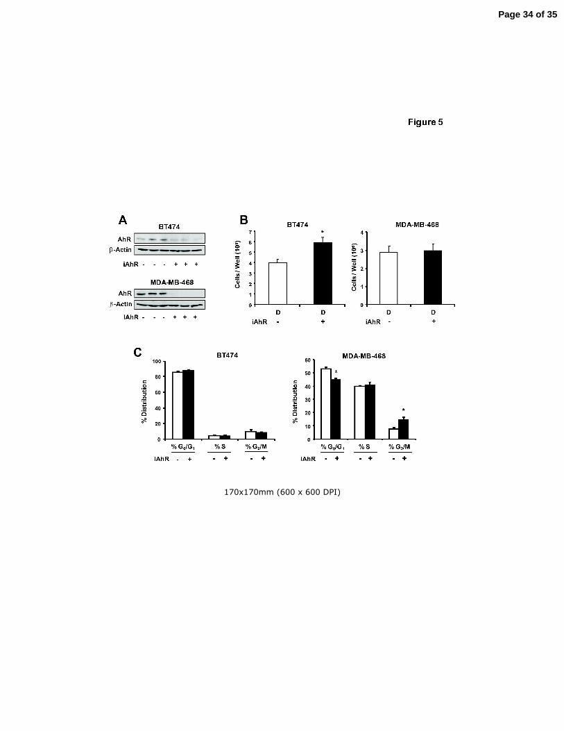
AhR (iAhR). Data in Figure 5A show that transfection with iAhR resulted in a > 80%
decrease in AhR expression in BT474 and MDA-MB-468 cells. Knockdown of the AhR
in BT474 cells resulted in a significant increase in cell proliferation compared to cells
transfected with iCtr (Fig. 5B). This indicated that in BT474 cells, basal expression of
the AhR inhibited cell proliferation, and similar results were previously reported in ER-
positive MCF-7 breast cancer cells (Abdelrahim et al. 2003). In contrast, a comparison
of cell numbers in MDA-MB-468 cells transfected with iCtr or iAhR treated with DMSO
indicated that basal expression of the AhR did not affect proliferation of this cell line
(Fig. 5B). The effects of AhR knockdown on distribution of BT474 and MDA-MB-468
cells in G0/G1, S and G2/M phases of the cell cycle were also determined (Fig. 5C). No
significant effects were observed in BT474 cells, whereas AhR knockdown in MDA-MB-
468 cells decreased cells in G0/G1 and induced a G2/M arrest.
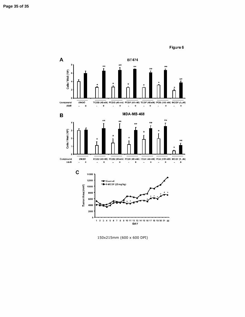
Due to the temporal limitations in AhR knockdown by RNA interference, we used
higher concentrations of AhR agonists in the short term inhibition of cell proliferation
study summarized in Figures 6A and 6B. Treatment of BT474 cells with 5 µM MCDF,
40 µM TCDD, 40 µM PCDD, 40 µM PCDF, 40 µM TCDF and 100 µM PCB all
significantly decreased BT474 cell proliferation. In contrast, after transfection with iAhR,
the antiproliferative effects of the AhR agonists were significantly inhibited and the
Page 12 of 35
Endocrine-Related Cancer 13
chlorinated aromatics (TCDD, PCDD, TCDF, PCDF and PCB) did not significantly
inhibit growth of BT474 cells compared to the solvent (DMSO) control. MCDF partially
inhibited BT474 cell growth, even after AhR knockdown, suggesting that the growth
inhibitory effects of this compound were both AhR-dependent and AhR-independent.
The effects of iAhR on MDA-MBA-468 cell proliferation after treatment with the same
set of compounds showed that the AhR agonist-dependent inhibition of growth was
blocked after AhR knockdown by RNA interference (Fig. 6B). Moreover, the results
obtained for MCDF were similar in MDA-MB-468 and BT474 cells, indicating an AhR-
dependent and AhR-independent mechanism of action for this compound in both cell
lines. Results in Figure 6C demonstrate that MCDF (25 mg/kg every second day) also
inhibited growth of tumors in athymic nude mice bearing MDA-MB-468 cells injected
directly into the mammary fat pad. Tumors derived from MDA-MB-468 cells grew slowly
and consistent differences in tumor area between control and MCDF treatment groups
were not observed until day 16 and significant (p < 0.05) differences were observed
from days 16 – 22. Treatment with MCDF did not significantly affect body, liver, uterine,
heart, spleen or kidney weight or expression of hepatic CYP1A1 (data not shown).
These results demonstrate the potential clinical applications of SAhRMs for treatment of
ER-negative breast cancer.
Page 13 of 35
Endocrine-Related Cancer 14
DISCUSSION
Breast cancer is a highly complex disease in which treatment options depend on
the staging of the tumor, localization or spreading of the tumor, and the molecular
characteristics of the tumor including its estrogen receptor status or expression of other
genes such as the ErbB2 (HER2/neu) oncogene (Moulder & Hortobagyi 2008; Buzdar
2003; Macaskill & Dixon 2007). Many early stage mammary tumors are ER-positive
and have been successfully treated with antiestrogens such as tamoxifen, raloxifene,
fulvestrant or aromatase inhibitors (Fisher et al. 2005; Vogel et al. 2006; Semiglazov et
al. 2007; Howell et al. 2005). Prolonged use of tamoxifen can result in development of
drug-resistant tumors and there is evidence that long term use of tamoxifen increases
the risk for endometrial cancer (Vogel et al. 2006; Clarke et al. 2001). Some early stage
and most later stage mammary tumors are ER-negative and patients with ER-negative
breast cancer do not respond well to endocrine therapy and successful adjuvant
chemotherapy requires the use of more highly cytotoxic drugs commonly used to treat
other endocrine-independent tumors (Semiglazov et al. 2007; Moulder & Hortobagyi
2008). These agents generally target some aspect of nuclear function or modulate
microtubule formation/breakdown and include compounds such as adriamycin,
cyclophosphamide, gemcitabine, taxanes (taxol and taxotere), and capecitabine, a
precursor of 5-FU (Moulder & Hortobagyi 2008). More recently, there has been an
increase in the applications and development of more targeted therapies that include
antibodies that interact with the angiogenic factor vascular endothelial growth factor
(VEGF). In addition, tyrosine kinase inhibitors that target VEGF receptor and growth
factor receptors have also been developed for clinical treatment of breast cancer
Page 14 of 35

Endocrine-Related Cancer 15
(Moulder & Hortobagyi 2008; Buzdar 2003; Macaskill & Dixon 2007; Hobday & Perez
2005; Demonty et al. 2007). Another important advance for breast cancer treatment
has been the increased use of combined agents which often target different pathways
responsible for tumor survival, growth, angiogenesis and metastasis. Herceptin or
trastuzumab is a monoclonal antibody directed against the extracellular domain of
ErbB2 and objective response rates of 25-40% are observed with this antibody in
patients that overexpress ErbB2 (Demonty et al. 2007).
Drugs such as MCDF that target the AhR are highly effective for inhibition of E2-
responsive tumor growth in carcinogen-induced female Sprague Dawley rats, and
MCDF and tamoxifen in combination synergistically blocked tumor formation and growth
(McDougal et al. 2001). Although the AhR is widely expressed in ER-negative and ER-
positive breast cancer cell lines (Wang et al. 1997; Wang et al. 1995), the potential
applications of AhR agonists for treatment of ER-negative breast cancer is not well
defined. One study in ER-negative MDA-MB-468 cells showed that TCDD inhibited
survival of these cells through the induction of TGFα which exhibits antiproliferative
activity in this cell line (Wang et al. 1997). Figure 1 shows that in addition to MDA-MB-
468 cells, at least six other ER-negative breast cancer cell lines including two that
overexpress ErbB2 (BT474 and MDA-MB-468 cells) were Ah-responsive, and TCDD
and five structurally related chlorinated aromatics induced CYP1A1-dependent EROD
activity. In this study, TCDD did not induce EROD activity in BT20 and MDA-MB-134
cells, and the reasons for the lack of Ah-responsiveness in these cells lines are
currently being investigated. We also examined the comparative effects of TCDD and
Page 15 of 35
Endocrine-Related Cancer 16
MCDF on survival of this panel of ER-negative breast cancer cells (Figs. 2 and 3) and
both compounds significantly decreased growth of the six Ah-responsive cell lines.
The AhR interacts with structurally diverse ligands including synthetic aromatics,
phytochemicals such as flavonoids and indole derivatives, drugs, pesticides,
endogenous biochemicals including bilirubin, and other polyaromatics (Denison & Nagy
2003). The structure-dependent potencies of chlorinated aromatics such as TCDD,
TCDF, PCDF, PCDD and PCBs as AhR agonists has been extensively investigated
(Van den Berg et al. 2006) and for some responses such as induction of CYP1A1, there
is a rank order correlation between their structure-AhR binding versus structure-activity
relationships. Results in Figures 4A and 4B show that the chlorinated aromatics and
MCDF induced luciferase activity in BT474 and MDA-MB-468 cells transfected with an
Ah-responsive DRE-luciferase construct. Moreover, treatment of the two cell lines with
the same set of compounds also induced CYP1A1 protein and western blot analysis of
whole cell lysates also showed that the AhR was expressed in BT474 and MDA-MB-468
cells (Fig. 4C). In previous studies with ErbB2-overexpressing BT474 and MDA-MB-
453 cells, we also showed that TCDD and MCDF inhibited cell proliferation but did not
affect ErbB2 or its phosphorylation, and downstream kinases were also unchanged
(unpublished results). However, results of the CYP1A1 induction studies coupled with
the structure-dependent effects of the chlorinated aromatics and MCDF on decreased
BT474 and MDA-MB-468 cell proliferation (Fig. 4D) are consistent with a role for the
AhR in mediating the effects of these compounds.
Endogenous expression of the AhR in cancer cell lines can affect cell growth
(Abdelrahim et al. 2003). Knockdown of the AhR in ER-positive MCF-7 breast cancer
Page 16 of 35

Endocrine-Related Cancer 17
cells enhanced cell proliferation, whereas in HepG2 liver cancer cells, AhR knockdown
decreased the rate of cell growth (Abdelrahim et al. 2003). In this study, iAhR
transfection in BT474 cells resulted in enhanced growth; however, this was not
accompanied by changes in the % distribution of cells in G0/G1, S or G2/M phases (Fig.
5C). Moreover, AhR agonists did not affect expression of ErbB2, phospho-ErbB2 or
downstream kinases (data not shown), and we are currently investigating how the AhR
and AhR agonists inhibit BT474 cell proliferation without changing the distribution of
cells in G0/G1, S and G2/M phases of the cell cycle. In contrast to BT474 cells, no
significant changes in proliferation were observed in MDA-MB-468 cells transfected with
iAhR (Fig. 5B) but these cells exhibited a decrease in G0/G1 and an arrest at G2/M.
Thus, the AhR differentially affects proliferation and % distribution of ER-negative breast
cancer cells in G0/G1, S or G2/M phases of the cycle, and current studies are
investigating the cell context-dependent modulation of Ah-responsive genes, proteins
and microRNAs that determine these responses. The growth inhibitory effects of the
chlorinated aromatic compounds in BT474 and MDA-MB-468 cells (Figs. 4 and 6) were
reversed in both cell lines after transfection with iAhR (Figs. 6A and 6B) and this was
consistent with the role of the ligand-activated AhR in mediating the decreased
proliferation of ER-negative breast cancer cell.
MCDF also decreased breast cancer cell survival and inhibited tumor growth in
athymic nude mice bearing MDA-MB-468 cells as xenografts (Fig. 6). These data
complement previous studies showing the effectiveness of this compound as a
mammary tumor growth inhibitor in carcinogen-induced female Sprague-Dawley rats
(McDougal et al. 2001). MCDF was initially characterized as an AhR antagonist
Page 17 of 35

Endocrine-Related Cancer 18
(McDougal et al. 2001) and studies with 125I-MCDF showed that this compound bound
the AhR and induced formation of a nuclear AhR complex in cancer cells (Piskorska-
Pliszczynska et al. 1991). However, results of RNA interference studies with iAhR
(Figs. 6A and 6B) demonstrate that loss of the AhR only partially reversed the
antiproliferative effects of MCDF on BT474 and MDA-MB-468 cells. Thus, the
anticancer activity of MCDF in ER-negative breast cancer cells is both AhR-dependent
and AhR-independent, and current studies are focused on the molecular mechanisms
associated with both pathways and application of MCDF and other SAhRMs for
treatment of ER-negative breast cancer.
Declaration of Interest: The authors declare that there are no conflicts of interest.
Funding: The financial assistance of the Dow Chemical Company, the National
Institutes of Health (ES04917), and Texas A&M AgriLife is gratefully
acknowledged.
Page 18 of 35
Endocrine-Related Cancer 19
REFERENCES
Abdelrahim M, Smith III R & Safe S 2003 Aryl hydrocarbon receptor gene silencing with
small inhibitory RNA differentially modulates Ah-responsiveness in MCF-7 and
HepG2 cancer cells. Mol. Pharmacol. 63 1373-1381.
Astroff B, Zacharewski T, Safe S, Arlotto MP, Parkinson A, Thomas P & Levin W 1988
6-Methyl-1,3,8-trichlorodibenzofuran as a 2,3,7,8- tetrachlorodibenzo-p-dioxin
antagonist: inhibition of the induction of rat cytochrome P-450 isozymes and
related monooxygenase activities. Mol. Pharmacol. 33 231-236.
Bannister R, Biegel L, Davis D, Astroff B & Safe S 1989 6-Methyl-1,3,8-
trichlorodibenzofuran (MCDF) as a 2,3,7,8- tetrachlorodibenzo-p-dioxin
antagonist in C57BL/6 mice. Toxicology 54 139-150.
Buzdar AU 2003 Advances in endocrine treatments for postmenopausal women with
metastatic and early breast cancer. Oncologist. 8 335-341.
Castro-Rivera E, Wormke M & Safe S 1999 Estrogen and aryl hydrocarbon
responsiveness of ECC-1 endometrial cancer cells. Mol. Cell. Endocrinol. 150
11-21.
Clarke R, Leonessa F, Welch JN & Skaar TC 2001 Cellular and molecular
pharmacology of antiestrogen action and resistance. Pharmacol. Rev. 53 25-71.
Demonty G, Bernard-Marty C, Puglisi F, Mancini I & Piccart M 2007 Progress and new
standards of care in the management of HER-2 positive breast cancer. Eur. J.
Cancer 43 497-509.
Page 19 of 35

Endocrine-Related Cancer 20
Denison MS & Nagy SR 2003 Activation of the aryl hydrocarbon receptor by structurally
diverse exogenous and endogenous chemicals. Annu. Rev. Pharmacol. Toxicol.
43 309-334.
Fisher B, Costantino JP, Wickerham DL, Cecchini RS, Cronin WM, Robidoux A, Bevers
TB, Kavanah MT, Atkins JN, Margolese RG, Runowicz CD, James JM, Ford LG
& Wolmark N 2005 Tamoxifen for the prevention of breast cancer: current status
of the National Surgical Adjuvant Breast and Bowel Project P-1 study. J. Natl.
Cancer Inst. 97 1652-1662.
Gonzalez FJ & Fernandez-Salguero P 1998 The aryl hydrocarbon receptor: studies
using the AHR-null mice. Drug Metab Dispos. 26 1194-1198.
Harris M, Zacharewski T, Astroff B & Safe S 1989 Partial antagonism of 2,3,7,8-
tetrachlorodibenzo-p-dioxin-mediated induction of aryl hydrocarbon hydroxylase
by 6-methyl-1,3,8-trichlorodibenzofuran: mechanistic studies. Mol. Pharmacol. 35
729-735.
Hobday TJ & Perez EA 2005 Molecularly targeted therapies for breast cancer. Cancer
Control 12 73-81.
Howell A, Pippen J, Elledge RM, Mauriac L, Vergote I, Jones SE, Come SE, Osborne
CK & Robertson JF 2005 Fulvestrant versus anastrozole for the treatment of
advanced breast carcinoma: a prospectively planned combined survival analysis
of two multicenter trials. Cancer 104 236-239.
Jana NR, Sarkar S, Ishizuka M, Yonemoto J, Tohyama C & Sone H 2000 Comparative
effects of 2,3,7,8-tetrachlorodibenzo-p-dioxin on MCF-7, RL95-2, and LNCaP
Page 20 of 35

Endocrine-Related Cancer 21
cells: role of target steroid hormones in cellular responsiveness to CYP1A1
induction. Mol. Cell. Biol. Res. Commun. 4 174-180.
Kimura A, Naka T, Nohara K, Fujii-Kuriyama Y & Kishimoto T 2008 Aryl hydrocarbon
receptor regulates Stat1 activation and participates in the development of Th17
cells. Proc. Natl. Acad. Sci. U. S. A 105 9721-9726.
Kociba RJ, Keyes DG, Beger JE, Carreon RM, Wade CE, Dittenber DA, Kalnins RP,
Frauson LE, Park CL, Barnard SD, Hummel RA & Humiston CG 1978 Results of
a 2-year chronic toxicity and oncogenicity study of 2,3,7,8- tetrachlorodibenzo-p-
dioxin (TCDD) in rats. Toxicol. Appl. Pharmacol. 46 279-303.
Koliopanus A, Kleeff J, Xiao Y, Safe S, Zimmerman A, Buchler MW & Friess H 2002
Increased aryl hydrocarbon receptor expression offers a potential therapeutic
target in pancreatic cancer. Oncogene 21 6059-6070.
Lawrence BP, Denison MS, Novak H, Vorderstrasse BA, Harrer N, Neruda W, Reichel
C & Woisetschlager M 2008 Activation of the aryl hydrocarbon receptor is
essential for mediating the anti-inflammatory effects of a novel low-molecular-
weight compound. Blood 112 1158-1165.
Macaskill EJ & Dixon JM 2007 Neoadjuvant use of endocrine therapy in breast cancer.
Breast J. 13 243-250.
McDougal A, Wilson C & Safe S 1997 Inhibition of 7,12-dimethylbenz[a]anthracene-
induced rat mammary tumor growth by aryl hydrocarbon receptor agonists.
Cancer Lett. 120 53-63.
Page 21 of 35

Endocrine-Related Cancer 22
McDougal A, Wormke M, Calvin J & Safe S 2001 Tamoxifen-induced
antitumorigenic/antiestrogenic action synergized by a selective Ah receptor
modulator. Cancer Res. 61 3901-3907.
Morrow D, Qin C, Smith III R & Safe S 2004 Aryl hydrocarbon receptor-mediated
inhibition of LNCaP prostate cancer cell growth and hormone-induced
transactivation. J. Steroid Biochem. Mol. Biol. 88 27-36.
Moulder S & Hortobagyi GN 2008 Advances in the treatment of breast cancer. Clin.
Pharmacol. Ther. 83 26-36.
Ohtake F, Baba A, Takada I, Okada M, Iwasaki K, Miki H, Takahashi S, Kouzmenko A,
Nohara K, Chiba T, Fujii-Kuriyama Y & Kato S 2007 Dioxin receptor is a ligand-
dependent E3 ubiquitin ligase. Nature 446 562-566.
Piskorska-Pliszczynska J, Astroff B, Zacharewski T, Harris M, Rosengren R, Morrison
V, Safe L & Safe S 1991 Mechanism of action of 2,3,7,8-tetrachlorodibenzo-p-
dioxin antagonists: characterization of [125I]-6-methyl-8-iodo-1,3-
dichlorodibenzofuran-Ah receptor complexes. Arch. Biochem. Biophys. 284 193-
200.
Poland A, Glover E & Kende AS 1976 Stereospecific, high affinity binding of 2,3,7,8-
tetrachlorodibenzo-p-dioxin by hepatic cytosol: evidence that the binding species
is receptor for induction of aryl hydrocarbon hydroxylase. J. Biol. Chem. 251
4936-4946.
Poland A & Knutson JC 1982 2,3,7,8-Tetrachlorodibenzo-p-dioxin and related
halogenated aromatic hydrocarbons. Examinations of the mechanism of toxicity.
Annu. Rev. Pharmacol. Toxicol. 22 517-554.
Page 22 of 35
Endocrine-Related Cancer 23
Quintana FJ, Basso AS, Iglesias AH, Korn T, Farez MF, Bettelli E, Caccamo M, Oukka
M & Weiner HL 2008 Control of Treg and TH17 cell differentiation by the aryl
hydrocarbon receptor. Nature 453 65-71.
Safe S 2005 2,3,7,8-Tetrachlorodibenzo-p-dioxin (TCDD) and related environmental
antiestrogens: characterization and mechanism of action. In Endocrine
Disruptors, edn 2nd, pp 249-287. Ed RK Naz. Boca Raton, FL: CRC Press.
Safe S & McDougal A 2002 Mechanism of action and development of selective aryl
hydrocarbon receptor modulators for treatment of hormone-dependent cancers.
Int. J. Oncol. 20 1123-1228.
Safe S, Qin C & McDougal A 1999 Development of selective aryl hydrocarbon receptor
modulators (SARMs) for treatment of breast cancer. Expert Opinion on
Investigational Drugs 8 1385-1396.
Safe S & Wormke M 2003 Inhibitory aryl hydrocarbon-estrogen receptor α cross-talk
and mechanisms of action. Chem. Res. Toxicol. 16 807-816.
Schmidt JV, Su GH, Reddy JK, Simon MC & Bradfield CA 1996 Characterization of a
murine Ahr null allele: involvement of the Ah receptor in hepatic growth and
development. Proc. Natl. Acad. Sci. U. S. A. 93 6731-6736.
Semiglazov VF, Semiglazov VV, Dashyan GA, Ziltsova EK, Ivanov VG, Bozhok AA,
Melnikova OA, Paltuev RM, Kletzel A & Berstein LM 2007 Phase 2 randomized
trial of primary endocrine therapy versus chemotherapy in postmenopausal
patients with estrogen receptor-positive breast cancer. Cancer 110 244-254.
Van den Berg M, Birnbaum LS, Denison M, DeVito M, Farland W, Feeley M, Fiedler H,
Hakansson H, Hanberg A, Haws L, Rose M, Safe S, Schrenk D, Tohyama C,
Page 23 of 35
Endocrine-Related Cancer 24
Tritscher A, Tuomisto J, Tysklind M, Walker N & Peterson RE 2006 The 2005
World Health Organization reevaluation of human and mammalian toxic
equivalency factors for dioxins and dioxin-like compounds. Toxicol. Sci. 93 223-
241.
Veldhoen M, Hirota K, Westendorf AM, Buer J, Dumoutier L, Renauld JC & Stockinger
B 2008 The aryl hydrocarbon receptor links TH17-cell-mediated autoimmunity to
environmental toxins. Nature 453 106-109.
Vogel VG, Costantino JP, Wickerham DL, Cronin WM, Cecchini RS, Atkins JN, Bevers
TB, Fehrenbacher L, Pajon ER, Jr., Wade JL, III, Robidoux A, Margolese RG,
James J, Lippman SM, Runowicz CD, Ganz PA, Reis SE, Caskill-Stevens W,
Ford LG, Jordan VC & Wolmark N 2006 Effects of tamoxifen vs raloxifene on the
risk of developing invasive breast cancer and other disease outcomes: the
NSABP Study of Tamoxifen and Raloxifene (STAR) P-2 trial. JAMA 295 2727-
2741.
Wang W, Porter W, Burghardt R & Safe S 1997 Mechanism of inhibition of MDA-MB-
468 breast cancer cell growth by 2,3,7,8-tetrachlorodibenzo-p-dioxin.
Carcinogenesis 18 925-933.
Wang X, Thomsen JS, Santostefano M, Rosengren R, Safe S & Perdew GH 1995
Comparative properties of the nuclear Ah receptor complex from several human
cell lines. Eur. J. Pharmacol. 293 191-205.
Whitlock JP, Jr. 1993 Mechanistic aspects of dioxin action. Chem. Res. Toxicol. 6 754-
763.
Page 24 of 35

Endocrine-Related Cancer 25
Whitlock JP, Okino ST, Dong L, Ko HP, Clarke-Katzenberg R, Ma Q & Li H 1996
Induction of cytochrome P4501A1: a model for analyzing mammalian gene
transcription. FASEB J. 10 809-818.
Wormke M, Castro-Rivera E, Chen I & Safe S 2000 Estrogen and aryl hydrocarbon
receptor expression and crosstalk in human Ishikawa endometrial cancer cells. J.
Steroid Biochem. Mol. Biol. 72 197-207.
Wormke M, Stoner M, Saville B, Walker K, Abdelrahim M, Burghardt R & Safe S 2003
The aryl hydrocarbon receptor mediates degradation of the estrogen receptor α
through activation of proteasomes. Mol. Cell. Biol. 23 1843-1855.
Yao C & Safe S 1989 2,3,7,8-Tetrachlorodibenzo-p-dioxin-induced porphyria in
genetically inbred mice: partial antagonism and mechanistic studies. Toxicol.
Appl. Pharmacol. 100 208-216.
Page 25 of 35

Endocrine-Related Cancer 26
FIGURE CAPTIONS
Figure 1. Ah-responsiveness of ER-negative breast cancer cells. Induction of EROD
activity by TCDD. ER-negative breast cancer cells were treated with DMSO or
different concentrations of TCDD and EROD activity was determined as
described in the Materials and Methods. Results are expressed as means ± SE
for 3 replicate determinations for each treatment group and significant (p < 0.05)
induction is indicated (*).
Figure 2. Antiproliferative activity of TCDD. Inhibition of ER-negative breast cancer cell
growth. ER-negative breast cancer cell lines were treated with DMSO or
different concentrations of TCDD for six days and cells were counted as
described in the Materials and Methods. Results are expressed as means ± SE
for at least 3 replicate determinations for each treatment group and significant (p
< 0.05) inhibition is indicated (*).
Figure 3. Antiproliferative activity of MCDF. Inhibition of ER-negative breast cancer cell
growth. ER-negative breast cancer cell lines were treated with DMSO or
different concentrations of MCDF for six days and cells were counted as
described in the Materials and Methods. Results are expressed as means ± SE
for 3 replicate determinations for each treatment group and significant (p < 0.05)
of cell proliferation is indicated (*).
Figure 4. Structure-dependent activation of AhR-dependent responses by chlorinated
aromatics in BT474 and MDA-MB-468 cells. Activation of DRE-luc in BT474 (A)
and MDA-MB-468 cells (B). Cells were transfected with the DRE-luc construct
and treated with DMSO or different concentrations of TCDD, PCDD, TCDF,
Page 26 of 35
Endocrine-Related Cancer 27
PCDF and PCB, and luciferase activity determined as described in the Materials
and Methods. Results are expressed as means ± SE for 3 replicate
determinations for each treatment group and significant (p < 0.05) induction is
indicated (*). Structure-dependent induction of CYP1A1 protein and AhR
expression (C) and growth inhibition (D) by AhR agonists. Cells were treated for
either 24 (C) or 96 hr (D) and whole cell lysates were analyzed by western blots
(C) or cells were counted (D) as described in the Materials and Methods.
Results in (D) are presented as means ± SE for at least 3 replicate
determinations for each treatment group and significant (p < 0.05) growth
inhibition is indicated (*).
Figure 5. RNA interference and FACS analysis. Effects of iAhR on AhR protein (A) and
cell proliferation (B). Cells were transfected with iAhR or non-specific
oligonucleotide and the effects on AhR protein and proliferation of BT474 and
MDA-MB-468 cells were determined as described in the Materials and Methods.
Replicate (3) experiments were carried out for each treatment and, for the cell
proliferation studies, results are expressed as means ± SE (after treatment for 96
hr) and significant (p < 0.05) effects of iAhR on cell proliferation are indicated (*).
(C) FACS analysis. The effects of iAhR on distribution of BT474 and MDA-MB-
468 cells in G0/G1, S and G2/M phases of the cell cycle were determined by
FACS analysis as described in the Materials and Methods. Results obtained in
cells transfected with iAhR are compared to cells transfected with a non-specific
oligonucleotide as indicated above in (A) and (B). Results are expressed as
Page 27 of 35

Endocrine-Related Cancer 28
means ± SE for three replicate experiments and significant changes after
transfection with iAhR are indicated (*).
Figure 6. Antiproliferative and antitumorigenic activity of AhR agonists. Role of the AhR
in mediating the antiproliferative effects of AhR agonists in BT474 (A) and MDA-
MB-468 (B) cells. Cells were transfected with non-specific scrambled
oligonucleotide (iCtr) or iAhR and treated with DMSO or different AhR agonists
for 4 days, and the number of cells were counted as described in the Materials
and Methods. Results are expressed as means ± SE for at least 3 replicate
determinations for each treatment group and significant (p < 0.05) inhibition of
cell growth by the AhR agonists (*) and reversal of this effect by iAhR (**) are
indicated. (C) Tumor growth inhibition. MDA-MB-468 cells were injected into
the mammary fat pad of athymic nude mice and after palpable tumors were
detected (7 - 10 days), mice were treated with corn oil (vehicle control) or MCDF
(25 mg/kg) every 48 hr. Tumor volumes were determined as described in the
Materials and Methods. Significant (p < 0.05) inhibition of tumor growth is
indicated by an asterisk. Body weights and liver, uterine, heart, spleen and
kidney weights as % body weight in control/MCDF-treated mice were 26±1/25±1,
5.2±0.3/5.3±0.1, 0.35±0.1/0.35±0.04, 0.46±0.01/0.49±0.01, 0.72±0.1/0.66±0.01
and 0.68±0.03/0.63±0.02, respectively.
Supplemental Figure 1. Ah-responsiveness and ERα expression in BT474 and MCF-7
cells. Cells were treated with DMSO (D), 10 nM TCDD, 5 µM MCDF, 10 µM
diindolylmethane (DIM), and 10 nM 17β-estradiol (E2, MCF-7 cells only) and
Page 28 of 35

Endocrine-Related Cancer 29
after 24 hr, whole cell lysates were analyzed by western blots for ERα, AhR and
CYP1A1 (loading control was β-actin).
Page 29 of 35

161x178mm (600 x 600 DPI)
Page 30 of 35
159x183mm (600 x 600 DPI)
Page 31 of 35
160x176mm (600 x 600 DPI)
Page 32 of 35

196x244mm (600 x 600 DPI)
Page 33 of 35
170x170mm (600 x 600 DPI)
Page 34 of 35
150x215mm (600 x 600 DPI)
Page 35 of 35