sorafenib decreases proliferation and induces...
TRANSCRIPT
Endocrine-Related Cancer (2012) 19 305–319
Sorafenib decreases proliferation andinduces apoptosis of prostate cancercells by inhibition of the androgenreceptor and Akt signaling pathways
Su Jung Oh, Holger H H Erb, Alfred Hobisch1, Frederic R Santer* andZoran Culig*
Division of Experimental Urology, Department of Urology, Innsbruck Medical University, Anichstrasse 35, A-6020 Innsbruck, Austria1Department of Urology, General Hospital Feldkirch, Carinagasse 35, A-6800 Feldkirch, Austria
(Correspondence should be addressed to Z Culig; Email: [email protected]; F R Santer; Email: [email protected])
*(F R Santer and Z Culig joint senior authors)
Abstract
Antihormonal and chemotherapy are standard treatments for nonorgan-confined prostate cancer.The effectivity of these therapies is limited and the development of alternative approaches isnecessary. In the present study, we report on the use of themultikinase inhibitor sorafenib in a panelof prostate cancer cell lines and their derivatives which mimic endocrine and chemotherapyresistance. 3H-thymidine incorporation assays revealed that sorafenib causes a dose-dependentinhibition of proliferation of all cell lines associated with downregulation of cyclin-dependent kinase2 and cyclinD1 expression. Apoptosiswas induced at 2 mMof sorafenib in androgen-sensitive cells,whereas a higher dose of the drug was needed in castration-resistant cell lines. Sorafenibstimulated apoptosis in prostate cancer cell lines through downregulation ofmyeloid cell leukemia-1(MCL-1) expression and Akt phosphorylation. Although concentrations of sorafenib required for theantitumor effect in therapy-resistant sublines were higher than those needed in parental cells, thedrug showed efficacy in cells which became resistant to bicalutamide and docetaxel respectively.Most interestingly, we show that sorafenib has an inhibitory effect on androgen receptor (AR) andprostate-specific antigen expression. In cells in which AR expression was downregulated byshort interfering RNA, the treatment with sorafenib increased apoptosis in an additive manner.In summary, the results of the present study indicate that there is a potential to use sorafenib inprostate cancers as an adjuvant therapy option to current androgen ablation treatments, but also inprogressed prostate cancers that become unresponsive to standard therapies.
Endocrine-Related Cancer (2012) 19 305–319
Introduction
Prostate cancer is the most common malignancy in
Western countries and the second leading cause of
cancer-related deaths in males (Jemal et al. 2010).
Patients diagnosed with localized disease can be cured
by either surgery or radiation therapy. In contrast,
advanced stages of the tumor are subjected to
androgen ablation treatment in order to reduce the
tumor-promoting effect of androgens. Standard
therapy approaches include administration of LH
releasing hormone analogs, nonsteroidal antiandrogens
Endocrine-Related Cancer (2012) 19 305–319
1351–0088/12/019–305 q 2012 Society for Endocrinology Printed in Great
This is an Open Access article distributed under the terms of the Society
commercial use, distribution, and reproduction in any medium, provided the
(e.g. bicalutamide), or surgical castration. However,
androgen-ablated tumors eventually develop resistance
to this therapy and progress toward castration-resistant
prostate cancer (CRPC), for which only palliative
treatment is available. Androgen receptor (AR) was
shown to play a critical role in progression of prostate
cancer (Grossmann et al. 2001). Activated AR interacts
with androgen response elements in the promoters of
target genes including prostate-specific antigen (PSA),
thereby regulating their transcription. PSA is the most
frequently used marker for monitoring response to
Britain
DOI: 10.1530/ERC-11-0298
Online version via http://www.endocrinology-journals.org
for Endocrinology’s Re-use Licence which permits unrestricted non-
original work is properly cited.
S J Oh et al.: Sorafenib and prostate cancer therapy resistance
prostate cancer treatment. Chemotherapy for prostate
cancer has been used for a number of years, however
only limited improvement in survival was observed in
CRPC with docetaxel-based therapies (Tannock et al.
2004). Nevertheless, apart from a relatively short
extension of survival, w50% of patients initially do
not respond to docetaxel treatment and are exposed to
significant toxicity. Therefore, novel targeted
approaches are in need to optimize the currently
available therapies for patients with androgen-sensitive
and CRPC.
One aim of therapies for various cancers including
that of the prostate is to increase the percentage of
tumor cells undergoing apoptosis. Increased expression
of endogenous inhibitors of programmed cell death is
one of the reasons for the development of therapy
resistance. One of these inhibitors is myeloid cell
leukemia-1 (MCL-1), an antiapoptotic member of the
Bcl-2 family, which was originally identified as an
early gene induced during differentiation of ML-1
myeloid leukemia cells (Kozopas et al. 1993). MCL-1
is overexpressed in various human malignancies and
has been implicated in resistance to anticancer drugs
(Craig 2002). Elevated expression of MCL-1 in
prostate cancer tissue compared to normal or hyper-
plastic tissue or prostate intraepithelial neoplasia
(Krajewska et al. 1996) suggests an involvement of
this protein in tumor initiation and progression.
Previously, we demonstrated the importance of
MCL-1 in mediating the prosurvival activity of
interleukin 6 (IL6) in prostate cancer (Cavarretta
et al. 2007). In view of its active role in protecting
prostate cancer cells from induction of apoptosis
(Cavarretta et al. 2007), targeting MCL-1 could be
considered a valid therapeutic approach.
Another potential therapy target is Akt (protein
kinase B), a serine–threonine protein kinase, which
plays a central role in phosphoinositide-3-kinase-
mediated signaling. Its activation has been implicated
in prostate cancer cell survival as well as in progression
to castration resistance and refractoriness to che-
motherapy (Nesterov et al. 2001). Akt is frequently
activated in advanced prostate cancer due to deletion or
mutation of the PTEN tumor suppressor gene (Sircar
et al. 2009). In clinical and preclinical studies,
overexpression and activation of Akt have been
associated with high preoperative levels of PSA,
higher Gleason grades, shorter relapses, and resistance
to treatment (Sircar et al. 2009). Activated Akt
phosphorylates and thereby inactivates its downstream
target glycogen synthase kinase-3b (GSK-3b). Conse-
quently, GSK-3b-mediated phosphorylation of MCL-1
promotes its binding to the E3 ligase b-TrCP and
306
degradation of MCL-1 by the proteasome (Ding et al.
2007). Furthermore, it has recently been reported that
Akt activity can positively regulate AR protein levels
(Ha et al. 2011).
Sorafenib (Nexavar, BAY 43-9006) is an oral
multikinase inhibitor that was initially developed in
an attempt to block Raf kinase, a well-studied serine–
threonine kinase regulating cell survival (Wilhelm
et al. 2004). It was revealed that sorafenib also targets a
number of receptor tyrosine kinases involved in
neoangiogenesis including vascular endothelial growth
factor receptor, platelet-derived growth factor receptor,
FLT3, Ret, and c-Kit (Wilhelm et al. 2004). Moreover,
sorafenib was found to induce apoptosis in several
human cancer cell lines by downregulating the
expression levels of MCL-1 (Rahmani et al. 2005).
Sorafenib has shown promising preclinical activity
against a variety of tumor types and is approved for the
treatment of hepatocellular and renal cell carcinoma
(Kane et al. 2006, Lang 2008). In prostate cancer, it
was shown that sorafenib treatment has a positive
outcome in clinical studies in combination with
antiangiogenic agents in CRPC (Steinbild et al. 2007,
Chi et al. 2008, Dahut et al. 2008). Although sorafenib
is undergoing phase II clinical evaluation for treatment
of prostate cancer, molecular events following inhi-
bition of its targets and regulation of the apoptotic
pathways have not been studied systematically. We
also hypothesized that sorafenib has a potential in the
treatment of endocrine- and chemotherapy-resistant
prostate cancer.
In this study, we demonstrate that sorafenib exerts
antiproliferative and proapoptotic activities in human
prostate cancer cells by targeting several regulators of
cell cycle progression and survival. We also evaluated
the antitumor efficacy of sorafenib in bicalutamide-
and docetaxel-resistant cell lines in order to test the
anticancer potential of sorafenib in therapy-resistant
prostate cancer.
Materials and methods
Cell lines
Prostate cancer cells PC3, LNCaP, and 22Rv1 were
obtained from ATCC (Rockville, MD, USA). Cell line
authenticity was confirmed by short tandem repeat
analysis. The LNCaP subline LNCaP-IL6C was
derived in the presence of IL6, as described elsewhere
(Hobisch et al. 2001). The therapy-resistant model
LNCaP-Bic was obtained by long-term treatment of
LNCaP cells with 10 pM R1881 and 1 mM bicaluta-
mide (Hobisch et al. 2006). The LNCaP-abl subline
www.endocrinology-journals.org
Endocrine-Related Cancer (2012) 19 305–319
was described previously (Culig et al. 1999). PC3-DR
cells were established by continuously treating PC3
cells in a dose escalation manner with docetaxel until
reaching a concentration of 12.5 nM in analogy to
Patterson et al. (2006). PC3 cells were cultured in
RPMI 1640 containing 10% FCS, 1% antibiotics, and
glutaMax. For LNCaP and 22Rv1 cell lines, media
were additionally supplemented with 1 mM sodium
pyruvate, 4.5 g/l glucose, and 10 mM HEPES buffer
(pH 7.2). LNCaP-IL6C cells were maintained in the
presence of 5 ng/ml of IL6. PC3-DR cells were
cultured in RPMI 1640 containing 10% FCS, 1%
antibiotics, and glutaMax supplemented with 12.5 nM
docetaxel. All treatments with sorafenib were per-
formed for 48 h in modified HITES medium (RPMI
medium supplemented with 10 nM hydrocortisone,
10 nM estradiol, and 1! insulin–transferrin–selenium
(Life Technologies, Vienna, Austria)).
Chemicals and plasmids
Sorafenib tosylate (BAY 43-9006) was provided
by Bayer and dissolved in dimethylsulfoxide
(DMSO) to a stock concentration of 10 mM. Bicalu-
tamide (Casodex was kindly provided by Astrazeneca
(Macclesfield, UK) and dissolved in DMSO to a stock
concentration of 10 mM. Controls were treated with
the corresponding volume of the vehicle. The MCL-1
expression vector was purchased from OriGene (Rock-
ville, MD, USA).
Proliferation assays
LNCaP-IL6C, LNCaP-Bic, PC3, and PC3-DR cells
were seeded at a density of 6!103 per well, and LNCaP
and 22Rv1 cells were seeded at a density of 1!104 per
well in triplicates onto 96 well plates. Plates for LNCaP
cells were previously coated with poly-D-lysine hydro-
bromide (30 mg/ml; Sigma–Aldrich). On the next day,
the cells were treated with increasing concentrations of
sorafenib (0–2 mM) alone or in combination with
docetaxel or bicalutamide for 48 h in modified HITES
medium. The cells were incubated for the last 16 h of
treatment with 37 kBq/well 3H-thymidine and DNA
was measured as described before (Puhr et al. 2010).
Western blotting
Western blot analysis was performed as described
previously (Cavarretta et al. 2007). The following
antibodies were used for western blots: anti-MCL-1
(1:500; Santa Cruz Biotechnology, Santa Cruz, CA,
USA), anti-glyceraldehyde-3-phosphate dehydro-
genase (GAPDH; 1:100 000; Chemicon International
www.endocrinology-journals.org
Inc., Billerica, MA, USA), anti-phospho Akt (S473;
1:1000; Cell Signaling Technology, Danvers, MA,
USA), anti-Akt (1:1000; Cell Signaling Technology),
anti-phospho GSK-3b (S9; 1:500; Cell Signaling
Technology), anti-GSK-3b (1:1000; Cell Signaling
Technology), anti-AR (1:500; Santa Cruz Bio-
technology), anti-cyclin-dependent kinase 2 (CDK2;
1:1000; Santa Cruz Biotechnology), and anti-cyclin D1
(1:1000; Neomarkers Inc., Fremont, CA, USA).
Short interfering RNA transfection
LNCaP and 22Rv1 cells were plated at low density in
the presence of 10% FCS onto six well tissue culture
plates previously coated with poly-D-lysine hydro-
bromide (30 mg/ml, for experiments with LNCaP cells;
Sigma–Aldrich). One day later, the cells were
transfected using Lipofectamine 2000 in serum- and
antibiotics-free medium with 10 nM ligand-binding
domain (LBD) short interfering RNA (siRNA) accor-
ding to the manufacturer’s protocol (Invitrogen). The
target sequence for AR LBD was published previously
(Desiniotis et al. 2010). A nontargeting siRNA pool
was used as a negative control and purchased from
Dharmacon (Lafayette, CO, USA). Six hours after
transfection, medium was changed to full growth
conditions for overnight. On the next day, treatment
with sorafenib (2 mM) was performed for 48 h in
serum-free HITES medium. Cells were harvested for
western blot analysis and caspase 3/7 activity assay.
Apoptosis assay
Cells were seeded onto six wells and treated with
sorafenib (0–4 mM) alone or in combination as described
above. After 48 h, the cells were harvested and
centrifuged. Apoptosis was measured by using the PE
Annexin V Apoptosis Detection Kit I in combination
with flow cytometry (Becton Dickinson, Schwechat,
Austria) according to the manufacturer’s protocols.
Assays for caspase 3/7 activity were performed with
the Caspase-Glo 3/7 assay kit (Promega) according to the
manufacturer’s protocols (Santer et al. 2011).
PSA measurements
Supernatants of LNCaP and LNCaP-Bic cells after the
treatment with sorafenib or bicalutamide for 48 h were
collected and PSA concentration was determined on an
Advia Centaur XP Immunoassay System (Siemens,
Vienna, Austria). The cells were trypsinized and
counted with a Casy Counter (Scharfe System
GmbH, Reutlingen, Germany). Secreted PSA concen-
trations were normalized to cell number.
307
S J Oh et al.: Sorafenib and prostate cancer therapy resistance
Statistical analysis
Student’s t-test was used to assess significant
differences between the control and the indicated
treated group and was encoded as follows: *P!0.05;
**P!0.01; ***P!0.001.
Results
Sorafenib inhibits proliferation of prostate cancer
cells in a dose-dependent manner and targets
cell cycle control proteins
In the first attempt we analyzed the consequences
of sorafenib treatment on prostate cancer cell
175
A
B
[3 H] T
hym
idin
e in
corp
orat
ion
(% o
f unt
reat
ed c
ells
)P
OI/G
AP
DH
(% o
f con
trol
)
150
150
125
100
100
75
50
25
0
50
0
PO
I/GA
PD
H(%
of c
ontr
ol)
150
100
50
0
0 0.25 0.5 1 1.5
*
**
*
*
***
*
***
LNCaP (*)22Rv1 (#)
###
2
Sorafenib (µM)
0 0.25 0.5 1 2
0 0.25 0.5 1 2
LNCaP
PC3
CDK2
Cyclin D1
GAPDH
C
CDK2
Cyclin D1
GAPDH
Sorafenib (µM)
Sorafenib (µM)
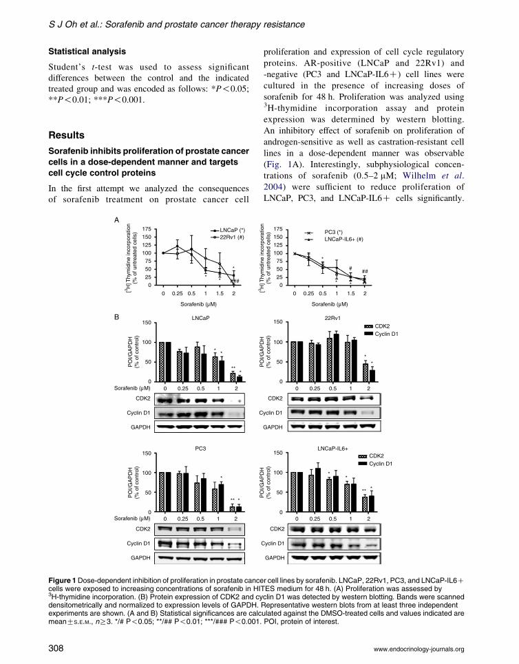
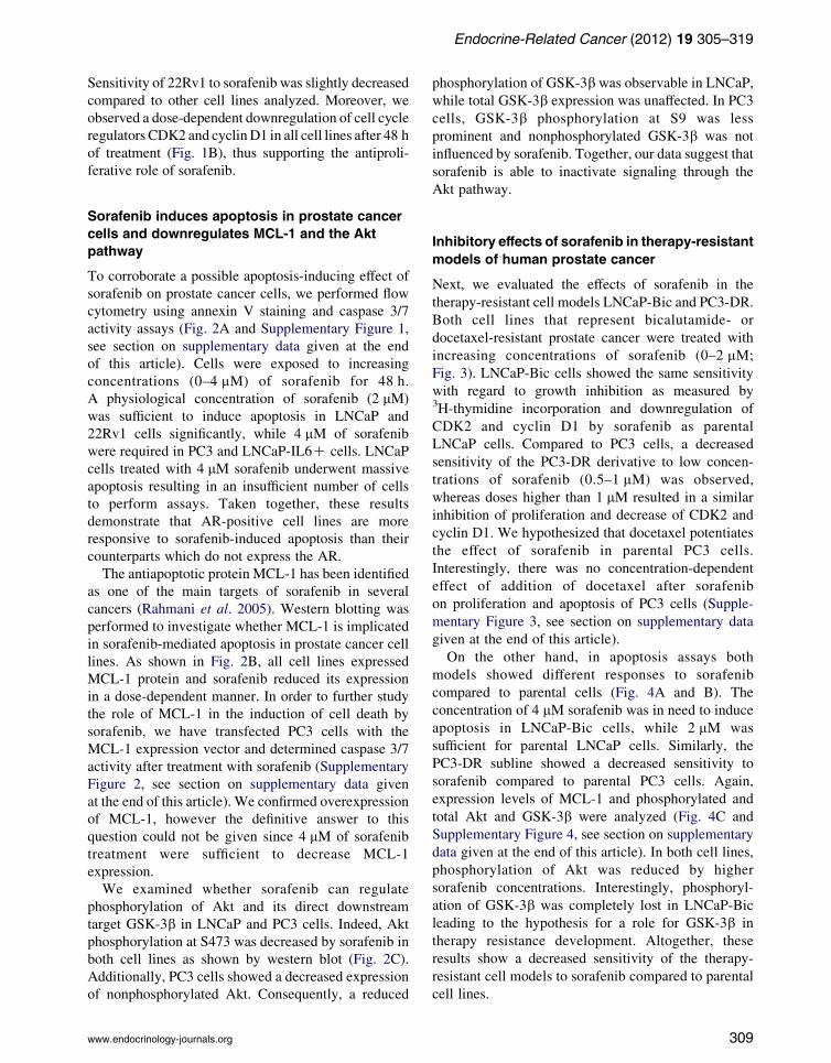
Figure 1Dose-dependent inhibition of proliferation in prostate cancecells were exposed to increasing concentrations of sorafenib in HIT3H-thymidine incorporation. (B) Protein expression of CDK2 and cydensitometrically and normalized to expression levels of GAPDH. Rexperiments are shown. (A and B) Statistical significances are calcmeanGS.E.M., nR3. */# P!0.05; **/## P!0.01; ***/### P!0.001.
308
proliferation and expression of cell cycle regulatory
proteins. AR-positive (LNCaP and 22Rv1) and
-negative (PC3 and LNCaP-IL6C) cell lines were
cultured in the presence of increasing doses of
sorafenib for 48 h. Proliferation was analyzed using3H-thymidine incorporation assay and protein
expression was determined by western blotting.
An inhibitory effect of sorafenib on proliferation of
androgen-sensitive as well as castration-resistant cell
lines in a dose-dependent manner was observable
(Fig. 1A). Interestingly, subphysiological concen-
trations of sorafenib (0.5–2 mM; Wilhelm et al.
2004) were sufficient to reduce proliferation of
LNCaP, PC3, and LNCaP-IL6C cells significantly.
175
[3 H] T
hym
idin
e in
corp
orat
ion
(% o
f unt
reat
ed c
ells
)
150
125
100
75
50
25
0
PO
I/GA
PD
H(%
of c
ontr
ol)
150
100
50
0
PO
I/GA
PD
H(%
of c
ontr
ol)
150
100
50
0
*
* * **
# ##
*
**
**
**
PC3 (*)LNCaP-IL6+ (#)
0 0.25 0.5 1 1.5 2
0 0.25 0.5 1 2
0 0.25 0.5 1 2
Sorafenib (µM)
LNCaP-IL6+
22Rv1
CDK2Cyclin D1
CDK2Cyclin D1
CDK2
yclin D1
GAPDH
CDK2
Cyclin D1
GAPDH
r cell lines by sorafenib. LNCaP, 22Rv1, PC3, and LNCaP-IL6CES medium for 48 h. (A) Proliferation was assessed byclin D1 was detected by western blotting. Bands were scannedepresentative western blots from at least three independent
ulated against the DMSO-treated cells and values indicated arePOI, protein of interest.
www.endocrinology-journals.org
Endocrine-Related Cancer (2012) 19 305–319
Sensitivity of 22Rv1 to sorafenib was slightly decreased
compared to other cell lines analyzed. Moreover, we
observed a dose-dependent downregulation of cell cycle
regulators CDK2 and cyclin D1 in all cell lines after 48 h
of treatment (Fig. 1B), thus supporting the antiproli-
ferative role of sorafenib.
Sorafenib induces apoptosis in prostate cancer
cells and downregulates MCL-1 and the Akt
pathway
To corroborate a possible apoptosis-inducing effect of
sorafenib on prostate cancer cells, we performed flow
cytometry using annexin V staining and caspase 3/7
activity assays (Fig. 2A and Supplementary Figure 1,
see section on supplementary data given at the end
of this article). Cells were exposed to increasing
concentrations (0–4 mM) of sorafenib for 48 h.
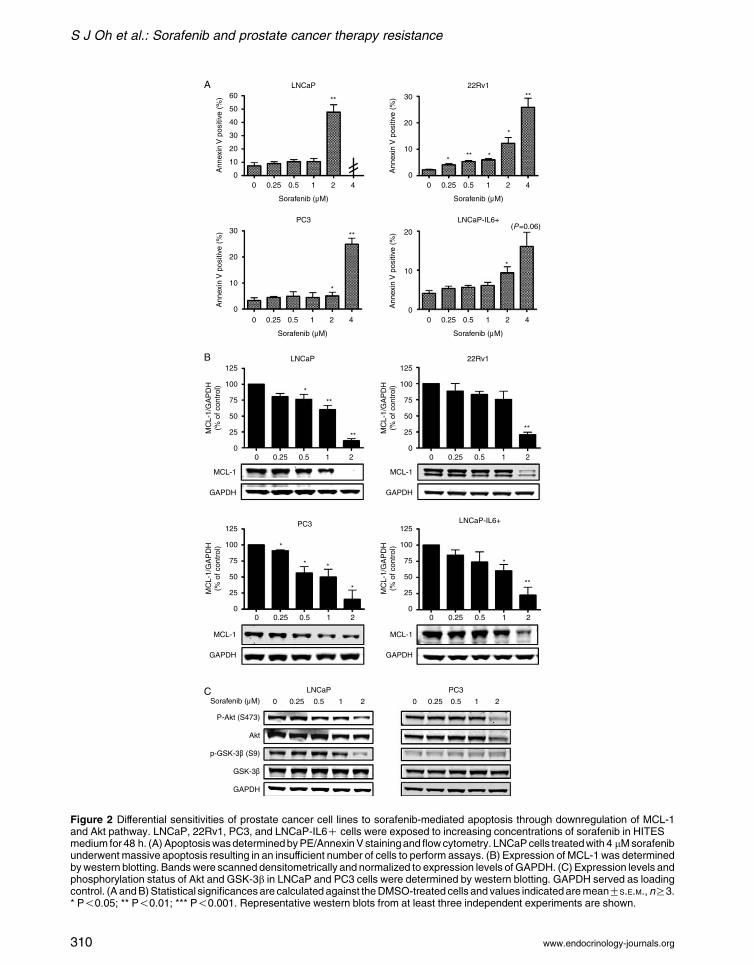
A physiological concentration of sorafenib (2 mM)
was sufficient to induce apoptosis in LNCaP and
22Rv1 cells significantly, while 4 mM of sorafenib
were required in PC3 and LNCaP-IL6C cells. LNCaP
cells treated with 4 mM sorafenib underwent massive
apoptosis resulting in an insufficient number of cells
to perform assays. Taken together, these results
demonstrate that AR-positive cell lines are more
responsive to sorafenib-induced apoptosis than their
counterparts which do not express the AR.
The antiapoptotic protein MCL-1 has been identified
as one of the main targets of sorafenib in several
cancers (Rahmani et al. 2005). Western blotting was
performed to investigate whether MCL-1 is implicated
in sorafenib-mediated apoptosis in prostate cancer cell
lines. As shown in Fig. 2B, all cell lines expressed
MCL-1 protein and sorafenib reduced its expression
in a dose-dependent manner. In order to further study
the role of MCL-1 in the induction of cell death by
sorafenib, we have transfected PC3 cells with the
MCL-1 expression vector and determined caspase 3/7
activity after treatment with sorafenib (Supplementary
Figure 2, see section on supplementary data given
at the end of this article). We confirmed overexpression
of MCL-1, however the definitive answer to this
question could not be given since 4 mM of sorafenib
treatment were sufficient to decrease MCL-1
expression.
We examined whether sorafenib can regulate
phosphorylation of Akt and its direct downstream
target GSK-3b in LNCaP and PC3 cells. Indeed, Akt
phosphorylation at S473 was decreased by sorafenib in
both cell lines as shown by western blot (Fig. 2C).
Additionally, PC3 cells showed a decreased expression
of nonphosphorylated Akt. Consequently, a reduced
www.endocrinology-journals.org
phosphorylation of GSK-3b was observable in LNCaP,
while total GSK-3b expression was unaffected. In PC3
cells, GSK-3b phosphorylation at S9 was less
prominent and nonphosphorylated GSK-3b was not
influenced by sorafenib. Together, our data suggest that
sorafenib is able to inactivate signaling through the
Akt pathway.
Inhibitory effects of sorafenib in therapy-resistant
models of human prostate cancer
Next, we evaluated the effects of sorafenib in the
therapy-resistant cell models LNCaP-Bic and PC3-DR.
Both cell lines that represent bicalutamide- or
docetaxel-resistant prostate cancer were treated with
increasing concentrations of sorafenib (0–2 mM;
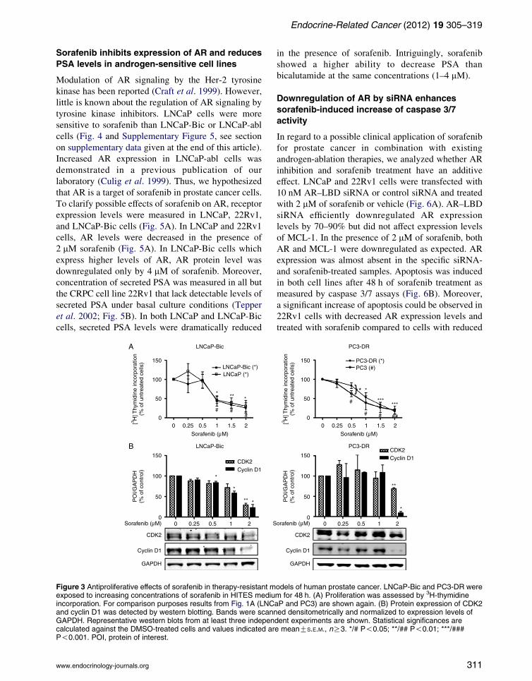
Fig. 3). LNCaP-Bic cells showed the same sensitivity
with regard to growth inhibition as measured by3H-thymidine incorporation and downregulation of
CDK2 and cyclin D1 by sorafenib as parental
LNCaP cells. Compared to PC3 cells, a decreased
sensitivity of the PC3-DR derivative to low concen-
trations of sorafenib (0.5–1 mM) was observed,
whereas doses higher than 1 mM resulted in a similar
inhibition of proliferation and decrease of CDK2 and
cyclin D1. We hypothesized that docetaxel potentiates
the effect of sorafenib in parental PC3 cells.
Interestingly, there was no concentration-dependent
effect of addition of docetaxel after sorafenib
on proliferation and apoptosis of PC3 cells (Supple-
mentary Figure 3, see section on supplementary data
given at the end of this article).
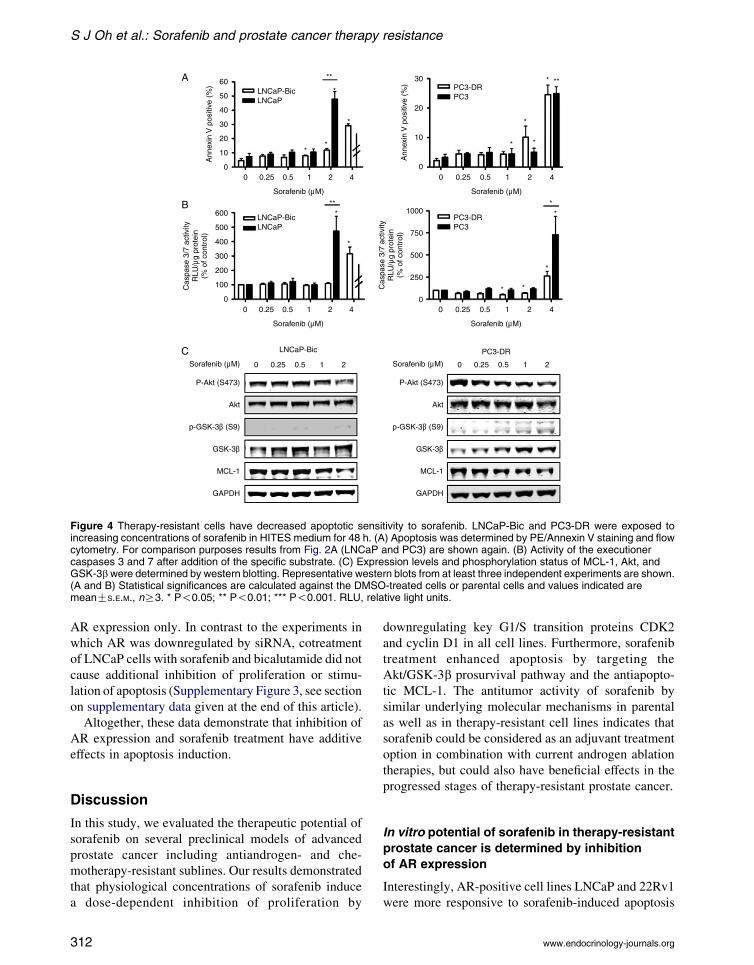
On the other hand, in apoptosis assays both
models showed different responses to sorafenib
compared to parental cells (Fig. 4A and B). The
concentration of 4 mM sorafenib was in need to induce
apoptosis in LNCaP-Bic cells, while 2 mM was
sufficient for parental LNCaP cells. Similarly, the
PC3-DR subline showed a decreased sensitivity to
sorafenib compared to parental PC3 cells. Again,
expression levels of MCL-1 and phosphorylated and
total Akt and GSK-3b were analyzed (Fig. 4C and
Supplementary Figure 4, see section on supplementary
data given at the end of this article). In both cell lines,
phosphorylation of Akt was reduced by higher
sorafenib concentrations. Interestingly, phosphoryl-
ation of GSK-3b was completely lost in LNCaP-Bic
leading to the hypothesis for a role for GSK-3b in
therapy resistance development. Altogether, these
results show a decreased sensitivity of the therapy-
resistant cell models to sorafenib compared to parental
cell lines.
309
60
50
40
Ann
exin
V p
ositi
ve (
%)
Ann
exin
V p
ositi
ve (
%)
MC
L-1/
GA
PD
H(%
of c
ontr
ol)
Ann
exin
V p
ositi
ve (
%)
Ann
exin
V p
ositi
ve (
%)
30
20
10
0 0.25 0.5 1 2
**
**
**
**
*
*
*
*
*
* *
*
*
**
**
****
(P=0.06)
**
LNCaPA
B
C
LNCaP-IL6+
LNCaP-IL6+
LNCaP
PC3
LNCaP
Sorafenib (µM)
P-Akt (S473)
p-GSK-3β (S9)
GSK-3β
GAPDH
Akt
PC3
MCL-1
GAPDH
MCL-1
GAPDH
MCL-1
GAPDH
MCL-1
GAPDH
PC3
22Rv1
22Rv1
4
Sorafenib (µM)
0 0.25 0.5 1 2 4
Sorafenib (µM)
0 0.25 0.5 1 2 4
Sorafenib (µM)
0 0.25 0.5 1 2 4
Sorafenib (µM)
0 0.25 0.5 1 2
0 0.25 0.5 1 2 0 0.25 0.5 1 2
0 0.25 0.5 1 2
0 0.25 0.5 1 2 0 0.25 0.5 1 2
0
30
20
10
0
125
100
75
50
25
0
MC
L-1/
GA
PD
H(%
of c
ontr
ol)
125
100
75
50
25
0
MC
L-1/
GA
PD
H(%
of c
ontr
ol)
125
100
75
50
25
0
MC
L-1/
GA
PD
H(%
of c
ontr
ol)
125
100
75
50
25
0
30
20
10
0
20
10
0
Figure 2 Differential sensitivities of prostate cancer cell lines to sorafenib-mediated apoptosis through downregulation of MCL-1and Akt pathway. LNCaP, 22Rv1, PC3, and LNCaP-IL6C cells were exposed to increasing concentrations of sorafenib in HITESmedium for 48 h. (A)ApoptosiswasdeterminedbyPE/AnnexinVstainingand flowcytometry. LNCaPcells treatedwith 4 mMsorafenibunderwent massive apoptosis resulting in an insufficient number of cells to perform assays. (B) Expression of MCL-1 was determinedbywestern blotting. Bandswere scanned densitometrically and normalized to expression levels of GAPDH. (C) Expression levels andphosphorylation status of Akt and GSK-3b in LNCaP and PC3 cells were determined by western blotting. GAPDH served as loadingcontrol. (A andB)Statistical significancesare calculated against theDMSO-treated cells and values indicatedaremeanGS.E.M.,nR3.* P!0.05; ** P!0.01; *** P!0.001. Representative western blots from at least three independent experiments are shown.
S J Oh et al.: Sorafenib and prostate cancer therapy resistance
www.endocrinology-journals.org310
Endocrine-Related Cancer (2012) 19 305–319
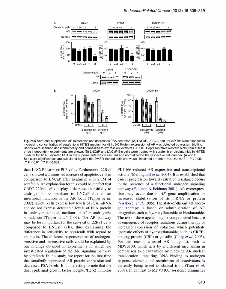
Sorafenib inhibits expression of AR and reduces
PSA levels in androgen-sensitive cell lines
Modulation of AR signaling by the Her-2 tyrosine
kinase has been reported (Craft et al. 1999). However,
little is known about the regulation of AR signaling by
tyrosine kinase inhibitors. LNCaP cells were more
sensitive to sorafenib than LNCaP-Bic or LNCaP-abl
cells (Fig. 4 and Supplementary Figure 5, see section
on supplementary data given at the end of this article).
Increased AR expression in LNCaP-abl cells was
demonstrated in a previous publication of our
laboratory (Culig et al. 1999). Thus, we hypothesized
that AR is a target of sorafenib in prostate cancer cells.
To clarify possible effects of sorafenib on AR, receptor
expression levels were measured in LNCaP, 22Rv1,
and LNCaP-Bic cells (Fig. 5A). In LNCaP and 22Rv1
cells, AR levels were decreased in the presence of
2 mM sorafenib (Fig. 5A). In LNCaP-Bic cells which
express higher levels of AR, AR protein level was
downregulated only by 4 mM of sorafenib. Moreover,
concentration of secreted PSA was measured in all but
the CRPC cell line 22Rv1 that lack detectable levels of
secreted PSA under basal culture conditions (Tepper
et al. 2002; Fig. 5B). In both LNCaP and LNCaP-Bic
cells, secreted PSA levels were dramatically reduced
[3 H] T
hym
idin
e in
corp
orat
ion
(% o
f unt
reat
ed c
ells
) 150
100
50
0
150
100
50
0
0 0.25 0.5 1 1.5 2
Sorafenib (µM)
Sorafenib (µM) 0 0.25 0.5 1 2 S
LNCaP (*)
****
###
LNCaP-Bic (*)
LNCaP-BicA
B LNCaP-Bic
PO
I/GA
PD
H(%
of c
ontr
ol)
Cyclin D1
CDK2
GAPDH
CDK2Cyclin D1
*
*
***
Figure 3 Antiproliferative effects of sorafenib in therapy-resistant mexposed to increasing concentrations of sorafenib in HITES mediuincorporation. For comparison purposes results from Fig. 1A (LNCand cyclin D1 was detected by western blotting. Bands were scannGAPDH. Representative western blots from at least three indepencalculated against the DMSO-treated cells and values indicated arP!0.001. POI, protein of interest.
www.endocrinology-journals.org
in the presence of sorafenib. Intriguingly, sorafenib
showed a higher ability to decrease PSA than
bicalutamide at the same concentrations (1–4 mM).
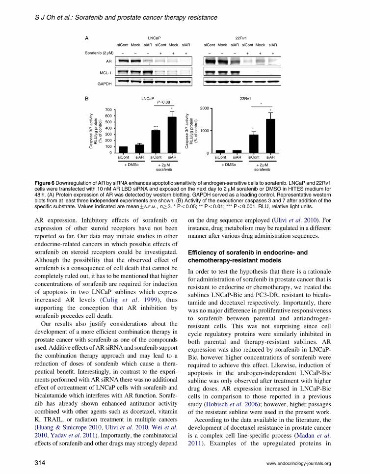
Downregulation of AR by siRNA enhances
sorafenib-induced increase of caspase 3/7
activity
In regard to a possible clinical application of sorafenib
for prostate cancer in combination with existing
androgen-ablation therapies, we analyzed whether AR
inhibition and sorafenib treatment have an additive
effect. LNCaP and 22Rv1 cells were transfected with
10 nM AR–LBD siRNA or control siRNA and treated
with 2 mM of sorafenib or vehicle (Fig. 6A). AR–LBD
siRNA efficiently downregulated AR expression
levels by 70–90% but did not affect expression levels
of MCL-1. In the presence of 2 mM of sorafenib, both
AR and MCL-1 were downregulated as expected. AR
expression was almost absent in the specific siRNA-
and sorafenib-treated samples. Apoptosis was induced
in both cell lines after 48 h of sorafenib treatment as
measured by caspase 3/7 assays (Fig. 6B). Moreover,
a significant increase of apoptosis could be observed in
22Rv1 cells with decreased AR expression levels and
treated with sorafenib compared to cells with reduced
[3 H] T
hym
idin
e in
corp
orat
ion
(% o
f unt
reat
ed c
ells
) 150
100
50
00 0.25 0.5 1 1.5 2
0 0.25 0.5 1 2
Sorafenib (µM)
orafenib (µM)
******
**#
# # ##
PC3 (#)PC3-DR (*)
PC3-DR
PC3-DR
150
100
50
0
PO
I/GA
PD
H(%
of c
ontr
ol)
Cyclin D1
CDK2
GAPDH
*
**
CDK2Cyclin D1
odels of human prostate cancer. LNCaP-Bic and PC3-DR werem for 48 h. (A) Proliferation was assessed by 3H-thymidineaP and PC3) are shown again. (B) Protein expression of CDK2ed densitometrically and normalized to expression levels ofdent experiments are shown. Statistical significances aree meanGS.E.M., nR3. */# P!0.05; **/## P!0.01; ***/###
311
60
50
40
Ann
exin
V p
ositi
ve (
%)
Cas
pase
3/7
act
ivity
RLU
/µg
prot
ein
(% o
f con
trol
)
Cas
pase
3/7
act
ivity
RLU
/µg
prot
ein
(% o
f con
trol
)
30
20
10
600 1000
750
500
250
0
500
400
300
200
100
0
A
B
C
0
Ann
exin
V p
ositi
ve (
%)
30
20
10
00 0.25 0.5 1 2
0 0.25 0.5 1 2
4
**
*
*
**
*
*
**
**
*
* *
* ***
**
Sorafenib (µM)
0 0.25 0.5 1 2 4
Sorafenib (µM)
0 0.25 0.5 1 2 4
Sorafenib (µM)
0 0.25 0.5 1 2 4
Sorafenib (µM)
Sorafenib (µM)
LNCaPLNCaP-Bic
LNCaP-Bic
LNCaPLNCaP-Bic
PC3PC3-DR
PC3-DR
PC3PC3-DR
P-Akt (S473)
p-GSK-3β (S9)
GSK-3β
GAPDH
MCL-1
Akt
0 0.25 0.5 1 2Sorafenib (µM)
P-Akt (S473)
p-GSK-3β (S9)
GSK-3β
GAPDH
MCL-1
Akt
Figure 4 Therapy-resistant cells have decreased apoptotic sensitivity to sorafenib. LNCaP-Bic and PC3-DR were exposed toincreasing concentrations of sorafenib in HITES medium for 48 h. (A) Apoptosis was determined by PE/Annexin V staining and flowcytometry. For comparison purposes results from Fig. 2A (LNCaP and PC3) are shown again. (B) Activity of the executionercaspases 3 and 7 after addition of the specific substrate. (C) Expression levels and phosphorylation status of MCL-1, Akt, andGSK-3bwere determined by western blotting. Representative western blots from at least three independent experiments are shown.(A and B) Statistical significances are calculated against the DMSO-treated cells or parental cells and values indicated aremeanGS.E.M., nR3. * P!0.05; ** P!0.01; *** P!0.001. RLU, relative light units.
S J Oh et al.: Sorafenib and prostate cancer therapy resistance
AR expression only. In contrast to the experiments in
which AR was downregulated by siRNA, cotreatment
of LNCaP cells with sorafenib and bicalutamide did not
cause additional inhibition of proliferation or stimu-
lation of apoptosis (Supplementary Figure 3, see section
on supplementary data given at the end of this article).
Altogether, these data demonstrate that inhibition of
AR expression and sorafenib treatment have additive
effects in apoptosis induction.
Discussion
In this study, we evaluated the therapeutic potential of
sorafenib on several preclinical models of advanced
prostate cancer including antiandrogen- and che-
motherapy-resistant sublines. Our results demonstrated
that physiological concentrations of sorafenib induce
a dose-dependent inhibition of proliferation by
312
downregulating key G1/S transition proteins CDK2
and cyclin D1 in all cell lines. Furthermore, sorafenib
treatment enhanced apoptosis by targeting the
Akt/GSK-3b prosurvival pathway and the antiapopto-
tic MCL-1. The antitumor activity of sorafenib by
similar underlying molecular mechanisms in parental
as well as in therapy-resistant cell lines indicates that
sorafenib could be considered as an adjuvant treatment
option in combination with current androgen ablation
therapies, but could also have beneficial effects in the
progressed stages of therapy-resistant prostate cancer.
In vitro potential of sorafenib in therapy-resistant
prostate cancer is determined by inhibition
of AR expression
Interestingly, AR-positive cell lines LNCaP and 22Rv1
were more responsive to sorafenib-induced apoptosis
www.endocrinology-journals.org
LNCaPA
B
LNCaP-Bic
LNCaP-Bic
LNCaP-BicLNCaP
DMSO 1 2
Bicalutamide(µM)
Sorafenib(µM)
4 1 2 4 DMSO 1 2
Bicalutamide(µM)
Sorafenib(µM)
4 1 2 4
22Rv1
22Rv1
0 0.25 0.5 1 2 0 0.25 0.5 1 2 0 0.25 0.5 1 2
0 0.25
0 2 4 0 2 4
0.5 1 2 0 0.25 0.5 1 2 0 0.25 0.5 1 2
Sorafenib (µM)
Sorafenib (µM)
AR
GAPDH
AR
GAPDH
*
* **
100
50
0
150
100
50
0
150
AR
/GA
PD
H(%
of c
ontr
ol)
100
50
0
150
AR
/GA
PD
H(%
of c
ontr
ol)
100
50
0
150
AR
/GA
PD
H(%
of c
ontr
ol)
Sec
rete
d P
SA
ng/m
l per
106 c
ells
Sec
rete
d P
SA
ng/m
l per
106 c
ells
125
100
75
25 ******
*
***
50
0
Figure 5 Sorafenib suppresses AR expression and decreases PSA secretion. (A) LNCaP, 22Rv1, and LNCaP-Bic were exposed toincreasing concentration of sorafenib in HITES medium for 48 h. (A) Protein expression of AR was detected by western blotting.Bands were scanned densitometrically and normalized to expression levels of GAPDH. Representative western blots from at leastthree independent experiments are shown. (B) LNCaP and LNCaP-Bic cells were treated with sorafenib or bicalutamide in HITESmedium for 48 h. Secreted PSA in the supernatants was measured and normalized to the respective cell number. (A and B)Statistical significances are calculated against the DMSO-treated cells and values indicated are meanGS.E.M., nR3. * P!0.05;** P!0.01; *** P!0.001.
Endocrine-Related Cancer (2012) 19 305–319
than LNCaP-IL6C or PC3 cells. Furthermore, 22Rv1
cells showed a diminished increase of apoptotic cells in
comparison to LNCaP after treatment with 2 mM of
sorafenib. An explanation for this could be the fact that
CRPC 22Rv1 cells display a decreased sensitivity to
androgen in comparison to LNCaP due to an
insertional mutation in the AR locus (Tepper et al.
2002). 22Rv1 cells express low levels of PSA mRNA
and do not express detectable levels of PSA protein
in androgen-depleted medium or after androgenic
stimulation (Tepper et al. 2002). The AR pathway
may be less important for the survival of 22Rv1 cells
compared to LNCaP cells, thus explaining the
difference in sensitivity to sorafenib with regard to
apoptosis. The different responsiveness of androgen-
sensitive and -insensitive cells could be explained by
our findings obtained in experiments in which we
investigated regulation of the AR signaling pathway
by sorafenib. In this study, we report for the first time
that sorafenib suppressed AR protein expression and
decreased PSA levels. It is interesting to note that the
dual epidermal growth factor receptor/Her-2 inhibitor
www.endocrinology-journals.org
PKI-166 reduced AR expression and transcriptional
activity (Mellinghoff et al. 2004). It is established that
cancer progression toward castration resistance occurs
in the presence of a functional androgen signaling
pathway (Feldman & Feldman 2001). AR overexpres-
sion may occur due to AR gene amplification or
increased stabilization of its mRNA or protein
(Visakorpi et al. 1995). The state-of-the-art antiandro-
gen therapy is based on administration of AR
antagonists such as hydroxyflutamide or bicalutamide.
The use of these agents may be compromised because
of emergence of receptor mutations during therapy or
increased expression of cofactors which potentiate
agonistic effects of hydroxyflutamide, such as CREB-
binding protein (CBP) or gelsolin (Culig et al. 2005).
For this reason, a novel AR antagonist, such as
MDV3100, which acts by a different mechanism in
comparison to bicalutamide by blocking AR nuclear
translocation, impairing DNA binding to androgen
response elements and recruitment of coactivators, is
currently being tested in clinical trials (Tran et al.
2009). In contrast to MDV3100, sorafenib diminishes
313
LNCaPA
B
AR
MCL-1
GAPDH
700
P=0.08
**
*
****
600
500
400
Cas
pase
3/7
act
ivity
RLU
/µg
prot
ein
(% o
f con
trol
)
Cas
pase
3/7
act
ivity
RLU
/µg
prot
ein
(% o
f con
trol
)
300
200
2000
100
1000
00
siCont Mock siAR
siCont
+ DMSo + 2µMsorafenib
siAR siCont siAR siCont
+ DMSo + 2µMsorafenib
siAR siCont siAR
siCont Mock siAR
– – – + + +
siCont Mock siAR siCont Mock siAR
– – – + + +
22Rv1
LNCaP 22Rv1
Sorafenib (2µM)
Figure 6 Downregulation of AR by siRNA enhances apoptotic sensitivity of androgen-sensitive cells to sorafenib. LNCaP and 22Rv1cells were transfected with 10 nM AR LBD siRNA and exposed on the next day to 2 mM sorafenib or DMSO in HITES medium for48 h. (A) Protein expression of AR was detected by western blotting. GAPDH served as a loading control. Representative westernblots from at least three independent experiments are shown. (B) Activity of the executioner caspases 3 and 7 after addition of thespecific substrate. Values indicated are meanGS.E.M., nR3. * P!0.05; ** P!0.01; *** P!0.001. RLU, relative light units.
S J Oh et al.: Sorafenib and prostate cancer therapy resistance
AR expression. Inhibitory effects of sorafenib on
expression of other steroid receptors have not been
reported so far. Our data may initiate studies in other
endocrine-related cancers in which possible effects of
sorafenib on steroid receptors could be investigated.
Although the possibility that the observed effect of
sorafenib is a consequence of cell death that cannot be
completely ruled out, it has to be mentioned that higher
concentrations of sorafenib are required for induction
of apoptosis in two LNCaP sublines which express
increased AR levels (Culig et al. 1999), thus
supporting the conception that AR inhibition by
sorafenib precedes cell death.
Our results also justify considerations about the
development of a more efficient combination therapy in
prostate cancer with sorafenib as one of the compounds
used. Additive effects of AR siRNA and sorafenib support
the combination therapy approach and may lead to a
reduction of doses of sorafenib which cause a thera-
peutical benefit. Interestingly, in contrast to the experi-
ments performed with AR siRNA there was no additional
effect of cotreatment of LNCaP cells with sorafenib and
bicalutamide which interferes with AR function. Sorafe-
nib has already shown enhanced antitumor activity
combined with other agents such as docetaxel, vitamin
K, TRAIL, or radiation treatment in multiple cancers
(Huang & Sinicrope 2010, Ulivi et al. 2010, Wei et al.
2010, Yadav et al. 2011). Importantly, the combinatorial
effects of sorafenib and other drugs may strongly depend
314
on the drug sequence employed (Ulivi et al. 2010). For
instance, drug metabolism may be regulated in a different
manner after various drug administration sequences.
Efficiency of sorafenib in endocrine- and
chemotherapy-resistant models
In order to test the hypothesis that there is a rationale
for administration of sorafenib in prostate cancer that is
resistant to endocrine or chemotherapy, we treated the
sublines LNCaP-Bic and PC3-DR, resistant to bicalu-
tamide and docetaxel respectively. Importantly, there
was no major difference in proliferative responsiveness
to sorafenib between parental and antiandrogen-
resistant cells. This was not surprising since cell
cycle regulatory proteins were similarly inhibited in
both parental and therapy-resistant sublines. AR
expression was also reduced by sorafenib in LNCaP-
Bic, however higher concentrations of sorafenib were
required to achieve this effect. Likewise, induction of
apoptosis in the androgen-independent LNCaP-Bic
subline was only observed after treatment with higher
drug doses. AR expression increased in LNCaP-Bic
cells in comparison to those reported in a previous
study (Hobisch et al. 2006); however, higher passages
of the resistant subline were used in the present work.
According to the data available in the literature, the
development of docetaxel resistance in prostate cancer
is a complex cell line-specific process (Madan et al.
2011). Examples of the upregulated proteins in
www.endocrinology-journals.org
Endocrine-Related Cancer (2012) 19 305–319
docetaxel resistance include but are not limited to
Pim-1 kinase, chemokine CCL2, and class III b tubulin
(Zemskova et al. 2008, Ploussard et al. 2010, Qian
et al. 2010). Identification of additional mechanisms
being responsible for resistance of the sublines derived
in our laboratory is at present under investigation.
However, although efficacy of growth inhibition and
apoptosis induction of PC3-DR is somewhat reduced
compared to parental cells, it is important to note that
PC3-DR could still be inhibited by sorafenib but no
longer by docetaxel. This finding may have clinical
implications especially when keeping in mind that the
duration of docetaxel response in prostate cancer
patients is limited to several months.
Antiapoptotic pathways in prostate cancer cells
are inhibited by sorafenib
In concordance to findings observed in other tumors,
inhibition of Akt phosphorylation by sorafenib was
also seen in our experiments in LNCaP and PC3 cells
(Chapuy et al. 2011). The Akt signaling pathway is
frequently activated in advanced prostate cancer due to
deletion or mutation of the PTEN tumor suppressor
gene (Sircar et al. 2009). In cell culture models, Akt is
constitutively active in LNCaP and PC3 cells due to
PTEN mutation (LNCaP) or deletion (PC3; Vlietstra
et al. 1998). In line with those data, Kreisberg et al.
(2004) showed that phosphorylation of Akt S473 is
a predictor of poor clinical outcome in prostate cancer.
Moreover, it is known that the Akt downstream target
GSK-3b mediates degradation of MCL-1 by the
proteasome. Interestingly, differences in phosphoryl-
ation of GSK-3b in prostate cancer after sorafenib
treatment were observed in a cell type-dependent
manner. GSK-3b is phosphorylated and inactivated by
phosphorylated Akt. Consequently, phosphorylation of
GSK-3b may lead to upregulation of MCL-1 in
multiple tumor cell lines and primary cancer samples
(Maurer et al. 2006). As an implication of sorafenib
treatment, downregulation of MCL-1 could be
achieved by a decrease of total or inactivated,
i.e. phosphorylated GSK-3b. It is known that MCL-1
is expressed at high levels in prostate cancer and is
important for mediating a survival function of the
proinflammatory cytokine IL6 (Krajewska et al. 1996,
Cavarretta et al. 2007). Taken together, our results
suggest the sorafenib-mediated modulation of the Akt/
GSK-3b/MCL-1 pathway in prostate cancer is clini-
cally relevant. Although the results of our over-
expression experiments cannot definitively answer
the question whether the presence of MCL-1 is
required for the antiapoptotic effect of sorafenib in
www.endocrinology-journals.org
prostate cancer cells, there is an evidence in the
scientific literature supporting this view. First, in K562
chronic myelogenous leukemia cells overexpression
of MCL-1 inhibited sorafenib-induced apoptosis
(Yu et al. 2005). In addition, in a recent study
performed in androgen-insensitive prostate cancer
cell lines sorafenib sensitized tumor cells to (K)-
gossypol through MCL-1 inhibition (Lian et al. 2012).
The perspective for further development of
sorafenib-based prostate cancer treatments
Three preclinical studies have addressed the drug
response of sorafenib on prostate cancer cells in vitro
(Dahut et al. 2008, Huang et al. 2010, Ullen et al.
2010). In contrast to our work, those reports were
focused on antiangiogenic and cytotoxic effects of
sorafenib. Moreover, they were performed in a single
prostate cancer cell line using concentrations of the
drug which were higher than the physiological
concentrations of 2–5 mM measured in sera of patients
after administration of 400 mg twice daily (Dahut et al.
2008). In one of those previous studies, decreased
phosphorylation of MAP kinases by sorafenib in PC3
and DU145 cells was observed (Ullen et al. 2010)
confirming the results in colon, pancreas, and breast
cancer cell lines (Wilhelm et al. 2004). However, other
signaling pathways were not investigated after sor-
afenib treatment in prostate cancer in previous reports.
Our results may have implications for development
of clinical prostate cancer therapies. Tannock et al.
(2004) documented that docetaxel-based chemother-
apy in combination with prednisone improved median
overall survival of patients with CRPC by 2.4 months.
However, because of limited benefits and significant
toxicity of docetaxel therapy, the search for a more
efficient treatment for CRPC is continued. On the basis
of a recent publication by de Bono et al. (2011) that
administration of the inhibitor of androgen synthesis
abiraterone in combination with prednisone in patients
pretreated with docetaxel prolonged survival to 450 vs
332 days, it could be concluded that targeting the
androgen signaling pathway in docetaxel-resistant
prostate cancer in vivo is nevertheless a worthy
therapeutic goal. The question whether a combinatorial
treatment on the basis of androgenic and multiple
kinase inhibition by sorafenib has a benefit in patients
with therapy-resistant prostate cancer needs to be
addressed in the future.
Clinical studies have reported benefits following
treatment with tyrosine kinase inhibitors erlotinib and
sunitinib in prostate cancer patients (Gravis et al. 2008,
Sonpavde et al. 2008). In other clinical trials, the
315
S J Oh et al.: Sorafenib and prostate cancer therapy resistance
investigators reported on a small number of patients in
which stabilization of the disease by sorafenib was
achieved (Chi et al. 2008, Dahut et al. 2008, Steinbild
et al. 2007, Aragon-Ching et al. 2009). On the other
hand, difficulties in correlating clinical response and
PSA measurements were observed. In the context of
the final analysis of a phase II trial, Aragon-Ching et al.
(2009) suggested that a selected population of patients
may benefit from sorafenib treatment. The absence of
adequate biomarkers for monitoring the therapeutic
success may be the reason why it is difficult to match
preclinical findings with clinical effects. It should be
mentioned that PSA measurements in vitro could not
be simply extrapolated in vivo since the patients’ data
also reflect the disruption of the basement membrane.
In a recently reported phase II clinical trial with
sorafenib and bicalutamide in patients with CRPC 47%
of patients presented with either PSA decrease or stable
disease (Beardsley et al. 2012). Those clinical findings
could be partly explained by our results showing
differences in responsiveness of prostate cancer
parental cells and sublines representing advanced
disease stages to sorafenib.
In summary, we demonstrate that the multitargeting
effects of sorafenib induce growth inhibition and
apoptosis in a variety of prostate cancer cell lines.
Most importantly, we found that sorafenib affects AR
expression and signaling, which is a previously
unknown mechanism of sorafenib. Our data also
suggest that maximal effect of sorafenib may be
expected in androgen-sensitive prostate cancer prior
to the development of resistance to castration and
chemotherapy. However, there may be also a rationale
for the use of sorafenib in docetaxel-resistant carci-
noma of the prostate. The evidence for differential
response of prostate cancer cell lines may explain why
sorafenib is beneficial in a selected population of
patients in clinical trials.
Supplementary data
This is linked to the online version of the paper at http://dx.
doi.org/10.1530/ERC-11-0298.
Declaration of interest
The authors declare that there is no conflict of interest that
could be perceived as prejudicing the impartiality of the
research reported.
Funding
This work was supported by the Austrian Science Fund
(FWF, grant number L544 to Z Culig), Austrian National
316
Bank (OENB, grant number 13952 to Z Culig), and Bayer
Austria. Research support by Bayer Austria (to Z Culig) was
received.
Author contribution statement
S J Oh performed research, analyzed data, wrote the first
version of the paper; H H H Erb performed research,
analyzed data; A Hobisch designed research; F R Santer
performed and supervised research, analyzed data, prepared
the final version of the paper; Z Culig designed and
supervised research, and prepared the final version of the
paper. All authors have participated in writing and approved
the final version of the paper.
Acknowledgements
We thank Ms Tanja Fuchs and Birgit Stenzel for PSA
measurements. We are grateful to all members of the Culig
laboratory for their discussions during preparation of the
manuscript, Dr Walther Parson for cell authentication,
Dr Dennis Healy and Mr Gerhard Briesch for providing
sorafenib.
References
Aragon-Ching JB, Jain L, Gulley JL, Arlen PM, Wright JJ,
Steinberg SM, Draper D, Venitz J, Jones E, Chen CC et al.
2009 Final analysis of a phase II trial using sorafenib for
metastatic castration-resistant prostate cancer. British
Journal of Urology International 103 1636–1640.
(doi:10.1111/j.1464-410X.2008.08327.x)
Beardsley EK, Hotte SJ, North S, Ellard SL, Winquist E,
Kollmannsberger C, Mukherjee SD & Chi KN 2012
A phase II study of sorafenib in combination with
bicalutamide in patients with chemotherapy-naive
castration resistant prostate cancer. Investigational
New Drugs [in press].
de Bono JS, Logothetis CJ, Molina A, Fizazi K, North S,
Chu L, Chi KN, Jones RJ, Goodman OB Jr, Saad F et al.
2011 Abiraterone and increased survival in metastatic
prostate cancer. New England Journal of Medicine 364
1995–2005. (doi:10.1056/NEJMoa1014618)
Cavarretta IT, Neuwirt H, Untergasser G, Moser PL, Zaki MH,
Steiner H, Rumpold H, Fuchs D, Hobisch A, Nemeth JA
et al. 2007 The antiapoptotic effect of IL-6 autocrine loop
in a cellular model of advanced prostate cancer is mediated
by Mcl-1. Oncogene 26 2822–2832. (doi:10.1038/sj.onc.
1210097)
Chapuy B, Schuelper N, Panse M, Dohm A, Hand E,
Schroers R, Truemper L & Wulff GG 2011 Multikinase
inhibitor sorafenib exerts cytocidal efficacy against
non-Hodgkin lymphomas associated with inhibition of
MAPK14 and AKT phosphorylation. British Journal of
Haematology 152 401–412. (doi:10.1111/j.1365-2141.
2010.08526.x)
www.endocrinology-journals.org
Endocrine-Related Cancer (2012) 19 305–319
Chi KN, Ellard SL, Hotte SJ, Czaykowski P, Moore M,
Ruether JD, Schell AJ, Taylor S, Hansen C, Gauthier I
et al. 2008 A phase II study of sorafenib in patients
with chemo-naive castration-resistant prostate cancer.
Annals of Oncology 19 746–751. (doi:10.1093/annonc/
mdm554)
Craft N, Shostak Y, Carey M & Sawyers CL 1999 A
mechanism for hormone-independent prostate cancer
through modulation of androgen receptor signaling by the
HER-2/neu tyrosine kinase. Nature Medicine 5 280–285.
(doi:10.1038/6495)
Craig RW 2002 MCL1 provides a window on the role of
the BCL2 family in cell proliferation, differentiation and
tumorigenesis. Leukemia 16 444–454. (doi:10.1038/sj.
leu.2402416)
Culig Z, Hoffmann J, Erdel M, Eder IE, Hobisch A, Hittmair A,
Bartsch G, Utermann G, Schneider MR, Parczyk K et al.
1999 Switch from antagonist to agonist of the androgen
receptor blocker bicalutamide is associated with prostate
tumour progression in a new model system. British
Journal of Cancer 81 242–251. (doi:10.1038/sj.bjc.
6690684)
Culig Z, Steiner H, Bartsch G & Hobisch A 2005
Mechanisms of endocrine therapy-responsive and
-unresponsive prostate tumours. Endocrine-Related
Cancer 12 229–244. (doi:10.1677/erc.1.00775a)
Dahut WL, Scripture C, Posadas E, Jain L, Gulley JL,
Arlen PM, Wright JJ, Yu Y, Cao L, Steinberg SM et al.
2008 A phase II clinical trial of sorafenib in androgen-
independent prostate cancer. Clinical Cancer Research
14 209–214. (doi:10.1158/1078-0432.CCR-07-1355)
Desiniotis A, Schafer G, Klocker H & Eder IE 2010
Enhanced antiproliferative and proapoptotic effects on
prostate cancer cells by simultaneously inhibiting
androgen receptor and cAMP-dependent protein kinase A.
International Journal of Cancer 126 775–789.
(doi:10.1002/ijc.24806)
Ding Q, He X, Hsu JM, Xia W, Chen CT, Li LY, Lee DF,
Liu JC, Zhong Q, Wang X et al. 2007 Degradation of
Mcl-1 by beta-TrCP mediates glycogen synthase kinase
3-induced tumor suppression and chemosensitization.
Molecular and Cellular Biology 27 4006–4017.
(doi:10.1128/MCB.00620-06)
Feldman BJ & Feldman D 2001 The development of
androgen-independent prostate cancer. Nature Reviews.
Cancer 1 34–45. (doi:10.1038/35094009)
Gravis G, Bladou F, Salem N, Goncalves A, Esterni B,
Walz J, Bagattini S, Marcy M, Brunelle S & Viens P
2008 Results from a monocentric phase II trial of
erlotinib in patients with metastatic prostate cancer.
Annals of Oncology 19 1624–1628. (doi:10.1093/
annonc/mdn174)
Grossmann ME, Huang H & Tindall DJ 2001 Androgen
receptor signaling in androgen-refractory prostate cancer.
Journal of the National Cancer Institute 93 1687–1697.
(doi:10.1093/jnci/93.22.1687)
www.endocrinology-journals.org
Ha S, Ruoff R, Kahoud N, Franke TF & Logan SK 2011
Androgen receptor levels are upregulated by Akt in
prostate cancer. Endocrine-Related Cancer 18 245–255.
(doi:10.1530/ERC-10-0204)
Hobisch A, Ramoner R, Fuchs D, Godoy-Tundidor S,
Bartsch G, Klocker H & Culig Z 2001 Prostate cancer
cells (LNCaP) generated after long-term interleukin 6
(IL-6) treatment express IL-6 and acquire an IL-6
partially resistant phenotype. Clinical Cancer Research
7 2941–2948.
Hobisch A, Fritzer A, Comuzzi B, Fiechtl M,
Malinowska K, Steiner H, Bartsch G & Culig Z 2006
The androgen receptor pathway is by-passed in
prostate cancer cells generated after prolonged
treatment with bicalutamide. Prostate 66 413–420.
(doi:10.1002/pros.20365)
Huang S & Sinicrope FA 2010 Sorafenib inhibits STAT3
activation to enhance TRAIL-mediated apoptosis in
human pancreatic cancer cells. Molecular Cancer
Therapeutics 9 742–750. (doi:10.1158/1535-7163.MCT-
09-1004)
Huang R, Chen XQ, Huang Y, Chen N & Zeng H 2010
The multikinase inhibitor sorafenib induces caspase-
dependent apoptosis in PC-3 prostate cancer cells.
Asian Journal of Andrology 12 527–534. (doi:10.1038/
aja.2010.21)
Jemal A, Siegel R, Xu J & Ward E 2010 Cancer statistics,
2010. CA: A Cancer Journal for Clinicians 60 277–300.
(doi:10.3322/caac.20073)
Kane RC, Farrell AT, Saber H, Tang S, Williams G, Jee JM,
Liang C, Booth B, Chidambaram N, Morse D et al. 2006
Sorafenib for the treatment of advanced renal cell
carcinoma. Clinical Cancer Research 12 7271–7278.
(doi:10.1158/1078-0432.CCR-06-1249)
Kozopas KM, Yang T, Buchan HL, Zhou P & Craig RW
1993 MCL1, a gene expressed in programmed myeloid
cell differentiation, has sequence similarity to BCL2.
PNAS 90 3516–3520. (doi:10.1073/pnas.90.8.3516)
Krajewska M, Krajewski S, Epstein JI, Shabaik A,
Sauvageot J, Song K, Kitada S & Reed JC 1996
Immunohistochemical analysis of bcl-2, bax, bcl-X,
and mcl-1 expression in prostate cancers. American
Journal of Pathology 148 1567–1576.
Kreisberg JI, Malik SN, Prihoda TJ, Bedolla RG, Troyer DA,
Kreisberg S & Ghosh PM 2004 Phosphorylation of Akt
(Ser473) is an excellent predictor of poor clinical outcome
in prostate cancer. Cancer Research 64 5232–5236.
(doi:10.1158/0008-5472.CAN-04-0272)
Lang L 2008 FDA approves sorafenib for patients with
inoperable liver cancer. Gastroenterology 134 379.
Lian J, Ni Z, Dai X, Su C, Smith AR, Xu L & He F 2012
Sorafenib sensitizes (K)-gossypol-induced growth
suppression in androgen-independent prostate cancer
cells via Mcl-1 inhibition and Bak activation. Molecular
Cancer Therapeutics 11 416–426. (doi:10.1158/1535-
7163.MCT-11-0559)
317

S J Oh et al.: Sorafenib and prostate cancer therapy resistance
Madan RA, Pal SK, Sartor O & Dahut WL 2011 Overcoming
chemotherapy resistance in prostate cancer. Clinical
Cancer Research 17 3892–3902. (doi:10.1158/1078-
0432.CCR-10-2654)
Maurer U, Charvet C, Wagman AS, Dejardin E & Green DR
2006 Glycogen synthase kinase-3 regulates mitochondrial
outer membrane permeabilization and apoptosis by
destabilization of MCL-1. Molecular Cell 21 749–760.
(doi:10.1016/j.molcel.2006.02.009)
Mellinghoff IK, Vivanco I, Kwon A, Tran C, Wongwipat J &
Sawyers CL 2004 HER2/neu kinase-dependent modu-
lation of androgen receptor function through effects on
DNA binding and stability. Cancer Cell 6 517–527.
(doi:10.1016/j.ccr.2004.09.031)
Nesterov A, Lu X, Johnson M, Miller GJ, Ivashchenko Y &
Kraft AS 2001 Elevated AKT activity protects the
prostate cancer cell line LNCaP from TRAIL-induced
apoptosis. Journal of Biological Chemistry 276
10767–10774. (doi:10.1074/jbc.M005196200)
Patterson SG, Wei S, Chen X, Sallman DA, Gilvary DL,
Zhong B, Pow-Sang J, Yeatman T & Djeu JY 2006 Novel
role of Stat1 in the development of docetaxel resistance
in prostate tumor cells. Oncogene 25 6113–6122.
(doi:10.1038/sj.onc.1209632)
Ploussard G, Terry S, Maille P, Allory Y, Sirab N, Kheuang L,
Soyeux P, Nicolaiew N, Coppolani E, Paule B et al. 2010
Class III beta-tubulin expression predicts prostate tumor
aggressiveness and patient response to docetaxel-based
chemotherapy. Cancer Research 70 9253–9264.
(doi:10.1158/0008-5472.CAN-10-1447)
Puhr M, Santer FR, Neuwirt H, Marcias G, Hobisch A &
Culig Z 2010 SOCS-3 antagonises the proliferative
and migratory effects of fibroblast growth factor-2
in prostate cancer by inhibition of p44/p42 MAPK
signalling. Endocrine-Related Cancer 17 525–538.
(doi:10.1677/ERC-10-0007)
Qian DZ, Rademacher BL, Pittsenbarger J, Huang CY,
Myrthue A, Higano CS, Garzotto M, Nelson PS & Beer TM
2010 CCL2 is induced by chemotherapy and protects
prostate cancer cells from docetaxel-induced cytotoxicity.
Prostate 70 433–442.
Rahmani M, Davis EM, Bauer C, Dent P & Grant S 2005
Apoptosis induced by the kinase inhibitor BAY 43-9006
in human leukemia cells involves down-regulation of
Mcl-1 through inhibition of translation. Journal of
Biological Chemistry 280 35217–35227.
(doi:10.1074/jbc.M506551200)
Santer FR, Hoschele PP, Oh SJ, Erb HH, Bouchal J,
Cavarretta IT, Parson W, Meyers DJ, Cole PA & Culig Z
2011 Inhibition of the acetyltransferases p300 and CBP
reveals a targetable function for p300 in the survival and
invasion pathways of prostate cancer cell lines. Molecular
Cancer Therapeutics 10 1644–1645. (doi:10.1158/1535-
7163.MCT-11-0182)
Sircar K, Yoshimoto M, Monzon FA, Koumakpayi IH,
Katz RL, Khanna A, Alvarez K, Chen G, Darnel AD,
Aprikian AG et al. 2009 PTEN genomic deletion is
318
associated with p-Akt and AR signalling in poorer
outcome, hormone refractory prostate cancer. Journal of
Pathology 218 505–513. (doi:10.1002/path.2559)
Sonpavde G, Hutson TE, Berry WR, Boehm KA & Asmar L
2008 Phase II trial of sunitinib for the therapy of
progressive metastatic castration-refractory prostate
cancer after previous docetaxel chemotherapy. Clinical
Genitourinary Cancer 6 134–137. (doi:10.3816/CGC.
2008.n.023)
Steinbild S, Mross K, Frost A, Morant R, Gillessen S, Dittrich C,
Strumberg D, Hochhaus A, Hanauske AR, Edler L et al.
2007 A clinical phase II study with sorafenib in patients with
progressive hormone-refractory prostate cancer: a study of
the CESAR Central European Society for Anticancer Drug
Research – EWIV.British Journal ofCancer 97 1480–1485.
(doi:10.1038/sj.bjc.6604064)
Tannock IF, de Wit R, Berry WR, Horti J, Pluzanska A,
Chi KN, Oudard S, Theodore C, James ND, Turesson I
et al. 2004 Docetaxel plus prednisone or mitoxantrone
plus prednisone for advanced prostate cancer. New
England Journal of Medicine 351 1502–1512.
(doi:10.1056/NEJMoa040720)
Tepper CG, Boucher DL, Ryan PE, Ma AH, Xia L, Lee LF,
Pretlow TG & Kung HJ 2002 Characterization of a
novel androgen receptor mutation in a relapsed CWR22
prostate cancer xenograft and cell line. Cancer Research
62 6606–6614.
Tran C, Ouk S, Clegg NJ, Chen Y, Watson PA, Arora V,
Wongvipat J, Smith-Jones PM, Yoo D, Kwon A et al.
2009 Development of a second-generation antiandrogen
for treatment of advanced prostate cancer. Science 324
787–790. (doi:10.1126/science.1168175)
Ullen A, Farnebo M, Thyrell L, Mahmoudi S, Kharaziha P,
Lennartsson L, Grander D, Panaretakis T & Nilsson S
2010 Sorafenib induces apoptosis and autophagy in
prostate cancer cells in vitro. International Journal of
Oncology 37 15–20. (doi:10.3892/ijo_00000648)
Ulivi P, Arienti C, Zoli W, Scarsella M, Carloni S, Fabbri F,
Tesei A, Chiadini E, Orlandi A, Passeri D et al. 2010
In vitro and in vivo antitumor efficacy of docetaxel and
sorafenib combination in human pancreatic cancer cells.
Current Cancer Drug Targets 10 600–610. (doi:10.2174/
156800910791859489)
Visakorpi T, Hyytinen E, Koivisto P, Tanner M, Keinanen R,
Palmberg C, Palotie A, Tammela T, Isola J & Kallioniemi OP
1995 In vivo amplification of the androgen receptor gene
and progression of human prostate cancer. Nature Genetics
9 401–406. (doi:10.1038/ng0495-401)
Vlietstra RJ, van Alewijk DC, Hermans KG, van Steenbrugge GJ
& Trapman J 1998 Frequent inactivation of PTEN in prostate
cancer cell lines and xenografts. Cancer Research 58
2720–2723.
Wei G, Wang M, Hyslop T, Wang Z & Carr BI 2010
Vitamin K enhancement of sorafenib-mediated HCC
cell growth inhibition in vitro and in vivo. International
Journal of Cancer 127 2949–2958. (doi:10.1002/
ijc.25498)
www.endocrinology-journals.org
Endocrine-Related Cancer (2012) 19 305–319
Wilhelm SM, Carter C, Tang L, Wilkie D, McNabola A,
Rong H, Chen C, Zhang X, Vincent P, McHugh M et al.
2004 BAY 43-9006 exhibits broad spectrum oral
antitumor activity and targets the RAF/MEK/ERK
pathway and receptor tyrosine kinases involved in tumor
progression and angiogenesis. Cancer Research 64
7099–7109. (doi:10.1158/0008-5472.CAN-04-1443)
Yadav A, Kumar B, Teknos TN & Kumar P 2011
Sorafenib enhances the antitumor effects of chemor-
adiation treatment by downregulating ERCC-1 and
XRCC-1 DNA repair proteins. Molecular Cancer
Therapeutics 10 1241–1251. (doi:10.1158/1535-7163.
MCT-11-0004)
Yu C, Bruzek A, Meng XW, Gores GJ, Carter CA, Kaufmann
SH & Adjei AA 2005 The role of Mcl-1 downregulation
www.endocrinology-journals.org
in the proapoptotic activity of the multikinase inhibitor
BAY 43-9006. Oncogene 24 6861–6869. (doi:10.1038/sj.
onc.1208841)
Zemskova M, Sahakian E, Bashkirova S & Lilly M 2008 The
PIM1 kinase is a critical component of a survival pathway
activated by docetaxel and promotes survival of doc-
etaxel-treated prostate cancer cells. Journal of Biological
Chemistry 283 20635–20644. (doi:10.1074/jbc.
M709479200)
Received in final form 15 February 2012Accepted 24 February 2012Made available online as an Accepted Preprint1 March 2012
319