the effect of moderate and severe 91 the effect of
TRANSCRIPT
The Effect of Moderate and Severe …… -91-
Zagazig J. Forensic Med.& Toxicol Vol.(16) No. (1) Jan 2018
THE EFFECT OF MODERATE AND SEVERE BURN INJURIES ON HUMAN LIVER,
KIDNEY & BLOOD (BIOCHEMICAL STUDY)
Samira M. Saleh* & Mohammed H. Hassan ** and Ahmed M. Tohamy***
Departments of Forensic Medicine & Clinical Toxicology*, Medical Biochemistry and Molecular Biology**
Faculty of medicine South Valley University *,** department of Plastic Surgery, Faculty of medicine, Assiut
University ***, Egypt
ABSTRACT The current study was done to determine changes in hematological, renal and hepatic parameters induced by
moderate and severe burn injuries. Patients and Methods: the study was done during the period from march
2016 to December 2016 on 60 adult patients, who were admitted at burn unit of Assiut university hospitals
with burn area ≥ 20% of body surface, and 20 healthy adults considered as control. The patients and control
were investigated for renal and hepatic functions, serum sodium (Na+) & potassium (K
+) & and blood
picture. Results: the blood urea and serum creatinine levels were significantly increased in burn patients in
comparison to control, the blood glucose showed significant increase on the first day then decrease gradually
on the 3rd
and 5th days post burn. The serum Na
+ & K
+ levels decreased significantly during the 3
rd and 5
th
days post burn, the blood picture showed a significant increase in white blood cells (WBCs) and Neutrophil
count ratio in burn patients in comparison to control, the Platelet count showed a significant reduction in
burn patients throughout the whole period of the study while hemoglobin (Hb) level showed significant
lowering during the 3rd
and 5th days post burn, the red blood cells (RBCs) count showed a significant
increased on the 1st day, then decreased significantly on the 5
th day post burn, the hematocrit (HCT) value
showed a significant decrease on the 3rd
and 5th days post burn. Regarding the liver functions, the serum
levels of liver enzymes showed a significant increase in burn patients in comparison to control, the serum
protein and albumin showed a significant decrease in burn patients, the prothrombin concentration was
reduced and Prothrombin time was prolonged significantly, while the (INR) showed a significant increase in
patients in comparison to control.
Key words: hematological parameters, kidney &liver function, serum electrolytes moderate & severe burn
Corresponding Author: Samira M. Saleh
Assistant professor of Forensic Medicine & Clinical Toxicology.
Faculty of Medicine, Qena.
South Vally university.Egypt.
INTRODUCTION
urns is the fourth most common type of
trauma worldwide, where 90% of burn
injuries occur in low and middle income
countries (Peck et al., 2013).
Thermal injuries which affect more than
20 % of total body surface area induce a
massive capillary leakage and autonomic
disturbances (Pham et al., 2008). Also the
burn injuries lead to disruption of sodium-
ATPase activity through cell membrane
resulting in intracellular retention of sodium,
osmotic shifts, and cellular edema.
Aggregation of Mast cells to the burn area
results in secretion of histamine, which leads
to extravasation of proteins and plasma fluid
from the intravascular compartment into the
tissues (Cakir and Yagen , 2004), (Al-
Muhammadi and Azeez, 2011).
The incidence of acute renal failure in
burn patients ranges from 1.3 to 38 %, and is
associated with high mortality rates (73 to
100 %). Early in the acute phase of burn
injury, renal blood flow (RBF) and
glomerular filtration rate (GFR) are decreased
due to the decrease in cardiac output. This
activates the rennin-angiotensin-aldosterone
system and stimulates release of antidiuretic
hormone. The overall effect is sodium and
water retention accompanied by a great loss
of potassium, calcium, and magnesium (Cakir
and Yagen , 2004).
The liver plays a very important role
in survival and recovery of burn patients
through its inflammatory, metabolic and
immune functions (Jeschke et al., 2007). Burn
patients may have significant reduction in
serum albumin level due to high vascular
permeability in the burn area and also due to
acute phase response of synthesis of plasma
proteins in the liver which occurs even in a
very small surface area burn (0.8%) (Kim et
al., 2003). Regarding the serum level of
hepatic enzymes {alanine transaminase
B
The Effect of Moderate and Severe …… -92-
Zagazig J. Forensic Med.& Toxicol Vol.(16) No. (1) Jan 2018
(ALT), aspartate transaminase (AST) and
alkaline phosphatase (ALP)} the researchers
found that their level raised gradually and
reach maximum on the 5th
day and declined
on the 10th
day after burn (Halkes et al.,
2002). The prothrombin time may be
prolonged in the early stage after burn due to
hepatic dysfunction, but it return to normal in
a few weeks (Lavrentieva 2016).
Hemoconcentration is one of the
earliest findings in burn injuries resulting
from the loss of circulating plasma volume.
However, after the initial resuscitation,
patients are more often anemic due to
hemodilution from fluid administration
(Hettiaratchy and Papini, 2004).
Over time, suppression of
erythropoietin production, blood loss from
frequent debridement and other surgical
procedures, and hemolysis of heat-damaged
red blood cells contribute to longstanding
anemia in this patient population.
Autoimmune hemolytic anemia may also
develop (Posluszny and Gamelli (2010).
Hypercoagulability that ensues many
burn injuries may predispose the patient to
disseminated intravascular coagulation and
episodes of vascular occlusion. White blood
cells counts (WBCs) are usually elevated
following burn injuries; however, severe
sepsis and silver sulfadiazine in wound
dressings may cause leukopenia, therefore
offsetting the leukocytosis (Hettiaratchy and
Papini , 2004).
Aim of work: is to evaluate the effect
of thermal injuries on liver and kidney
functions, blood glucose, serum electrolytes
(Na+ and K
+) and complete blood count.
PATIENTS AND METHODS
This study was a case control hospital
based study conducted on 60 patients, who
admitted at the burn unit of Assiut university
hospitals, and were selected according to the
inclusion criteria, also 20 apparently healthy
age and sex matched persons selected as
control group. The study was approved by the
ethical committee of our institute and
informed consent from the included patients
and control persons was obtained, the study
was carried out during the period from march
the 1st 2016 to December the 30
th 2016.
Inclusion criteria: male & female Patients
aging between 18 to 60 years old, with 2nd
and 3rd
degree thermal injuries including dry
burn, scalds and electrical burn, with burn
area ≥ 20% of total body surface area.
Exclusion criteria: Cardiac, hepatic and
renal patients, diabetes mellitus, hypertension,
bleeding tendencies, and chronic debilitating
diseases.
Methodology:
1-Taking history from the patients to
determine the etiology and the time of the
burn, exclude patients below 18 and patient
above 60 years, and exclude patients with
chronic diseases mentioned in the exclusion
criteria.
2-Clinical examination of all the included
patients to determine the degree and type of
burn, and surface area affected.
3-The followings biochemical parameters
were measured in patient at time of admission
(Burn A), then repeated at 3rd
day post burn
(Burn B), and on 5th
day from admission
(Burn C), they include:
-Spectrophotometric measurements of
blood glucose, liver functions (ALT, AST,
ALP, total proteins and albumin), kidney
functions (blood urea and serum
creatinine) using Cobas C311 (Roche
diagnostics, Germany).
-Serum electrolytes (Na+ and K
+), using
Easylyte Medica- USA.
-Complete blood count, using Cell Dyn
1800-Abbott diagnostics- USA.
Statistical analysis: the data obtained from
the patients under the study and the control
persons were analyzed using statistical
package for the social science (SPSS, version
22) software and presented as mean ± SD.
The blood and serum parameters of burn
patients were compared with those of healthy
control, and the results were considered
significant when p value was lower than 0.05
RESULTS
The study was done over ten months, sixty
patients were involved, table (1) shows burn
characteristics of the studied patients were
most of them (76.7%) have got thermal burns
due to flame, followed by scalds (16.6% ) and
lastly the electrical burns (6.7%). Regarding
the degree of burn, most of cases (60%) have
got 2nd
and 3rd
degree burns, followed by 3rd

The Effect of Moderate and Severe …… -93-
Zagazig J. Forensic Med.& Toxicol Vol.(16) No. (1) Jan 2018
degree burns in (20%) of cases. The total
body surface area (TBSA%) affected in most
of the patients (51.6%) was (20- 25%), while
many patients (43.3%) showed TBSA% of
burn (25 - 50%). Most of the burned patients
(55%) had improved, 31.7% deteriorated, and
13.3% died during the study.
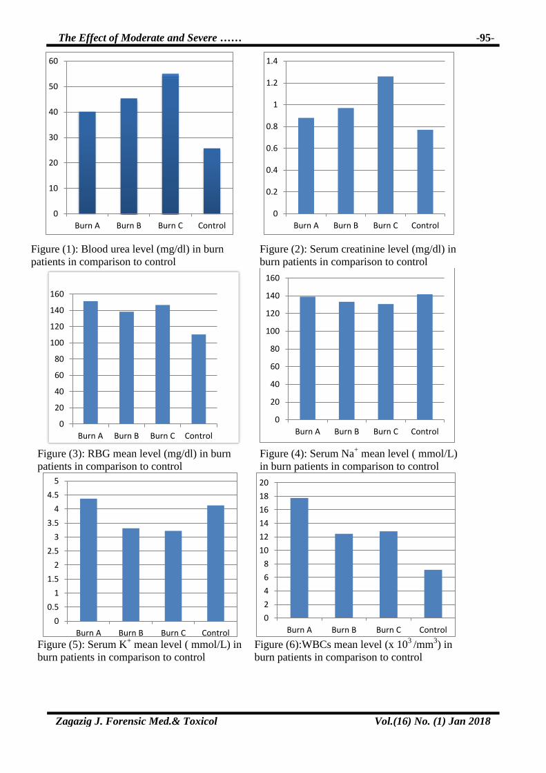
In table 2 and Fig (1-5), the study revealed
that there was a significant increase in blood
urea and serum creatinine mean values in
burned patients, compared to healthy control
specially during the third and the fifth days
post burn. The random blood glucose (RBG)
level showed significant difference where
they showed maximum increase during the
first day post burn compared to control, while
the 3rd
and 5th
days post burn showed less
significant increase in comparison to control
level. Regarding serum electrolytes, the serum
Na+ & K+ levels declined in a significant
manner during the 3rd
and 5th
days post burn,
while its level did not show any significant
differences during the 1st day post burn.
Table 3 and fig (6-11), show a significant
increase in (WBCs) and Neutrophils count
during the 1st, 3
rd and 5
th days post burn
compared to the control group, where the
highest increase was noticed during the 1st
day post burn. The Platelets count showed
significant decrease in burn patients
throughout the whole period of monitoring
with the great lowering was in the 3rd
day.
Hemoglobin (Hb) level showed no significant
difference during the 1st day whereas it
showed significant decrease during the 3rd
and
5th
days post burn. Red blood cells (RBCs)
count increased in a significant manner during
the 1st day, then decreased significantly on the
5th
day post burn. The hematocrit value
(HCT) decreased significantly on the 3rd
and
5th
days post burn.
Table 4 and fig (12-19) show the mean
values of the liver functions, regarding liver
transaminases (ALT & AST) and alkaline
phosphatase (ALP) the mean values showed
significant increase after burn specially on the
5th
day. The serum levels of total protein and
albumin decreased in a significant manner
during the whole days post burn under the
study. Regarding the coagulation profile, the
prothrombin concentration (PC) was lowered
and Prothrombin time (PT) was prolonged in
a significant manner in burn patients during
the whole period of the study. The
International Normalized Ratio (INR) showed
a significant increase in patients over the
control value on the 1st, 3
rd and 5
th days post
burn.
Table (1): Burn characteristics of the studied patients Burn characteristics No. (n= 60) %
Cause of burn:
Flame 46 76.7
Hot liquid 10 16.6
Electric current 4 6.7
Type of burn:
Dry 46 76.7
Scald 10 16.6
Electric 4 6.7
Degree of Burn:
First and Second 8 13.3
Second 4 6.7
Second and Third 36 60.0
Third 12 20.0
TBSA%:
< 25% 31 51.6
25 - 50% 26 43.3
50 - 75% 3 5.1
Mean ± SD 35.87 ± 2.55
Outcome of injury:
Improved 33 55.0
Deteriorated 19 31.7
Dead 8 13.3

The Effect of Moderate and Severe …… -94-
Zagazig J. Forensic Med.& Toxicol Vol.(16) No. (1) Jan 2018
Table (2): comparison between blood urea, serum creatinine, RBG, sodium and potassium levels in
burn patients and control
Burn (A)
N=60
Burn (B) Burn (C) Control
N=20 P-
value1
P-
value2
P-
value3
Mean ± SD Mean ± SD Mean ± SD Mean ± SD
Urea (mg/dl) 40.15 ± 22.32 45.37 ± 52.43 53.29 ± 24.57 25.71 ± 6.45 0.017* 0.004* 0.001*
Creatinine (mg/dl) 0.88 ± 0.26 0.97 ± 0.42 1.26 ± 0.39 0.77 ± 0.26 0.134 0.05* 0.01*
RBG (mg/dl) 151.39 ± 58.40 138.54 ± 45.66 146.84 ± 61.73 110.54 ± 12.29 0.003* 0.003* 0.043*
Na+ ( mmol/L) 138.96 ± 4.53 136.23 ± 3.27 134.40 ± 4.69 141.80 ± 4.08 0.064 0.000* 0.000*
K+ ( mmol/L) 4.37 ± 0.53 3.75 ± 0.49 3.69 ± 0.41 4.13 ± 0.36 0.091 0.03* 0.01*
RBG (random blood glucose) Na (sodium) K (potassium) N= number of cases
Student-t test where P<0.05= significant value p 1=comparison between control & Burn (A),
p 2=comparison between control & Burn (B), p 3=comparison between control & Burn (C)
Table (3): complete blood picture parameters in burn patients compared to control
Burn (A)
N=60
Burn (B) Burn (C) Control
N=20 P-value1 P-value
2
P-
value3
Mean ± SD Mean ± SD Mean ± SD Mean ± SD
WBCs (x 103
/mm3)
17.74 ± 7.51 12.45 ± 4.92 12.82 ± 3.90 7.13 ± 2.22 0.000* 0.000* 0.000*
Neutrophils (%) 82.11 ± 6.84 74.89 ± 6.77 76.44 ± 5.76 65.53 ± 6.08 0.000* 0.000* 0.000*
PLTs (x 103 mm
3) 237.78 ± 35.42 213.24 ± 32.12 235.00 ± 42.97 303.33 ± 50.42 0.001* 0.000* 0.009*
Hb ( gm/dl) 14.79 ± 2.79 11.17 ± 2.21 10.30 ± 1.22 13.66 ± 1.12 0.118 0.000* 0.000*
RBCs (x 106 /mm
3) 5.46 ± 0.98 4.19 ± 0.78 3.99 ± 0.53 4.31 ± 0.47 0.000* 0.804 0.038*
HCT (%) 45.12 ± 10.21 33.65 ± 6.53 32.07 ± 4.11 39.09 ± 2.82 0.079 0.000* 0.000*
WBCs (white blood cells) - PLTs (platelets) - Hb (hemoglobin) - RBCs (red blood cells) - HCT
(hematocrit value) N= number of cases Student-t test where P<0.05= significant value
p 1=comparison between control & Burn (A) p 2=comparison between control & Burn (B), p
3=comparison between control & Burn (C)
Table (4): comparison between liver functions in burn patients and control
Burn (A)
N=60
Burn (B) Burn (C) Control
N=20 P-value1 P-value
2 P-value
3
Mean ± SD Mean ± SD Mean ± SD Mean ± SD
ALT (IU/L) 29.71 ± 14.38 38.78 ± 22.22 49.36 ± 20.16 22.80 ± 5.35 0.021* 0.013* 0.000*
AST (IU/L) 33.05 ± 10.84 34.13 ± 14.87 41.71 ± 15.02 25.47 ± 3.29 0.014* 0.013* 0.000*
ALP (IU/L) 67.93 ± 13.65 74.53 ± 13.70 85.67 ± 15.09 55.07 ± 11.00 0.04* 0.015* 0.001*
Total protein (g/dl) 4.76 ± 0.70 4.46 ± 0.60 4.53 ± 0.84 7.12 ± 0.45 0.007* 0.000* 0.000*
Albumin (g/dl) 2.60 ± 0.45 2.41 ± 0.40 2.25 ± 0.52 3.83 ± 0.41 0.005* 0.000* 0.000*
PC (%) 82.91 ± 8.82 76.09 ± 9.28 76.02 ± 9.32 99.13 ± 1.13 0.03* 0.000* 0.000*
PT (Sec) 13.93 ± 1.03 14.59 ± 1.16 14.76 ± 1.12 12.05 ± 0.07 0.000* 0.000* 0.000*
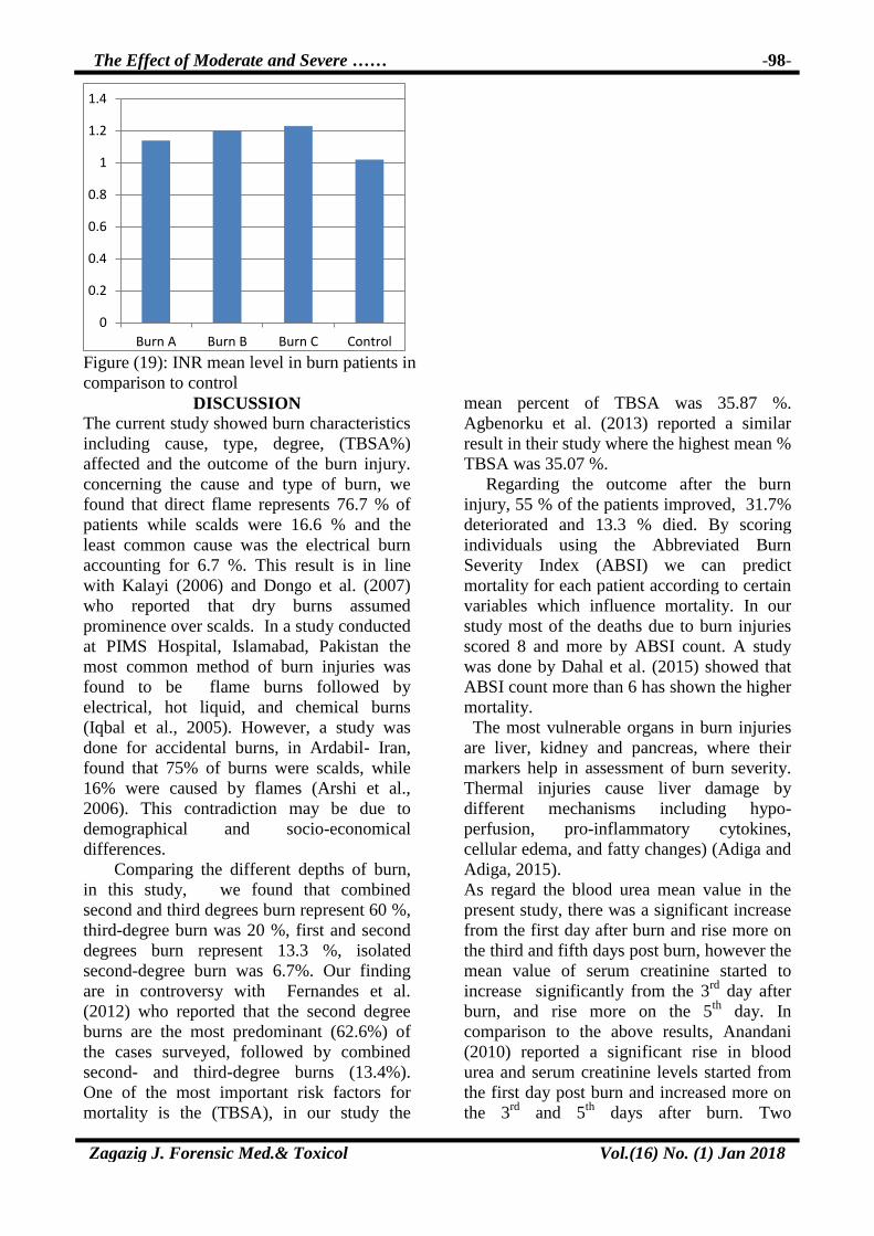
INR 1.14 ± 0.09 1.20 ± 0.10 1.23 ± 0.10 1.02 ± 0.03 0.04* 0.01* 0.001*
ALT (alanine transaminase); AST (aspartate transaminase); ALP (alkaline phosphatase)
PC (prothrombin concentration); PT (prothrombin time); INR (international normalized
ratio); N (number of cases) Student-t test where P<0.05=
significant value p 1=comparison between control & Burn (A), p 2=comparison between
control & Burn (B), p 3=comparison between control & Burn (C)
The Effect of Moderate and Severe …… -95-
Zagazig J. Forensic Med.& Toxicol Vol.(16) No. (1) Jan 2018
Figure (1): Blood urea level (mg/dl) in burn
patients in comparison to control
Figure (2): Serum creatinine level (mg/dl) in
burn patients in comparison to control
Figure (3): RBG mean level (mg/dl) in burn
patients in comparison to control
Figure (4): Serum Na+ mean level ( mmol/L)
in burn patients in comparison to control
Figure (5): Serum K
+ mean level ( mmol/L) in
burn patients in comparison to control
Figure (6):WBCs mean level (x 103
/mm3) in
burn patients in comparison to control
0
10
20
30
40
50
60
Burn A Burn B Burn C Control
0
0.2
0.4
0.6
0.8
1
1.2
1.4
Burn A Burn B Burn C Control
0
20
40
60
80
100
120
140
160
Burn A Burn B Burn C Control
0
20
40
60
80
100
120
140
160
Burn A Burn B Burn C Control
0
0.5
1
1.5
2
2.5
3
3.5
4
4.5
5
Burn A Burn B Burn C Control
0
2
4
6
8
10
12
14
16
18
20
Burn A Burn B Burn C Control

The Effect of Moderate and Severe …… -96-
Zagazig J. Forensic Med.& Toxicol Vol.(16) No. (1) Jan 2018
Figure (7): Neutrophils (%) mean level in burn
patients in comparison to control
Figure (8): Platelet count mean level in burn
patients in comparison to control
Figure (9): Hb mean levels in burn
patients in comparison to control
Figure (10): RBCs mean levels in burn patients
in comparison to control
Figure (11): HCT mean levels in burn
patients in comparison to control
Figure (12): ALT mean levels in burn patients
in comparison to control
0%
10%
20%
30%
40%
50%
60%
70%
80%
90%
Burn A Burn B Burn C Control
0
50
100
150
200
250
300
350
Burn A Burn B Burn C Control
0
2
4
6
8
10
12
14
16
Burn A Burn B Burn C Control
0
1
2
3
4
5
6
Burn A Burn B Burn C Control
0%
5%
10%
15%
20%
25%
30%
35%
40%
45%
50%
Burn A Burn B Burn C Control
0
10
20
30
40
50
60
Burn A Burn B Burn C Control

The Effect of Moderate and Severe …… -97-
Zagazig J. Forensic Med.& Toxicol Vol.(16) No. (1) Jan 2018
Figure (13): AST mean level in burn
patients in comparison to control
Figure (14): ALP mean level in burn patients
in comparison to control
Figure (15): total Protein mean level in burn
patients in comparison to control
Figure (16): Serum Albumin mean level in
burn patients in comparison to control
Figure (17): Prothrombin Concentration mean
level in burn patients in comparison to control
Figure (18): Prothrombin Time mean level
in burn patients in comparison to control
0
5
10
15
20
25
30
35
40
45
Burn A Burn B Burn C Control51
52
53
54
55
56
57
58
59
60
61
62
Burn A Burn B Burn C Control
0
1
2
3
4
5
6
7
8
Burn A Burn B Burn C Control 0
0.5
1
1.5
2
2.5
3
3.5
4
4.5
Burn A Burn B Burn C Control
0%
20%
40%
60%
80%
100%
120%
Burn A Burn B Burn C Control
0
2
4
6
8
10
12
14
16
Burn A Burn B Burn C Control
The Effect of Moderate and Severe …… -98-
Zagazig J. Forensic Med.& Toxicol Vol.(16) No. (1) Jan 2018
Figure (19): INR mean level in burn patients in
comparison to control
DISCUSSION
The current study showed burn characteristics
including cause, type, degree, (TBSA%)
affected and the outcome of the burn injury.
concerning the cause and type of burn, we
found that direct flame represents 76.7 % of
patients while scalds were 16.6 % and the
least common cause was the electrical burn
accounting for 6.7 %. This result is in line
with Kalayi (2006) and Dongo et al. (2007)
who reported that dry burns assumed
prominence over scalds. In a study conducted
at PIMS Hospital, Islamabad, Pakistan the
most common method of burn injuries was
found to be flame burns followed by
electrical, hot liquid, and chemical burns
(Iqbal et al., 2005). However, a study was
done for accidental burns, in Ardabil- Iran,
found that 75% of burns were scalds, while
16% were caused by flames (Arshi et al.,
2006). This contradiction may be due to
demographical and socio-economical
differences.
Comparing the different depths of burn,
in this study, we found that combined
second and third degrees burn represent 60 %,
third-degree burn was 20 %, first and second
degrees burn represent 13.3 %, isolated
second-degree burn was 6.7%. Our finding
are in controversy with Fernandes et al.
(2012) who reported that the second degree
burns are the most predominant (62.6%) of
the cases surveyed, followed by combined
second- and third-degree burns (13.4%).
One of the most important risk factors for
mortality is the (TBSA), in our study the
mean percent of TBSA was 35.87 %.
Agbenorku et al. (2013) reported a similar
result in their study where the highest mean %
TBSA was 35.07 %.
Regarding the outcome after the burn
injury, 55 % of the patients improved, 31.7%
deteriorated and 13.3 % died. By scoring
individuals using the Abbreviated Burn
Severity Index (ABSI) we can predict
mortality for each patient according to certain
variables which influence mortality. In our
study most of the deaths due to burn injuries
scored 8 and more by ABSI count. A study
was done by Dahal et al. (2015) showed that
ABSI count more than 6 has shown the higher
mortality.
The most vulnerable organs in burn injuries
are liver, kidney and pancreas, where their
markers help in assessment of burn severity.
Thermal injuries cause liver damage by
different mechanisms including hypo-
perfusion, pro-inflammatory cytokines,
cellular edema, and fatty changes) (Adiga and
Adiga, 2015).
As regard the blood urea mean value in the
present study, there was a significant increase
from the first day after burn and rise more on
the third and fifth days post burn, however the
mean value of serum creatinine started to
increase significantly from the 3rd
day after
burn, and rise more on the 5th
day. In
comparison to the above results, Anandani
(2010) reported a significant rise in blood
urea and serum creatinine levels started from
the first day post burn and increased more on
the 3rd
and 5th
days after burn. Two
0
0.2
0.4
0.6
0.8
1
1.2
1.4
Burn A Burn B Burn C Control

The Effect of Moderate and Severe …… -99-
Zagazig J. Forensic Med.& Toxicol Vol.(16) No. (1) Jan 2018
mechanisms of renal failure have been
described in burn patients. The first one
occurs early after the injury and is due to
hypovolemia, low cardiac output, and
systemic vasoconstriction, and the second one
is the presence of some degree of
myoglobinuria which can destroy tubular cells
(Schneider et al., 2012).
In the present study, there was a significant
increase in the mean random serum glucose
level early on the first day post burn. This
could be explained by stress due to severe
pain & anxiety in many cases of burn, where
the body respond by excessive secretion of
glucagon, growth hormone, catecholamine,
and glucocorticoid (Marti and Leitman,
2013).
The present study showed a significant
decrease in serum Na+ level on the 3rd and
5th days post burn while its level showed
non-significant decrease on the 1st day post
burn. In contrary to our findings, Kaddoura et
al. (2003), Al-Muhammadi and Azeez, (2011)
reported that a significant hyponatremia
started in the first day of burn before starting
resuscitation therapy and increased after
resuscitation. A retrospective study done by
Stewart et al. (2013) revealed that
hyponatremia occurred in 6.8% and
hypernatremia in 9.9% of burn patients.
In the present study, serum K+ mean value
showed non significant increase during the 1st
day post burn however, it showed a
significant decline on the 3rd and 5th days
post burn. This result is in agreement with
Rainer et al. (1999), who reported that
hypokalaemia is well recognized after stress
conditions due to the combined effect of
adrenaline and insulin, which stimulates
receptors on skeletal muscle fibers with
subsequent uptake of potassium from the
circulation. The hypokalemia in the early
post-resuscitation period between 2-5 days of
burns' patients may be due to increased
potassium losses (urinary, gastric or fecal). In
disagreement with our findings, Kaddoura et
al. (2017), Al-Muhammadi and Azeez (2011)
reported that significant hyperkalemia
occurred early in burn patients prior to
resuscitation therapy.
The White Blood Cells count and the ratio
of neutrophilic count showed a significant
increase during the 1st, 3
rd and 5
th days post
burn where the maximum increase was
recorded on the 1st day post burn. Our results
are similar to those of El-Sonbaty and El –
Otiefy (1996) who reported significant
leukocytosis from the first day after burn ,
also the results of the present study are in line
with Kim et al. (2011) and Belba et al. (2015)
who reported that thermal injuries stimulate
changes in hematopoiesis by inducing early
acute phase response, and the WBC count is
important hallmark in the evaluation of burn
injuries. Our findings disagree with those of
Barati et al. (2008) who reported that no
significant differences were found in the total
WBC count, in burn patients with or without
bacterial infection. This controversy may be
explained by the fact that the inflammatory
systemic signs (changes in body temperature,
tachycardia and leukocytosis) are used for
diagnosis of sepsis, but sometimes this may
be misleading, because critically ill burn
patients often manifest a systemic
inflammatory response syndrome without
infection (Mokline et al., 2015).
Platelets count monitored in the present
study showed a significant decline throughout
the period of monitoring, this result is in line
with Pavic and Milevoj (2007) who reported a
significant decrease in platelets count which
was observed in patients with moderate and
severe burn injuries. A retrospective study
done by Kim et al. (2011) on 265 burned
patients showed that the platelet count started
to increase immediately after thermal injury
reaching the peak within 12 hours, then
decreased gradually. Sarda et al. (2005)
concluded that platelet count decreases
initially in all cases of burn sepsis, then it
gradually rises to normal in improving
patients and declines gradually in
deteriorating patients. The decrease in platelet
count is caused by multiple factors, increased
Platelet destruction, hemodilution and
reduced platelet production (Pavic and
Milevoj, 2007).
Regarding (RBCs) count, it increased in a
significant manner during the 1st day post
burn, then decreased significantly on the 5th
day post burn. Hemoglobin (Hb) and (HCT)
levels showed a significant decrease during
the 3rd
and 5th
days post burn. This study

The Effect of Moderate and Severe …… -100-
Zagazig J. Forensic Med.& Toxicol Vol.(16) No. (1) Jan 2018
agrees with Al-Muhammadi and Azeez
(2011), EI-Sonbaty and EI-Otiefy (1996),
who stated that (RBCs) count, hemoglobin
concentration and hematocrit value showed
significantly high levels immediately after
burn, then decreased gradually to below the
control level on the 4th
day post-burn. The
decline of hematocrit value could be expected
due to adequate fluid resuscitation, but may
also be a sign of occult bleeding. Anemia
which complicate severe burn injuries,
usually starts few days after burn due to
morphological changes in (RBCs) leading to
increased destruction, and also due to
decreased erythropoietin production and
frequent surgical maneuvers done to those
patients (Posluszny and Gamelli (2010). Al-
Muhammadi and Azeez (2011) found that
hematocrit value decreased when major blood
loss treated by plasma volume replacement or
due to pre-existing anemia or hypervolemia.
Regarding serum levels of liver enzymes,
the present study showed a highly significant
increase in ALT, AST and ALP levels in burn
patients, with the highest recordings on the 5th
day post burn. In agreement with our
findings, Bhagwat et al. (2007) and Jeschke et
al. (2007) reported elevation of liver enzymes
in burn patients which reach maximum level
by day 5. The excess release of hepatic
enzymes could be explained by that burns
lead to increase in edema formation that leads
to cell damage (Adiga and Adiga, 2015).
The findings of the present study revealed
significantly lower serum total proteins and
serum albumin levels from the 1st up to the 5
th
days post burn in comparison with healthy
control, also there were significant decrease in
prothrombin concentrations and significant
increase in prothrombin time and INR. These
results were in accordance with Al-
Muhammadi and Azeez (2011), Aguayo-
Becerra et al. (2013) and Deepthi et al.
(2015). The mechanism of lowered values of
serum proteins in the acute phase is due to
local inflammatory cytokines which enter
circulation in case of moderate and severe
burn injuries resulting in systemic
inflammatory response leading to generalized
micro-vascular leak and permitting fluid &
protein loss from intra-vascular to extra-
vascular compartments (Al-Muhammadi and
Azeez, 2011). After the initial phase, hypo-
proteinemia and hypo-albuminemia persist
because of the lack of their synthesis, loss
through damaged skin areas, and lack of
nutritional support (Kumar, 2010). The
decreased prothrombin concentration and the
increase in prothrombin time and INR
represent one of the liver synthetic function
which affected by burn injury
CONCLUSION Mild and moderate burn injuries result in
renal and hepatic dysfunction, in addition to
disturbances in hematological parameters and
bleeding profiles, so that estimation of
hematological and serum biochemical
markers in burn patients is very important for
helping the health care team to expect the
prognosis in burn patients and how the body
is responding to the different therapeutic
lines.
RECOMMENDATIONS
careful monitoring of liver and kidney
functions, serum electrolytes and complete
blood picture is very important in patients
with moderate and severe burn injuries to
prevent complications and improve outcome.
We recommend follow up for longer period (4
weeks) in patients with altered biochemical
parameters.
REFERENCES
-Adiga U and Adiga S (2015): Biochemical
Changes in Burns. International Journal of
Research Studies in Biosciences; 3 (7):88-
91.
-Agbenorku P, Agbenorku M and Fiifi-
Yankson PK (2013): Pediatric burns
mortality risk factors in a developing
country’s tertiary burns intensive care unit.
International Journal Burns Trauma.;
3(3):151–8.
-Aguayo-Becerra OA, Torres-Garibay C,
Macı´as-Amezcua MD et al. (2013): Serum
albumin level as a risk factor for mortality in
burn patients. Clinics. 68(7):940-45.
-Al-Muhammadi MO and Azeez HA (2011):
Some Physiological Changes in Burn
Patients. Medical Journal of Babylon; 8(3):
303-19.
-Anandani JH (2010): Impact of thermal
injury on hematological and biochemical

The Effect of Moderate and Severe …… -101-
Zagazig J. Forensic Med.& Toxicol Vol.(16) No. (1) Jan 2018
parameters in burn patients. Biosci. Biotech
Res. Comm; 3 (1): 97-100.
-Arshi S, Sadeghi-Bazargani H, Mohammadi
R et al. (2006): Prevention oriented
epidemiologic study of accidental burns in
rural areas of Ardabil, Iran. Burns,
32(3):366-71.
-Barati M, Alinejad F, Bahar MA et al.
(2008): Comparison of WBC, ESR, CRP
and PCT serum levels in septic and non-
septic burn cases. Burns, 34(6): 770-74.
-Belba M, Aleksi A, Nezha I et al. (2015):
Impact of Severe Burns in Hematological
Parameters. AJMHS, 46 (3): 59-69
-Bhagwat VR, Subrahmanyam M and Pujari
KN (2007): Serum enzymes in thermal
injury. Indian Journal. Clinical
Biochemistry. 22 (2): 154-57.
-Cakir B and Yegen BC (2004): Systemic
responses to burn injury. Turk Journal Med
Sci. (34): 215–26.
-Dahal P, Ghimire S, Maharjan NK et al.
(2015): Baux’s and Abbreviated Burn
Severity Score for the Prediction of
Mortality in Patients with Acute Burn
Injury. Journal of College of Medical
Sciences-Nepal, Vol-11, No 4, Oct-Dec 015.
-Deepthi SKD and Narayan GAR (2015):
Evaluation of serum albumin levels and its
relation to burn size in burn patients. World
journal of Pharmacy and Pharmaceutical
Sciences 4(8): 1462-5.
- Dongo, AE, Irekpita, EE, Oseghale, LO et
al. (2007): A five-year review of burn
injuries in Irrua. BMC Health Serv Res.;
7:171.
-El-Sonbaty MA and El –Otiefy MA (1996):
Hematological changes in severely Burned
Patients . Annals of Burns and Fire Disasters
9(4): 1-4.
-Fernandes FMFA, Torquato IMB, Dantas
MAS et al. (2012): Burn injuries in children
and adolescents: clinical and
epidemiological characterization. Rev
Gaúcha Enferm. 33(4):133-41.
-Halkes S, van den Berg A, Hoekstra M
(2002): Transaminase and alkaline
phosphatase activity in serum of burn
patients treated with highly purified tannic
acid. Acta Chir Plast. 28: 449-53.
-Hettiaratchy S and Papini R (2004): Initial
management of a major burn: II—
assessment and resuscitation. BMJ.
329(7457): 101–3.
-Iqbal T, Rashid R and Ibrahim M (2005):
Incidence of burn injury admission at PIMS,
Islamabad. Ann Pak Inst Med Sci. 1:194‑5.
-Jeschke MG, Micak RP, Finnerty CC et al.
(2007): Changes in liver function and size
after a severe thermal injury. Shock 28:172–
7.
-Kaddoura I, Sittah GA, Karamanoukian R et
al. (2017): Burn injury: review of
pathophysiology and therapeutic modalities
in major burns. Ann Burns Fire Disasters.
Jun 30; 30(2): 95–102
-Kalayi GD (2006): Mortality from burns in
Zaria: an experience in a developing
economy. East Afr Med Journal 83: 461–64.
-Kim HS, Kwon HW, Yang HT et al. (2011):
A Serial Study of Hematologic Change in
Burned Patients. Journal Lab Med Qual
Assur. Jun; 33(1): 9-16.
-Kim GH, Oh KH and Yoon JW (2003):
Impact of burn size and initial serum
albumin level on acute renal failure
occurring in major burn. Am Journal
Nephrol 23: 55-60.
-Kumar P (2010): Grading of severity of the
condition in burn patients by serum protein
and albumin/globulin studies. Ann Plast
Surg.; 65(1):74-9.
- Lavrentieva A, Depetris N and Kaimakamis
E (2016): Monitoring and treatment of
coagulation abnormalities in burn patients.
An international survey on current practices.
Ann Burns Fire Disasters. 29: 172-77.
-Mahmood S, Prabhakar M, Anees S et al.
(2016): Hypoproteinemia, Hypocalcemia,
Hyponatraemia, Hyperkalaemia and Trace
Elements in Patients with Burn Injuries.
International journal of scientific research
5(10): 588-9.
-Marti JL and Leitman IM (2013):
Understanding the causes of hyperglycemia
in burn patients. Journal of surgical research
182. 205-6.
-Mokline A, Garsallah L, Rahmani I et al.
(2015): Procalcitonin: a diagnostic and
prognostic biomarker of sepsis in burned
patients. Ann Burns Fire Disasters. Jun 30;
28 (2): 116-20

The Effect of Moderate and Severe …… -102-
Zagazig J. Forensic Med.& Toxicol Vol.(16) No. (1) Jan 2018
-Pavic M and Milevoj L (2007): Platelet
count monitoring in burn patients.
Biochemia Medica 17(2): 212–19.
-Peck M and Pressman MA (2013): The
correlation between burn mortality rates
from fire and flame and economic status of
countries. Burns 39:1054.
-Pham TN, Cancio LC and Gibran NS (2008):
American Burn Association practice
guidelines: Burn shock resuscitation. Journal
Burn Care Res 29(1): 257– 66.
--Posluszny JA and Gamelli RL (2010):
Anemia of thermal injury: combined acute
blood loss anemia and anemia of critical
illness. Journal Burn Care Res. 31: 229–42.
-Rainer TH, Beattie T, Crofton P et al. (1999):
Systemic hormonal, electrolyte, and
substrate changes after non-thermal limb
injury in children. Journal Accid. Emerg.
Med. March; 16(2): 104–7.
-Sarda DK, Dagwade AM and Lohiya S
(2005): Evauation of platelet count as a
prognostic indicator in early detection of
post burn septicaemia. Bombay Hosp
Journal 47(3): 36.
-Schneider DF, Dobrowolsky A, Shakir IA et
al. (2012): Predicting acute kidney injury
among burn patients in the 21st century: a
classification and regression tree analysis.
Journal Burn Care Res. 33 (2):242–51.
-Stewart IJ, Morrow BD, Tilley MA et al.
(2013): Dysnatremias and survival in adult
burn patients: a retrospective analysis. Am
Journal Nephrol. 37:59–64 .