the effectiveness of subdural drains using urokinase after burr … · tion, and general anesthesia...
TRANSCRIPT

Copyright © 2016 Korean Neurotraumatology Society 101
Introduction
A hematoma formed between the dura mater and arach-noid is called a subdural hematoma (SDH), and it is known
to occur in 5% to 29% of all head trauma patients.1,6,19) It can even occur in patients with no trauma or minor trau-ma, such as those who take anticoagulants, have a blood disease, or have received a hydrocephalus shunt.7,14,15,19) The hematoma is often formed by the rupture of the artery or vein of the cerebral cortex or the bridging vein between the cerebral cortex and the arteries and veins.4)
A subacute SDH (SASDH) is a hematoma occurring be-tween 4 and 20 days after injury.6,9) Surgical treatment is required if neurological symptoms progress from mild to moderate or severe due to injury during the subacute stage (e.g., decreased consciousness, motor nerve palsy, head-ache), while patients receive conservative treatment for mild neurological symptoms due to acute SDH.6) For SASDH, a
The Effectiveness of Subdural Drains Using Urokinase after Burr Hole Evacuation of Subacute Subdural Hematoma in Elderly Patients : A Prelimilary Report
Chang-Gi Yeo, MD1, Woo-Yeol Jeon, MD2, Seong-Ho Kim, MD1, Oh-Lyong Kim, MD1, and Min-Su Kim, MD1
1Department of Neurosurgery, Yeungnam University College of Medicine, Daegu, Korea 2Department of Neurosurgery, Good Morning Hospital, Daegu, Korea
Objective: A subdural drain using urokinase after a burr hole hematoma evacuation was performed for subacute subdural hematoma (SASDH), and its effectiveness and safety in elderly patients were evaluated. Methods: Between January 2013 and May 2015, subdural drains using urokinase after burr hole hematoma evacuation were performed in 19 elderly patients. The inclusion criteria were as follows: 1) a subdural hematoma occurring between 4 and 20 days after injury; 2) worsening neurological symptoms, from mild to moderate or severe, due to injury during the subacute stage; 3) a mix of solid clots (high-density lighter shadow) and fluid hematoma (low-density darker shadow) on the computed tomography (CT) scan; 4) a score of ≥9 on the Glasgow Coma Scale (GCS) assessed immediately before sur-gery; and 5) an age of ≥65 years. When the majority of the hematoma was evacuated on the CT, we removed the catheter.Results: Under local anesthesia, a catheter was inserted into the hematoma through a burr hole. The mean age of the pa-tients was 73.7 years (range, 65-87 years). The mean preoperative GCS score was 11.2 (range, 10-13), and the mean Glasgow Outcome Scale score for all patients was 5 at discharge. No recurrences of hematomas or surgical complications were observed.Conclusion: A subdural drain using urokinase after burr hole hematoma evacuation under local anesthesia is thought to be an effective and safe method of blood clot removal with low morbidity. This surgical method is less invasive for treating elderly patients with SASDH. (Korean J Neurotrauma 2016;12(2):101-106)
KEY WORDS: Subdural hematoma ㆍElderly patients ㆍDrainage ㆍUrokinase.
Received: February 17, 2016 / Revised: March 22, 2016Accepted: April 25, 2016Address for correspondence: Min-Su KimDepartment of Neurosurgery, Yeungnam University College of Medicine, 170 Hyeonchung-ro, Nam-gu, Daegu 42415, KoreaTel: +82-53-620-3790, Fax: +82-53-620-3770E-mail: [email protected] cc This is an Open Access article distributed under the terms of Cre-ative Attributions Non-Commercial License (http://creativecommons.org/licenses/by-nc/3.0/) which permits unrestricted noncommercial use, distribution, and reproduction in any medium, provided the original work is properly cited.
CLINICAL ARTICLEKorean J Neurotrauma 2016;12(2):101-106
pISSN 2234-8999 / eISSN 2288-2243
https://doi.org/10.13004/kjnt.2016.12.2.101
102 Korean J Neurotrauma 2016;12(2):101-106
Burr Hole Drainage Using Urokinase for Subacute Subdural Hematoma
burr hole or craniotomy should be considered. With burr holes, it is often difficult to remove hematomas completely. Although hematomas can be removed completely with cra-niotomies, they increase morbidity and mortality because of the blood loss, long operating time, postoperative infec-tion, and general anesthesia associated with the invasive surgery.10,18)
Therefore, we propose that a subdural drain using uro-kinase after a burr hole hematoma evacuation under local anesthesia would be less invasive and more effective for elderly patients with SASDHs. Although stereotactic he-matoma aspiration using urokinase is commonly practiced for spontaneous intracerebral hemorrhages,2,11) no studies have been published on its use for SASDH. The effective-ness and safety of a subdural drain using urokinase after a burr hole hematoma evacuation for these patients and their outcomes are reported.
Materials and Methods
Patient selectionBetween January 2013 and May 2015, 19 elderly patients
with SASDH who underwent subdural drains using uroki-nase after burr hole hematoma evacuation were analyzed.
The inclusion criteria were as follows: 1) a SDH occur-ring between 4 and 20 days after injury; 2) worsening symp-toms, from mild to moderate, due to injury during the sub-acute stage while receiving conservative treatment for mild symptoms due to acute SDH upon admission; 3) a mix of solid clots (high-density lighter shadow) and fluid hemato-ma (low-density darker shadow) on the computed tomog-raphy (CT) scan; 4) a score of ≥9 on the Glasgow Coma Scale (GCS) assessed immediately before surgery; and 5) an age of ≥65 years. The exclusion criteria were as follows: 1) co-occurrence with other head trauma, including brain contusion, epidural hematoma, intracerebral hemorrhage, intraventricular hemorrhage, and traumatic subarachnoid hemorrhage; 2) the occurrence of the SDH as a complica-tion from previous neurosurgery, including craniotomy, ventricular drainage, or ventricle peritoneal shunt; 3) a score of <8 on the GCS assessed immediately before surgery; and 4) an age of <65 years.
Surgical treatment and evaluationUnder local anesthesia, a catheter was inserted into the
hematoma through a burr hole in all patients. Surgery was conducted with the patient in the supine position with the head raised by approximately 15 degrees and the site for the burr hole raised the highest. The fenestration site was
planned for the thickest part of the hematoma. Local anes-thesia with 1% lidocaine was applied on the scalp, a 3-cm incision was made, and then fenestration was performed with an electric drill. After incising the dura mater and identifying the membrane of the hematoma, we identified the hematoma from the subdural space and inserted a 9-French drainage tube to a depth of about 5 cm in the fron-tal area. After injecting a mixture of 3 mL of normal saline and urokinase (1,000 units) using the drainage tube, we closed off the tube for two hours and then opened the tube again to allow the hematoma to be drained naturally by gravity into the drainage pocket. Urokinase was injected into the hematoma every 12 hours. The drainage tube was removed on the second or third day after surgery based on the amount of remaining hematoma seen on the CT per-formed on the second day after surgery.
We collected the following clinical information on par-ticipants: age, gender, symptoms, head trauma history, underlying systemic disease, antiplatelet or anticoagulant medication use, and blood test results. The patient condi-tions were evaluated using the GCS and assessed immedi-ately before surgery. The treatment outcomes of all patients were evaluated using the Glasgow Outcome Scale (GOS).
This study was approved by the Institutional Review Board (IRB) of the Yeungnam University Hospital (IRB No. YUMC 2015-03-004).
Results
Patient demographic characteristics and clinical features
The patient demographic characteristics and clinical fea-tures are summarized in Table 1. The mean age of the pa-tients was 73.7 years (range, 65-87 years). There were 13 male and 6 female patients. The causes of injury were slips in 18 patients and a fall from a height in one patient. Under-lying diseases included hypertension in five patients, dia-betes in eight patients, cerebral infarction in six patients, and myocardial infarction in one patient. Nine patients were taking antiplatelet medication on admission. The blood test results for all patients were normal for prothrombin time, partial thromboplastin time, and platelet counts as-sociated with blood coagulation. The clinical symptoms observed immediately before surgery were drowsy men-tality in 10 patients, stupor mentality in two patients, hemi-paresis in four patients, gait disturbance in one patient, and sensory aphasia in two patients. Ten of the SASDHs were on the right side, seven were on the left side, and two were bilateral. The mean period in which symptoms worsened
Chang-Gi Yeo, et al.
http://www.kjnt.org 103
after head trauma was 13.6 days (range, 4-20 days). The mean preoperative GCS score was 11.2 (range, 10-13).
Surgical outcomesThe hematoma catheter was in place for a median dura-
tion of 2.5 days (range, 2-13 days). The mean GOS score for all patients was 5 at discharge. The mean follow-up period was 17.7 months (range, 12-24 months). During the follow-up period, no recurrences of hematomas or surgery-relat-ed complications, such as infection or intracranial hemor-rhage, were observed in any of the patients. No bleeding in other organs from the use of urokinase or blood test ab-normalities was found.
Illustrative cases
Case 2A 71-year-old female patient was admitted to the emer-
gency room (ER) complaining of headache due to a head trauma after a slip and fall. She showed no neurological abnormalities. The initial CT results showed an acute SDH in the right frontal and temporal regions, resulting in slight pressure on the right ventricle (Figure 1A). She had hyper-tension, diabetes mellitus, and cerebral infarction in her
medical history. She was taking antiplatelet medication on admission. No abnormalities were found on the laboratory analysis. She was initially treated conservatively because of her old age and minor symptoms. On the 17th day from admission, the patient showed symptoms of increased in-tracranial pressure, including paralysis of the upper and lower-left limbs, an increase in the SDH on the CT, increased pressure on the right ventricle, and aggravation of the midline shift toward the left. The CT showed a mixture of solid blood clots and fluid hematoma in the SDH (Figure 1B). A subdural drain using urokinase after a burr hole he-matoma evacuation was performed (Figure 1C). The cath-eter was removed two days after the surgery based on a CT that showed sufficient removal of the hematoma. The CT at postoperative day 7 showed no recurrence of the hema-toma, and the GOS score was 5 at discharge (Figure 1D).
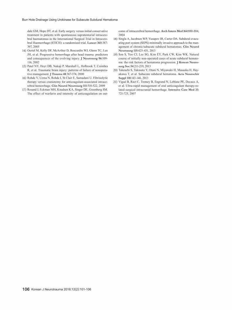
Case 6A 79-year-old male patient was admitted to the ER com-
plaining of sensory aphasia and cognitive deterioration due to head trauma after a slip and fall. The initial CT showed an acute SDH in the left frontal and temporal regions, resulting in slight pressure on the left ventricle (Figure 2A). His med-ical history was unremarkable. No abnormalities were found
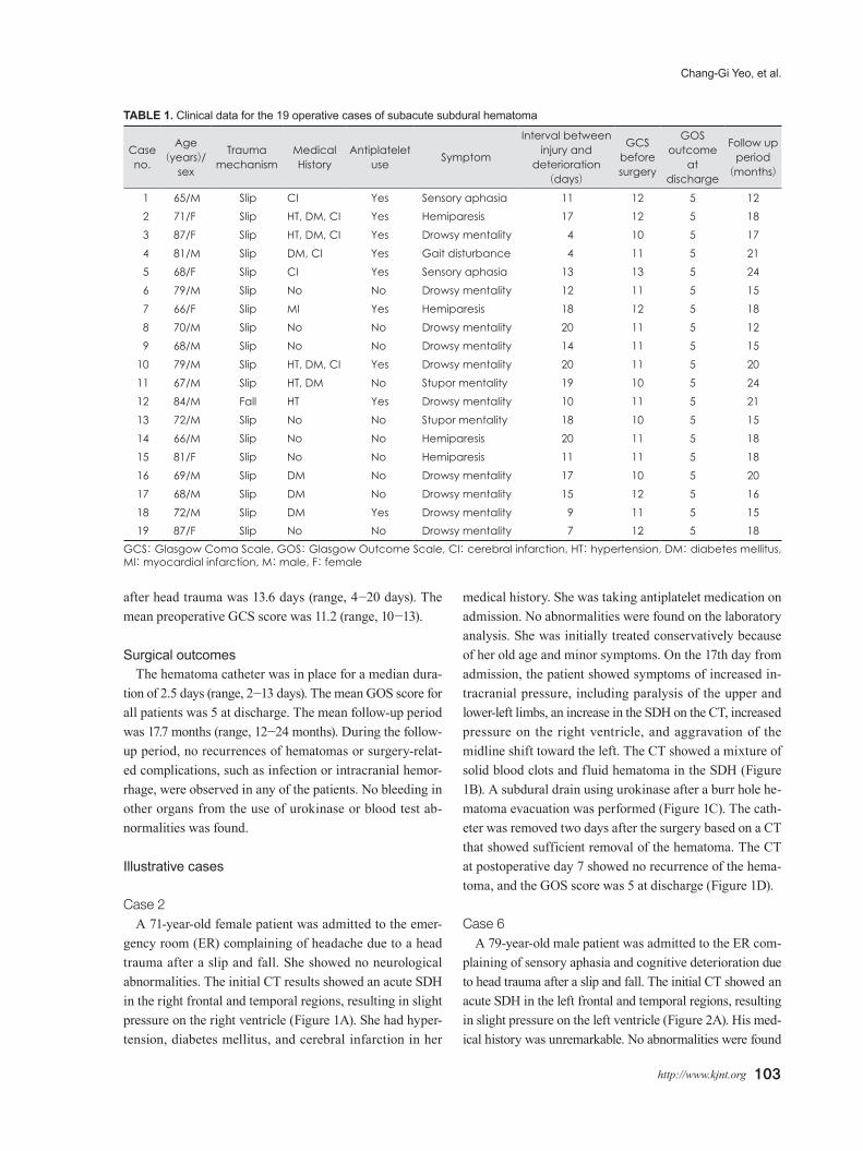
TABLE 1. Clinical data for the 19 operative cases of subacute subdural hematoma
Caseno.
Age(years)/
sex
Trauma mechanism
Medical History
Antiplatelet use Symptom
Interval betweeninjury and
deterioration(days)
GCS before surgery
GOS outcome
at discharge
Follow up period
(months)
1 65/M Slip CI Yes Sensory aphasia 11 12 5 122 71/F Slip HT, DM, CI Yes Hemiparesis 17 12 5 183 87/F Slip HT, DM, CI Yes Drowsy mentality 4 10 5 174 81/M Slip DM, CI Yes Gait disturbance 4 11 5 215 68/F Slip CI Yes Sensory aphasia 13 13 5 246 79/M Slip No No Drowsy mentality 12 11 5 157 66/F Slip MI Yes Hemiparesis 18 12 5 188 70/M Slip No No Drowsy mentality 20 11 5 129 68/M Slip No No Drowsy mentality 14 11 5 15
10 79/M Slip HT, DM, CI Yes Drowsy mentality 20 11 5 2011 67/M Slip HT, DM No Stupor mentality 19 10 5 2412 84/M Fall HT Yes Drowsy mentality 10 11 5 2113 72/M Slip No No Stupor mentality 18 10 5 1514 66/M Slip No No Hemiparesis 20 11 5 1815 81/F Slip No No Hemiparesis 11 11 5 1816 69/M Slip DM No Drowsy mentality 17 10 5 2017 68/M Slip DM No Drowsy mentality 15 12 5 1618 72/M Slip DM Yes Drowsy mentality 9 11 5 1519 87/F Slip No No Drowsy mentality 7 12 5 18
GCS: Glasgow Coma Scale, GOS: Glasgow Outcome Scale, CI: cerebral infarction, HT: hypertension, DM: diabetes mellitus, MI: myocardial infarction, M: male, F: female

104 Korean J Neurotrauma 2016;12(2):101-106
Burr Hole Drainage Using Urokinase for Subacute Subdural Hematoma
on laboratory analysis. He was initially treated conserva-tively because of his old age. On the 12th day from admis-sion, the patient showed drowsy mentality, an increase in the SDH on the CT, increased pressure on the left ventri-cle, and aggravation of the midline shift toward the right (Figure 2B). The CT showed a mixture of solid blood clots and fluid hematoma in the SDH. The tube was removed two days after the surgery based on a CT that showed suffi-cient removal of the hematoma (Figure 2C). The CT at post-operative day 7 showed no recurrence of the hematoma, and the GOS score was 5 at discharge (Figure 2D).
Discussion
Due to the increase in the elderly population, the number of elderly patients with acute SDHs caused by minor head trauma is growing. In particular, a high prevalence of acute SDHs has been reported among elderly patients with hem-orrhagic tendencies caused by antiplatelet agents or antico-agulants.7,14,15,17,19)
SDHs are divided into three stages: acute, subacute, and chronic. In terms of the findings on the form of SDHs on brain CT scans, those in the solid form of a blood clot are in the acute stage, those in a mixed form (a solid blood clot and fluid hematoma) are in the subacute stage, and those in the fluid form are in the chronic stage.5,20) In terms of time, acute SDHs occur within 72 hours of receiving an injury, SASDHs occur within 4 to 20 days of receiving an injury, and chronic SDHs occur within three weeks or longer of receiving an injury.6) Acute and chronic SDHs are easy to differentiate, but subacute and chronic SDHs are difficult to differentiate clearly.
When an acute SDH is small and there are no signs of increased intracranial pressure, conservative treatment is provided, whereas when a hematoma is large and the neu-rological condition is serious due to the increased intra-cranial pressure, it should be removed immediately via a craniotomy.1) A large chronic SDH over 1 cm thick requires surgical treatment, and unlike a solid acute hematoma, a chronic fluid hematoma can be treated by removing the
FIGURE 1. Case 2 was a 71-year-old female patient who presented with headache after a traumatic head injury caused by a slip. (A) At admission, a computed tomography (CT) scan showed a hyperdense hematoma in the subdural space in the right frontotem-poral convexity. (B) She had sudden onset left-sided hemiparesis at 17 days after admission. The follow-up CT showed a mixed hypodense and hyperdense subdural hematoma (SDH) and midline shift progression. (C) An immediately postoperative CT scan showed a catheter in the subdural space and a significant amount of residual hematoma with mass effect. (D) A CT scan at postop-erative 7 days showed a marked decrease of the SDH and resolution of the mass effect.
A B C D
FIGURE 2. Case 6 was a 79-year-old male patient who presented with sensory aphasia and cognitive deterioration after a traumat-ic head injury caused by a slip. (A) At admission, a computed tomography (CT) scan showed a hyperdense hematoma in the sub-dural space in the left frontotemporal convexity. (B) He experienced aggravation of his symptoms at 12 days after admission. The follow-up CT showed a mixed hypodense and hyperdense subdural hematoma (SDH) and midline shift progression. (C) An imme-diately postoperative CT scan showed a catheter in the subdural space and the partial removal of the hematoma. (D) A CT scan at postoperative 7 days showed a marked decrease of the SDH and resolution of the mass effect.
A B C D

Chang-Gi Yeo, et al.
http://www.kjnt.org 105
hematoma using burr hole drainage.8,18) During conserva-tive treatment of an acute SDH, the hematoma can increase suddenly in the subacute stage, requiring surgical treat-ment.1)
In the case of an SASDH with a mix of a solid blood clot and fluid, burr hole drainage can remove the fluid, but it is difficult to remove the solid blood clot.8,20) In particular, for a thick, widely distributed solid hematoma, craniotomy should be considered, as it can completely remove the he-matoma, but it is associated with complications from the general anesthesia and the surgery itself. In particular, high-risk craniotomies can be difficult to perform on elderly pa-tients with a high risk of complications from general anes-thesia due to internal medicine diseases, such as heart or respiratory diseases, or on patients with a high risk of ex-cessive blood loss due to the use of antiplatelet agents or anticoagulants that result in decreased hemostasis.16,21) He-matoma drainage using a burr hole and urokinase is gen-erally performed to decrease the intracranial pressure by draining the cerebrospinal fluid in spontaneous acute ven-tricular intracerebral hemorrhages and removing the solid hematoma.2,3,11)
In cerebral hemorrhages associated with anticoagulants, large amounts of blood can be lost during surgery, and the risk of hemorrhage recurrence is high.13) In these patients, hematoma drainage using a burr hole and urokinase has been reported to be a good micro-invasive treatment, as it has a lower risk of mortality and complications than crani-otomy.16) Good outcomes have been reported for hemato-ma drainage using a burr hole and urokinase in traumatic epidural hematomas with minor neurological abnormali-ties that do not require the reduction of the intracranial pres-sure by removing the hematoma through craniotomy.12)
Urokinase liquefied the solid hematoma that was diffi-cult to drain and rapid SDH drainage was possible within 3 days, the patient’s symptom was improved quickly and early ambulation was possible so this surgical treatment could reduce the length of hospital stay.
In this study, the patients’ mean age was 73.3 years, and there was no mortality or morbidity in any of the patients. Therefore, it is thought that a subdural drain using uroki-nase after a burr hole hematoma evacuation is useful for elderly patients with SASDHs. However, our report is on a small number of cases, and it is a retrospective study. Thus, more cases with accumulated clinical experience are need-ed for the further analysis of reliability and outcomes. In addition, it will be necessary to evaluate the effectiveness and safety of our procedure compared with craniotomy for hematoma removal in elderly patients with SASDHs.
Conclusion A subdural drain using urokinase after burr hole hema-
toma evacuation under local anesthesia is thought to be an effective and safe method of blood clot removal with low morbidity. This surgical method is less invasive for treating elderly patients with SASDHs. In particular, it may be a good alternative treatment to craniotomy for elderly pa-tients with a high risk of complications from general anes-thesia due to internal medicine diseases, such as heart or respiratory diseases, or for patients with a high risk of exces-sive blood loss due to the use of antiplatelet agents or anti-coagulants.
■ The authors have no financial conflicts of interest.
REFERENCES1) Bullock MR, Chesnut R, Ghajar J, Gordon D, Hartl R, Newell
DW, et al. Surgical management of acute subdural hematomas. Neurosurgery 58:S16-S24, 2006
2) Chen X, Chen W, Ma A, Wu X, Zheng J, Yu X, et al. Frameless stereotactic aspiration and subsequent fibrinolytic therapy for the treatment of spontaneous intracerebral haemorrhage. Br J Neu-rosurg 25:369-375, 2011
3) Gaberel T, Montagne A, Lesept F, Gauberti M, Lemarchand E, Orset C, et al. Urokinase versus Alteplase for intraventricular hem-orrhage fibrinolysis. Neuropharmacology 85:158-165, 2014
4) Han SB, Choi SW, Song SH, Youm JY, Koh HS, Kim SH, et al. Prediction of chronic subdural hematoma in minor head trauma patients. Korean J Neurotrauma 10:106-111, 2014
5) Izumihara A, Orita T, Tsurutani T, Kajiwara K. Natural course of non-operative cases of acute subdural hematoma: sequential com-puted tomographic study in the acute and subacute stages. No Shinkei Geka 25:307-314, 1997
6) Izumihara A, Yamashita K, Murakami T. Acute subdural hema-toma requiring surgery in the subacute or chronic stage. Neurol Med Chir (Tokyo) 53:323-328, 2013
7) Kawamata T, Takeshita M, Kubo O, Izawa M, Kagawa M, Takaku-ra K. Management of intracranial hemorrhage associated with anticoagulant therapy. Surg Neurol 44:438-442; discussion 443, 1995
8) Kenning TJ, Dalfino JC, German JW, Drazin D, Adamo MA. Analysis of the subdural evacuating port system for the treatment of subacute and chronic subdural hematomas. J Neurosurg 113: 1004-1010, 2010
9) Kuwahara S, Fukuoka M, Koan Y, Miyake H, Ono Y, Moriki A, et al. Diffusion-weighted imaging of traumatic subdural hemato-ma in the subacute stage. Neurol Med Chir (Tokyo) 45:464-469, 2005
10) Lind CR, Lind CJ, Mee EW. Reduction in the number of repeat-ed operations for the treatment of subacute and chronic subdural hematomas by placement of subdural drains. J Neurosurg 99:44-46, 2003
11) Liu L, Shen H, Zhang F, Wang JH, Sun T, Lin ZG. Stereotactic as-piration and thrombolysis of spontaneous intracerebellar hemor-rhage. Chin Med J (Engl) 124:1610-1615, 2011
12) Liu W, Ma L, Wen L, Shen F, Sheng H, Zhou B, et al. Drilling skull plus injection of urokinase in the treatment of epidural hae-matoma: a preliminary study. Brain Inj 22:199-204, 2008
13) Mendelow AD, Gregson BA, Fernandes HM, Murray GD, Teas-
106 Korean J Neurotrauma 2016;12(2):101-106
Burr Hole Drainage Using Urokinase for Subacute Subdural Hematoma
dale GM, Hope DT, et al. Early surgery versus initial conservative treatment in patients with spontaneous supratentorial intracere-bral haematomas in the International Surgical Trial in Intracere-bral Haemorrhage (STICH): a randomised trial. Lancet 365:387-397, 2005
14) Oertel M, Kelly DF, McArthur D, Boscardin WJ, Glenn TC, Lee JH, et al. Progressive hemorrhage after head trauma: predictors and consequences of the evolving injury. J Neurosurg 96:109-116, 2002
15) Patel NY, Hoyt DB, Nakaji P, Marshall L, Holbrook T, Coimbra R, et al. Traumatic brain injury: patterns of failure of nonopera-tive management. J Trauma 48:367-374, 2000
16) Rohde V, Uzma N, Rohde I, St Clair E, Samadani U. Fibrinolytic therapy versus craniotomy for anticoagulant-associated intrace-rebral hemorrhage. Clin Neurol Neurosurg 111:518-522, 2009
17) Rosand J, Eckman MH, Knudsen KA, Singer DE, Greenberg SM. The effect of warfarin and intensity of anticoagulation on out-
come of intracerebral hemorrhage. Arch Intern Med 164:880-884, 2004
18) Singla A, Jacobsen WP, Yusupov IR, Carter DA. Subdural evacu-ating port system (SEPS)-minimally invasive approach to the man-agement of chronic/subacute subdural hematomas. Clin Neurol Neurosurg 115:425-431, 2013
19) Son S, Yoo CJ, Lee SG, Kim EY, Park CW, Kim WK. Natural course of initially non-operated cases of acute subdural hemato-ma: the risk factors of hematoma progression. J Korean Neuro-surg Soc 54:211-219, 2013
20) Takeuchi S, Takasato Y, Otani N, Miyawaki H, Masaoka H, Hay-akawa T, et al. Subacute subdural hematoma. Acta Neurochir Suppl 118:143-146, 2013
21) Vigué B, Ract C, Tremey B, Engrand N, Leblanc PE, Decaux A, et al. Ultra-rapid management of oral anticoagulant therapy-re-lated surgical intracranial hemorrhage. Intensive Care Med 33: 721-725, 2007