the history of eukaryotes
TRANSCRIPT
1
The History of
Eukaryotes
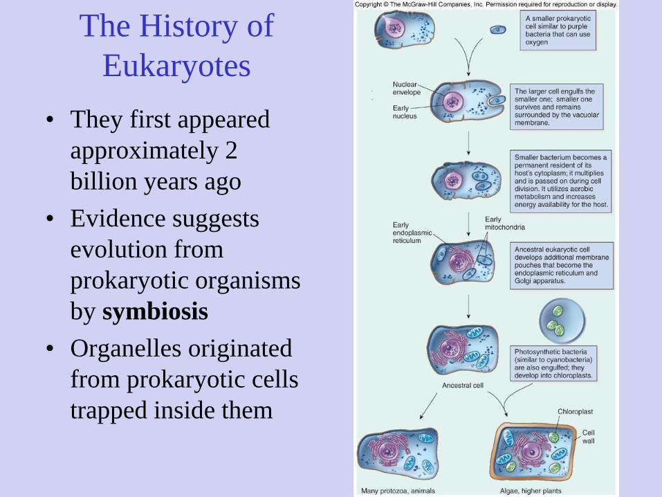
• They first appeared
approximately 2
billion years ago
• Evidence suggests
evolution from
prokaryotic organisms
by symbiosis
• Organelles originated
from prokaryotic cells
trapped inside them

2

3
4
5
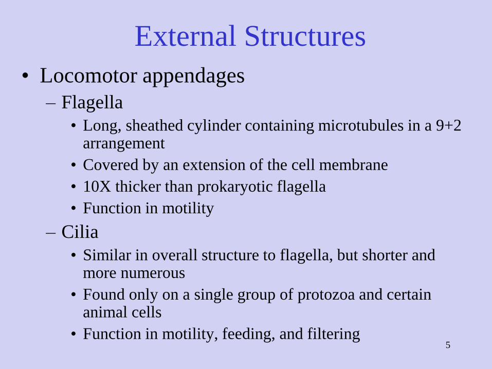
External Structures
• Locomotor appendages
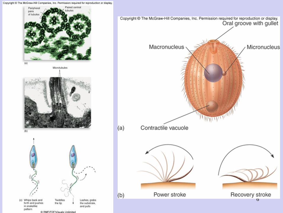
– Flagella
• Long, sheathed cylinder containing microtubules in a 9+2 arrangement
• Covered by an extension of the cell membrane
• 10X thicker than prokaryotic flagella
• Function in motility
– Cilia
• Similar in overall structure to flagella, but shorter and more numerous
• Found only on a single group of protozoa and certain animal cells
• Function in motility, feeding, and filtering
6
7
External Structures• Glycocalyx
– An outermost boundary that comes into direct contact with environment
– Usually composed of polysaccharides
– Appears as a network of fibers, a slime layer or a capsule
– Functions in adherence, protection, and signal reception
– Beneath the glycocalyx
• Fungi and most algae have a thick, rigid cell wall
• Protozoa, a few algae, and all animal cells lack a cell wall and have only a membrane

8
External Boundary Structures
• Cell wall
– Rigid, provides structural support and shape
– Fungi have thick inner layer of polysaccharide
fibers composed of chitin or cellulose and a thin
layer of mixed glycans
– Algae – varies in chemical composition;
substances commonly found include cellulose,
pectin, mannans, silicon dioxide, and calcium
carbonate

9
External Boundary Structures
• Cytoplasmic (cell) membrane
– Typical bilayer of phospholipids and proteins
– Sterols confer stability
– Serves as selectively permeable barrier in
transport
– Eukaryotic cells also contain membrane-bound
organelles that account for 60-80% of their
volume
10
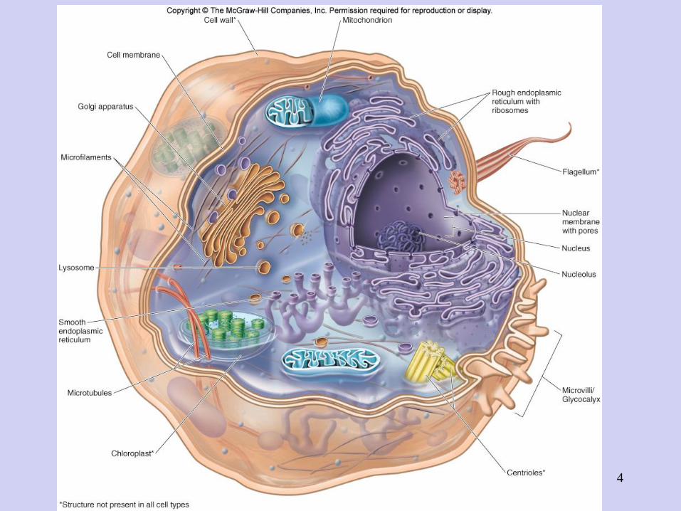
Internal Structures
• Nucleus
– Compact sphere, most prominent organelle of
eukaryotic cell
– Nuclear envelope composed of two parallel
membranes separated by a narrow space and is
perforated with pores
– Contains chromosomes
– Nucleolus – dark area for rRNA synthesis and
ribosome assembly
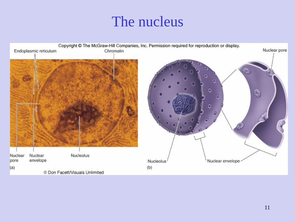
The nucleus
11

Mitosis
12
Internal Structures- Endoplasmic
reticulum
• Rough endoplasmic reticulum (RER) –originates from the outer membrane of the nuclear envelope and extends in a continuous network through cytoplasm; rough due to ribosomes; proteins synthesized and shunted into the ER for packaging and transport; first step in secretory pathway
• Smooth endoplasmic
reticulum (SER) – closed
tubular network without
ribosomes; functions in
nutrient processing,
synthesis, and storage of
lipids
13

Rough endoplasmic reticulum
14

15
Internal Structures
• Golgi apparatus
– Modifies, stores, and
packages proteins
– Consists of a stack of
flattened sacs called cisternae
– Transitional vesicles from the
ER containing proteins go to
the Golgi apparatus for
modification and maturation
– Condensing vesicles transport
proteins to organelles or
secretory proteins to the
outside

16nucleus RER Golgi vesicles secretion
17
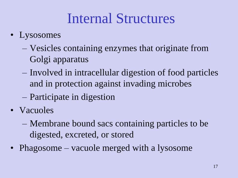
Internal Structures• Lysosomes
– Vesicles containing enzymes that originate from
Golgi apparatus
– Involved in intracellular digestion of food particles
and in protection against invading microbes
– Participate in digestion
• Vacuoles
– Membrane bound sacs containing particles to be
digested, excreted, or stored
• Phagosome – vacuole merged with a lysosome

18

19
Internal Structures
• Mitochondria
– Function in energy production
– Consist of an outer membrane and an inner
membrane with folds called cristae
– Cristae hold the enzymes and electron carriers of
aerobic respiration
– Divide independently of cell
– Contain DNA and prokaryotic ribosomes

Structure of mitochondrion
20

21
Internal Structures
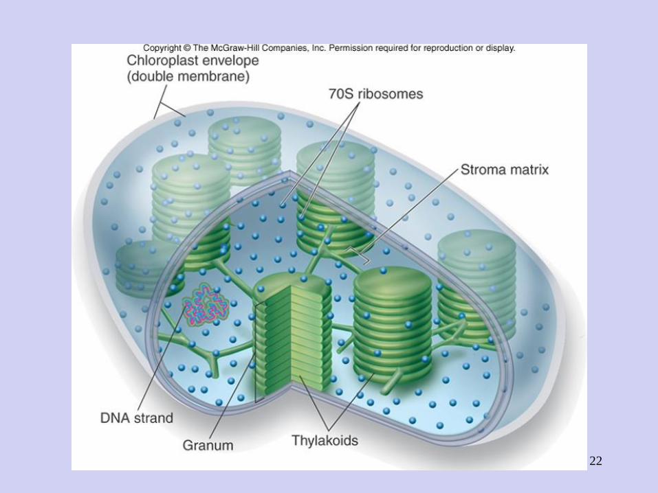
• Chloroplast
– Convert the energy of sunlight into chemical energy through photosynthesis
– Found in algae and plant cells
– Outer membrane covers inner membrane folded into sacs, thylakoids, stacked into grana
– Larger than mitochondria
– Contain photosynthetic pigments
– Primary producers of organic nutrients for other organisms
22

23
Internal Structures
• Ribosomes
– Composed of rRNA and proteins
– Scattered in cytoplasm or associated with RER
– Larger than prokaryotic ribosomes
– Function in protein synthesis

24
Internal Structures
• Cytoskeleton
– Flexible framework of proteins, microfilaments
and microtubules form network throughout
cytoplasm
– Involved in movement of cytoplasm, amoeboid
movement, transport, and structural support
25
Survey of Eukaryotic Microbes
• Fungi
• Algae
• Protozoa
• Parasitic worms

26
Kingdom Fungi
• 100,000 species divided into 2 groups:
– Macroscopic fungi (mushrooms, puffballs, gill
fungi)
– Microscopic fungi (molds, yeasts)
– Majority are unicellular or colonial; a few have
cellular specialization

27
Microscopic Fungi• Exist in two morphologies:
– Yeast – round ovoid shape, asexual reproduction
– Hyphae – long filamentous fungi or molds
• Some exist in either form – dimorphic – characteristic of
some pathogenic molds
28
Fungal Nutrition
• All are heterotrophic
• Majority are harmless saprobes living off dead plants and animals
• Some are parasites, living on the tissues of other organisms, but none are obligate
– Mycoses – fungal infections
• Growth temperature 20o-40oC
• Extremely widespread distribution in many habitats

Nutritional sources for fungi
29

30
Fungal Organization
• Most grow in loose associations or colonies
• Yeast – soft, uniform texture and appearance
• Filamentous fungi – mass of hyphae called
mycelium; cottony, hairy, or velvety texture
– Hyphae may be divided by cross walls – septate
– Vegetative hyphae – digest and absorb nutrients
– Reproductive hyphae – produce spores for
reproduction

31
Figure 5.18

32
Fungal Reproduction
• Primarily through spores formed on reproductive
hyphae
• Asexual reproduction – spores are formed
through budding or mitosis; conidia or
sporangiospores

33
Fungal Reproduction
• Sexual reproduction – spores are formed
following fusion of two different strains and
formation of sexual structure
– Zygospores, ascospores, and basidiospores
• Sexual spores and spore-forming structures
are one basis for classification

34
Formation of zygospores

35
Production of ascospores

36
Figure 5.22 Formation of basidiospores in a mushroom

37
Fungal Classification
Kingdom Eumycota is subdivided into several
phyla based upon the type of sexual
reproduction:
1. Zygomycota – zygospores; sporangiospores and some
conidia
2. Ascomycota – ascospores; conidia
3. Basidiomycota – basidiospores; conidia
4. Chytridomycota – flagellated spores
5. Fungi that produce only Asexual Spores (Imperfect)
38
Fungal Identification
• Isolation on specific media
• Macroscopic and microscopic observation of:
– Asexual spore-forming structures and spores
– Hyphal type
– Colony texture and pigmentation
– Physiological characteristics
– Genetic makeup

39
Roles of Fungi
• Adverse impact
– Mycoses, allergies, toxin production
– Destruction of crops and food storages
• Beneficial impact
– Decomposers of dead plants and animals
– Sources of antibiotics, alcohol, organic acids, vitamins
– Used in making foods and in genetic studies

40

41
Kingdom Protista
• Algae - eukaryotic organisms, usually
unicellular and colonial, that
photosynthesize with chlorophyll a
• Protozoa - unicellular eukaryotes that lack
tissues and share similarities in cell
structure, nutrition, life cycle, and
biochemistry

42
Algae
• Photosynthetic organisms
• Microscopic forms are
unicellular, colonial,
filamentous
• Macroscopic forms are
colonial and multicellular
• Contain chloroplasts with
chlorophyll and other
pigments
• Cell wall
• May or may not have flagella

43
Algae
• Most are free-living in fresh and marine water –plankton
• Provide basis of food web in most aquatic habitats
• Produce large proportion of atmospheric O2
• Dinoflagellates can cause red tides and give off toxins that cause food poisoning with neurological symptoms
• Classified according to types of pigments and cell wall
• Used for cosmetics, food, and medical products
44
Protozoa
• Diverse group of 65,000 species
• Vary in shape, lack a cell wall
• Most are unicellular; colonies are rare
• Most are harmless, free-living in a moist habitat
• Some are animal parasites and can be spread by insect
vectors
• All are heterotrophic – lack chloroplasts
• Cytoplasm divided into ectoplasm and endoplasm
• Feed by engulfing other microbes and organic matter

45
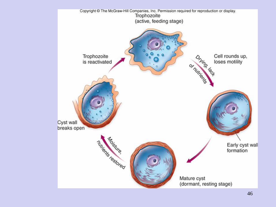
Protozoa
• Most have locomotor structures – flagella, cilia, or
pseudopods
• Exist as trophozoite – motile feeding stage
• Many can enter into a dormant resting stage when
conditions are unfavorable for growth and feeding –
cyst
• All reproduce asexually, mitosis or multiple fission;
many also reproduce sexually – conjugation
46
47
Protozoan Identification
• Classification is difficult because of diversity
• Simple grouping is based on method of motility,
reproduction, and life cycle
1. Mastigophora – primarily flagellar motility, some flagellar
and amoeboid; sexual reproduction
2. Sarcodina – primarily amoeba; asexual by fission; most
are free-living
3. Ciliophora – cilia; trophozoites and cysts; most are free-
living, harmless
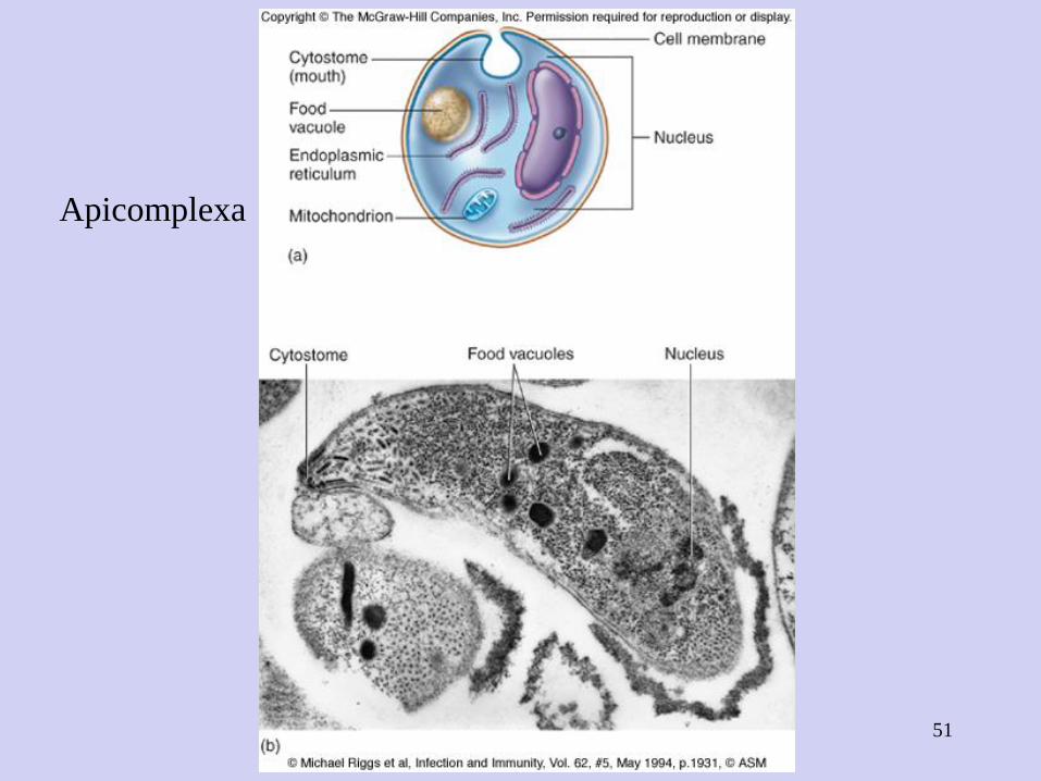
4. Apicomplexa – motility is absent except male gametes;
sexual and asexual reproduction; complex life cycle – all
parasitic

48
Mastigophora

49
Sarcodina

50
Ciliophora
51
Apicomplexa
52
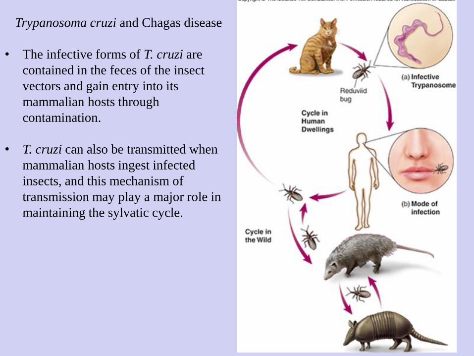
Trypanosoma cruzi and Chagas disease
• The infective forms of T. cruzi are
contained in the feces of the insect
vectors and gain entry into its
mammalian hosts through
contamination.
• T. cruzi can also be transmitted when
mammalian hosts ingest infected
insects, and this mechanism of
transmission may play a major role in
maintaining the sylvatic cycle.

53
Entamoeba histolytica

54
Parasitic Helminths
• Multicellular animals, organs for reproduction, digestion, movement, protection
• Parasitize host tissues
• Have mouthparts for attachment to or digestion of host tissues
• Most have well-developed sex organs that produce eggs and sperm
• Fertilized eggs go through larval period in or out of host body

55
Major Groups of Parasitic Helminths
1. Flatworms – flat, no definite body cavity; digestive tract a blind pouch; simple excretory and nervous systems
• Cestodes (tapeworms)
• Trematodes or flukes, are flattened, nonsegmented worms with sucking mouthparts
2. Roundworms (nematodes) – round, a complete digestive tract, a protective surface cuticle, spines and hooks on mouth; excretory and nervous systems poorly developed

56
Helminths
• Acquired through ingestion of larvae or
eggs in food; from soil or water; some are
carried by insect vectors
• Afflict billions of humans

57
Parasitic Flatworms

58
59
Helminth Classification and Identification
• Classify according to shape, size, organ
development, presence of hooks, suckers, or
other special structures, mode of reproduction,
hosts, and appearance of eggs and larvae
• Identify by microscopic detection of adult
worm, larvae, or eggs
60
Distribution and Importance of
Parasitic Worms
• Approximately 50 species parasitize humans
• Distributed worldwide; some restricted to
certain geographic regions with higher
incidence in tropics