the in situ - jcp.bmj.com · it is suggested that the erythrasma microorganism secretes a...
TRANSCRIPT
J. clin. Path., 1972, 25, 799-803
The erythrasma microorganism in situ: studiesusing the skin surface biopsy techniqueR. MARKS, N. D. RAMNARAIN, B. BHOGAL, AND N. T. MOORE
From the Institute of Dermatology, St. John's Hospital for Diseases of the Skin, London, and the Depart-ment of Crystallography, Birkbeck College, London
SYNOPSIS The skin surface biopsy technique has been used to investigate the erythrasma organismin situ in the stratum corneum in 11 patients. Staining by PAS and Gram stain showed the presenceof a large number of organisms arranged haphazardly in some areas and in microcolonies in others.With the scanning electron microscope it was possible to see that smooth filamentous chains ofmicroorganisms had penetrated horn cells and caused disturbance of the surface structure of thesecells.Enzyme histochemical tests showed that the erythrasma microorganism possessed a strong react-
ivity for NAD diaphorase and other mitochondrial enzymes. The reactivity was focal confirminga complex subcellular organization of organelles.
It is suggested that the erythrasma microorganism secretes a mucopolysaccharide sheath in somecircumstances.
Erythrasma is a chronic scaly dermatosis affectingthe body flexures and intertriginous areas of adults.Previously it was believed to be due either to a typeof dermatophyte (Microsporum minutissimum) or anactinomycete (Nocardia minutissimum). In 1969, adiphtheroid termed Corynebacterium minutissimumwas isolated from lesions of erythrasma and thoughtto be the causative agent (Sarkany, Taplin, andBlank, 1961). Still more recently Somerville (1970)suggested that several strains of fluorescent diphther-oid organisms were responsible for the clinicaldisorder that is recognized as erythrasma. In thisstudy we describe the results of our investigationsinto the causative organism of erythrasma using theskin surface biopsy technique, which removes anintact layer of stratum corneum with a cyano-acrylate adhesive (Marks and Dawber, 1971). Ageneral account of the use of this method fordemonstrating pathogenic microorganisms in thestratum corneum is documented elsewhere (Marksand Dawber, 1972).
Methods
PATIENTSThe clinical details of the 11 patients with erythrasmawho were investigated in this study are as follows:Received for publication 8 June 1972.
all 11 patients were men, the average age was 48 andthe age range 24 to 63. In nine the groins wereinvolved, in four the toe webs were involved, and infour the axillae were involved. It can be seen that theusual male predominance of the disease is evidentin our group of patients. A coral pink fluorescencewas detected in the involved sites when they wereviewed in ultraviolet light and small fluorescingcolonies of C. minutissimum were isolated on 199plates from skin scales in all patients.
SKIN SURFACE BIOPSIESThese specimens were obtained using ethyl cyano-acrylate (Permabond, Staines, Middx). Drops of theadhesive were placed on glass microscope slides orcoverslips. Samples taken onto coverslips werestored in the deep freeze at -40'C before histo-chemical examination. Samples on microscope slideswere examined by light microscopy after alcoholfixation (two minutes) and staining with periodicacid Schiff reagent (periodic acid 10 minutes,Schiff reagent 20 minutes) or Gram stain (methylviolet two minutes, Gram's iodine two minutes, withdifferentiation in equal parts of alcohol and acetonefor four seconds and counterstaining in neutral redtwo minutes). These preparations were made per-manent by mounting them with a coverslip andneutral mounting medium.
799
copyright. on 26 M
ay 2019 by guest. Protected by
http://jcp.bmj.com
/J C
lin Pathol: first published as 10.1136/jcp.25.9.799 on 1 S
eptember 1972. D
ownloaded from
R. Marks, N. D. Ramnairaini, B. Bhogal, and N. T. Moore
ENZYME HISTOCHEMICAL INVESTIGATIONS
Skin surface biopsies from six patients were takenonto coverslips instead of microscope slides and were
tested with the following enzyme histochemicalreactions: (a) non-specific esterase (indoxyl acetateesterase method of Holt, 1954); (b) leucine amino-peptidase (sing L-leucyl 4 methoxy - naphthyla-mide as substrate and the method of Nachlas,Crawford, and Seligman, 1957); (c) NAD diaphorase,lactic dehydrogenase, succinic dehydrogenase, andbeta-hydroxybutyric dehydrogenase (using NBTmethod of Pearse, 1960); and (d) adenosine tri-phosphatase (using method ofWachstein, Meisel,andNiedzwiedz, 1960).
SCANNING ELECTRON MICROSCOPE STUDIES
Skin surface biopsies from three of the subjects were
taken onto glass microscope slides. Areas from thespecimen-bearing sites of these slides were cut tosize, mounted on 'stubs', and then coated withcarbon in an Edwards coating unit. The coatedspecimens were then viewed in a Cambridgestereoscan scanning electron microscope (SEM) at anaccelerating voltage of 20kv at a 45° tilt.
Results
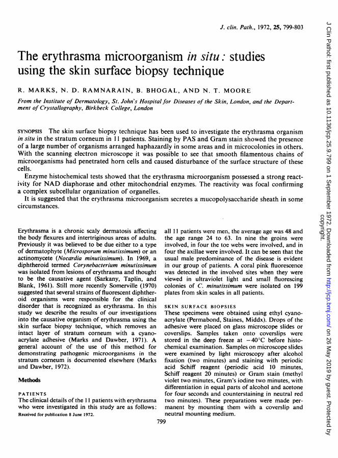
Skin surface biopsies from the affected sites stainedwith PAS reagent or Gram stain demonstrated thepresence of numerous fusiform microorganismswhich were arranged in clusters, chains, or singly,and scattered over variously sized areas (Figs. 1 a andb). The chains that were seen usually comprised
three, four, or occasionally more, individualbacterial cells. The microorganisms appearedapproximately three to four times as long as theywere broad. With the PAS stain there was noticeablediffuse staining of the sites containing the micro-organisms which was visible macroscopically. Theredid not appear to be a particular accentuation aroundhair follicles or sweat gland openings.The results of the enzyme histochemical in-
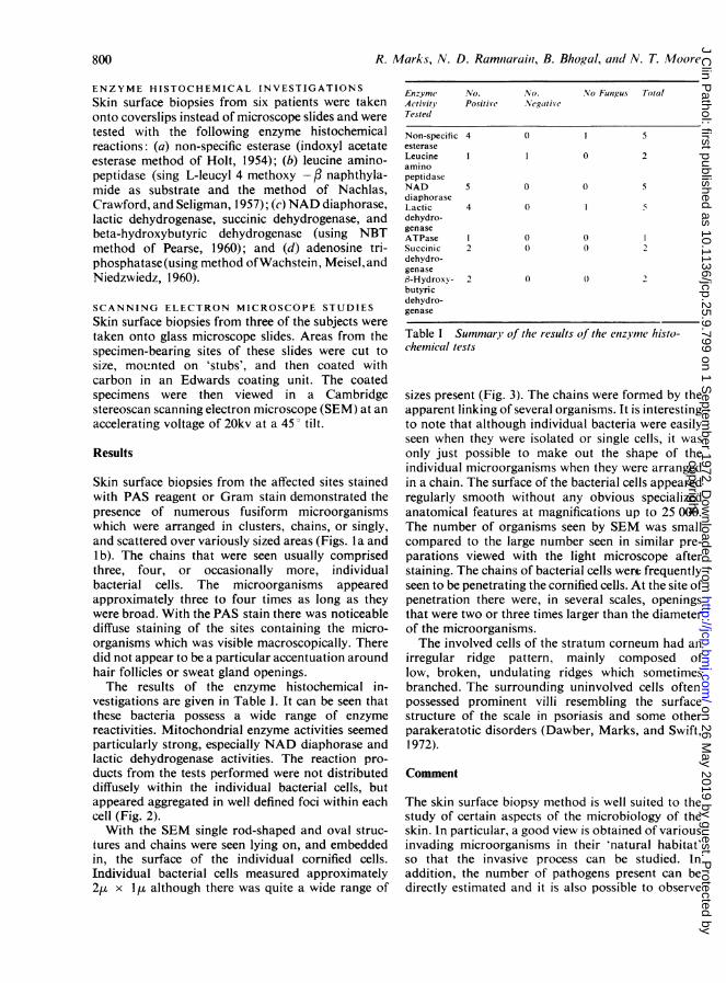
vestigations are given in Table 1. It can be seen thatthese bacteria possess a wide range of enzymereactivities. Mitochondrial enzyme activities seemedparticularly strong, especially NAD diaphorase andlactic dehydrogenase activities. The reaction pro-
ducts from the tests performed were not distributeddiffusely within the individual bacterial cells, butappeared aggregated in well defined foci within eachcell (Fig. 2).With the SEM single rod-shaped and oval struc-
tures and chains were seen lying on, and embeddedin, the surface of the individual cornified cells.Individual bacterial cells measured approximately2, x 1Is although there was quite a wide range of
Enzyme No. No. No Futngus TotalActivity Positive NegativeTested
Non-specific 4 0 1 5esteraseLeucine 1 1 0 2aminopeptidaseNAD 5 0 0 5diaphoraseLactic 4 0 1 5dehydro-genaseATPase I 0 0 1Succinic 2 0 2dehydro-genase3-Hydroxy- 2 0 0 2butyricdehydro-genase
Table I Summary of the results of the enzyme histo-chemical tests
sizes present (Fig. 3). The chains were formed by theapparent linking of several organisms. It is interestingto note that although individual bacteria were easilyseen when they were isolated or single cells, it wasonly just possible to make out the shape of theindividual microorganisms when they were arrangedin a chain. The surface of the bacterial cells appearedregularly smooth without any obvious specializedanatomical features at magnifications up to 25 000.The number of organisms seen by SEM was smallcompared to the large number seen in similar pre-parations viewed with the light microscope afterstaining. The chains of bacterial cells were frequentlyseen to be penetrating the cornified cells. At the site ofpenetration there were, in several scales, openingsthat were two or three times larger than the diameterof the microorganisms.The involved cells of the stratum corneum had an
irregular ridge pattern, mainly composed oflow, broken, undulating ridges which sometimesbranched. The surrounding uninvolved cells oftenpossessed prominent villi resembling the surfacestructure of the scale in psoriasis and some otherparakeratotic disorders (Dawber, Marks, and Swift,1972).
Comment
The skin surface biopsy method is well suited to thestudy of certain aspects of the microbiology of theskin. In particular, a good view is obtained of variousinvading microorganisms in their 'natural habitat'so that the invasive process can be studied. Inaddition, the number of pathogens present can bedirectly estimated and it is also possible to observe
800
copyright. on 26 M
ay 2019 by guest. Protected by
http://jcp.bmj.com
/J C
lin Pathol: first published as 10.1136/jcp.25.9.799 on 1 S
eptember 1972. D
ownloaded from

The erythrasma microorganism in situ: studies using the skin surface biopsy technique
Fig. Ia Gram-stainedpreparation to show C.minutissimum (x 250).
Fig. lb Gram-stainedpreparation to show C.minutissimum (x 480).
801copyright.
on 26 May 2019 by guest. P
rotected byhttp://jcp.bm
j.com/
J Clin P
athol: first published as 10.1136/jcp.25.9.799 on 1 Septem
ber 1972. Dow
nloaded from

R. Marks, N. D. Ramnarain, B. Bhogal, and N. T. Moore
Fig. 2 Photomicrograph from skin surface biopsy testedwith NAD diaphorase reaction. Note focal aggregationofformazan reaction product ( x 250).
any morphological or histochemical changes causedby therapeutic measures.
In this study of erythrasma, several features havecome to light that are worthy of comment. Therewas a remarkable disparity between the numbers ofbacteria seen in stained preparations with the lightmicroscope and the numbers seen by SEM. This isprobably a reflection of the fact that the 'surface'scanned by SEM using the skin surface biopsymethod is five or six cell layers down into thestratum corneum, while with the light microscope thewhole thickness of the specimen removed is exa-mined. Montes, Black, and McBride (1967) demon-strated by conventional EM that the great majorityof the microorganisms of erythrasma did not invadefurther than the superficial part of the stratumcorneum. This must also be a major contributingreason for the relatively small number of micro-organisms seen by SEM in this study.The surfaces of the horn cells into which the
microorganisms had penetrated possessed a dis-organized ridge pattern, suggesting the possibility of
Fig. 3 Scanning electronmicrograph from skin surfacebiopsy of erythrasma. Notefilamentous chain of micro-organisms (A) emerging froma scale with a disorganizedscale pattern (B) ( x 4000).
802
.::. a .. .gk.
copyright. on 26 M
ay 2019 by guest. Protected by
http://jcp.bmj.com
/J C
lin Pathol: first published as 10.1136/jcp.25.9.799 on 1 S
eptember 1972. D
ownloaded from

The erythrasma microorganism in situ: studies using the skin surface biopsy technique
disruption of the system of tonofflament-desmosomecomplexes. In addition, the impression was gainedthat at the site of penetration the opening was, insome cases, much wider than the microorganisms.This might suggest that penetration was accomplishedby a chemical dissolution rather than by purelyphysical pressure. Montes et al (1967) noted dis-organization of 'keratin fibrils' in horn cells invadedby C. minutissimum and this would be in accord withour observations.
Strong enzyme reactivity of individual bacterialcells was detected and to our knowledge thismetabolic activity has not been reported in this waybefore with the erythrasma organism. Some of thereaction products were obviously aggregated in welldefined areas, confirming a complex subcellulararrangement akin to mammalian cells. A similararrangement has been noted with enzyme reactions inthe dermatophyte fungi (Meinhof, 1968).We believe that the smooth exterior of the
organisms observed by SEM, with only a slightconstriction at the site of the junctions betweenmicroorganisms in a chain, plus the PAS reactivityof the microorganism (and its immediate environ-ment) reflects the secretion of a mucopolysaccharidesheath by the organism in some situations. Somer-ville (1972) discussed the microbiology of thecutaneous diphtheroids and stated that the diph-theroids responsible for trichomycosis axillarisproduced secretions that stick them together andto the hair, and cause, in addition, destruction of the
hair keratin. A similar material may well be producedby C. minutissimum.
We are grateful to Dr Y. M. Clayton and the staff ofthe Mycology Department for help in this investi-gation, and to Drs W. C. Noble and D. A. Somer-ville for their helpful suggestions.R.M. is in receipt of a grant from the MRC.
ReferencesDawber, R. P. R., Marks, R., and Swift, J. A. (1972). Scanning
electron microscopy ofthe Stratum corneum. Brit. J. Derm., 86,272-281.
Holt, S. J. (1954). A new approach to the cytochemical localization ofenzymes. Proc. roy. Soc. B., 142, 160-169.
Marks, R., and Dawber, R. P. R. (1971). Skin surface biopsy. Brit. J.Derm., 84,117-123.
Marks, R., and Dawber, R. P. R. (1972). In situ microbiology of thestratum corneum. Arch. Derm., 105, 216-221.
Meinhof, W. (1968). Zum histochemischen Nachweis von Enzymen desenergieliefernden Stoffwechsels in Dermatophyten derGattungen Microsporum Epidermophyton und Keratinomyces.Arch. klin. exp. Derm., 232, 279-294.
Montes, L. F., Black, S. H., and McBride, M. E. (1967). Bacterialinvasion of the Stratum corneum in erythrasma. J. invest.Derm., 49, 474-485.
Nachlas, M. M., Crawford, D. T., and Seligman, A. M. (1957). Thehistochemical demonstration of leucine aminopeptidase. J.Histochem. Cytochem., 5, 264-278.
Pearse, A. G. E. (1960). Histochemistry; Theoretical and Applied,2nd ed. Churchill, London.
Sarkany, I., Taplin, D., and Blank, H. (1961). The etiology andtreatment of erythrasma. J. invest. Derm., 37, 283-290.
Som:rville, D. A. (1972). The microbiology of the cutaneousdiphtheroids. Brit. J. Derm., 86, Suppl. 8, 16-20.
Somerville, D. A. (1970). Erythrasma in normal young adults. J. med.Microbiol., 3, 57-64.
Wachstein, M., Meisel, E., and Niedzwiedz, A. (1960). Histochemicaldemonstration of mitochondrial adenosine triphosphatasewith the lead-adenosine triphosphate technique. J. Histochem.Cytochem., 8, 387-388.
803
copyright. on 26 M
ay 2019 by guest. Protected by
http://jcp.bmj.com
/J C
lin Pathol: first published as 10.1136/jcp.25.9.799 on 1 S
eptember 1972. D
ownloaded from