the merit of histological evaluation of clinically...
TRANSCRIPT

84
The Merit of Histological Evaluation of
Clinically Successful Reconstructive Therapies in
Periodontology and Related Fields
PhD Thesis
Dr. Attila Horváth
Semmelweis University
PhD School of Clinical Medicine
Consultant:
Dr. István Gera PhD
Official reviewers:
Dr. István Gorzó PhD Dr. Árpád Joób-Fancsaly PhD
Final Examination Board:
Dr. Ida Nyárasdy PhD (Head) Dr. Vilmos Tóth PhD
Dr. Szabolcs Gyulai-Gaál PhD
Budapest 2014

1
“The more I learn,
the more I learn how little I know.”
Socrates
(469-399 BC)

2
TABLE OF CONTENTS
1. LIST OF ABBREVIATIONS……………………...........………………...…......….4
2. INTRODUCTION………………………………………..........…………...….........6
2.1. Tissue regeneration in periodontology. History and principles..........................6
2.2. Current regenerative considerations of the non-contained periodontal defects....11
2.3. Rationale of peri-implant dehiscence therapy...................................................14
2.4. Provision of post-extraction alveolar sockets....................................................18
3. OBJECTIVES.……………………………………………….......……………........20
4. METHODS AND MATERIALS…………………………………....…....………..21
4.1 Overview of the applied methods........................................................................21
4.2 Regeneration of periodontal defects……............................................................23
Clinical and histological evaluation of the effect of a novel biphasic synthetic bone substitute (Straumann BoneCeramic®) and enamel matrix derivative (Emdogain®) on human intrabony periodontal defects
Clinical and histological evaluation of the effect of a novel unsintered nanocrystalline synthetic bone substitute (Ostim®) on human intrabony periodontal defects
4.3 Regeneration of peri-implant dehiscence defects……........................................30
Clinical and histological evaluation of the effect of a novel biodegradable synthetic hydrogel membrane (Straumann MembraGel®) in combination with a novel biphasic synthetic bone substitute (Straumann BoneCeramic®) on porcine peri-implant dehiscence defects
4.4 Regeneration of post-extraction alveolar sockets……...............................….....38
Clinical and histological evaluation of the effect of alveolar ridge preservation on human extraction sockets
5. RESULTS………………………………………………..............................………43
5.1. Periodontal regeneration..............…………..................................…………….43
5.1.1. Treatment with EMD and SBC………....…………………...................43
5.1.2. Treatment with nano-HA......………......….……...………….................45
5.2. Provision of peri-implant dehiscence defects with SBC and PEG..........................47
5.3. The effect of ARP on extraction sockets..……................…..............……........56

3
6. DISCUSSION………………………………………………………..................…..77
6.1. Limits of periodontal regeneration...................………………............................77
6.1.1. Treatment with EMD and SBC………....……………...........................77
6.1.2. Treatment with nano-HA.....…......….……...………….…....................80
6.2. Peri-implant dehiscence defect therapy with SBC and PEG..........................................83
6.3. Current options for the treatment of extraction sockets by ARP................…..88
6.4. Overall considerations........................................................................................96
7. CONCLUSIONS………………………………………………………………….100
8. SUMMARY……………………………………………………………………….102
9. ÖSSZEFOGLALÁS………………………………………………………………103
10. REFERENCES……………………………………………………………………104
11. LIST OF PUBLICATIONS……...…..……………………………………………129
12. ACKNOWLEDGEMENTS………………………………..……………………...131

4
1. LIST OF ABBREVIATIONS
3D Three-Dimensional
AAP American Academy of Periodontology
ABB Autogenous Bone Block
ABP Autogenous Bone Particulates
ANOVA Analysis of Variance
AR Alveolar Ridge
ARP Alveolar Ridge Preservation
BCP Biphasic Calcium Phosphate
BS Bone Surface Area
β-TCP Beta-Tricalcium Phosphate
BG Bioactive Glass
BIC Bone-to-Implant Contact
BMP Bone Morphogenetic Protein
BoP Bleeding on Probing
cAMP Cyclic Adenosine Monophosphate
CBCT Cone-Beam Computerised Tomography
CEJ Cementoenamel Junction
CENTRAL The Cochrane Central Register of Controlled Trials
CI Confidence Interval
CONSORT Consolidated Standards of Reporting Trials
DBBM Deproteinized Bovine Bone Mineral
DFDBA Demineralized Freeze-Dried Bone Allograft
EDTA Ethylene Diamine Tetraacetic Acid
EMD Enamel Matrix Derivative
e-PTFE Expanded Polytetrafluoroethylene
FMBS Full Mouth Bleeding Score
FMPS Full Mouth Plaque Score
GBR Guided Bone Regeneration
GS Graft Surface Area
GTR Guided Tissue Regeneration
HA Hydroxyapatite
5
IL-6 Interleukin-6
INTRA Depth of the Intrabony Component of the Periodontal Defect
IOPA Intraoral Periapical Radiograph
ISRCTN International Standard Randomised Controlled Trial Number Register
LILACS Literatura Latino-Americana e do Caribe em Ciências da Saúde
LJE Long Junctional Epithelium
LRAP Leucine-Rich Amelogenin Peptide
Nano-HA Unsintered, Nanocrystalline, Phase-Pure Hydroxyapatite
OFD Open Flap Debridement
PDL Periodontal Ligaments
PEG Polyethylene Glycol-Based Hydrogel
PGPL Polyglycolide/Polylactide
PPD Pocket Probing Depth
PRGF Plasma Rich in Growth Factor
RCT Randomised Controlled Clinical Trial
REC Gingival Recession
rhBMP-2 Recombinant Human Bone Morphogenetic Protein-2
ROI Region of Interest
SLA Sandblasted, Large-grit, Acid-etched
SLActive Modified (Hydrophilic) Sandblasted, Large-grit, Acid-etched
TCP Tricalcium Phosphate
TGF-β Transforming Growth Factor-Beta
TRAP Tyrosine-Rich Amelogenin Peptide

6
2. INTRODUCTION
2.1. Tissue regeneration in periodontology. History and principles
Periodontal disease has been considered as one of the major causes of tooth loss, being
accountable for 30-35% of extractions [1]. A widely accepted measure to determine the
severity of the disease is the periodontal pocket probing depth (PPD). Advanced
periodontitis (PPD>5 mm) has been observed in 2-40% of the population in Europe [1]
and 7.4% in Hungary, according to a recent survey [2]. The treatment of periodontitis
involves non-surgical as well as surgical means, beyond the inevitable enforcement of
individual oral hygiene. The main goal of treatment is alleviating inflammation,
arresting disease progression and establishing a stable periodontal condition that is
maintenable for long run by the patient’s self-performed oral hygiene.
The principles above that were laid down by the great pioneers of periodontology in
Scandinavia and North America remain valid today [3]. Nevertheless, the outcome of
the conservative (i.e. non-surgical) periodontal therapy or even of the
resective/explorative surgical treatment are often associated with gingival recession and
very limited regeneration of the periodontium [4]. Despite the clinically stable
periodontal conditions of such cases (i.e. PPD≤4 mm, negative bleeding on probing
(BoP)), histological results are mostly characterised by reparative tissues. In such type
of healing, the genuine attachment apparatus (acellular root cementum – inserting
Sharpey fibres – bundle bone) that was lost as a result of periodontitis, has been
predominantly replaced by long junctional epithelium (LJE) instead. Such epithelial
attachment merely represents a secondary (inferior) connection in terms of quality and
tissue adhesion [5]. Optimal treatment outcome would incorporate the reconstitution of
the lost periodontal tissues (i.e. cementum, functionally oriented periodontal ligament,
alveolar bone and gingiva). In the Seventies, Melcher suggested that periodontal healing
might be characterized by the type of tissues first repopulating the periodontal defect
following flap surgery [6]. Based on this assumption, it was hypothesized that
precluding the undesired cells (epithelium, gingiva, etc.) from the healing hub of the
intrabony periodontal pocket by a mechanical barrier device, would enable the cells
from the remaining periodontal ligament to repopulate the defect. Consequently, the
7
principles of Guided Tissue Regeneration (GTR) and subsequently Guided Bone
Regeneration (GBR) were conceived and described in Scandinavian research centres by
Jan Gottlow, Thorkild Karring, Jan Lindhe, Sture Nyman, Christer Dahlin and many
others [7] [8] [9] [10]. Thenceforth, the theory of GTR and GBR was broadly
investigated and proved by sound evidence. However, the “mechanical concept” that the
clue of regeneration is purely based on the cell-occlusive barrier function of the
membrane is being challenged nowadays. Emerging evidence suggests a rather
biological way of thinking that the intent of a membrane might be (i) to protect and
stabilize the wound by neutralizing and deflecting any wound rupturing forces away
from the root surface, (ii) to provide a secluded space to release the native potential for
regeneration and therefore, (iii) to maintain the structural integrity of a maturing blood
clot [11] [12] [5].
Over the past decades, tremendous efforts have been made by scientists and clinicians to
develop novel techniques and materials in order to improve the outcome of regenerative
procedures. Treatment of supra-alveolar defects or intrabony defects with missing
buccal and/or lingual wall (i.e. non-containing defects) is often associated with least
predictable outcome. For such augmentations a combinative approach was suggested
and successfully utilized by means of different ‘bone fillers’ in combination with
GTR/GBR [13]. Several grafts, bone substitutes and biomaterials were introduced and
widely employed to fill such defects in order to exploit their possible osteoconductive,
osteoinductive or even osteogenic potentials. However, the additional benefit of the
application of these materials has not been unanimously supported by the literature [14]
[15] [16]. As described above, the volume of regenerated tissue may rest on the wound
stability and the space provided by the barrier membrane beneath the tension-free
approximated mucoperiosteal flaps. Therefore, the main mechanism behind the use of
bone replacements seems to be blood clot stabilization through their scaffolding
architecture, thus space provision, rather than osteoconduction or induction [17].
The use of barrier membranes, with or without bone substitutes, is, however, frequently
associated with side effects, such as membrane exposure. It may arise from the
compromised re-vascularization of the flaps above an implanted “foreign body”, as well
as from the tension of the flaps caused by the increased volume of the augmentation. In
the end of the Nineties a biologic device was developed in Sweden by Lars Heijl, Lars

8
Hammarström and co-workers that promotes regeneration without the side effects and
the handling difficulties of the GTR technique [18] [19] [20]. The biological active
component of this regenerative gel originates from amelogenin, an enamel-specific
protein of porcine tooth buds. Enamel matrix derivative (EMD), as it is predominantly
quoted in the literature, refers to amelogenin fraction following a purification process.
EMD in combination with a propylene glycol alginate vehicle results in Emdogain®, the
brand name of this biological active device. In the past fifteen years a large number of
preclinical and clinical trials have proven its ability to regenerate periodontal ligaments
(PDL), cementum and alveolar bone in some extent [21].
However, more than a decade following the first publications of EMD, the biologic
cascade of the effects of EMD on wound healing at subcellular level has not yet been
fully understood. According to data from in vitro studies EMD may modulate wound
healing by stimulating the proliferation of preosteoblasts and the differentiation of
osteoblasts via upregulating cyclic adenosine monophosphate (cAMP) levels, inducing
the synthesis and secretion of transforming growth factor-beta (TGF-β) and interleukin-6
(IL-6) in gingival fibroblasts and PDL [22] [23] [24] [25]. In addition, EMD may retard
epithelial cell proliferation and may confound dental plaque homeostasis [26] [27] [28].
Hence, it seems that EMD may stimulate periodontal wound healing by an indirect effect
on the release of growth factors and by retarding the downgrowth of junctional
epithelium. A very recent in vitro investigation of amelogenin peptides, conducted by the
research group of Nikos Donos at the UCL Eastman Dental Institute, elucidated novel
possible pathways of the effect of different fractions of EMD on bone repair and
regeneration [29]. It was demonstrated that low- and high-molecular-weight fractions of
EMD, namely leucine-rich amelogenin peptide (LRAP) upregulated osteogenic
differentiation and enhanced terminal differentiation of bone-forming cells, meanwhile
tyrosine-rich amelogenin peptide (TRAP) suppressed the formation of bone-like
mineralized nodules. This indicates that fractions of EMD might play a therapeutic role
not only in periodontal or alveolar bone regeneration, but also in orthopaedic repair
(LRAP), or in the opposite, treatment of pathologic or even malignant bone formation
(TRAP). A more recent publication of the same group demonstrated that EMD could
modulate the differentiation of PDL cells in vitro, such as up-regulating chondrogenic,
neovasculogenic and osteogenic genes, but suppressing adipogenesis, gliogenesis and
neurogenesis [30]. A very recent review of the effect of EMD at cellular level concluded
9
that EMD elicited a regenerative response in periodontal tissues through a complex
cascade of gene expression, protein production, proliferation and differentiation of
various cells, particularly periodontal ligament and osteoblastic cell types [31].
Our research group at the Semmelweis University, led by Péter Windisch and Anton
Sculean, has considerably contributed to the understanding of the effect of EMD on
human periodontal defect, by illumination of its histological outcomes [32] [33] [34]
[35] [36]. Although the regenerative potential of EMD per se was demonstrated both at
preclinical and clinical levels [21] [37], when treating large or non-containing defects,
clinicians are often facing the challenge of space provision and prevention of flap
collapse. For such cases, bone fillers (in combination with EMD) are suggested to
overcome these problems. Data from controlled clinical trials have demonstrated greater
clinical improvements following treatment of EMD combined with certain types of
bone grafts, such as autogenous bone particles (ABP), or deproteinized bovine bone
mineral (DBBM) compared to EMD alone [33] [38] [39]. Nevertheless, other
substitutes, such as demineralized freeze-dried bone allograft (DFDBA), or bioactive
glass (BG) did not demonstrate significant clinical benefit in terms of PPD reduction
and CAL gain over EMD alone [40] [41]. Moreover, the histological evidence seems to
be even more conflicting, since beside the formation of new cementum, periodontal
ligament and alveolar bone up to a various extent, the implanted bone substitute was
frequently encapsulated in connective tissue [34] [35] [42].
It has also been investigated over the decades, whether bone grafts/substitutes alone (i.e.
without the additional effect of GTR or EMD) might bear the potential to restore the
lost periodontal attachment apparatus. The use of certain types of grafting materials
such as ABP, DFDBA or DBBM has been shown to result not only in substantial
clinical improvements, evidenced by PPD reduction, defect fill and CAL gain, but also
to promote, at least to some extent, formation of a new connective tissue attachment (i.e.
new cementum with inserting collagen fibres) and of new alveolar bone [43] [44] [45]
[46] [47]. However, the use of autografts for regenerative periodontal therapy is limited
by its source and increases donor site morbidity. Furthermore, most types of DBBM
possess a fairly slow resorption potential, resulting in a mixed hard tissue formation in
periodontal osseous defects [48]. In addition, the use of grafts from bovine (e.g. DBBM)
or human origin (e.g. DFDBA), still comport the risk (at least theoretical) for

10
antigenicity and disease transmission [49] [50]. Furthermore, the animal origin of these
materials keeps raising concerns for some patients, due to religious purposes or animal
right considerations.
To overcome these drawbacks, synthetic materials in conjunction with open flap
debridement were tested. The currently available data suggest that in intrabony defects,
the implantation of various types of such alloplasts may lead to significant
improvements of the investigated clinical parameters [51] [52] [53] [54] [55] [56]. On
the other hand, the available histological evidence indicates that in human intrabony
defects, the healing following implantation of alloplasts is predominantly characterized
by epithelial proliferation, connective tissue encapsulation and limited periodontal
regeneration [57] [58] [59] [53] [60] [61] [62] [63] [64] [65].
The above inconsistent findings on the adjunctive benefit of the application of bone
replacement biomaterials highlight the current criteria of successful regeneration. In
terms of clinical success, beyond arresting inflammation, we aimed at reconstituting the
lost periodontal attachment apparatus. Although we are more aware nowadays that the
complete reconstruction may remain a daydream, the therapist must possess certain
clinical measures that support the evaluation of the treatment outcome. Such measures
are PPD reduction, CAL gain, decreased furcation involvement or sometimes reduced
gingival recession, discontinued tooth mobility, etc. Albeit the improvement of all of
these measurements undoubtedly indicates a level success, none of them is able to
demonstrate the presence of true regeneration (i.e. new cementum, inserting Sharpey-
fibres and new bundle/alveolar bone). The reduction of PPD for instance may well be a
result of reparative attachment (i.e. LJE) between the root surface and the bone
substitute that may not bear long-term stability. In order to display the presence of new
bone, standardised intraoral periapical radiograph (IOPA) is commonly used. One
should bear in mind though, that based on an radiographic image, we will not be able (i)
to distinguish the new bone and the implanted, non/slow resorbable radiopaque bone
substitute, (ii) to find out the type of connection to the root cementum and (iii) to
ascertain, whether the substitute is connected to the newly formed bone, or encapsulated
in connective tissue, which is not regarded as functional bonding. Therefore, these
measurements are inappropriate for demonstrating the formation of new cementum, new
bone and new periodontal ligaments, merely to unveil the presence of the radiopaque

11
biomaterial. Thus, from the biological point of view, it seems reasonable that the
selection of bone substitutes for the treatment of periodontal defects should be based on
histological evidence, indicating positive biological properties of the biomaterial (with
or without GTR technique or in combination with biologically active agent) [66].
In order to supply the clinician with generalizable guidelines, the World Workshop in
Periodontics of the American Academy of Periodontology (AAP) summarized the
criteria for a periodontal treatment to be considered a regenerative procedure [67]:
- Controlled animal histologic studies demonstrating formation of new cementum,
periodontal ligament and bone (in absence of human histologic data retrieved
from controlled trials)
- Human histologic specimens demonstrating formation of new cementum,
periodontal ligament and bone coronal to the former defect base
- Controlled human clinical trials demonstrating improved clinical probing
attachment and bone levels.
In addition to these criteria, a further requirement has been proposed, namely, the
regenerative procedure should be based on a concept that explains why the treatment
resulted in regeneration [68].
Nowadays, the market of regenerative materials offers an overwhelming number of
products, making the right choice for the right clinical scenario extremely difficult. A
meticulous analysis of the literature reveals that only a few materials meet the above
cited criteria. There are, in fact, several data on clinical and radiographic success, but
the histological evidence behind is limited. Furthermore, just a few studies have
evaluated human histology and ultimately, there are very limited data on controlled
human histological trials on periodontal regeneration, such as the one our group
published recently [69] [70].
2.2. Current regenerative considerations of the non-contained periodontal defects
As discussed above it has been proven that EMD alone possesses the biological
properties to regenerate PDL and cementum in periodontal intrabony defect with
supportive anatomy. However, in a wide, non-contained defect EMD gel cannot provide
sufficient support for the flap to prevent its collapse, therefore various combinations of

12
EMD and different types of scaffolds (bone grafts/substitutes), such as ABP, DFDBA,
DBBM, BG, or beta-tricalcium phosphate (β-TCP), have been employed [77] [38] [40]
[39] [34] [41].
Synthetic bone substitutes bear the advantage of unlimited source, lack of donor site
morbidity, lack of disease transmission and lack of religious concerns. Recently a new
composite bone substitute was introduced for periodontal and peri-implant
reconstructions. This biphasic calcium phosphate (BCP), namely Straumann
BoneCeramic®, consists of 60% hydroxyapatite (HA) and 40% β-TCP in a particulate
form. Preclinical evidence suggested that this HA/β-TCP ratio may allow for optimised
control of bioabsorbability, thus resulting in accelerated new bone formation [60] [78]
[50] [79].
Therefore, the use of a combination of EMD and BCP might be of clinical and biologic
relevance in the treatment of advanced, non-contained periodontal defects, since the
absorption pace of BCP placeholder might be in line with the regenerative process
stimulated by EMD. However, as previously underscored, we cannot declare the
possible regenerative features of a novel material or combination of materials, until
affirmed by human histology.
To the best of our knowledge, no human histologic data have been available so far
evaluating the effect of OFD combined with EMD and BCP on intrabony periodontal
defects.
Apart from investigating the above treatment combination (Study 1), we wanted to go
one step further to examine, whether a synthetic bone substitute per se, without the
regenerative potential of EMD would be able to reconstitute periodontium. It would
simplify and accelerate the surgical procedure, as well as reduce treatment cost for the
sake of the patient.
Therefore, it would be valuable to seek for an osteoconductive scaffold that is able on
its own to (i) function as a space-providing buttress for the flap; (ii) foster blood clot
stabilization; (iii) induce proliferation, differentiation and maturation/mineralization of
periodontal cells; (iv) resorb totally, in harmony with the formation of new bone.

13
Ideally, this material is synthetic, does not inflict allergic reaction, inexpensive and
originates from an unlimited source.
Studies investigating hydroxyapatite (HA) and/or tricalcium phosphates (TCP) in
reconstructive periodontal therapy reported promising clinical, but variable histological
results [52]. [58] [59] [54] [60] [62] [63] [65]. Incomplete resorption and implant
encapsulation in soft tissue seem to be common drawbacks of the sintered HA. In a
recent case report where dense hydroxyapatite material (surface area 59 m2/g) was
placed in postextraction socket, the implant particles were observable in the histologic
sections even 20 years after implantation [80]. This might be the consequence of the
sintering process at high temperature (1200°C). Therefore, the surface structure of the
biomaterial could not attract osteoclasts, resulting in an undesirable slow resorption
pace [81].
Recently, a new, fully synthetic, unsintered, nanocrystalline, phase-pure hydroxyapatite
(nano-HA) has been developed (Ostim®; Heraeus Kulzer, Hanau, Germany). The
nanometre sized crystalline structure results in a large, 106 m2/g surface, due to the lack
of the high temperature sintering phase [82]. It may be a promising candidate for
enhancing periodontal and bone regeneration, since its chemical composition and
nanocrystalline structure correspond to the calcium phosphate component of natural
bone. Ostim® is supplied in a ready-to-use, sterile capsule, containing the injectable,
white, aqueous suspension of 35% nano-HA. According to the manufacturer’s
instructions, application of biologic barrier above the implanted nano-HA is not
required. Hence, the possibility of incomplete primary closure and flap adaptation, thus
membrane exposure and infection is reduced to minimum. In comparison with
hydroxyapatite porcelains (sintered HA), nano-HA has greater potential for resorption.
The nanocrystals can be ingested and broken down by phagocytosis, which did not
appear to increase of the serum calcium level [83] [84]. Depending on the site and
volume, it is resorbed within few months [85].
Results of in vitro studies indicate that nano-HA bears the potential to stimulate the
migration, adhesion, differentiation and proliferation of PDL cells [86] [87] [88] [89]. In
preclinical experiments nano-HA demonstrated accelerated angiogenesis,
microvascularisation and new bone formation without inducing inflammatory response

14
[90] [91] [92] [93]. Rapid resorption was observed via phagocytosis by mononuclear
macrophages and multinuclear giant cells [85]. Promising clinical and histological
results were reported in the field of orthopaedic trauma and reconstructive surgery
following nano-HA implantation [94] [95] [96] [97]. In the field of dentistry, case
reports have demonstrated substantial clinical improvements following the use of nano-
HA, as a filler of jaw cysts, extraction sockets, sinus and ridge augmentation,
periodontal and peri-implant defects [98] [99] [100] [101] [102] [103] [104]. Human
histology demonstrated the presence of the alloplast surrounded by woven bone six or
even 36 months following implantation in lateral ridge augmentation or sinus floor
elevation [101] [103].
Furthermore, the use of nano-HA for the surgical treatment of intrabony periodontal
defects demonstrated statistically significantly higher clinical improvements compared
to OFD alone [105] [106].
In spite of these promising results, to the best of our knowledge, no data are available
from human histological studies on the healing of intrabony defects following
perodontal surgery with nano-HA. Thus, at the time being, it is virtual y unknown to
what extent this material may promote periodontal wound healing/regeneration in
humans.
2.3. Rationale of peri-implant dehiscence therapy
Comprehensive restorative dentistry that includes the placement of dental implants is
considered as a safe and successful therapy aimed at restoring fully or partially
edentulous alveolar ridges [126] [127]. However, sufficient quantity and quality of
alveolar bone is required for the longevity of a dental implant [128]. Adequate amount
of bone is seldom found prior to implantation due to periodontal disease, periapical
pathology and trauma before or even during tooth removal. Moreover, the jawbone
undergoes atrophy as part of a natural remodelling after tooth extraction [129] [130]
[76] [131]. This resorption requires the restoration of the remaining alveolar ridge [132],
in order to meet the contemporary demand of the three-dimensional, prosthetically
driven implant placement, as a prerequisite of long term success in function and
aesthetics [133].

15
Several surgical methods have been proposed over the past decades to rebuild the
alveolar ridge. These procedures comprise the use of autogenous bone block (ABB),
ABP, bone substitutes (allografts, xenografts, alloplasts), GBR alone or with grafting,
sinus floor augmentation, forced eruption, as well as ridge expansion techniques
utilizing “split” osteotomy or distraction osteogenesis [127].
Among these procedures, GBR has found to be one of the most effective according to
the current scientific evidence [127] [134] [135] [136]. Successful bone regeneration
has been observed when GBR was used alone or in combination with bone grafts, either
prior to placement of dental implants (i.e. two stages procedure) [137] [138], or
simultaneously with the placement of implants (i.e. one stage procedure) [139] [140]
[141] [142]. In addition, the survival rate of implants placed in the augmented alveolar
ridge is comparable to that of implants placed in pristine sites [127] [143].
As per definition, GBR requires the placement of an occlusive barrier that prevents the
invasion of non-bone-forming cells from the surrounding soft tissues into the defect. At
the same time it allows sufficient time and space for bone forming cells to repopulate
the defect [144] [145] [146] [147].
One of the most employed and researched non-resorbable barrier, which had proven to
be effective in bone regeneration is the expanded polytetrafluoroethylene membrane (e-
PTFE) [148] [149] [150] [151] [152]. However, exposure and inflammation, resulting
in soft tissue dehiscence, premature membrane removal, thus compromising bone
regeneration, were frequently reported [149] [150] [153] [154] [155].
The main disadvantage of non-resorbable materials is the need for a second surgical
procedure to remove the device. This led to the development of bioabsorbable barrier
membranes, which did not require a re-entry surgery. Resorbable membranes, such as
collagen or glycolide and trimethylene carbonate, have shown improved tissue healing,
decreased morbidity, complete resorption and in case of exposure, the risk of bacterial
contamination is reduced [156] [157] [158] [112] [159]. On the other hand, some of the
resorbable materials may elicit tissue reactions, have uncontrolled resorption rate and
show poor resistance to collapse [147] [160] [161].
16
An ideal membrane is (i) highly biocompatible (does not elicit adverse tissue reactions);
(ii) totally resorbable in a predictable rate (reliable maturation of the newly formed
tissue beneath); (iii) easy to handle (predictable result even in the hand of less
experienced surgeons); (iv) inexpensive and synthetic (available for patients with less
financial resources and with concern of animal or human origin).
In order to meet the above demand, a novel synthetic bio-degradable polyethylene
glycol hydrogel (PEG) membrane (MembraGel®; Straumann; Basel, Switzerland) was
developed recently. PEG membrane is composed of two liquid PEG compounds that
react upon mixing and form a hydrogel. PEG has been shown to be highly
biocompatible and it is presently approved for several pharmaceutical applications or as
medical device [162] [163]. Polyethylene glycol hydrogel degrades by hydrolysis and
experimental studies have shown that this process is complete within 4–6 months,
therefore a second surgery to remove the membrane is not required [164] [165]. This
material, applied as a membrane, has been shown to be cell-occlusive and to be able to
prevent soft tissue ingrowth and collapse [165] [166]. Recent experimental studies have
also demonstrated positive results in bone regeneration with PEG membranes in bone
defects and for the treatment of dehiscence defects around implants [167] [168] [169]
[170] [166]. Since its biodegradation is significantly slower compared to standard
resorbable collagen membranes, the required barrier function may last longer [164].
This novel barrier material is easy to handle, since the two component of the hydrogel is
delivered in an automix syringe, thus the amalgamated membrane gel could simply be
placed on the top of the defect or bone graft/substitute. The time of application is shown
to be significantly reduced, compared to a conventional collagen membrane, hence the
length of the surgery is reduced [169].
In order to stabilize the blood clot and provide space-maintenance below the barrier, the
use of a recent developed synthetic biphasic bone substitute seemed to be beneficial.
The BCP we used (Straumann BoneCeramic®; Straumann; Basel, Switzerland) had
been shown to accelerate bone formation in standard, dehiscence bone defects [78] [79].
There is not much evidence in the literature evaluating the response of regenerated bone
to functional loading. It has been perceived that once the protection of the secluded
space created by a membrane is removed and the newly formed bone is not functionally
loaded, then some bone resorption might take place [171] [172]. The influence of

17
loading on the outcome of GBR in peri-implant dehiscence defects was investigated in a
preclinical study [173]. At the loaded sites significant decrease in bone fill was occurred
between the three and nine months healing period (loading), whereas no change was
observed at non-loaded sites. On the other hand, the one year data of an ongoing five
years RCT conducted by our group, failed to demonstrate significant differences
between the loaded (immediately provisionalized) and the non-loaded (healing
abutments) groups [174].
When looking into the literature for the desired histological evidences of regenerative
procedures in other disciplines in the neighbourhood of periodontology, such as ridge
augmentation prior to placement of a dental implant, it has to be realised that alike in
periodontology, only a limited number of histological studies could be identified. The
explanation is presumably the cumbersome patient and case selection, which must be
under any circumstances in coherence with the current ethical guidelines. Furthermore,
the concomitant cost of such histological analysis either in preclinical, or in clinical
setting, prerequisites a wealthy sponsor or department. Finally, a human histological
trial usually encompasses a compensative treatment for the patient that extends the
overall treatment time and cost.
If we would like to investigate a regenerative method and material with simultaneous
dental implant placement, which is quite a frequent clinical scenario in the daily
practice, we could face enormous difficulties with a human histological trial design. For
such study, an experimental narrow implant should be placed in an experimental defect
that would be regenerated with the experimental biomaterial. Then, it is removed for
histological evaluation with some surrounding hard and soft tissues resulting in a much
larger bony defect that should be eventually restored with a corresponding size
compensatory definitive implant. The chance to obtain a positive ethical authorization
or to recruit the sufficient number of patients for such experiment is meagre. Thus,
designing such a human histological trial could practically be beyond the bounds of
possibilities.
18
2.4. Provision of post-extraction alveolar sockets
Various methods of alveolar bone augmentation could predictably support the
prosthetically driven, three-dimensional implant placement that is a contemporary
requirement for long-term success in implant therapy [133]. Simultaneous augmentation
procedures can be implemented with high predictability, provided that three intact bone
walls are present and the implant location is inside the alveolar envelope [139] [140]
[141] [142]. However, in case of extensive ridge resorption, simultaneous bone
augmentation becomes less predictable [155]. Two-stages implant placement, which is
preceded by GBR, block grafting procedure or ridge expansion techniques, could be
implemented with success, although the concomitant treatment time is extended [137]
[138] [155]. Moreover, this intervention often comprises intraoral bone harvesting as
well as extensive soft tissue manipulation aiming at tensionless flap approximation,
which considerably increases morbidity and patient’s intra and postoperative
discomfort. Therefore, it is desirable to preserve, rather than reconstruct post extraction
ridge dimension, thus minimizing morbidity and discomfort.
The reduction of alveolar ridge (AR) dimension may originate in various reasons.
Firstly, periodontal disease, periapical pathology and mechanical trauma could result in
bone loss prior to tooth removal [131]. Secondly, indelicate extraction has also been
associated with additional bone deficit. Finally, alveolar bone undergoes additional
atrophy as a result of natural bone remodeling following tooth removal [129] [130]
[197] [198] [199]. This process begins subsequently after extraction and continues for
years resulting in even 50% reduction of alveolar ridge width [131]. According to a
recent review, the horizontal and vertical components of this resorption may amount to
3.87 and 1.67 mm, respectively [200].
Prevention of alveolar bone resorption would apparently maintain acceptable ridge
contour for pontics in areas of aesthetic concern. Nowadays, great emphasis is placed on
preserving adequate dimensions of alveolar bone in order to facilitate implant placement
in prosthetically driven positions [133] [192] [201]. In order to prevent alveolar ridge
atrophy, thereby omit extensive augmentative surgeries, alveolar ridge preservation
(ARP) procedures have been introduced. This would reduce treatment time, cost and
complexity. Again, such treatment should only be regarded as a valid regenerative

19
therapy, if human histological results corroborated the possible positive clinical benefits.
Several methods have already been investigated for ARP in preclinical models [14] [202]
[203] [204] and clinical studies, such as socket grafting with autogenous bone [205],
DFDBA [205] [206] [207], xenografts, DBBM [208], alloplasts [209]. GBR with or
without bone grafts has also been evaluated [210] [211] [212] [213] [214] [192] [201]. In
addition, biologically active molecules, like bone morphogenic proteins (BMP) were also
tested [215] and for the improvement of early soft tissue closure socket sealing technique
was also recommended [216].
Although some of the above procedures were able to limit the resorption of post-extraction
alveolar ridge up to a certain extent based on clinical assessments, the quality of the new
tissue in the socket varied broadly. The remnants of the graft materials often interfered with
the normal healing process according to preclinical results [205] [206] [207] [217].
Due to the increasing interest in ARP technique, an overwhelming number of original
articles, as well as some reviews on ARP were published in the last decade [218] [219]
[220] [221] [222] [223]. However, a systematic assessment of the nature and quality of the
newly formed tissue alongside evaluation of methodological quality and risk of bias of the
studies has not been carried out to the best of our knowledge. Furthermore, non-controlled
prospective and retrospective studies, as well as case series and solitaire case reports were
also included in most of the previous reviews without the comparison to the control group
of unassisted socket healing [224] [225] [226] [227]. Conclusions from such articles might
not reflect accurately the available highest evidence.
To summarize the background of the present thesis, we can conclude that several
‘regenerative’ methods and bone substitute materials have been claimed as successful in the
field of reconstructive periodontology and implantology over the past decades. However,
they do not always appear to promote the formation of new bone, periodontal tissue or
functional attachment, according to the only reliable histological evidences. In addition,
several data sets seem to be contradictory, indicating that some of these materials may even
hinder tissue regeneration. Hence, it would be essential to support the clinician’s daily
decision-making with evidence-based methods. Within its limits, the present thesis would
like to contribute to this decision-making even with a limited extent, with illuminating the
true regenerative properties of some of the available materials and methods.

20
3. OBJECTIVES
The aim of the studies included the present thesis was to investigate the histological
appearances and properties of some representative materials and methods in the field of
regenerative periodontology and implant dentistry, which had shown favourable clinical
outcomes already.
In other words, are the treatments and employed materials that were claimed clinically
successful in tissue regeneration really effective in the light of histology?
Regeneration of periodontal defects
The aims of the human clinical and histological case series were to evaluate the healing
of periodontal intrabony defects following surgical treatment with either a combination
of EMD and BCP, or with the nano-HA.
Regeneration of peri-implant dehiscence defects
The aim of the preclinical study was to evaluate histologically the effect of a (i) novel
BCP (ii) with or without the application of the novel PEG barrier and (iii) the effect of
functional loading on buccal peri-implant dehiscence defects.
Regeneration of post-extraction alveolar sockets
Our objective was to methodically collect, meticulously scrutinise and systematically evaluate the evidence available in the literature on the effect of ARP on the residual alveolar ridge dimensions and on histological characteristics, compared to unassisted socket healing.

21
4. METHODS AND MATERIALS
4.1. Overview of the applied methods
In order to answer the above questions we aimed at investigating the histological
healing characteristics alongside clinical outcomes of selected materials and treatments
used for reconstructive periodontal and implant therapy. Therefore, three scenarios were
set up.
Firstly, we have examined clinically and histologically the efficiency of two novel bone
substitute biomaterials in periodontal settings. It is based on sound grounds in the
literature that the highest proof of the possible regenerative properties of a material (i.e.
human histology) could be investigated in human intrabony periodontal defects.
Furthermore, over the past years, an accepted protocol has been establish and published
for such human histological investigations in our Department of Periodontology at the
Semmelweis University. [34] [71] [35] [65] [69] [72] [36] [70]
Regeneration of periodontal defects
STUDY 1
To examine the clinical and histological healing characteristics of EMD
combined with a new alloplastic defect filler (Straumann BoneCeramic®) for
the treatment of human intrabony periodontal pockets. (Clinical case series) [73]
STUDY 2
To examine the clinical and histological healing characteristics of a new
synthetic nanocrystalline bone substitute (Ostim®) that may per se promote
healing, in absence of any mechanical barrier, for the treatment of human
periodontal intrabony pockets. (Clinical case series)[74]
Secondly, we aimed at assess the regenerative potential of the same bone substitute with
a novel hydrogel barrier membrane in the field of reconstructive implant dentistry.
Practically, it is not feasible to conduct a human histological study to investigate the
performance of these materials in case of human peri-implant dehiscence defect, due to
obvious ethical considerations. Therefore, a preclinical protocol was established to

22
assess the regeneration of a critical size, peri-implant dehiscence defect model on
Göttingen minipigs.
Regeneration of peri-implant dehiscence defects
STUDY 3
Clinical and histological evaluation of the effect of a novel biodegradable
synthetic hydrogel membrane (Straumann MembraGel®) in combination with a
novel biphasic synthetic bone substitute (Straumann BoneCeramic®) on critical
size porcine peri-implant dehiscence defects. (Preclinical randomised controlled
trial) [75]
Finally, we wanted to investigate whether or not the bone loss, associated with tooth
extraction, could be prevented with any material or method, hence to limit the need for
peri-implant bone augmentation, with its inherent cost, discomfort and morbidity. The
pilot search in the literature resulted in overwhelming number of studies in the field of
alveolar ridge preservation. However, a meticulous analysis of these data with
structured methodology, especially the assessment the risks of bias and the histological
healing characteristics in light of clinical results was lacking. Therefore, instead of
conducting yet another human clinical and histological trial, we have decided to
perform a systematic review in order to extract the available histological and clinical
data on ARP, thus to obtain the highest level of evidence in this field.
Regeneration of post-extraction alveolar sockets
STUDY 4
Clinical and histological evaluation of the effect of alveolar ridge preservation
on human extraction sockets. (Systematic review of human controlled trials)[76]

23
4.2. Regeneration of periodontal defects (Study 1, 2)
Experimental design and subject population
Both trials were designed as prospective, single arm, human histological study in
accordance with the latest amendment of Declaration of Helsinki and were approved by
the Regional Ethical Committee of Semmelweis University, Budapest, Hungary. Prior
to signing the informed consent form each volunteered, enrolled patient received verbal
and written explanations of the research protocol, its purpose, risks and the possibility to
withdraw at any time without further consequences. All patients were recruited and
treated in the Department of Periodontology, Semmelweis University.
Ten (Study 1) and six (Study 2) patients with advanced chronic periodontitis were
enrolled in the trials in 2005 (Study 1) and in 2007 (Study 2). Subjects presented with
one advanced intrabony defect around the teeth scheduled for extraction due to
advanced destruction of the periodontal attachment apparatus and further prosthetic
considerations in conjunction with the overall treatment plan (Fig. 1a, 2a). PPD of at
least 6 mm and intrabony component of at least 3 mm were present as visualized on the
intraoral radiographs (Fig. 1b, 2b). The selected teeth had some potential for
regeneration of lost attachment apparatus as diagnosed clinically and radiographically.
In every case, the decision of inclusion was based upon agreement of two clinicians,
who were entirely independent of the study.
Furthermore, the following inclusion criteria were set: (i) 20-70 years of age; (ii)
completed initial phase of periodontal therapy at least six weeks prior to surgery; (iii)
good level of oral hygiene as evidenced by a plaque index <1 [107] (Study 1); (iv)
FMPS≤20% [108], FMBS≤20% [109] (Study 2); (v) good compliance to follow up
visits and to maintain self-performed oral hygiene; (vi) legal ability to sign informed
consent form; (vii) absence of untreated endodontic lesion, hypermobility and occlusal
overload.
Exclusion criteria were: (i) general medical history that contraindicates elective surgery
and may affect treatment outcome (e.g. uncontrolled diabetes, osteoporosis,
immunodeficiency); (ii) medication that may affect treatment outcome (e.g. high dose
steroid, hormone replacement therapy, bisphosphonate, chemotherapy,
immunosuppressant); (iii) systemic antibiotic treatment within three months prior to the

24
current study; (iv) pregnancy during experimental period; (v) heavy smoking within the
past five years (more than 10 cigarettes per day or equivalent); (vi) history of irradiation
in head and neck region; (vii) previous periodontal surgery at the selected site.
Clinical parameters and outcome assessment
The primary outcome of the studies was the histological evaluation of the healing.
Secondary outcomes were the change in PPD, CAL and REC measured at baseline and
before biopsy with the same type of periodontal probe (PCPUNC 15; Hu-Friedy;
Chicago, IL, USA) and by the same calibrated examiner. Furthermore, plaque and
bleeding scores and in Study 2 the depth of the intrabony component (INTRA) were also
calculated. CEJ was used as the reference point. Where CEJ was not visible, the
restoration margin was considered instead. Clinical recordings were made at six sites
per tooth (mesio-buccal, mid-buccal, disto-buccal, mesio-lingual, mid-lingual and disto-
lingual). The studies report the measurements at the same site (deepest at baseline) of
the selected defect. Measurements were rounded up to the nearest millimetre. Examiner
calibration included CAL and PPD measurements on five periodontal patients with
similar disease severity, but other, than the patients enrolled in the study. Data were
captured from six sites per tooth from all quadrants by the same way and same type of
probe as described above. Measurements were repeated alike, 90 minutes apart.
Calibration was accepted, if at least 90% of the collected figures were reproduced
within a millimetre difference.
In addition, standardized long cone radiographs were taken at baseline, after three
months and before biopsy for the radiological evaluation, utilizing commercial plastic
film holder individualized by silicone putty impression material [110].
Reconstructive periodontal surgery
All surgeries were performed by the same, experienced periodontist in both trials,
respectively. Prior to surgery clinical parameters were recorded as described above.
Patients were asked to rinse with 0.2% chlorhexidine (Curasept ADS 220; Curaden,
Kriens, Switzerland) for two minutes before perioral disinfection. In local anaesthesia
(articain 80 mg + epinephrine 0.024 mg; Ultracain D-S forte; Aventis Pharma,
Frankfurt am Main, Germany) full thickness mucoperiosteal flaps were reflected
following intracrevicular incisions at the investigated site with additional one to two
teeth apart. No releasing incisions were deemed necessary in Study 2, whereas vertical

25
releasing incisions were implemented if necessary for improve access in Study 1.
Granulation tissue was removed and the roots were meticulously debrided by means of
ultrasonic and hand instruments (Fig. 1c, 2c).
The following measurements were then made in Study 1: distance from CEJ to the
bottom of the defect (CEJ–BD) and distance from CEJ to the most coronal extension of
the alveolar bone crest (CEJ–BC). The intrabony component of the defect was defined
as (CEJ–BD) − (CEJ–BC). INTRA was measured in Study 2 thereafter. By using a
round diamond bur (1 mm diameter) a notch was placed at the bottom of the defect on
the test tooth which was previously scheduled for extraction (Fig. 1d, 2d). Thus, any
PDL tissue which later developed coronally to this notch on the root surface would be
de novo formed connective tissue and clearly distinguishable in histological sections of
biopsies.
After defect debridement, in Study 1, the involved root surfaces were conditioned for 2
minutes with Ethylene Diamine Tetraacetic Acid (EDTA) gel (PrefGel; Straumann;
Basel, Switzerland) in order to remove the smear layer, according to the manufacturer’s
instruction [111]. The defect, the underlying bone and the surrounding mucoperiosteal
flaps were thoroughly rinsed with sterile saline to remove all EDTA residues. Following
root conditioning, EMD (Emdogain®; Straumann; Basel, Switzerland) was applied on
the root surfaces (Fig. 1e). The defects were then filled with a mixture of EMD + BCP
(Straumann BoneCeramic®; Straumann; Basel, Switzerland) (Fig. 1f, g). Where needed,
the periosteum, at the base of the mucoperiosteal flaps was incised to allow tension-free
flap closure in coronal position.
26
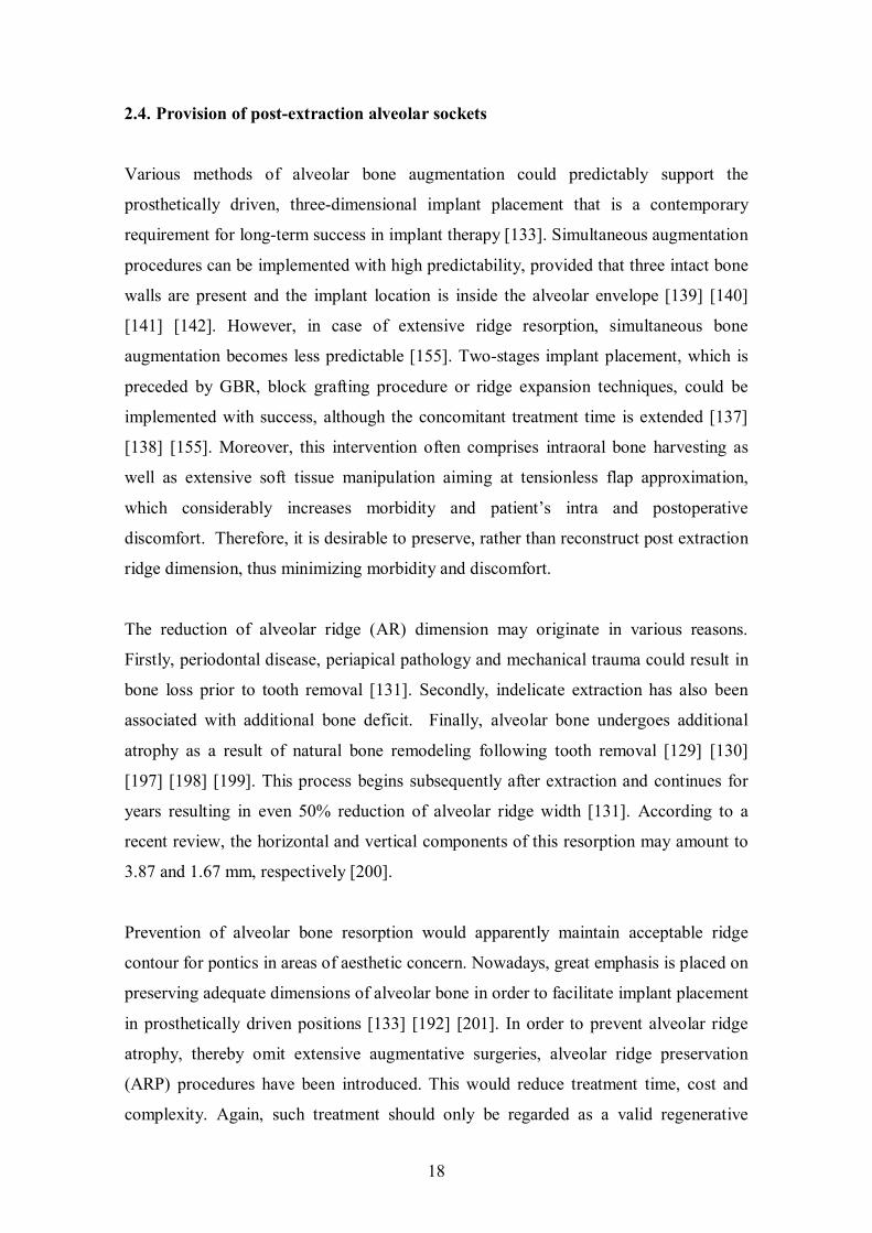
Fig. 1 Reconstructive periodontal surgery with the use of EMD + BCP. (a) preoperative clinical measurement demonstrates a PPD of 9 mm; (b) preoperative radiograph demonstrates the presence of a deep intrabony defect; (c) intraoperative measurement following debridement; (d) placement of the notch by means of a round bur indicating the bottom of the intrabony defect; (e) application of EMD; (f) BCP mixed with EMD prior to application; (g) periodontal defect filled with EMD + BCP.
No root conditioning or any other surface modifications were applied in Study 2. The
intrabony defect was subsequently filled with the nano-HA paste (Ostim®; Heraeus
Kulzer; Hanau, Germany) and adjusted to the alveolar crest, according to the
manufacturer’s instruction (Fig. 2e, f).
The mucoperiosteal flaps were then repositioned and secured with a combination of
suspended vertical mattress and single interrupted sutures (non-resorbable,
monofilament; Dafilon 5/0; Braun Aesculap; Tuttlingen, Germany) in order to achieve
tensionless flap closure. Teeth were splinted in case of extreme mobility.
Postoperative care
All patients were postoperatively administered antibiotics (amoxicillin 500 mg +
clavulanic acid 125 mg; Augmentin 625; GlaxoSmithKline; Brentford, Middlesex, UK)

27
Fig. 2 Reconstructive periodontal surgery with the use of nano-HA. (a) preoperative clinical measurement demonstrates a PPD of 10 mm; (b) preoperative radiograph demonstrates the presence of a deep intrabony defect; (c) the intraoperative situation revealed a deep one- and two-wall intrabony defect following debridement; (d) placement of the notch by means of a round bur indicating the bottom of the intrabony defect; (e) application of nano-HA; (f) periodontal defect filled with nano-HA.
three times daily for seven days and painkiller (diclofenac 75mg; Diclofenac Duo,
Pharmavit, Veresegyház, Hungary) according to individual need. Subjects were advised
not to brush the surgical area but rinse with 0.2 % chlorhexidine two times daily for 90
seconds during the postoperative four weeks. Subjects were asked to refrain any
mechanical plaque control at the surgical site. Sutures were removed 10 to 14 days after
surgery. Then the patients resumed tooth cleaning with the use of a soft brush.
Additional appointments including oral hygiene instructions and professional
supragingival tooth cleaning were performed fortnightly during the first twelve
postoperative weeks. After this period and until biopsy removal, recall appointments
were scheduled monthly. Neither subgingival instrumentation nor periodontal probing
was performed during the entire experimental period.

28
Re entry, biopsy and histological procedure
After a healing period of nine (Study 1) or seven months (Study 2) the teeth were
removed together with some of their surrounding periodontal tissues following the same
type of local anaesthesia and presurgical preparations (Fig. 3a-c, 4a-c).
Fig. 3 Re-entry and biopsy after nine months of healing following treatment of EMD +BCP. (a) stable-looking soft tissue conditions; (b) radiograph indicates radiopaque tissue in the former defect; (c) decoronated tooth with some of the surrounding tissues
Fig. 4 Re-entry and biopsy after seven months of healing following treatment of nano-HA. (a) at 7 months following surgery, a substantial reduction of PPD was measured; (b) radiograph indicates mineralisation in the former defect; (c) removed tooth with some of the surrounding tissues
Specimens were subsequently placed in 10% buffered formalin for fixation. The
extraction sites were then augmented with the use of various types of bone substitutes
and barrier membranes (Study 1) and with DBBM (TutoDent Microchips; Tutogen;
Neunkirchen, Germany) in conjunction with a resorbable collagen membrane of bovine
pericardium origin (TutoDent Membrane; Tutogen; Neunkirchen, Germany) in Study 2.
29
According to local need, connective tissue graft was transplanted in order to increase the
volume of keratinised gingiva [112]. After the healing of the extraction sites, subjects
received either implant-based crowns or fixed partial dentures as part of the prosthetic
rehabilitation.
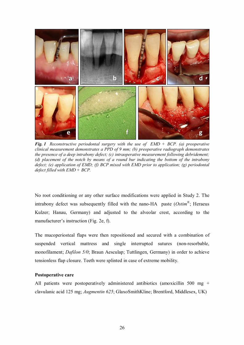
The block biopsies were fixed in 10 % buffered formalin, decalcified in EDTA for a
period of 4–6 weeks (depending on tooth/root volume) and dehydrated in graded series
of ethanol. Immediately prior to embedding in paraffin, the roots/teeth were split in two
along their long axis either in bucco-oral or mesiodistal direction (depending on the
location of the deepest site of the defect) exactly at the notch indicating the site of
interest. Thus, each biopsy provided two specimen blocks without the need for
extensive cutting. Twenty sections from each of the two blocks per specimen were
obtained with the microtome, set at 5 to 8 μm, and subsequently stained with
hematoxylin–eosin and further with the oxone-aldehyde-fuchsin-Halmi staining method
in Study 2. One experienced examiner measured the parameters by means of a
computer-assisted toolbox. The field of view in the light microscope (Olympus DH 50;
Olympus Denmark AS; Ballerup, Denmark) was examined on an LCD flat screen
monitor through live streaming, which were also captured by a digital camera (Olympus
DP 71; Olympus Denmark AS; Ballerup, Denmark). The following parameters were
measured: (i) height of cementum regeneration: distance between the apical extension
of the defect (bottommost point of the notch) and the coronal extension of a continuous
layer of new cementum or cementum-like deposit on the planed root (millimetres); (ii)
bone regeneration height: distance between the bottommost point of the notch and the
coronal extension of regenerated alveolar bone along the planed root (millimetres) (The
coronal extension of regenerated bone was defined as the most coronal level where the
PDL space had an almost normal width); (iii) root resorption: combined linear heights
of distinct resorption lacunae on the planed root (millimetres); (iv) ankylosis: combined
linear heights of ankylotic union between the regenerated alveolar bone and the planed
root (millimetres). Digital images were evaluated using a software program (SIS
AnalySIS Auto Software 3.2; Soft Imaging System; Münster, Germany). The
histomorphometric evaluation was carried out under ×25 to ×100 magnification. In the
absence of a visible notch, the apical extension of instrumentation was used as landmark
for the histomorphometric measurements.

30
Sources of support
Study 1 was supported by Institute Straumann, Basel, Switzerland. Study 2 was self-
funded and supported by a research grant from Heraeus Kulzer, Hanau, Germany.
Candidate’s contribution to the studies
Fine-tuning of the protocol, study co-ordination, patient recruitment, assessment, data
acquisition, patient management, compensatory treatment provision, revision and
approval of the manuscript (Study 1); study design and protocol, study co-
ordination, patient recruitment, surgical treatment (both stages), patient management,
compensatory treatment provision, analysis and interpretation of data, preparation of the
manuscript were carried out by the candidate of the present thesis. (Study 2)
4.3. Regeneration of peri-implant dehiscence defects (Study 3)
Experimental model and management
In the present experiment twelve female Göttingen minipigs (20 months of age and
average body weight of 35 kg) were used. Before each surgical procedure, the animals
were fasted overnight, weighed and pre-medicated. Each subjects received an injection
of analgesia (Temgesic®; Schering-Plough; Brussels, Belgium) on the day of the surgery
and on the next 3 days. StreptocillinVet® 250 + 200 mg/ml (Boehringer Ingelheim;
Copenhagen, Denmark) was started one day before the procedure and was continued for
seven days. The animals were anaesthetized according to a standard procedure using
ketamine (Ketalar® 50 gm/ml; Pfizer; Sollentuna, Sweeden) and midazolam (Hameln 5
mg/ml; Pharmaceuticals GmbH; Hameln, Germany). After disinfection of the surgical
site with 0.2% chlorohexidine solution (Corsodyl®; GlaxoSmithKline; Brentford,
Middlesex, UK), local infiltration anaesthesia (Lidocaine 2% with 1:80,000 of
epinefrine; HenrySchein Inc; Port Washington, NY, USA) was given. During the
operation, additional 10 ml of ketamine and 1.5 ml of midazolam were administered
when needed. Soft diet was provided daily during the whole course of the study. For
documentation purposes digital photography and radiographs of the inserted implants
were taken. The experimental protocol was approved by the Ethical Committee of the
University of Lund (Sweden).
31
Study sequence
All surgical procedures were performed by two experienced periodontists. The
treatment sequences in the study were:
1. Extractions and creation of “chronic” defect. Day 0/Baseline
2. Randomised implantation with/without creation of acute defect, with/without the
use of test materials (4 groups: T1, T2, N, P). 3 Months
3. Uncovering of the implants and connection of abutments in different length (2
groups: loaded/non-loaded). 6 Months
4. Termination, histological analysis. 8 Months
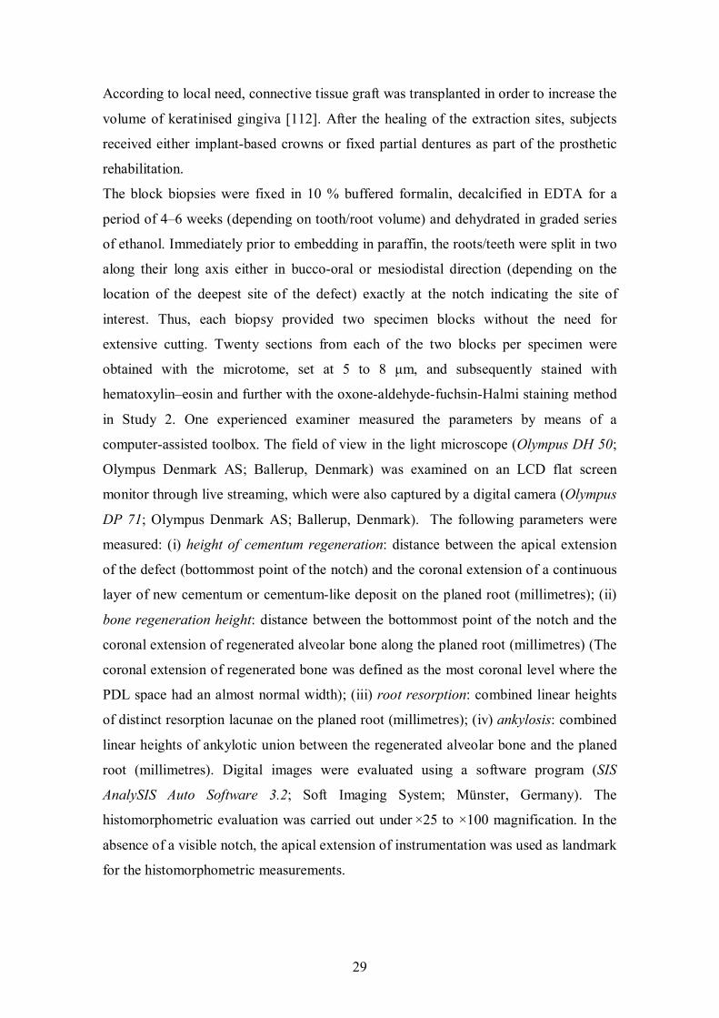
Extraction and creation of chronic defect – Day 0/Baseline
Following intracrevicular incisions and careful elevation of a full thickness muco-
periosteal flap, the premolars and the first molar were extracted in both hemi-mandibles
in all animals (Fig. 9a). Particular care was taken to ensure that no root remnants were
left in the extraction sockets. Following tooth extraction, bone defect was created on
both sides of the mandible by removal of the buccal plate with a chisel, on a length of
40 mm and a height of 6 mm (Fig. 9b). The lingual cortical bone wall was left intact.
The surgical area was then rinsed with saline. Flap closure was achieved with
absorbable sutures (Vicryl® 4–0; Ethicon GmbH; Norderstedt, Germany) (Fig. 9c) and
the defect was allowed to heal for three months (Fig. 9d).

32
Fig. 9 (a) Day 0: Extraction of P2, P3, P4 and M1. (b) Day 0: Removal of the buccal plate by a chisel. (c) Day 0: Flap closure and suturing. (d) Three months: Healing following teeth extraction and buccal plate removal (’chronic defect’).
Implantation with/without creation of acute defect, with/without the use of test
materials (first stage surgery) – 3 months
After a healing period of three months, following midcrestal incision and full thickness
flap reflection, novel, titanium, bone level, 4.1x8 mm dental implants with a modified
hydrophilic surface (Straumann®, Bone Level, SLActive implants; Straumann, Basel,
Switzerland) were inserted either into the reduced thickness alveolar ridge (chronic
defect and pristine site), or into a combined chronic and a surgically created
standardised acute buccal dehiscence-type defect. Triangular shaped acute defects
(height: 6 mm, apical mesio-distal width: 12 mm, bucco-lingual width: 2 mm) were
prepared with a rotating surgical drill with ample saline irrigation. Defects were created
in order to mimic real clinical circumstances of a partially edentulous ridge.
Consequently, the coronal 6 mm of the implants were exposed on the buccal aspect.
Only the apical 2 mm of the implants was in the alveolar bone around the whole
circumference (Fig. 10). In total, 48 bone level implants with a diameter of 4.1 mm
and a length of 8 mm were placed in 12 minipigs (four implants/animal; two in each
hemi-mandible). All implants were inserted according to the guidelines of the
manufacturer. Profile drilling (without tapping) was performed and the implants were
inserted in such a way that the implant shoulder was aimed to match the bone crest
level. Insertion torque was always higher than 35 Ncm.
Fig. 10 Schematic drawing of the chronic and acute, standardised, peri-implant, critical size dehiscence defect.
According to a computer-generated randomization scheme, four groups were created:
P (12 implants) – In the positive control group, twelve implants were inserted into the

33
chronic defects resulting from the 3-month healing following buccal bone plate removal
at the time of teeth extraction (pristine sites) (Fig. 11).
Fig. 11 Three months after baseline: Implant placement in group P (pristine site). Minor buccal and/or lingual dehiscence defects were occasionally present.
N (12 implants) – In the negative
control group the chronic and
acute dehiscence defects were left untreated as such the buccal surface of the implant
remained exposed (negative control) (Fig. 12).
Fig. 12 Three months after baseline: Creation of standardized acute dehiscence-type defect. (a) Standardized defects were created around the osteotomy by removing part of the buccal bone. (b) The resulting dehiscence defects presented triangular-shaped base and the following dimensions: 6 mm apico-coronally, 12 mm mesio-distally, 2 mm bucco-lingually.
T1 (12 implants) – The exposed implant surface in the dehiscence defects was treated
with BCP (Straumann BoneCeramic®; particle size 500–1000 µm; Straumann; Basel,
Switzerland). The BCP particles were mixed with autologous blood collected in the
surgical site. The graft material was then applied in the dehiscence defect in contact
with the exposed implant threads. Care was taken to avoid overfilling or coverage of the
implant head (Fig. 13).

34
Fig. 13 Three months after baseline: Group T1: (a) BCP was mixed with autologous blood from the surgical site; (b) The exposed implant surface in the dehiscence defects was covered with the BCP.
T2 (12 implants) – BCP and the novel PEG membrane (Straumann MembraGel®;
Straumann; Basel, Switzerland) was used for the treatment of the dehiscence defects.
The synthetic bioresorbable polyethylene glycol hydrogel membrane was activated by
mixing its components according to the manufacturer’s guidelines. The membrane was
applied as a gel with a continuous flow over the bone substitute and the adjacent bone.
(Fig. 14a-c). After the cross-linking reaction of the membrane components was
completed and the membrane set, the margins were smoothed with a sharp scalpel
blade. The membrane extended on the adjacent bone surface approximately 3 mm
further than the grafted area and covered completely the implant head (Fig. 14d).
Fig. 14 Three months after baseline: Group T2: (a) BCP was placed onto the defect; (b) The two component of the hydrogel was mixed prior to application; (c) The polyethylene glycol hydrogel membrane (PEG) was applied on and around the BCP and the implant; (d) The set, polimerised membrane in place.
35
All implants (48) of all groups received closure screws and were fully covered by the
mucoperiosteal flaps for submerged healing. The surgical area was carefully sutured
with absorbable material using single interrupted and vertical mattress sutures.
Implant uncover and abutment connection according to two different loading
protocols – 3 months after implant placement
At 3 months after implant placement (6 months after extraction), a second stage surgery
was performed to uncover all implants. Midcrestal incisions were performed on the sites
of implant placement. Minor buccal and lingual flap elevation was performed to limit
periosteal exposure. The distance from the implant shoulder to the first bone-to-implant
contact was measured on the buccal side with a UNC 15 periodontal probe. According
to a computer generated randomization scheme, two different abutments were
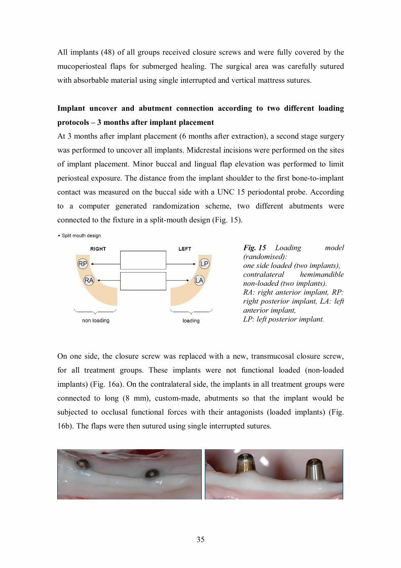
connected to the fixture in a split-mouth design (Fig. 15).
Fig. 15 Loading model (randomised): one side loaded (two implants), contralateral hemimandible non-loaded (two implants). RA: right anterior implant, RP: right posterior implant, LA: left anterior implant, LP: left posterior implant.
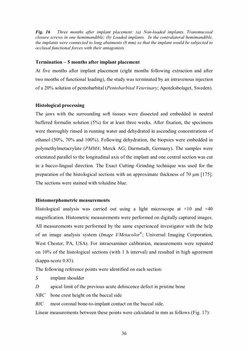
On one side, the closure screw was replaced with a new, transmucosal closure screw,
for all treatment groups. These implants were not functional loaded (non-loaded
implants) (Fig. 16a). On the contralateral side, the implants in all treatment groups were
connected to long (8 mm), custom-made, abutments so that the implant would be
subjected to occlusal functional forces with their antagonists (loaded implants) (Fig.
16b). The flaps were then sutured using single interrupted sutures.
36
Fig. 16 Three months after implant placement: (a) Non-loaded implants. Transmucosal closure screws in one hemimandible; (b) Loaded implants. In the contralateral hemimandible, the implants were connected to long abutments (8 mm) so that the implant would be subjected to occlusal functional forces with their antagonists.
Termination – 5 months after implant placement
At five months after implant placement (eight months following extraction and after
two months of functional loading), the study was terminated by an intravenous injection
of a 20% solution of pentobarbital (Pentobarbital Veterinary; Apoteksbolaget, Sweden).
Histological processing
The jaws with the surrounding soft tissues were dissected and embedded in neutral
buffered formalin solution (5%) for at least three weeks. After fixation, the specimens
were thoroughly rinsed in running water and dehydrated in ascending concentrations of
ethanol (50%, 70% and 100%). Following dehydration, the biopsies were embedded in
polymethylmetacrylate (PMMA; Merck AG; Darmstadt, Germany). The samples were
orientated parallel to the longitudinal axis of the implant and one central section was cut
in a bucco-lingual direction. The Exact Cutting–Grinding technique was used for the
preparation of the histological sections with an approximate thickness of 70 μm [175].
The sections were stained with toluidine blue.
Histomorphometric measurements
Histological analysis was carried out using a light microscope at ×10 and ×40
magnification. Histometric measurements were performed on digitally captured images.
All measurements were performed by the same experienced investigator with the help
of an image analysis system (Image I/Metacolor®; Universal Imaging Corporation,
West Chester, PA, USA). For intraexaminer calibration, measurements were repeated
on 10% of the histological sections (with 1 h interval) and resulted in high agreement
(kappa-score 0.83).
The following reference points were identified on each section:
S implant shoulder
D apical limit of the previous acute dehiscence defect in pristine bone
NBC bone crest height on the buccal side
BIC most coronal bone-to-implant contact on the buccal side.
Linear measurements between these points were calculated in mm as follows (Fig. 17):
37
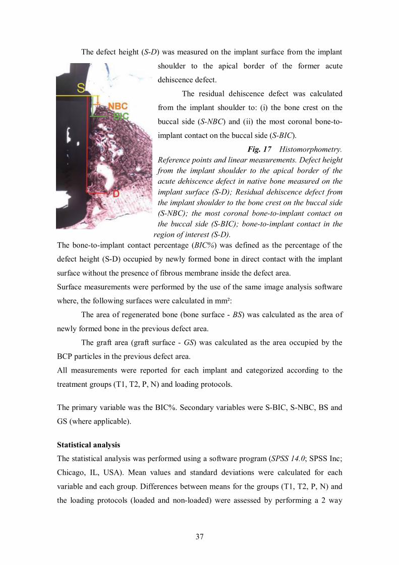
The defect height (S-D) was measured on the implant surface from the implant
shoulder to the apical border of the former acute
dehiscence defect.
The residual dehiscence defect was calculated
from the implant shoulder to: (i) the bone crest on the
buccal side (S-NBC) and (ii) the most coronal bone-to-
implant contact on the buccal side (S-BIC).
Fig. 17 Histomorphometry. Reference points and linear measurements. Defect height from the implant shoulder to the apical border of the acute dehiscence defect in native bone measured on the implant surface (S-D); Residual dehiscence defect from the implant shoulder to the bone crest on the buccal side (S-NBC); the most coronal bone-to-implant contact on the buccal side (S-BIC); bone-to-implant contact in the
region of interest (S-D). The bone-to-implant contact percentage (BIC%) was defined as the percentage of the
defect height (S-D) occupied by newly formed bone in direct contact with the implant
surface without the presence of fibrous membrane inside the defect area.
Surface measurements were performed by the use of the same image analysis software
where, the following surfaces were calculated in mm²:
The area of regenerated bone (bone surface - BS) was calculated as the area of
newly formed bone in the previous defect area.
The graft area (graft surface - GS) was calculated as the area occupied by the
BCP particles in the previous defect area.
All measurements were reported for each implant and categorized according to the
treatment groups (T1, T2, P, N) and loading protocols.
The primary variable was the BIC%. Secondary variables were S-BIC, S-NBC, BS and
GS (where applicable).
Statistical analysis
The statistical analysis was performed using a software program (SPSS 14.0; SPSS Inc;
Chicago, IL, USA). Mean values and standard deviations were calculated for each
variable and each group. Differences between means for the groups (T1, T2, P, N) and
the loading protocols (loaded and non-loaded) were assessed by performing a 2 way

38
repeated measures analysis of variance (ANOVA) for each variable, with repeated
measures over loading sites and treatments. The assumptions of normality and constant
variance underlying the ANOVA were checked by a study of the residuals. If the
assumptions were not fulfilled, a square root transformation was applied to the data.
When a statistically significant difference between the group means was indicated in the
ANOVA, a post-hoc Bonferroni test was used to determine which group means differed
significantly. Result was considered statistically significant if P < 0.05.
Source of support
The present study was supported by Institute Straumann, Basel, Switzerland.
Candidate’s contributions to the study
Fine-tuning of the design and protocol, study co-ordination, surgical treatment (all the
three stages), revision and approval of the manuscript were carried out by the candidate
of the present thesis.
4.4. Regeneration of post-extraction alveolar sockets (Study 4)
Prior to commencement of the study a detailed protocol was developed and agreed upon
by the authors based on the Cochrane Collaboration guidelines and previous reviews
published by our research group [127] [228] [229] [230] [231].
Focused question
Following tooth/root extraction in humans, what is the effect of ridge preservation on
the residual alveolar ridge dimension and on histological characteristics, compared to
unassisted socket healing?
Definition
Whilst ‘socket preservation’ has widely been employed to depict a certain procedure,
we believe that the objective of these interventions is to preserve the dimension of the
AR. Therefore, we have used the term ‘Alveolar Ridge Preservation’ to define such
procedures.
39
Types of studies
Longitudinal prospective studies were included i.e. RCTs, CCTs and cohort studies with
control group.
Populations of studies
Healthy individuals, without any age limit, who underwent any type of ridge
preservation following permanent tooth extraction, were included. Smokers and patients
with history of periodontal disease were not excluded. The minimum number of
subjects per group was five. However, no limit was set for study follow-up period.
Types of Interventions
Test groups
Studies reporting on any of the following types of interventions were included:
socket grafting (autograft, allograft, xenograft, alloplastic materials); socket sealing
(soft tissue grafts); GBR (resorbable/non-resorbable barriers); biological active
materials (growth factors) and combinations of the above techniques/materials.
Control groups
The control groups of the included studies comprised empty sockets, i.e. unassisted
socket healing.
Outcome variables
The primary outcome was the change in oro-facial (horizontal) and apico-coronal
(vertical) AR dimensions. Secondary outcomes were the followings: (i) change in
buccal plate thickness; (ii) bone volume alteration following extraction; (iii)
complications; (iv) histological healing characteristics; (v) site eligibility for placement
of an adequate size dental implant with or without further augmentation; (vi) patient-
reported outcomes, such as quality of life and (vii) health economics.
Risk of bias and methodological quality assessment
In order to evaluate the methodological quality and risk of bias of individual studies, we
used a combination of parameters from the Cochrane Collaboration and Consolidated
Standards of Reporting Trials (CONSORT) statement. The following parameters were
assessed and taken into consideration in the final analysis: sample size calculation,
statement of eligibility criteria, ethics approval, informed consent, baseline
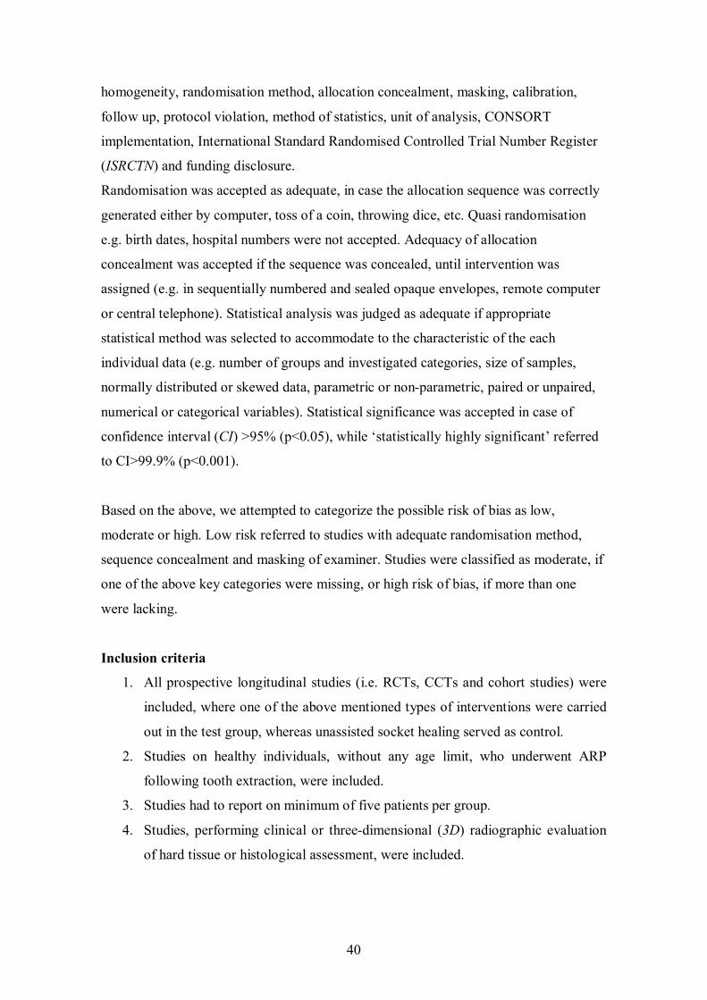
40
homogeneity, randomisation method, allocation concealment, masking, calibration,
follow up, protocol violation, method of statistics, unit of analysis, CONSORT
implementation, International Standard Randomised Controlled Trial Number Register
(ISRCTN) and funding disclosure.
Randomisation was accepted as adequate, in case the allocation sequence was correctly
generated either by computer, toss of a coin, throwing dice, etc. Quasi randomisation
e.g. birth dates, hospital numbers were not accepted. Adequacy of allocation
concealment was accepted if the sequence was concealed, until intervention was
assigned (e.g. in sequentially numbered and sealed opaque envelopes, remote computer
or central telephone). Statistical analysis was judged as adequate if appropriate
statistical method was selected to accommodate to the characteristic of the each
individual data (e.g. number of groups and investigated categories, size of samples,
normally distributed or skewed data, parametric or non-parametric, paired or unpaired,
numerical or categorical variables). Statistical significance was accepted in case of
confidence interval (CI) >95% (p<0.05), while ‘statistically highly significant’ referred
to CI>99.9% (p<0.001).
Based on the above, we attempted to categorize the possible risk of bias as low,
moderate or high. Low risk referred to studies with adequate randomisation method,
sequence concealment and masking of examiner. Studies were classified as moderate, if
one of the above key categories were missing, or high risk of bias, if more than one
were lacking.
Inclusion criteria
1. All prospective longitudinal studies (i.e. RCTs, CCTs and cohort studies) were
included, where one of the above mentioned types of interventions were carried
out in the test group, whereas unassisted socket healing served as control.
2. Studies on healthy individuals, without any age limit, who underwent ARP
following tooth extraction, were included.
3. Studies had to report on minimum of five patients per group.
4. Studies, performing clinical or three-dimensional (3D) radiographic evaluation
of hard tissue or histological assessment, were included.

41
Exclusion criteria
1. Case reports, case series, retrospective analyses were excluded.
2. Studies without a control group comprising unassisted socket healing were
excluded.
3. Studies on medically compromised patients e.g. uncontrolled diabetes mellitus
or cancer were excluded.
4. Studies reporting on immediate placement of dental implant were excluded.
5. Studies describing extraction of third molars were excluded.
Search strategy
A sensitive search strategy was designed as we anticipated that relevant studies might
be difficult to locate. The search strategy incorporated both electronic and hand
searches. The following electronic databases were utilized in Apr 2010: (i) MEDLINE
In-Process & Other Non-Indexed Citations and MEDLINE 1950 to present via Ovid
interface; (ii) EMBASE Classic + EMBASE 1947 to present via Ovid interface; (iii)
The Cochrane Central Register of Controlled Trials (CENTRAL); (iv) Literatura Latino-
Americana e do Caribe em Ciências da Saúde (LILACS).
The electronic search strategy used the following combination of key words and MeSH
terms:
(“tooth extraction” OR “tooth removal” OR “socket” OR “alveol$” OR “ridge” OR
“crest” OR “tooth socket” OR “alveolar bone loss” OR “bone resorption” OR “bone
remodeling”) AND (“preserv$” OR “reconstruct$” OR “augment$” OR “fill$” OR
“seal$” OR “graft$” OR “repair$” OR “alveolar ridge augmentation” OR “bone
regeneration” OR “bone substitutes” OR “transplantation”).
Cochrane search filters for RCTs and CCTs were implemented. In addition, cohort trials
were also searched. The results were limited to humans only.
An extensive hand search was also performed encompassing the bibliographies of the
included papers and review articles. Furthermore the following journals were screened
from 2001 to April 2010: Clinical Oral Implants Research, Clinical Implant Dentistry
and Related Research, European Journal of Oral Implantology, Implant Dentistry,
International Journal of Oral and Maxillofacial Implants, International Journal of
Periodontics and Restorative Dentistry, Journal of Clinical Periodontology, Journal of
Dental Research, Journal of Oral and Maxillofacial Surgery, Journal of

42
Periodontology, Oral Surgery, Oral Medicine, Oral Radiology, Oral Pathology and
Endodontics, Periodontology 2000. No language restrictions were applied. Translations
were carried out as necessary by two reviewers.
The extracted data were copied into Reference Manager 10 software (Thomson Reuters;
New York, NY, USA). Thus, the further steps of screening were performed on this
interface. A three-stage selection of the resulted hits was performed independently and
in duplicate by two reviewers. In order to reduce errors and bias, a calibration exercise
was performed with the first 500 titles, resulting in 96.4% agreement. In case of
disagreement at the title selection stage, the trial was included in the abstract stage. At
the abstract and full text selection any disagreements between the above reviewers were
resolved by discussion. If unresolved, a third reviewer was involved for arbitration. The
reasons for exclusion were recorded either in the Reference Manager (abstract stage) or
in a specific data extraction form (full text stage). The level of agreement was
determined by Kappa score calculation.
Research synthesis
Studies were grouped by research design and their chief characteristics. Outcomes were
recorded in evidence tables. In view of the marked heterogeneity, no meta-analysis was
conducted. Instead, a narrative synthesis was undertaken.
Sources of support
The present study was self-funded and supported by the Research Discretionary
Account of the Unit of Periodontology, UCL Eastman Dental Institute, which received a
proportion of funding from the UK Department of Health’s NIHR Biomedical Research
Centres funding scheme.
Candidate’s contributions to the study
Study design and protocol, study co-ordination, selection and review of all included and
excluded trials, data collection, analysis and interpretation of data, manuscript
preparation were carried out by the candidate of the present thesis.

43
5. RESULTS
5.1. Periodontal regeneration
5.1.1. Treatment with EMD and SBC
All ten subjects (six women and four men), aged between 27 and 62 years (mean age,
48.6 years) completed the study. Postoperative healing period was uneventful in all
cases. No complications, such as allergic reactions, abscesses or infections were
observed throughout the duration of the study.
In one of the 10 subjects, the clinical evaluation and radiographs demonstrated excellent
clinical healing. Therefore, putting patient’s benefit first, a decision was made not to
remove the tooth and to perform only a re-entry. In this particular case, the re-entry
revealed that the intrabony defect, which was located at the mesial aspect of tooth #24,
presented with almost complete healing with a newly formed hard tissue resistant to
probing.
The clinical measurements demonstrated a reduction in mean PPD from 8.6 ± 1.9 mm to
5.3 ± 2.0 mm at baseline and at nine months, respectively. The mean CAL changed
from 10.8 ± 2.0 mm to 7.8 ± 1.7 mm resulting in a mean CAL gain of 3.0 ± 1.6 mm
(Table 1).
Table 1 Clinical and histologic results following treatment of human intrabony periodontal defects with EMD + BCP (mm)
In six out of nine biopsies, the histological findings indicated the formation of new
cementum with inserting collagen fibres to a varying extent (Fig 5a, b). The newly
formed cementum was a mixed acellular and cellular type in all specimens. Collagen
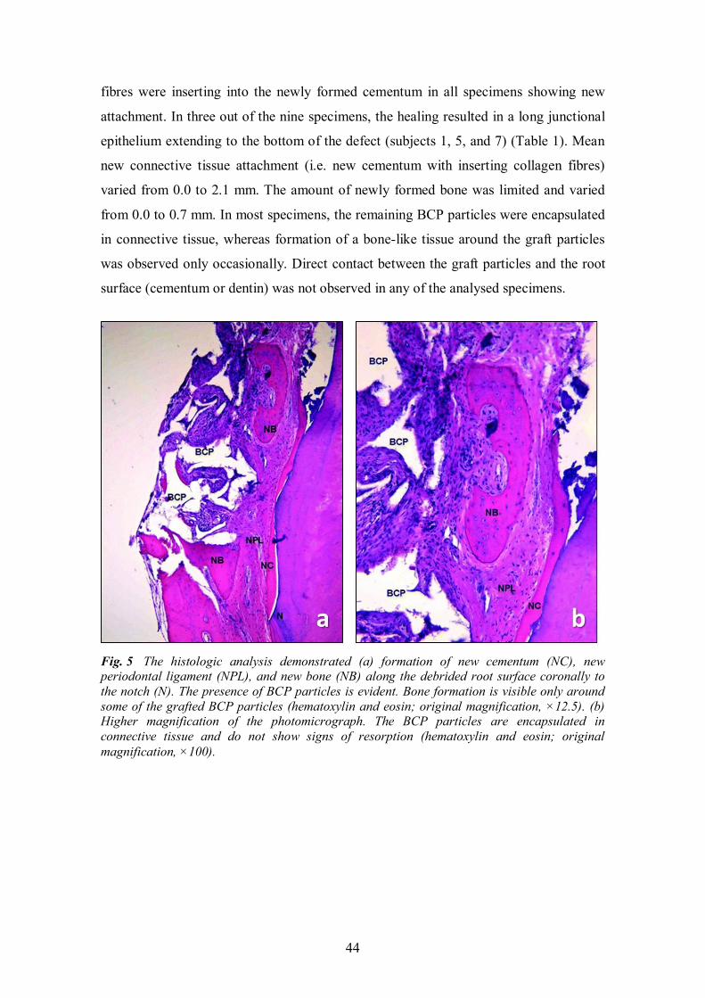
44
fibres were inserting into the newly formed cementum in all specimens showing new
attachment. In three out of the nine specimens, the healing resulted in a long junctional
epithelium extending to the bottom of the defect (subjects 1, 5, and 7) (Table 1). Mean
new connective tissue attachment (i.e. new cementum with inserting collagen fibres)
varied from 0.0 to 2.1 mm. The amount of newly formed bone was limited and varied
from 0.0 to 0.7 mm. In most specimens, the remaining BCP particles were encapsulated
in connective tissue, whereas formation of a bone-like tissue around the graft particles
was observed only occasionally. Direct contact between the graft particles and the root
surface (cementum or dentin) was not observed in any of the analysed specimens.
Fig. 5 The histologic analysis demonstrated (a) formation of new cementum (NC), new periodontal ligament (NPL), and new bone (NB) along the debrided root surface coronally to the notch (N). The presence of BCP particles is evident. Bone formation is visible only around some of the grafted BCP particles (hematoxylin and eosin; original magnification, ×12.5). (b) Higher magnification of the photomicrograph. The BCP particles are encapsulated in connective tissue and do not show signs of resorption (hematoxylin and eosin; original magnification, ×100).
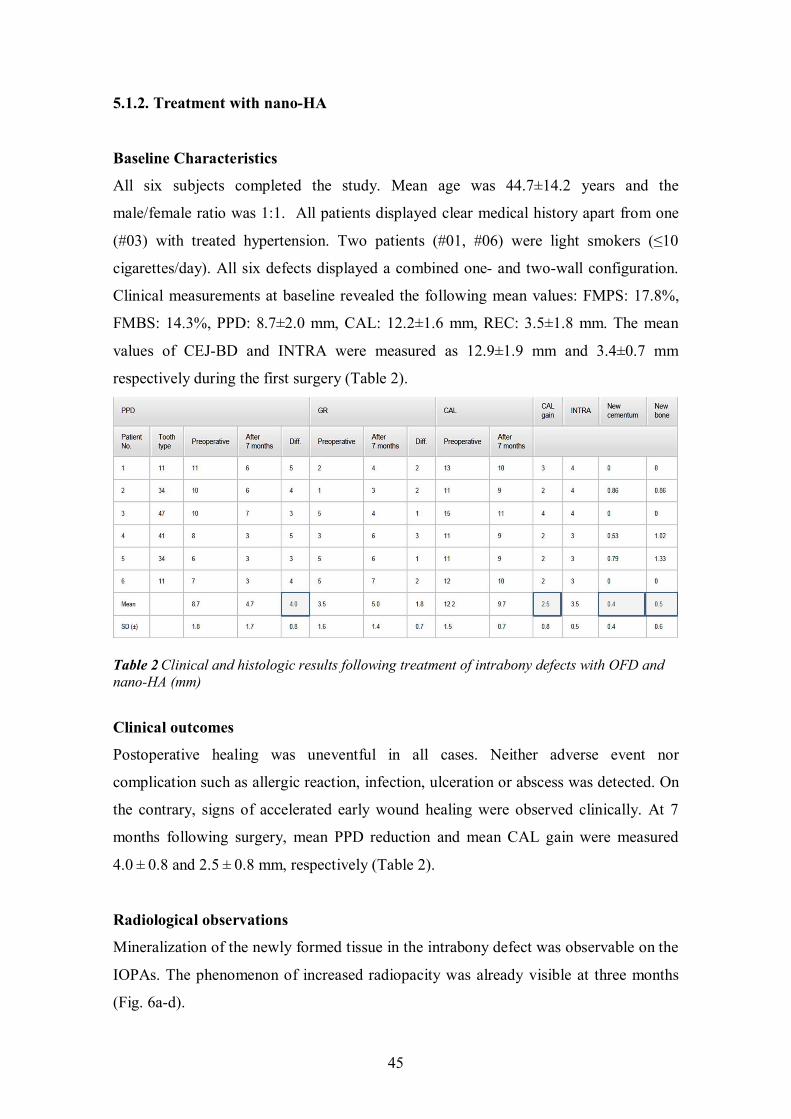
45
5.1.2. Treatment with nano-HA
Baseline Characteristics
All six subjects completed the study. Mean age was 44.7±14.2 years and the
male/female ratio was 1:1. All patients displayed clear medical history apart from one
(#03) with treated hypertension. Two patients (#01, #06) were light smokers (≤10
cigarettes/day). All six defects displayed a combined one- and two-wall configuration.
Clinical measurements at baseline revealed the following mean values: FMPS: 17.8%,
FMBS: 14.3%, PPD: 8.7±2.0 mm, CAL: 12.2±1.6 mm, REC: 3.5±1.8 mm. The mean
values of CEJ-BD and INTRA were measured as 12.9±1.9 mm and 3.4±0.7 mm
respectively during the first surgery (Table 2).
Table 2 Clinical and histologic results following treatment of intrabony defects with OFD and nano-HA (mm)
Clinical outcomes
Postoperative healing was uneventful in all cases. Neither adverse event nor
complication such as allergic reaction, infection, ulceration or abscess was detected. On
the contrary, signs of accelerated early wound healing were observed clinically. At 7
months following surgery, mean PPD reduction and mean CAL gain were measured
4.0 ± 0.8 and 2.5 ± 0.8 mm, respectively (Table 2).
Radiological observations
Mineralization of the newly formed tissue in the intrabony defect was observable on the
IOPAs. The phenomenon of increased radiopacity was already visible at three months
(Fig. 6a-d).

46
Fig. 6 Increasing density of the former defect is observable on the radiographs (a) prior to surgery; (b) right after surgery; (c) after three months of healing; (d) after seven months of healing.
Histological outcomes
The histological analysis revealed that in three out of the six defects, the healing
occurred through formation of a long junctional epithelium along the debrided root
surfaces extending until the most apical part of the defects (Fig. 7a, b). In the remaining
three specimens limited formation of new cementum with inserting connective tissue
fibres and new bone were observed with a magnitude varying from 0.53 to 0.86 mm and
from 0.86 to 1.33 mm, respectively (Table 2). Neither ankylosis, nor root resorption was
observed in any of the biopsies. In two out of the six biopsies, some remnants of the
grafting material were visible (Fig. 8a, b). The nano-HA particles were predominantly
surrounded by connective tissue, without signs indicating a potential to promote
periodontal or bone regeneration.
Fig. 7 The histological evaluation revealed (a) healing predominantly characterized by a long junctional epithelium and limited regeneration of cementum and bone. Red arrowhead: coronal extension of new cementum, blue arrowhead: coronal extension of new bone, red dotted line: apical extension of the notch. Original magnification × 5; (b) Higher magnification of the defect. Formation of new cementum (NC) and new periodontal ligament (PDL) was confined to the area of the notch. White dotted line: margin between the newly formed bone and old bone, red asterisk: artifact. Original magnification × 25

47
Fig. 8 (a) Representative photomicrograph revealing limited periodontal regeneration (case 5 from Table 1). Red dotted line: apical extension of the notch, red arrowhead: coronal extension of new cementum, blue arrowhead: coronal extension of new bone. Original magnification × 25. (b) Higher magnification of the defect. Formation of new cementum (NC), new periodontal ligament (PDL) and new bone (B) was confined to the area of the notch. White dotted line: margin between the newly formed bone and old bone, red asterisk: artefact, white asterisk: remnant nano-HA particle encapsulated in connective tissue. Original magnification × 50
5.2. Provision of peri-implant dehiscence defects with SBC and PEG
Clinical observations
Extractions and creation of chronic defect – Day 0/Baseline
Following extractions and removal of the buccal plate, all sites were closed by primary
intention and healed without complications.
Creation of acute dehiscence defects (T1, T2, N) and implantation (T1, T2, P, N) –
First stage surgery 3 months after baseline
As a result of the previous buccal plate removal, a remarkable decrease in the bucco-
lingual ridge width, which resulted in thin (knife-edge) ridge, was observed in all these
cases (Fig. 18). Consequently, in group P, the implant placement was occasionally

48
associated with the presence of buccal and/or lingual dehiscence around the 4.1 mm
implants. Dense (type 1) bone was observed in all sites during implantation.
Fig. 18 Removal of the buccal plate after extraction resulted in knife–edge ridge.
Good primary implant stability was achieved at all sites of all experimental groups,
despite the extensive dehiscence defects that were created. The final insertion torque
was always higher than 35 Ncm for all implants.
In group T2, the membrane was applied successfully in all cases. However, in 4/12
sites, the membrane had to be reconstructed prior to flap closure, because of membrane
rupture (three cases) or instability on the underlying graft/bone (one case) (Fig. 19).
Primary closure of the flaps was achieved in all sites of all groups.
Fig. 19 Three months after baseline. Rupture of the PEG membrane above the implant head, before flap closure is visible.
Implant uncover and abutment connection – 3 months after implant placement
Exposure of the cover screw of the implant was observed in 17/48 implants. This
occurred in all groups with similar frequency (four exposures in groups N, T1, T2 and

49
five exposures in group P). One implant was found to be mobile and was removed
(Group T2). All other sites healed uneventfully.
The distance from the implant shoulder to the first clinical bone-to-implant contact was
measured on the buccal side in all groups.
In group T1, after flap reflection, graft granules were found to be encapsulated in the
soft tissue flap in 3/12 sites. No attempt was made to remove the mobile graft particles.
The mean clinical distance from the implant shoulder to the first clinical bone-to-
implant contact was 2.55 mm (range: 0-6 mm).
In group T2, no remnant of the PEG membrane was visible in any of the sites during
second stage surgery. In this group, no loose graft particles were observed. The mean
clinical distance from the implant shoulder to the first clinical bone-to-implant contact
was 2.1 mm (range: 2-6 mm).
In group P, the mean clinical distance was 1.25 mm (range: 1-3 mm).
In group N, the mean clinical distance was 3.35 mm (range: 1-7 mm).
Histological analysis
Histological measurements and the relevant statistical analysis were performed in a total
of 47 of the 48 implants initially placed.
These assumptions of normality and constant variance were satisfied for all the
ANOVAs apart from that for GS when the constant variance assumption was violated.
In this case, a square root transformation of GS was taken and the ANOVA was
repeated on the transformed data.
Descriptive histology
Group P
One implant (loaded) showed complete lack of osseointegration. In this implant, a layer
of loose connective tissue was in contact with the implant surface around its

50
circumference. In the 11 remaining implant sites, the new bone crest (S-NBC) was
always located within 3 mm apical to the implant shoulder (in 7 sites, the bone crest was
located 0–2 mm from the implant shoulder). Similarly, the most coronal bone-to-
implant contact was located within 4 mm apical to the implant shoulder in 10/11 cases.
Implant osseointegration was achieved in both groups (Fig. 20).
Fig. 20 Representative histological views of wound healing at 5 months after implant placement. Group P: Non-loaded (left) and loaded implants (right). Implant osseointegration was observed in 11 specimens irrespective of the loading (Toluidine blue, original magnification ×10).
Group N
This group exhibited significant bone growth in the defect area. None of the specimens
showed complete resolution of the defect with newly formed bone, indicating that the
created defect met the criteria of ‘critical size’.
Non-loaded implant sites
New bone formation was observed in connection with significant bone-to-implant
contact alongside SLActive implant surface in most of the specimens, varying from 6%
to 67% (mean 40.5%). The newly formed bone crest always corresponded (within
1 mm) to the most coronal bone-to-implant contact. The bone formation was continuous
with the floor of the defect and it resembled trabecular bone (Fig. 21).
Loaded implant sites
The histological observations at the loaded implants were not significantly different
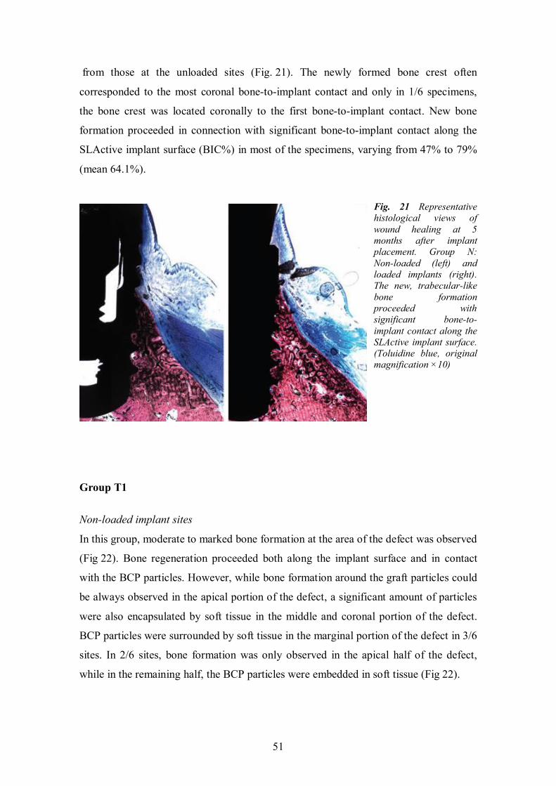
51
from those at the unloaded sites (Fig. 21). The newly formed bone crest often
corresponded to the most coronal bone-to-implant contact and only in 1/6 specimens,
the bone crest was located coronally to the first bone-to-implant contact. New bone
formation proceeded in connection with significant bone-to-implant contact along the
SLActive implant surface (BIC%) in most of the specimens, varying from 47% to 79%
(mean 64.1%).
Fig. 21 Representative histological views of wound healing at 5 months after implant placement. Group N: Non-loaded (left) and loaded implants (right). The new, trabecular-like bone formation proceeded with significant bone-to-implant contact along the SLActive implant surface. (Toluidine blue, original magnification ×10)
Group T1
Non-loaded implant sites
In this group, moderate to marked bone formation at the area of the defect was observed
(Fig 22). Bone regeneration proceeded both along the implant surface and in contact
with the BCP particles. However, while bone formation around the graft particles could
be always observed in the apical portion of the defect, a significant amount of particles
were also encapsulated by soft tissue in the middle and coronal portion of the defect.
BCP particles were surrounded by soft tissue in the marginal portion of the defect in 3/6
sites. In 2/6 sites, bone formation was only observed in the apical half of the defect,
while in the remaining half, the BCP particles were embedded in soft tissue (Fig 22).
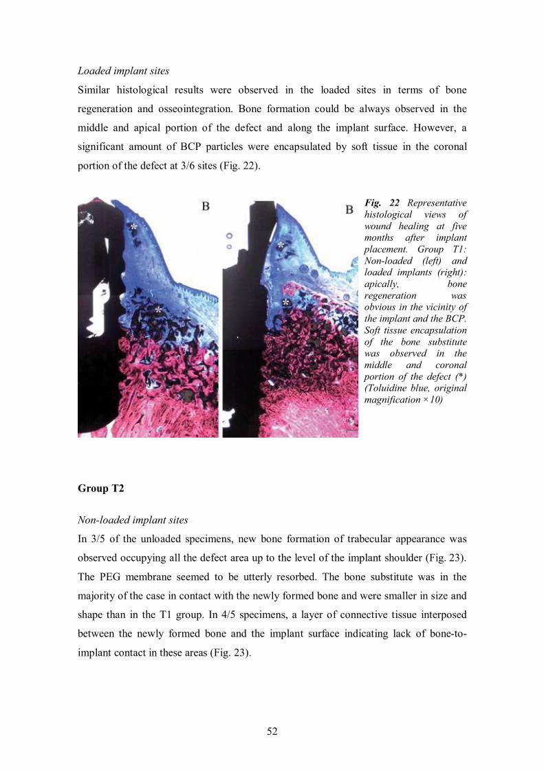
52
Loaded implant sites
Similar histological results were observed in the loaded sites in terms of bone
regeneration and osseointegration. Bone formation could be always observed in the
middle and apical portion of the defect and along the implant surface. However, a
significant amount of BCP particles were encapsulated by soft tissue in the coronal
portion of the defect at 3/6 sites (Fig. 22).
Fig. 22 Representative histological views of wound healing at five months after implant placement. Group T1: Non-loaded (left) and loaded implants (right): apically, bone regeneration was obvious in the vicinity of the implant and the BCP. Soft tissue encapsulation of the bone substitute was observed in the middle and coronal portion of the defect (*) (Toluidine blue, original magnification ×10)
Group T2
Non-loaded implant sites
In 3/5 of the unloaded specimens, new bone formation of trabecular appearance was
observed occupying all the defect area up to the level of the implant shoulder (Fig. 23).
The PEG membrane seemed to be utterly resorbed. The bone substitute was in the
majority of the case in contact with the newly formed bone and were smaller in size and
shape than in the T1 group. In 4/5 specimens, a layer of connective tissue interposed
between the newly formed bone and the implant surface indicating lack of bone-to-
implant contact in these areas (Fig. 23).

53
Loaded implant sites
Similar histological results were observed in the loaded sites compared to the non-
loaded sites. New bone formation of trabecular appearance was observed occupying the
defect area in 4/6 specimens. An extensive soft tissue downgrowth interposed between
the newly formed bone and the SLActive implant surface was observed in 5/6
specimens (Fig. 23).
Fig. 23 Representative
histological views of wound healing at five months after implant placement. Group T2: Non-loaded (left) and loaded implants (right): no remnants of the PEG were observed. The small BCP particles were in the majority of the case in contact with the newly formed bone (*). Soft tissue was observed between the newly formed bone and the implant surface in some specimens (Toluidine blue, original magnification ×10).
Histometric results
Linear measurements
Defect height
S-D was calculated in groups T1, T2, and N. The mean height was measured 5.95 mm
ranging from 4.3 to 7.4 mm. No statistically significant differences were observed
among these three group means (P = 0.82). No statistically significant difference was
observed between the means of the loaded and non-loaded groups (P = 0.47) (Table 3).
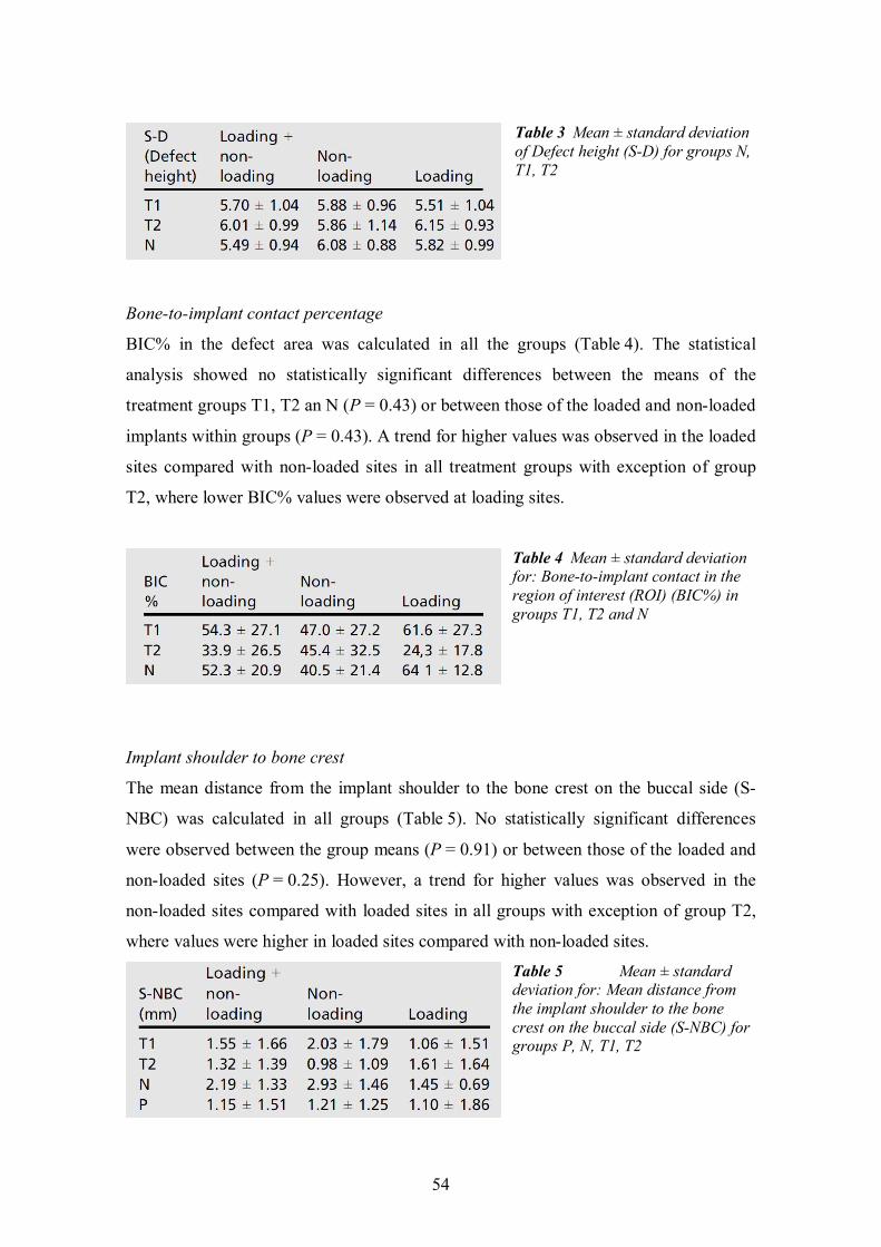
54
Table 3 Mean ± standard deviation of Defect height (S-D) for groups N, T1, T2
Bone-to-implant contact percentage
BIC% in the defect area was calculated in all the groups (Table 4). The statistical
analysis showed no statistically significant differences between the means of the
treatment groups T1, T2 an N (P = 0.43) or between those of the loaded and non-loaded
implants within groups (P = 0.43). A trend for higher values was observed in the loaded
sites compared with non-loaded sites in all treatment groups with exception of group
T2, where lower BIC% values were observed at loading sites.
Table 4 Mean ± standard deviation for: Bone-to-implant contact in the region of interest (ROI) (BIC%) in groups T1, T2 and N
Implant shoulder to bone crest
The mean distance from the implant shoulder to the bone crest on the buccal side (S-
NBC) was calculated in all groups (Table 5). No statistically significant differences
were observed between the group means (P = 0.91) or between those of the loaded and
non-loaded sites (P = 0.25). However, a trend for higher values was observed in the
non-loaded sites compared with loaded sites in all groups with exception of group T2,
where values were higher in loaded sites compared with non-loaded sites.
Table 5 Mean ± standard deviation for: Mean distance from the implant shoulder to the bone crest on the buccal side (S-NBC) for groups P, N, T1, T2
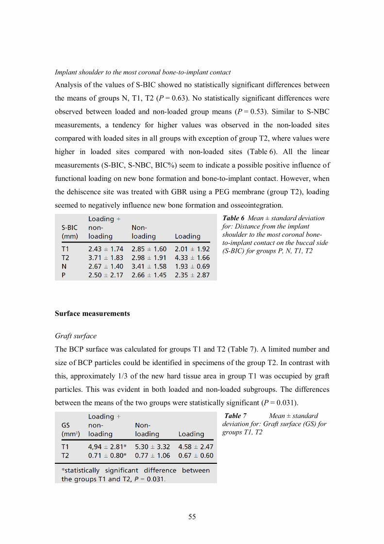
55
Implant shoulder to the most coronal bone-to-implant contact Analysis of the values of S-BIC showed no statistically significant differences between
the means of groups N, T1, T2 (P = 0.63). No statistically significant differences were
observed between loaded and non-loaded group means (P = 0.53). Similar to S-NBC
measurements, a tendency for higher values was observed in the non-loaded sites
compared with loaded sites in all groups with exception of group T2, where values were
higher in loaded sites compared with non-loaded sites (Table 6). All the linear
measurements (S-BIC, S-NBC, BIC%) seem to indicate a possible positive influence of
functional loading on new bone formation and bone-to-implant contact. However, when
the dehiscence site was treated with GBR using a PEG membrane (group T2), loading
seemed to negatively influence new bone formation and osseointegration. Table 6 Mean ± standard deviation for: Distance from the implant shoulder to the most coronal bone-to-implant contact on the buccal side (S-BIC) for groups P, N, T1, T2
Surface measurements
Graft surface
The BCP surface was calculated for groups T1 and T2 (Table 7). A limited number and
size of BCP particles could be identified in specimens of the group T2. In contrast with
this, approximately 1/3 of the new hard tissue area in group T1 was occupied by graft
particles. This was evident in both loaded and non-loaded subgroups. The differences
between the means of the two groups were statistically significant (P = 0.031).
Table 7 Mean ± standard deviation for: Graft surface (GS) for groups T1, T2

56
Bone surface
Bone surface area was calculated in groups T1, T2 and N in both loaded and non-loaded
subgroups (Table 8), with group T2 exhibiting the largest amount of bone formation
among the three groups (P < 0.05). There was a trend for higher BS values, in groups
T1 and N when functional loading was applied. However, in group T2, the newly
formed bone surface was higher in the non-loading group. This was in accordance with
what was observed with the linear measurements.
Table 8 Mean ± standard deviation for: Bone surface (BS) for groups N, T1, T2
Summated surfaces
The total surface occupied by bone and substitute (BS + GS, hard tissue surface) was
also calculated for group T1, T2 and N (Table 9). Groups T1 and T2 showed similar
amounts of hard tissue formation (14.6 and 13.9 mm² respectively). Limited hard tissue
formation was observed in group N too.
Table 9 Mean ± standard deviation for: Hard tissue surface (BS + GS) for groups N, T1, T2
5.3. The effect of ARP on extraction sockets
Search sequence
The electronic search yielded 6,216 relevant hits after removal of duplicates (Fig. 24).
Subsequently, 157 titles were selected for the abstract stage. Following investigation of
the abstracts, 42 articles qualified for full text evaluation. Four extra papers were then
added as a result of the hand search. Assessment of these articles resulted in the
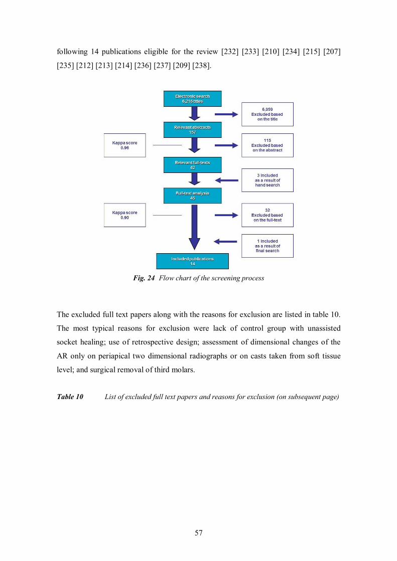
57
following 14 publications eligible for the review [232] [233] [210] [234] [215] [207]
[235] [212] [213] [214] [236] [237] [209] [238].
Fig. 24 Flow chart of the screening process
The excluded full text papers along with the reasons for exclusion are listed in table 10.
The most typical reasons for exclusion were lack of control group with unassisted
socket healing; use of retrospective design; assessment of dimensional changes of the
AR only on periapical two dimensional radiographs or on casts taken from soft tissue
level; and surgical removal of third molars.
Table 10 List of excluded full text papers and reasons for exclusion (on subsequent page)
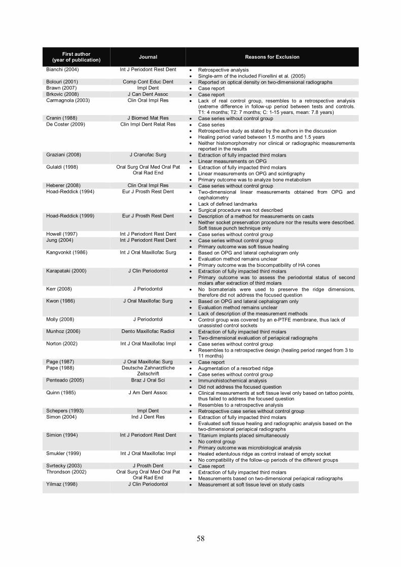
58
First author (year of publication) Journal Reasons for Exclusion
Bianchi (2004) Int J Periodont Rest Dent Retrospective analysis Single-arm of the included Fiorellini et al. (2005)
Bolouri (2001) Comp Cont Educ Dent Reported on optical density on two-dimensional radiographs Brawn (2007) Impl Dent Case report Brkovic (2008) J Can Dent Assoc Case report Carmagnola (2003) Clin Oral Impl Res Lack of real control group, resembles to a retrospective analysis
(extreme difference in follow-up period between tests and controls. T1: 4 months; T2: 7 months; C: 1-15 years, mean: 7.8 years)
Cranin (1988) J Biomed Mat Res Case series without control group De Coster (2009) Clin Impl Dent Relat Res Case series
Retrospective study as stated by the authors in the discussion Healing period varied between 1.5 months and 1.5 years Neither histomorphometry nor clinical or radiographic measurements
reported in the results Graziani (2008) J Cranofac Surg Extraction of fully impacted third molars
Linear measurements on OPG Gulaldi (1998) Oral Surg Oral Med Oral Pat
Oral Rad End Extraction of fully impacted third molars Linear measurements on OPG and scintigraphy Primary outcome was to analyze bone metabolism
Heberer (2008) Clin Oral Impl Res Case series without control group Hoad-Reddick (1994) Eur J Prosth Rest Dent Two-dimensional linear measurements obtained from OPG and
cephalometry Lack of defined landmarks Surgical procedure was not described
Hoad-Reddick (1999) Eur J Prosth Rest Dent Description of a method for measurements on casts Neither socket preservation procedure nor the results were described.
Soft tissue punch technique only Howell (1997) Int J Periodont Rest Dent Case series without control group Jung (2004) Int J Periodont Rest Dent Case series without control group
Primary outcome was soft tissue healing Kangvonkit (1986) Int J Oral Maxillofac Surg Based on OPG and lateral cephalogram only
Evaluation method remains unclear Primary outcome was the biocompatibility of HA cones
Karapataki (2000) J Clin Periodontol Extraction of fully impacted third molars Primary outcome was to assess the periodontal status of second
molars after extraction of third molars Kerr (2008) J Periodontol No biomaterials were used to preserve the ridge dimensions,
therefore did not address the focused question Kwon (1986) J Oral Maxillofac Surg Based on OPG and lateral cephalogram only
Evaluation method remains unclear Lack of description of the measurement methods
Molly (2008) J Periodontol Control group was covered by an e-PTFE membrane, thus lack of unassisted control sockets
Munhoz (2006) Dento Maxillofac Radiol Extraction of fully impacted third molars Two-dimensional evaluation of periapical radiographs
Norton (2002) Int J Oral Maxillofac Impl Case series without control group Resembles to a retrospective design (healing period ranged from 3 to
11 months) Page (1987) J Oral Maxillofac Surg Case report Pape (1988) Deutsche Zahnarztliche
Zeitschrift Augmentation of a resorbed ridge Case series without control group
Penteado (2005) Braz J Oral Sci Immunohistochemical analysis Did not address the focused question
Quinn (1985) J Am Dent Assoc Clinical measurements at soft tissue level only based on tattoo points, thus failed to address the focused question
Resembles to a retrospective analysis Schepers (1993) Impl Dent Retrospective case series without control group Simon (2004) Ind J Dent Res Extraction of fully impacted third molars
Evaluated soft tissue healing and radiographic analysis based on the two-dimensional periapical radiographs
Simion (1994) Int J Periodont Rest Dent Titanium implants placed simultaneously No control group Primary outcome was microbiological analysis
Smukler (1999) Int J Oral Maxillofac Impl Healed edentulous ridge as control instead of empty socket No compatibility of the follow-up periods of the different groups
Svrtecky (2003) J Prosth Dent Case report Throndson (2002) Oral Surg Oral Med Oral Pat
Oral Rad End Extraction of fully impacted third molars Measurements based on two-dimensional periapical radiographs
Yilmaz (1998)
J Clin Periodontol
Measurement at soft tissue level on study casts

59
The Kappa score for agreement between the reviewers at the abstract and full text
selection level, was 0.96 and 0.90, respectively, indicating a high level of agreement
[239].
Study characteristics
In the 14 included articles (eight RCTs and six CCTs) the efficacy of ARP techniques
was evaluated clinically by means of direct measurements of the residual alveolar ridge
dimensions during re-entry procedures, radiographically by means of computer
tomography or histologically from trephine biopsies taken at re-entry during
osteotomies for implant placement (Tables 11-13). Limited data were reported on
confounding factors, such as periodontitis, smoking, systemic disease and medication.
The extraction site distribution was fairly heterogeneous. In some studies ARP was
performed only in maxillary anterior sockets [232] [236] [237], whereas such restriction
was not employed in other studies. The residual bone volume around the investigated
sockets, e.g. the presence/absence and width of the buccal bone plate varied from
severely compromised [215] [236], to completely intact, buccal bone (Table 12) [207]
[210] [232].
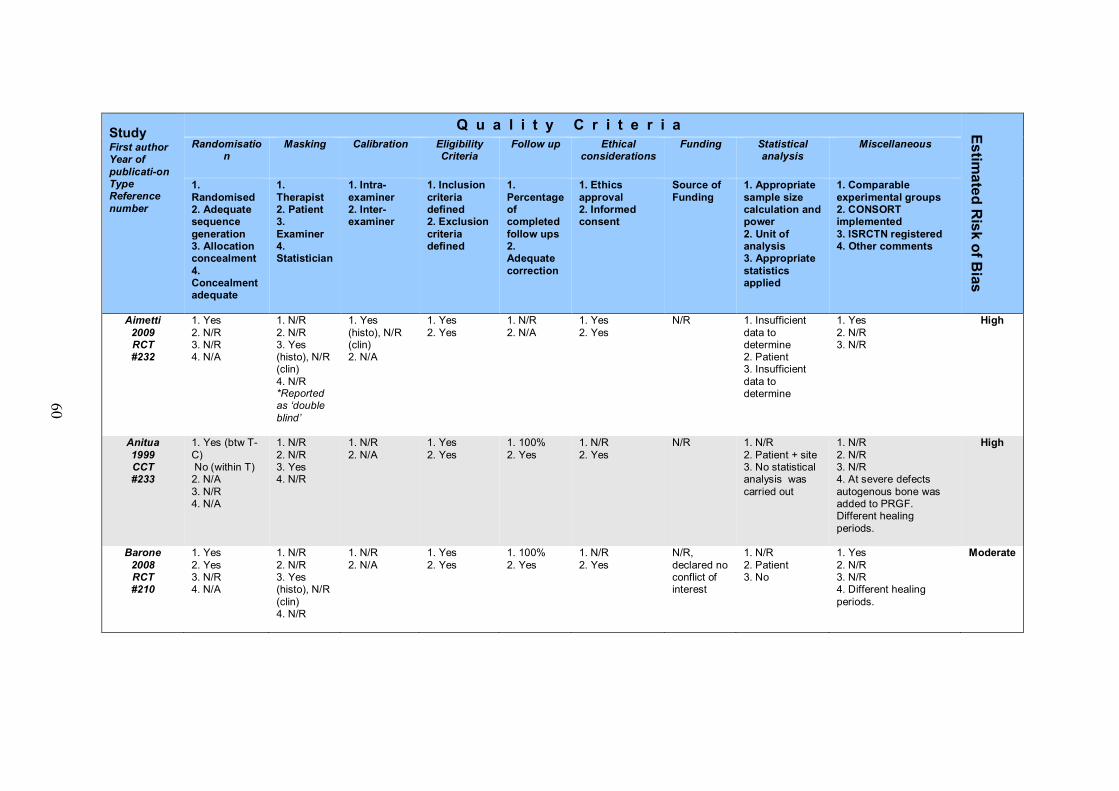
84
Study First author Year of publicati-on Type Reference number
Q u a l i t y C r i t e r i a Estimated R
isk of Bias
Randomisatio
n
Masking
Calibration
Eligibility Criteria
Follow up Ethical considerations
Funding Statistical analysis
Miscellaneous
1. Randomised 2. Adequate sequence generation 3. Allocation concealment 4. Concealment adequate
1. Therapist 2. Patient 3. Examiner 4. Statistician
1. Intra-examiner 2. Inter-examiner
1. Inclusion criteria defined 2. Exclusion criteria defined
1. Percentage of completed follow ups 2. Adequate correction
1. Ethics approval 2. Informed consent
Source of Funding
1. Appropriate sample size calculation and power 2. Unit of analysis 3. Appropriate statistics applied
1. Comparable experimental groups 2. CONSORT implemented 3. ISRCTN registered 4. Other comments
Aimetti 2009 RCT #232
1. Yes 2. N/R 3. N/R 4. N/A
1. N/R 2. N/R 3. Yes (histo), N/R (clin) 4. N/R *Reported as ‘double blind’
1. Yes (histo), N/R (clin) 2. N/A
1. Yes 2. Yes
1. N/R 2. N/A
1. Yes 2. Yes
N/R
1. Insufficient data to determine 2. Patient 3. Insufficient data to determine
1. Yes 2. N/R 3. N/R
High
Anitua 1999 CCT #233
1. Yes (btw T-C) No (within T) 2. N/A 3. N/R 4. N/A
1. N/R 2. N/R 3. Yes 4. N/R
1. N/R 2. N/A
1. Yes 2. Yes
1. 100% 2. Yes
1. N/R 2. Yes
N/R
1. N/R 2. Patient + site 3. No statistical analysis was carried out
1. N/R 2. N/R 3. N/R 4. At severe defects autogenous bone was added to PRGF. Different healing periods.
High
Barone 2008 RCT #210
1. Yes 2. Yes 3. N/R 4. N/A
1. N/R 2. N/R 3. Yes (histo), N/R (clin) 4. N/R
1. N/R 2. N/A
1. Yes 2. Yes
1. 100% 2. Yes
1. N/R 2. Yes
N/R, declared no conflict of interest
1. N/R 2. Patient 3. No
1. Yes 2. N/R 3. N/R 4. Different healing periods.
Moderate
60

61
Camargo 2000 CCT #234
N/A
1. N/R 2. N/R 3. N/R 4. N/R
1. N/R 2. N/A
1. Yes 2. Yes
1. 100% 2. Yes
1. Yes 2. Yes
Industry
1. N/R 2. Patient 3. Insufficient data to determine
1. N/R 2. N/R 3. N/R
High
Fiorellini 2005 RCT #215
1. Yes 2. N/R 3. N/R 4. N/A
1. N/R 2. N/R 3. Yes (CT scans) 4. N/R * Reported as ‘double blind’
1. N/R 2. Yes
1. No 2. No
1. 100% 2. Unclear
1. Yes 2. Yes
Industry
1. Insufficient data to determine 2. Patient 3. No
1. N/R 2. N/R 3. N/R 4. Standardisation of CT scans N/R. Final number of sockets, patients remain unclear.
High
Froum 2002 RCT #207
1. Yes 2. Yes 3. N/R 4. N/A
1. N/R 2. N/R 3. Yes 4. N/R
1. N/R 2. N/A
1. Yes 2. Yes
1. 100% 2. Unclear
1. Yes 2. Yes
Industry
1. N/R 2. Site 3. No
1. N/R 2. N/R 3. N/R 4. Different healing periods. Enrolment of sites of subjects inconsistent.
Moderate
Guarnieri 2004 CCT #235
N/A
1. N/R 2. N/R 3. N/R 4. N/R
1. N/R 2. N/A
1. Yes 2. No
1. N/R 2. N/A
1. Yes 2. Yes
Government; institution
1. N/R 2. Site 3. No
1. N/R 2. N/R 3. N/R
High
Iasella 2003 RCT #212
1. Yes 2. Yes 3. N/R 4. N/A
1. N/R 2. N/R 3. Yes 4. N/R
1. Yes 2. N/A
1. Yes 2. Yes
1. 100% 2. Yes
1. Yes 2. Yes
N/R
1. Insufficient data to determine 2. Patient 3. Insufficient data to determine
1. Yes 2. N/R 3. N/R
Moderate
Lekovic 1997 CCT #213
N/A
1. N/R 2. N/R 3. N/R 4. N/R
1. N/R 2. N/A
1. No 2. No
1. 70% (premature exposure of e-PTFE barrier in 3/10) 2. Yes
1. Yes 2. N/R
N/R
1. N/R 2. Patient 3. Insufficient data to determine
1. Yes 2. N/R 3. N/R
High
61

62
N/A not applicable; N/R not reported, T test; C control; RCT randomised controlled trial; CCT controlled clinical trial; PRGF platelet-rich growth factor; CONSORT Consolidated Standards of
Reporting Trials; ISRCTN International Standard Randomised Controlled Trial Number Register
Table 11 Quality assessment of the included studies (see above)
Lekovic 1998 RCT #214
1. Yes 2. Yes 3. N/R 4. N/A
1. N/R 2. N/R 3. Yes 4. Yes
1. N/R 2. N/A
1. No 2. No
1. 100% 2. Yes
1. Yes 2. Yes
N/R
1. N/R 2. Patient 3. Insufficient data to determine
1. Yes 2. N/R 3. N/R
Moderate
Nevins 2006 RCT #236
1. Yes 2. N/R 3. N/R 4. N/A
1. N/R 2. N/R 3. N/R 4. N/R
1. N/R 2. N/A
1. Yes 2. Yes
1. 100% 2. Yes
1. N/R 2. N/R
N/R
1. N/R 2. Site 3. No
1. Yes 2. N/R 3. N/R 4. Standardisation of CT scans N/R. Test material radiopaque. Different healing periods.
High
Pelegrine 2010 RCT #237
1. Yes 2. N/R 3. N/R 4. N/A
1. N/R 2. N/R 3. N/R 4. N/R
1. N/R 2. N/A
1. Yes 2. Yes
1. 100% 2. Yes
1. Yes 2. Yes
N/R
1. N/R 2. Patient 3. Yes
1. N/R 2. N/R 3. N/R
High
Serino 2003 CCT #209
N/A
1. N/R 2. N/R 3. N/R 4. N/R
1. N/R 2. N/A
1. Yes 2. No
1. 80% 2. Unclear
1. Yes 2. Yes
N/R
1. N/R 2. Site 3. No
1. N/R 2. N/R 3. N/R 4. Molars only in T.
High
Serino 2008 CCT #238
N/A
1. N/R 2. N/R 3. N/R 4. N/R
1. N/R 2. N/A
1. Yes 2. No
1. 80% 2. Unclear
1. Yes 2. Yes
Government; institution
1. N/R 2. Patient 3. Insufficient data to determine
1. N/R 2. N/R 3. N/R
High
62

63
First author Year of publication Type Design Methodology Reference number
Trial characteristics 1. Country 2. Number of centres 3. Setting
Population characteristics 1. Age range (mean) in years 2. Number of patients (sockets)
Confounding factors 1. Smoking 2. Periodontitis
Defect characteristics 1. Socket location 2. Defect morphology
Test material (number of sockets/ subjects)
Control (number of sockets/ subjects)
Surgical management 1. Type of flap 2. Soft tissue closure 3. Postoperative antimicrobials
Follow-up 1. Healing period 2. Number of drop-outs 3. Adverse events
Alveolar ridge dimension changes in horizontal width Mean/median mm 1. Whole ridge 2. Buccal plate
Alveolar ridge dimension changes in vertical height Mean/median mm (reference point) 1. Mid-buccal 2. Mesial 3. Distal 4. Socket Fill
Implant 1. Feasibility of implant placement 2. Necessity of simultaneous augmentation
Aimetti 2009 RCT
Parallel Clin + Histo
#232
1. Italy 2. 1 3. University
1. 36-68 (51.27 ±8.4) 2. 40 (40)
1. No 2. N/R
1. Maxillary anterior 2. 4-wall configuration
Calcium sulphate Hemihydrate (22/22)
Empty (18/18)
1. Flapless 2. No primary closure 3. Amoxicillin 2g/day for 5 days, Chlorexidine 0.12% for 2 weeks
1. 3 months 2. N/R 3. Uneventful healing
1. T: -2.0 ± 1.1** C: -3.2 ± 1.8**, *** 2. N/R
1. T: -0.5±1.1*, C: -1.2±0.6**, *** 2. T: -0.2±0.6, C: -0.5±0.9 3. T: -0.4±0.9, C: -0.5±1.1 4. T: 11.3±2.8**, C: 10.0±2.3** (Acrylic stent)
1. Implants were inserted 2. N/R
Anitua 1999 CCT
Parallel + Split-mouth Histo #233
1. Spain 2. 1 3. Private practice
1. T: 35-55 (41) C: 38-54 (42) 2. 23 (26)
1. Yes 2. Yes
1. Any 2. Variable
T1: PRGF (5+3/5+3) T2: PRGF+Autologous bone (5/5)
Empty (10+3/10+3)
1. Full-thickness 2. Primary closure 3. Amoxicillin 1.5g/day for 5 days
1. 2.5 – 4 months 2. 0 3. N/R
N/A
N/A
1. N/R 2. N/R
Barone 2008 RCT
Parallel Clin + Histo
#210
1. Italy 2. 1 3. Hospital
1. 26-69 2. 40 (40)
1. <10/day 2. Yes (treated)
1. Non-molars 2. 4-wall configuration
Corticocancellous porcine bone+ collagen membrane (20/20)
Empty (20/20)
1. Full-thickness 2. Primary closure 3. Amoxicillin 2g/day for 4 days + Chlorexidine 0.12% for 3 weeks
1. 7-9 months 2. 0 3. Uneventful healing (pain, swelling)
1. T: -2.5 ± 1.2*, C: -4.5 ± 0.8*, *** 2. N/R
1. T: -0.7±1.4*, C: -3.6±1.5*, *** 2. T: -0.2±0.8, C: -0.4±1.2 3. T: -0.4±0.8, C: -0.5±1.0 4. N/R (Acrylic stent)
1. ‘Implants were inserted in both groups’ 2. Some GBR were needed due to buccal dehiscence in the control group
Camargo 2000 CCT
Split-mouth Clin #234
1. USA, Yugoslavia 2. N/R 3. University
1. 28-60 (44±15.9) 2. 16 (32)
1. N/R 2. N/R
1. Maxillary anterior, premolars 2. N/R
Bioactive glass +covered by calcium sulphate layer (16/8)
Empty (16/8)
1. Full-thickness with 4 vertical releasing incisions 2. No primary closure 3. Penicillin 1.5g/day for 7 days + Chlorexidine 0.12% for 2 weeks
1. 6 months 2. N/R 3. Uneventful healing
1. T: -3.48±2.68**, C: -3.06±2.41** 2. N/R
1. T: -0.38±3.18, C: -1.00±2.25 (titanium tack) 2. N/R 3. N/R 4. T: 6.43±2.78**, C: 4.00±2.33**, *** (to buccal bone crest)
1. Reentry only 2. N/A

64
Fiorellini 2005 RCT
Parallel Radiogr + Histo
#215
1. USA 2. 8 centres 3. University
1. 47.4 2. 80 (95)
1. N/R 2. N/R
1. Maxillary anterior, premolars 2. ≥50% buccal bone loss
T1: 1.5mg/ml rhBMP-2 (?/21?) T2: 0.75mg/ml rhBMP-2 (?/22?) T3: Collagen sponge (?/17?)
Empty (?/20?)
1. Full-thickness with vertical incisions 2. Primary closure 3. Penicillin (?mg) for 7-10 days + Chlorexidine 0.12%
1. 4 months 2. No drop-outs reported. (3 patients incorrectly randomized, 1 patient received different graft) 3. 250 (T>C)
1. Coronal: T1: +3.27±2.53*, T2: +1.76±1.67*, T3: +0.82±1.40, C: +0.57±2.56, *** (T1 vs T2/T3/C) 2. N/R
1. T1: -0.02 ± 1.2, T2: -0.62±1.39*, T3: -1.00±1.40*, C: -1.17± 1.23*, *** (T1 vs C) 2. N/R 3. N/R 4. N/R
1. N/R 2. No need for augmentation T1: 18/21 (86%) T2: 12/22 (55%) T3: 10/17 (59%) C: 9/20 (45%) (T1 vs T2/C)***
Froum 2002 RCT
Split mouth Histo #207
1. USA 2. Single centre 3. University
1. 35-77 (54.9±11.9) 2. 19 (30)
1. No 2. N/R
1. Any 2. 4-wall configuration, ≤2mm buccal plate loss
T1: Bioactive glass (10/8) T2: DFDBA (10/8)
Empty (10/10)
1. Full-thickness without vertical incisions 2. Primary closure 3. Doxycycline 100mg/day for 13 days + Chlorexidine 0.12% for 30 days
1. 6-8 months 2. 0 3. Uneventful healing
N/A
N/A
1. ‘An implant of appropriate size was placed in the healed sockets.’ 2. N/R
Guarnieri 2004 CCT
Parallel + Split mouth Histo #235
1. Italy 2. N/R 3. N/R
1. 35-58 2. 10 (25)
1. N/R 2. Yes
1. Maxillary, mandibular anteriors, premolars 2. socket with ridge resorption ≥50% were excluded
Calcium sulphate Hemihydrate (10/10)
Empty (5/5)
1. Full-thickness without vertical incisions 2. Primary closure 3. Amoxicillin (?mg) for 1 week + Chlorexidine 0.2% for 2 weeks
1. 3 months 2. N/R 3. N/R
N/A
N/A
1. ‘Bucco-lingual dimensions of the alveolar ridge enabled safe insertion of titanium implant.’ 2. N/R
Iasella 2003 RCT
Parallel Clin + Histo
#212
1. USA 2. N/R 3. N/R
1. 28-76 (51.5±13.6) 2. 24 (24)
1. Yes 2. N/R
1. Maxillary anteriors, premolars and mandibular premolars 2. N/R
Tetracycline hydrated FDBA + collagen membrane (12/12)
Empty (12/12)
1. Full-thickness without vertical incisions 2. No primary closure 3. Doxycyclin 200mg/day for 1 week + Chlorexidine 0.12% for 2 weeks
1. 4 or 6 months (combined) 2. 0 3. N/R
1. T: -1.2 ± 0.9*, C: -2.6 ± 2.3* 2. N/R
1. T: +1.3±2.0, C: -0.9±1.6*** 2. T: -0.1±0.7, C:-1.0±0.8*** 3. T: -0.1±0.7, C: -0.8±0.8*** 4. N/R (Acrylic stent)
1. Implants successfully placed at all sites 2. Some sites had slight dehiscence and required further augmentation
Lekovic 1997 CCT
Split-mouth Clin #213
1. Yugoslavia / USA 2. N/R (presumably single centre) 3. University
1. (49.8) 2. 10 (20)
1. N/R 2. N/R
1. Maxillary and mandibular anteriors, premolars 2. N/R
e-PTFE membrane (10/10)
Empty (10/10)
1. Full-thickness with 4 vertical releasing incisions 2. Primary closure 3. Penicillin 1g/day for 7 days + Chlorexidine 0.2%
1. 6 months 2. 3/10 drop-outs due to premature membrane exposure 3. 3/10 exposed, 7/10 no infection
1. 10/10:, T: -1.80±0.51, C: -4.40±0.61**, *** 7/10: T: -1.71±0.75, C: -4.43±0.72**, *** 3/10: T: -2.00±0.00, C: -4.33±0.88* 2. N/R
1. 10/10: T: -0.5±0.22, C: -1.2±0.13**, *** 7/10: T: -0.28±0.18, C: -1.0±0.0**, *** 3/10: T: -1.0±0.58, C: -1.66±0.33 (titanium tack) 2. N/R 3. N/R 4. 10/10: T: 4.9±0.86*, C: 3.0±0.63, *** 7/10: T: 5.43±1.1*, C:2.92±1.61, *** 3/10: T: 3.66±1.20, C: 4.33±1.45 (to buccal bone crest)
1. Reentry only 2. N/A
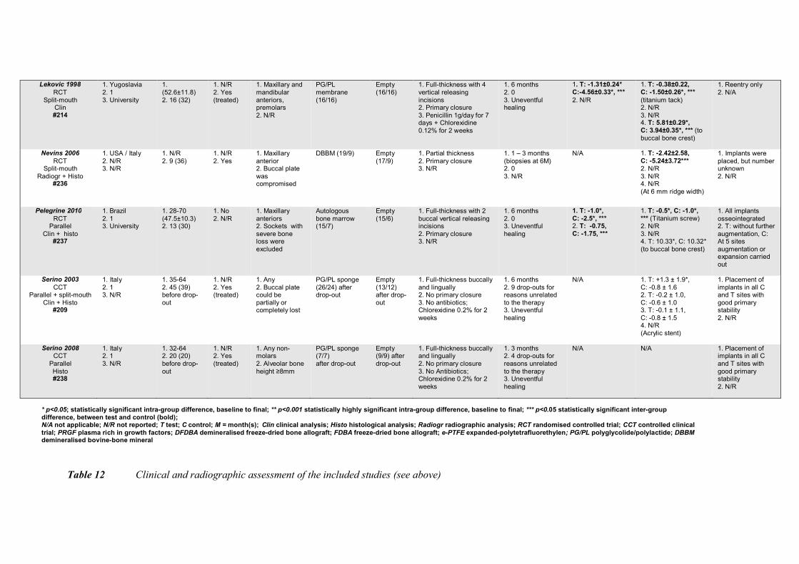
65
Lekovic 1998 RCT
Split-mouth Clin #214
1. Yugoslavia 2. 1 3. University
1. (52.6±11.8) 2. 16 (32)
1. N/R 2. Yes (treated)
1. Maxillary and mandibular anteriors, premolars 2. N/R
PG/PL membrane (16/16)
Empty (16/16)
1. Full-thickness with 4 vertical releasing incisions 2. Primary closure 3. Penicillin 1g/day for 7 days + Chlorexidine 0.12% for 2 weeks
1. 6 months 2. 0 3. Uneventful healing
1. T: -1.31±0.24* C:-4.56±0.33*, *** 2. N/R
1. T: -0.38±0.22, C: -1.50±0.26*, *** (titanium tack) 2. N/R 3. N/R 4. T: 5.81±0.29*, C: 3.94±0.35*, *** (to buccal bone crest)
1. Reentry only 2. N/A
Nevins 2006 RCT
Split-mouth Radiogr + Histo
#236
1. USA / Italy 2. N/R 3. N/R
1. N/R 2. 9 (36)
1. N/R 2. Yes
1. Maxillary anterior 2. Buccal plate was compromised
DBBM (19/9)
Empty (17/9)
1. Partial thickness 2. Primary closure 3. N/R
1. 1 – 3 months (biopsies at 6M) 2. 0 3. N/R
N/A
1. T: -2.42±2.58, C: -5.24±3.72*** 2. N/R 3. N/R 4. N/R (At 6 mm ridge width)
1. Implants were placed, but number unknown 2. N/R
Pelegrine 2010 RCT
Parallel Clin + histo
#237
1. Brazil 2. 1 3. University
1. 28-70 (47.5±10.3) 2. 13 (30)
1. No 2. N/R
1. Maxillary anteriors 2. Sockets with severe bone loss were excluded
Autologous bone marrow (15/7)
Empty (15/6)
1. Full-thickness with 2 buccal vertical releasing incisions 2. Primary closure 3. N/R
1. 6 months 2. 0 3. Uneventful healing
1. T: -1.0*, C: -2.5*, *** 2. T: -0.75, C: -1.75, ***
1. T: -0.5*, C: -1.0*, *** (Titanium screw) 2. N/R 3. N/R 4. T: 10.33*, C: 10.32* (to buccal bone crest)
1. All implants osseointegrated 2. T: without further augmentation, C: At 5 sites augmentation or expansion carried out
Serino 2003 CCT
Parallel + split-mouth Clin + Histo
#209
1. Italy 2. 1 3. N/R
1. 35-64 2. 45 (39) before drop-out
1. N/R 2. Yes (treated)
1. Any 2. Buccal plate could be partially or completely lost
PG/PL sponge (26/24) after drop-out
Empty (13/12) after drop-out
1. Full-thickness buccally and lingually 2. No primary closure 3. No antibiotics; Chlorexidine 0.2% for 2 weeks
1. 6 months 2. 9 drop-outs for reasons unrelated to the therapy 3. Uneventful healing
N/A
1. T: +1.3 ± 1.9*, C: -0.8 ± 1.6 2. T: -0.2 ± 1.0, C: -0.6 ± 1.0 3. T: -0.1 ± 1.1, C: -0.8 ± 1.5 4. N/R (Acrylic stent)
1. Placement of implants in all C and T sites with good primary stability 2. N/R
Serino 2008 CCT
Parallel Histo #238
1. Italy 2. 1 3. N/R
1. 32-64 2. 20 (20) before drop-out
1. N/R 2. Yes (treated)
1. Any non-molars 2. Alveolar bone height ≥8mm
PG/PL sponge (7/7) after drop-out
Empty (9/9) after drop-out
1. Full-thickness buccally and lingually 2. No primary closure 3. No Antibiotics; Chlorexidine 0.2% for 2 weeks
1. 3 months 2. 4 drop-outs for reasons unrelated to the therapy 3. Uneventful healing
N/A
N/A
1. Placement of implants in all C and T sites with good primary stability 2. N/R
* p<0.05; statistically significant intra-group difference, baseline to final; ** p<0.001 statistically highly significant intra-group difference, baseline to final; *** p<0.05 statistically significant inter-group difference, between test and control (bold); N/A not applicable; N/R not reported; T test; C control; M = month(s); Clin clinical analysis; Histo histological analysis; Radiogr radiographic analysis; RCT randomised controlled trial; CCT controlled clinical trial; PRGF plasma rich in growth factors; DFDBA demineralised freeze-dried bone allograft; FDBA freeze-dried bone allograft; e-PTFE expanded-polytetrafluorethylen; PG/PL polyglycolide/polylactide; DBBM demineralised bovine-bone mineral
Table 12 Clinical and radiographic assessment of the included studies (see above)

66
First author
Year of publication
Type Healing period
Reference number
Number of biopsies
(test material)
Histomorphology Histomorphometry (mean or median %)
Statistical difference between test and
control
Test
Control
Aimetti 2009 RCT 3 M #232
T: N/R 22? (MGCSH) C: N/R 18?
No residual graft material. No inflammatory infiltrate. New bone formation in all specimens, 100% living trabecular bone with woven and lamellar structure.
100% living bone (mostly woven) in all biopsies. Lamellar bone remodeling was starting.
Trabecular bone: T: 58.8±3.5 C: 47.2±7.7
Residual substitute material: T: 0.0 C: N/A
Woven bone: Coronal: T: 83.6±6.6 C: 88.9±7.6 Middle: T: 59.6±13.2 C: 81.1±7.6 Apical: T: 56.4±10.9 C: 77.8±8.1
Lamellar bone: Coronal: T: 16.4±6.6 C: 11.1±7.6 Middle: T: 40.4±13.2 C: 18.9±7.6 Apical: T: 43.6±10.9 C: 22.2±8.1
T vs C*
Anitua 1999 CCT
2.5 – 4 M #233
T: N/R (PRGF± autogen bone) C: N/R
Compact mature bone with well-organized trabeculae and morphology in 8/10 patients. Connective tissue with non-organized trabeculae in 2/10 patients. Significant intra-group differences 10 vs. 16 weeks!
Connective tissue fills the main part of the defect. No mature bone.
N/R
Barone 2008 RCT
7 – 9 M #210
T: 20 (Corticocancellous porcine bone+ collagen membrane) C: 20
Residual graft material embedded in newly formed bone in all specimens. Complete bone fill.
Typically trabecular bone pattern. Large marrow spaces filled with adipocytes. Lamellar bone was also present within the bone marrow.
Total bone volume: T: 35.5±10.4 C: 25.7±9.5
Connective tissue: T: 36.6±12.6 C: 59.1±10.4
Residual graft material: T: 29.2±10.1 C: N/A
Bone: T>C* Connective tissue: T<C*
Fiorellini 2005 RCT 4 M #215
T1: 16 (rhBMP-2 1.5mg/ml) T2: 15 (0,75mg/ml) T3: 11 (Collagen sponge) C: 14
No evidence of inflammation or residual graft. Trabecular bone formation in 2/3 of the samples. Mineralized tissue formation presented with different level of remodeling. Minor osteoclastic activity. No comparison reported between T and C!
N/R
66

67
Froum 2002 RCT
6 – 8 M #207
T1: 10 (Bioactive glass) T2: 10 (DFDBA) C: 10
T1: New bone formation. Osteoid surrounded and penetrated the bioactive glass particles. T2: Varying degrees of reossification around DFDBA.
N/R
Vital bone: T1: 59.5 T2: 34.7 C: 32.4
Connective tissue: T1: 35.3 T2: 51.6 C: 67.0
Residual bone substitute: T1: 5.5 T2: 13.5 C: N/A
Connective tissue: T1<T2 or C*
Guarnieri 2004 CCT 3 M #235
T: 10 (MGCSH) C: 5
Almost complete absence of MGCSH. Absence of connective tissue and inflammatory cells. In all sections trabecular bone formation with no differences between the apical, middle and coronal levels.
Less bone formation compared to test sites.
Trabecular bone area: T: Coronal: 58.6±9.2 Middle: 58.1±6.2 Apical: 58.3±7.8 C: ≤ 46
No statistical significance could be drawn due to small number of control specimens.
Iasella 2003 RCT
4 – 6 M #212
T: 4M: 5, 6M:7 (Tetracycline hydrated FDBA + Collagen membrane) C: 4M: 5, 6M: 5
Residual graft particles surrounded by woven bone or by connective tissue.
Similar amount of total bone and trabecular spaces as in test. (No biopsy from 2 C sites due to minimal bone fill)
Vital bone: 4M T: 31±9 C: 58±11 6M T: 25±17 C: 50±14 Combined T: 28±14 C: 54±12
Non-vital bone: 4M T: 32±19 C: N/A 6M T: 41±18 C: N/A Combined T: 37±18 C: N/A
N/R
Nevins 2005 RCT 6 M #236
T: 5 (DBBM) C: 5
DBBM granules present. Apically integrated in cancellous bone but coronally in soft tissue. No signs of inflammation or foreign body reaction.
New bone formation
No comparison made.
Pelegrine 2010 RCT 6 M #237
T: 7 (Autologous bone marrow) C: 6
Mineralized bone: T: 45.0 C: 43.75
No significant difference.
67
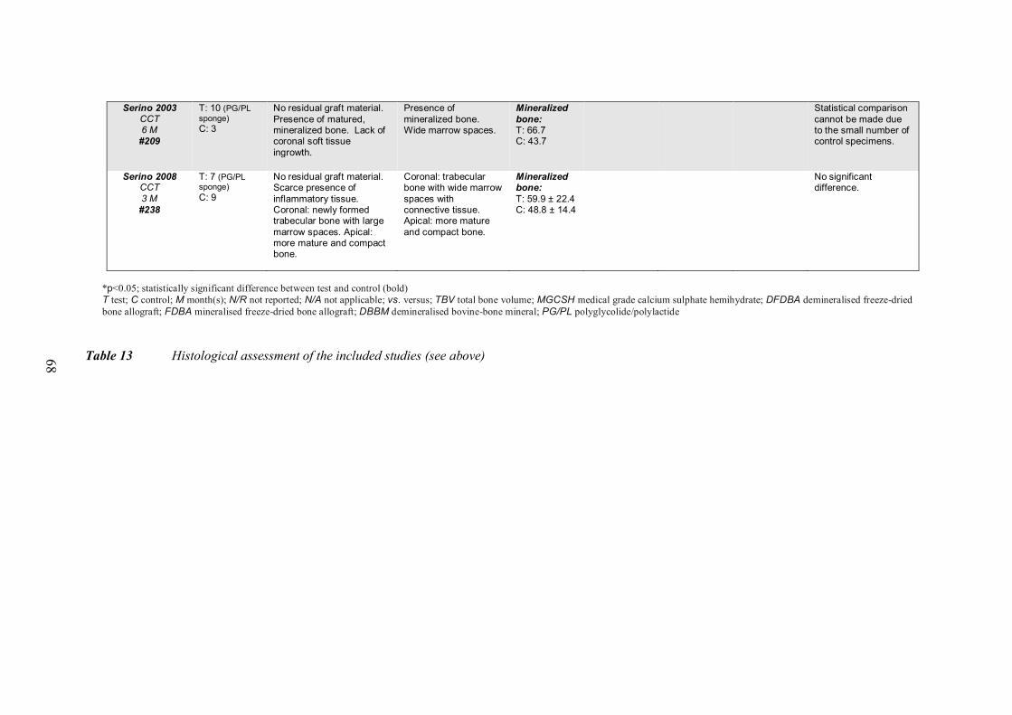
68
Serino 2003 CCT 6 M #209
T: 10 (PG/PL sponge) C: 3
No residual graft material. Presence of matured, mineralized bone. Lack of coronal soft tissue ingrowth.
Presence of mineralized bone. Wide marrow spaces.
Mineralized bone: T: 66.7 C: 43.7
Statistical comparison cannot be made due to the small number of control specimens.
Serino 2008 CCT 3 M #238
T: 7 (PG/PL sponge) C: 9
No residual graft material. Scarce presence of inflammatory tissue. Coronal: newly formed trabecular bone with large marrow spaces. Apical: more mature and compact bone.
Coronal: trabecular bone with wide marrow spaces with connective tissue. Apical: more mature and compact bone.
Mineralized bone: T: 59.9 ± 22.4 C: 48.8 ± 14.4
No significant difference.
*p<0.05; statistically significant difference between test and control (bold) T test; C control; M month(s); N/R not reported; N/A not applicable; vs. versus; TBV total bone volume; MGCSH medical grade calcium sulphate hemihydrate; DFDBA demineralised freeze-dried bone allograft; FDBA mineralised freeze-dried bone allograft; DBBM demineralised bovine-bone mineral; PG/PL polyglycolide/polylactide
Table 13 Histological assessment of the included studies (see above)68

69
Intervention characteristics
With regard to the techniques or materials used for ARP, the included studies were
grouped into three categories (Table 12);
1. Bone grafts/substitutes
2. GBR
3. Biological active materials.
In the majority of the included studies various bone grafts were utilised, such as
autologous bone marrow [237], plasma rich in growth factor (PRGF) with or without
autologous bone [233], DFDBA [207], DBBM [236], calcium sulphate hemihydrates
[232] [235] and bioactive glass [207]. Alloplastic polyglycolide/polylactide (PGPL)
sponge was also employed [209] [238]. GBR technique was applied using non-
resorbable e-PTFE [213] or resorbable (PGPL) barrier [214]. Resorbable collagen
membrane was also employed in combination with FDBA [233] or corticocancellous
porcine bone [210]. Biological active material, namely recombinant human bone
morphogenetic protein-2 (rhBMP-2) was used on a collagen sponge carrier in one study
[215]. Neither the ‘soft tissue punch’/socket seal, nor the ‘ice-cream cone’/bio-col
techniques were identified among the included studies [216] [240]. Primary flap closure
was achieved in nine out of 14 studies, while the sockets left uncovered in the rests.
Various types and amounts of antibiotics and antiseptic rinses were administered for
different duration in studies reporting on postoperative care. Finally, average healing
period ranged from one to nine months.
Outcome characteristics
Clinical outcomes
Eight out of the 14 included studies investigated the efficacy of various ARP techniques
to preserve the pre-extraction ridge dimensions using intra-surgical hard tissue
measurements taken during re-entry procedure [209] [215] [212] [213] [214] [232]
[234] [237]. In these studies ARP was performed in 137 sockets of 119 patients and
compared to 120 sockets that left to heal without any treatment in a total of 92 patients
(Table 12, Fig. 25, 26).

70
Fig. 25 Clinical horizontal
dimension changes.
Fig. 26 Clinical vertical
dimension changes.
Bone ‘graft’
Four studies evaluated changes in AR dimensions following grafting of the socket. Two
studies were RCTs [232] [237] and two were CCTs [209] [234]. Healing time varied
from three to six months [209] [234] [232] [236]. The horizontal (bucco-lingual)
changes of the alveolar ridge were assessed in three studies [234] [232] [237]. The AR
reduced in width from baseline to re-entry between -1.0 mm and -3.5±2.7 mm following
ARP (p<0.05) and between -2.5 mm and -3.2±1.8 mm in the control groups (p<0.05). In
two out of the three studies the width reduction was statistically significantly smaller in
the test groups compared to the controls [232] [237] .
Four studies investigated the mean change in ridge height at the mid-buccal aspect [237]
[209] [234] [232]. The AR height changed from baseline to re-entry between +1.3±1.9
and -0.5±1.1 mm following ARP, and between -0.8±1.6 and -1.2±0.6 mm in the control
groups. The height reduction between baseline and re-entry was not statistically
significant in one study in both test and control groups [234] , while one study reported
an increase in height instead of loss following ARP with a PGPL sponge (p<0.05
[209]). In two out of the four studies the height reduction was statistically significantly
smaller in the test groups compared to the controls [232] [237].
71
The vertical dimension changes at the mesial and distal aspects of the socket were
measured in two studies [232] [209] and did not present any statistically significant
difference for both groups.
Three studies captured data on socket fill and reported statistically significant
differences between baseline and re-entry in both groups [232] [234] [237] , but only
one reported statistically significantly higher socket fill, where bioactive glass was
covered by calcium sulphate, compared to the unassisted healing [234] .
GBR
Four studies evaluated changes in AR dimensions following ARP with GBR alone
[213] [214], or in combination with bone graft [210] [212] . Three studies were RCTs
[210] [212] [214] and one was CCT [213]. Healing time varied between four and nine
months.
Horizontal (bucco-lingual) changes of the AR were assessed in all four studies. AR
width reduction from baseline to re-entry varied between -1.2±0.9 and -2.5±1.2 mm in
the GBR treated sockets and between -2.6±2.3 and -4.6±0.3 mm in the control groups.
With the exception of one study [212], a statistically significantly smaller reduction of
the alveolar ridge width was observed when e-PTFE [213] , PGPL [214] , or collagen
membranes in combination with xenograft [210] were used.
All the four studies investigated the mean change in AR height at the mid-buccal aspect.
The AR height changed from baseline to re-entry between +1.3±2.0 and -0.7±1.4 mm in
the ARP groups and between -0.9±1.6 and -3.6±1.5 mm in the control groups. The
resorption in the ARP group was not statistically significant in three out of four studies
[212] [213] [214] . All studies reported statistically significantly less post extraction
reduction in AR height when the socket was treated by GBR compared to unassisted
healing.
Vertical dimension changes at mesial and distal aspects of the socket were measured in
two studies [210] [212] . The observed differences between baseline and re-entry were
not statistically significant in both groups. In one out of the two studies the height
72
reduction was statistically significantly smaller in the test group compared to the control
[212].
Two studies captured data on the socket fill [213] [214] and reported statistically
significant socket fill in both groups between baseline and re-entry, as well as between
tests and controls.
No data were found on either initial buccal plate thickness or alteration of bone volume.
However, one study measured the buccal bone thickness loss and reported statistically
significantly less reduction in the ARP group [237].
Radiographic measurements
Two RCTs, reporting on 3D radiographic assessment, met the inclusion criteria [215]
[236]. The healing time varied from one to four months. In one study, where the post
extraction socket was grafted with a radiopaque material (DBBM), treatment resulted in
significantly less reduction in radiographic AR height compared to unassisted socket
healing [236]. The test group in the other study, where the higher concentration
(1.5mg/ml) of rhBMP-2 was utilised [215], resulted in a mean increase of the
radiographic AR width by 3.27±2.53 mm at the most coronal part, compared to the
0.57±2.56 mm increase in the group of unassisted healing. AR height was reduced by
0.02±1.2 mm in the same test group and by 1.17±1.23 mm in the control group (Table
12). The differences between test and control were statistically significant.
Histological results
Eleven studies carried out a histological analysis based on trephine biopsies retrieved at
re-entry [232] [233] [210] [215] [207] [235] [212] [236] [237] [209] [238]. Seven
studies were RCTs [232] [207] [215] [210] [212] [236] [237] and four were CCTs
[209] [238] [233] [235]. In these studies, ARP was performed in 181 sockets of 158
patients and compared to 149 sockets that left to heal without any treatment in 131
patients (Table 13). Only two studies reported statistically significantly higher
trabecular bone volume following ARP in comparison to unassisted socket healing
[232] [210] and two studies reported statistically significantly more connective tissue in
the post extraction socket when no ARP was performed [210] [207]. On the contrary,
one study reported more vital bone in the unassisted socket healing group compared to
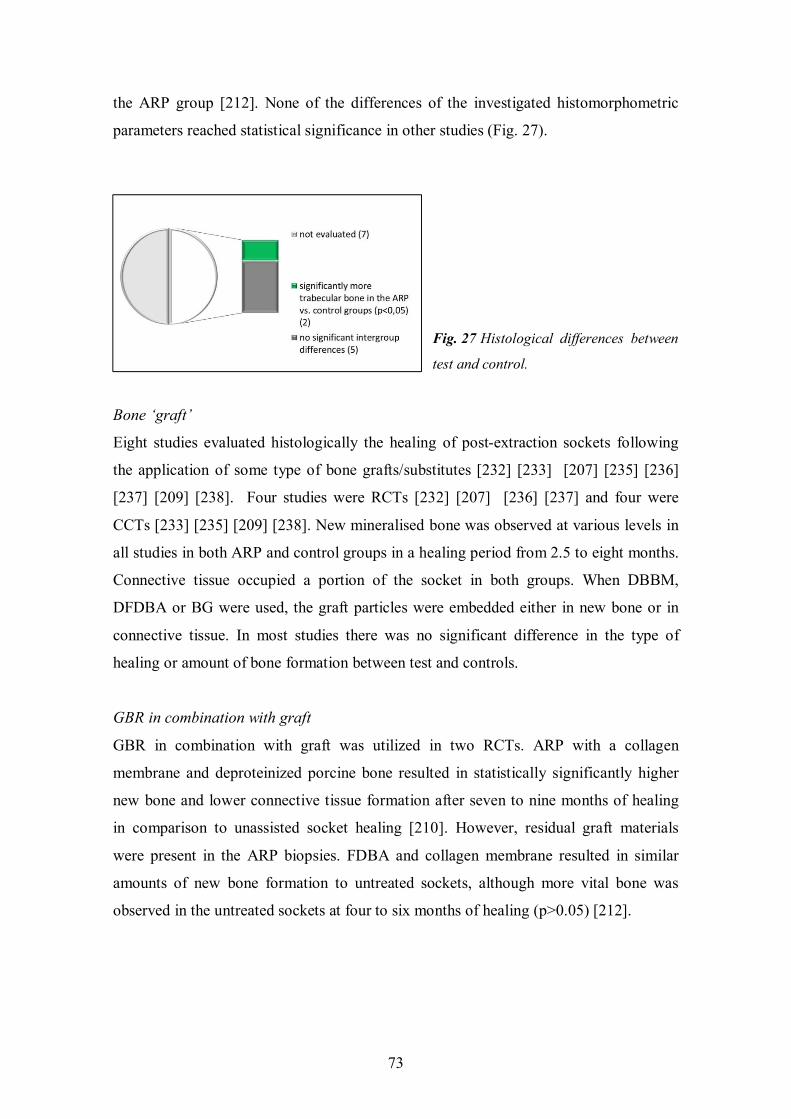
73
the ARP group [212]. None of the differences of the investigated histomorphometric
parameters reached statistical significance in other studies (Fig. 27).
Fig. 27 Histological differences between
test and control.
Bone ‘graft’
Eight studies evaluated histologically the healing of post-extraction sockets following
the application of some type of bone grafts/substitutes [232] [233] [207] [235] [236]
[237] [209] [238]. Four studies were RCTs [232] [207] [236] [237] and four were
CCTs [233] [235] [209] [238]. New mineralised bone was observed at various levels in
all studies in both ARP and control groups in a healing period from 2.5 to eight months.
Connective tissue occupied a portion of the socket in both groups. When DBBM,
DFDBA or BG were used, the graft particles were embedded either in new bone or in
connective tissue. In most studies there was no significant difference in the type of
healing or amount of bone formation between test and controls.
GBR in combination with graft
GBR in combination with graft was utilized in two RCTs. ARP with a collagen
membrane and deproteinized porcine bone resulted in statistically significantly higher
new bone and lower connective tissue formation after seven to nine months of healing
in comparison to unassisted socket healing [210]. However, residual graft materials
were present in the ARP biopsies. FDBA and collagen membrane resulted in similar
amounts of new bone formation to untreated sockets, although more vital bone was
observed in the untreated sockets at four to six months of healing (p>0.05) [212].

74
Biological active material
RhBMP-2 in a collagen sponge carrier was completely resorbed at four months
following ARP regardless of the concentration of the growth factor [215]. Mineralised
tissue was found and trabecular bone formation was noticed in two third of both the test
and control biopsies in the RCT.
Adverse events, complications
Adverse events were reported in six RCTs [215] [207] [210] [214] [232] [237] and four
CCTs [209] [213] [234] [238] including oedema, pain, erythema and membrane
exposure/infection. In two studies more adverse events i.e. oedema, erythema [215] or
membrane exposure [213] were observed in the ARP group compared to the natural
socket healing. No comparison between tests and controls were reported in the other
studies (Table 12).
Feasibility of implant placement
Seven studies [207] [209] [212] [232] [235] [236] [238] reported that implant placement
in the previous sockets were successful, but no differences between the ARP and
untreated sites were revealed. The outcome of implant placement remained unclear in
one article [233] and only re-entry without implantation was performed in three trials
[213] [214] [234]. Three studies reported that at the stage of implant placement less
need of further augmentation was observed in the ARP group compared to the controls
(i.e. less [215] or no sites [210] [237] in the ARP group presented with residual
dehiscence or fenestration defects around the inserted implants) (Table 12, Fig. 28).
Fig. 28 Necessity of
simultaneous
augmentation at
implant placement

75
Patient-reported outcome and health economics
No data were found for patient-reported outcome measures or health economic
evaluation.
Quality assessment
Considerable heterogeneity was found among the studies in terms of methodological
quality. Detailed description of the quality assessment of the included studies is
presented in table 11. Among the 14 included controlled studies, eight were randomised
[232] [210] [215] [207] [212] [214] [236] [237], although in four of them the
randomisation technique was not reported [215] [232] [236] [237]. None of the RCTs
reported the method of allocation concealment. Masking of the examiner was reported
at the clinical level in two out of eight [212] [214], at radiological level in one out of
two [215] and at histological level in four out of 11 studies [232] [233] [210] [207].
Examiner calibration was declared in three articles [232] [215] [212], whilst inclusion
and exclusion criteria were defined in seven publications [207] [210] [212] [232] [233]
[236] [237]. Apart from three studies [233] [210] [236], all the other reported the
approval of the ethical committee. Three studies were funded by industry [234] [207]
[215], two studies by academic institution [235] [238] and the remaining nine did not
report the source of funding.
Nine trials implemented patient-based analysis [232] [234] [215] [210] [212] [213]
[214] [237] [238], whilst the extraction site served as unit of analysis in the remaining
five investigations [207] [209] [233] [235] [236]. Sample size calculation was reported
only in three studies [232] [215] [212], although with insufficient data to evaluate the
validity of the calculations. Statistical analysis was appropriately carried out and
described in one study only [237]. Appropriate statistics were either not carried out
[207] [209] [215] [210] [233] [236] [235], or the reported data were insufficient to
determine the validity [232] [212] [213] [214] [233] [238]. In addition, no RCTs were
either registered with ISRCTN or reported using the CONSORT guidelines (Table 11).
Risk of bias
Four studies were classified as moderate risk of bias [210] [207] [212] [214] and the
rest were categorised as high risk of bias (Table 11, Fig. 29).

76
Fig. 29 Quality
assessment of the included
trials
77
6. DISCUSSION
6.1. Limits of periodontal regeneration
The main focus of the present investigations were to provide histological insight on the
regenerative potential of EMD combined with BCP (Study 1) or nano-HA (Study 2) for
the treatment of human periodontal intrabony defects.
6.1.1. Treatment with EMD and SBC
This study demonstrated that treatment of intrabony defects with a combination of EMD
+ BCP may lead to improvement of clinical parameters, such as PPD reduction and
CAL gain (Table 1). The lack of adverse reactions observed in all treated subjects is in
accordance with findings from previous clinical and histological studies that showed
that EMD and BCP are well tolerated and do not elicit allergic or foreign body reactions
[113] [77] [20] [78] [114] [60] [79] [115] [21] [116] [117] [36]. The mean CAL gain
of 3.0 mm obtained in this study is in line with previous findings from human histologic
studies evaluating the treatment of advanced intrabony defects with EMD alone or in
combination with DBBM or BG [33] [34] [41] [117] [36].
Considerations of the clinical outcomes
In contrast to our findings, the increased recession is an often observed phenomena
following surgical periodontal therapy. It suggests that in very advanced deep and wide
intrabony defects the additional use of graft material may prevent flap collapse and
consequently soft tissue recession. This finding corroborates the results from controlled,
clinical studies comparing the use of EMD in conjunction with ABP, DFDBA or
DBBM in the treatment of large intrabony defects [38] [40] [39]. The combined
treatment in these studies resulted in significantly less recession and consequently
greater CAL gain compared to treatment with EMD alone that points to the potential
clinical indication of a combination approach. BCP was employed to fill the intrabony
periodontal defect in a recent case-control study [118]. The authors reported a mean
CAL gain of 4.6 mm (3.0 mm in our trial) following the treatment with a combination

78
of BCP and guided tissue regeneration, showing the biocompatibility and promising
clinical properties of BCP.
To the best of our knowledge only two recent clinical trials investigated the treatment of
periodontal defects with a combination of EMD+BCP. In a randomized controlled
multicenter trial EMD+BCP (test; n=38) was compared to EMD alone (control; n=35)
[119]. At six months after surgery all investigated clinical parameters represented
significant improvements in both groups (PPD: 1.93±1.8 vs. 2.55±1.8; CAL: 1.31±1.8
vs. 1.83±1.6; REC: 0.62±1.1 vs. 0.72±1.1; bone sounding: 2.01±2.1 vs. 2.07±1.2; when
compared test to control, in millimeters, respectively). These results are slightly inferior,
but still comparable to the clinical outcomes of our investigation. The intergroup
differences failed to show statistical significance. Their results did not support the
theory that the additional bone substitute scaffold might improve the clinical outcome,
especially the hard tissue fill of the intrabony defect, compared to EMD alone.
Nevertheless, the investigators concluded that the null hypothesis that the applied
combined treatment of EMD+BCP is inferior to EMD alone, was refuted.
Another, more recent, randomised controlled clinical trial (RCT) that was conducted in
collaboration with the University of Szeged, evaluated the long-term effect of
EMD+BCP [120]. The authors reported similar outcomes in the test (EMD+BCP;
n=12), compared to the control group (EMD; n=12) at 12 months. Both groups lead to
significant improvement in terms of PPD reduction (from 8.8±1.5 to 4.3±0.9 mm vs.
from 8.8±1.0 to 4.1±0.5 mm) and CAL gain (from 10.8±1.6 to 7.4±1.6 mm vs. from
10.4±1.3 to 6.9±1.0 mm) in the test compared to control, respectively. No significant
differences were reported for any of the variables between the two groups indicating
that the added BCP did not additionally improve the outcomes obtained with EMD
alone. Slight, but not statistically significant deterioration of the investigated clinical
parameters between 1 and 4 years was reported, concluding that the results, irrespective
of the treatment modalities, were sustainable for four years.
Histological considerations
Human histological studies evaluating the regenerative potential of BCP alone or in
combination with EMD or other regenerative materials (e.g. barrier membranes), in
surgical treatment of periodontal intrabony defects are currently not available; therefore,
direct comparison is not possible. The histological evaluation of our experiment showed
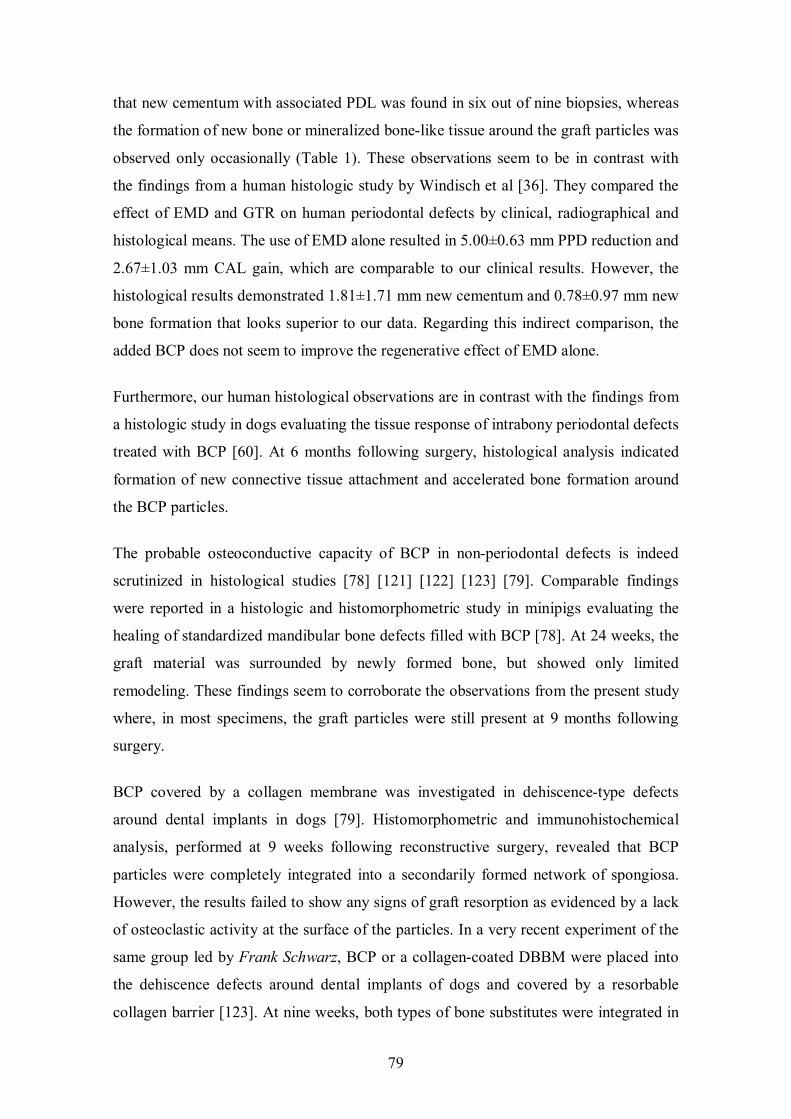
79
that new cementum with associated PDL was found in six out of nine biopsies, whereas
the formation of new bone or mineralized bone-like tissue around the graft particles was
observed only occasionally (Table 1). These observations seem to be in contrast with
the findings from a human histologic study by Windisch et al [36]. They compared the
effect of EMD and GTR on human periodontal defects by clinical, radiographical and
histological means. The use of EMD alone resulted in 5.00±0.63 mm PPD reduction and
2.67±1.03 mm CAL gain, which are comparable to our clinical results. However, the
histological results demonstrated 1.81±1.71 mm new cementum and 0.78±0.97 mm new
bone formation that looks superior to our data. Regarding this indirect comparison, the
added BCP does not seem to improve the regenerative effect of EMD alone.
Furthermore, our human histological observations are in contrast with the findings from
a histologic study in dogs evaluating the tissue response of intrabony periodontal defects
treated with BCP [60]. At 6 months following surgery, histological analysis indicated
formation of new connective tissue attachment and accelerated bone formation around
the BCP particles.
The probable osteoconductive capacity of BCP in non-periodontal defects is indeed
scrutinized in histological studies [78] [121] [122] [123] [79]. Comparable findings
were reported in a histologic and histomorphometric study in minipigs evaluating the
healing of standardized mandibular bone defects filled with BCP [78]. At 24 weeks, the
graft material was surrounded by newly formed bone, but showed only limited
remodeling. These findings seem to corroborate the observations from the present study
where, in most specimens, the graft particles were still present at 9 months following
surgery.
BCP covered by a collagen membrane was investigated in dehiscence-type defects
around dental implants in dogs [79]. Histomorphometric and immunohistochemical
analysis, performed at 9 weeks following reconstructive surgery, revealed that BCP
particles were completely integrated into a secondarily formed network of spongiosa.
However, the results failed to show any signs of graft resorption as evidenced by a lack
of osteoclastic activity at the surface of the particles. In a very recent experiment of the
same group led by Frank Schwarz, BCP or a collagen-coated DBBM were placed into
the dehiscence defects around dental implants of dogs and covered by a resorbable
collagen barrier [123]. At nine weeks, both types of bone substitutes were integrated in
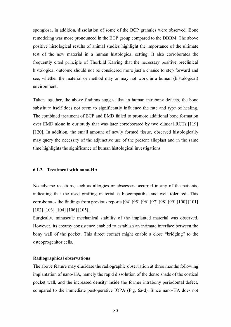
80
spongiosa, in addition, dissolution of some of the BCP granules were observed. Bone
remodeling was more pronounced in the BCP group compared to the DBBM. The above
positive histological results of animal studies highlight the importance of the ultimate
test of the new material in a human histological setting. It also corroborates the
frequently cited principle of Thorkild Karring that the necessary positive preclinical
histological outcome should not be considered more just a chance to step forward and
see, whether the material or method may or may not work in a human (histological)
environment.
Taken together, the above findings suggest that in human intrabony defects, the bone
substitute itself does not seem to significantly influence the rate and type of healing.
The combined treatment of BCP and EMD failed to promote additional bone formation
over EMD alone in our study that was later corroborated by two clinical RCTs [119]
[120]. In addition, the small amount of newly formed tissue, observed histologically
may query the necessity of the adjunctive use of the present alloplast and in the same
time highlights the significance of human histological investigations.
6.1.2 Treatment with nano-HA
No adverse reactions, such as allergies or abscesses occurred in any of the patients,
indicating that the used grafting material is biocompatible and well tolerated. This
corroborates the findings from previous reports [94] [95] [96] [97] [98] [99] [100] [101]
[102] [103] [104] [106] [105].
Surgically, minuscule mechanical stability of the implanted material was observed.
However, its creamy consistence enabled to establish an intimate interface between the
bony wall of the pocket. This direct contact might enable a close “bridging” to the
osteoprogenitor cells.
Radiographical observations
The above feature may elucidate the radiographic observation at three months following
implantation of nano-HA, namely the rapid dissolution of the dense shade of the cortical
pocket wall, and the increased density inside the former intrabony periodontal defect,
compared to the immediate postoperative IOPA (Fig. 6a-d). Since nano-HA does not

81
bear high radiopacity, such as sintered HA ceramics, ß-TCP, ABP or DBBM, the
observable increased radiopacity could be explained by the rapid mineralization of the
newly formed tissue in the former pocket.
Clinical results
The clinical findings correspond to those reported in the randomized controlled studies
[106] [105]. In the first RCT intrabony defects were treated either with OFD alone or
combined with nano-HA [105]. The additional application of the alloplast yielded a
mean PPD reduction of 3.9 mm and a mean CAL gain of 3.6 mm compared to 2.6 mm
and 1.8 mm following OFD alone. The intergroup comparison demonstrated
significantly more PPD reduction (P = 0.012) and CAL gain (P = 0.005) in the nano-HA
group compared to the OFD group. A subsequent study has evaluated the treatment of
intrabony defects by papilla preservation flap surgery with or without the application of
nano-HA [106]. At 6 months after surgery, both treatments yielded significant
improvements compared to baseline. Intergroup comparison demonstrated statistically
significantly higher mean PPD reduction (4.3 ± 1.6 mm) and mean probing bone level
gain (4.3 ± 1.4 mm) in the nano-HA group compared to the controls (2.9 ± 1.1 and
2.6 ± 1.4 mm, respectively).
In a recent randomised controlled split mouth clinical study, nano-HA was compared to
ABP in periodontal intrabony defects as adjunct to OFD [124]. Six months after surgery
both therapies resulted in statistically significant PPD reduction (3.21 vs. 3.37 mm) and
CAL gain (2.62 mm vs. 2.38 mm) in the nano-HA vs. ABP group, respectively.
The PPD changes reported in the aforementioned studies well concord to those obtained
in our investigation (i.e. 4.0 mm versus 3.9, 4.3 and 3.21 mm) (Table 2). The detected
slight discrepancies in CAL gain (i.e. 2.5 mm versus 3.6, 4.3 and 2.6 mm) may be
related to differences in defect configuration. In the present study the majority of the
defects displayed a predominantly one- and two-wall configuration, while the defects in
the clinical RCTs presented with either two or three-wall configuration. It is well known
that three-walled defects have a significantly higher healing potential compared to the
one-walled ones [72].
Histological considerations
The histological evaluation has, however, indicated that in all six biopsies the healing
was predominantly characterized by formation of a long junctional epithelium along the
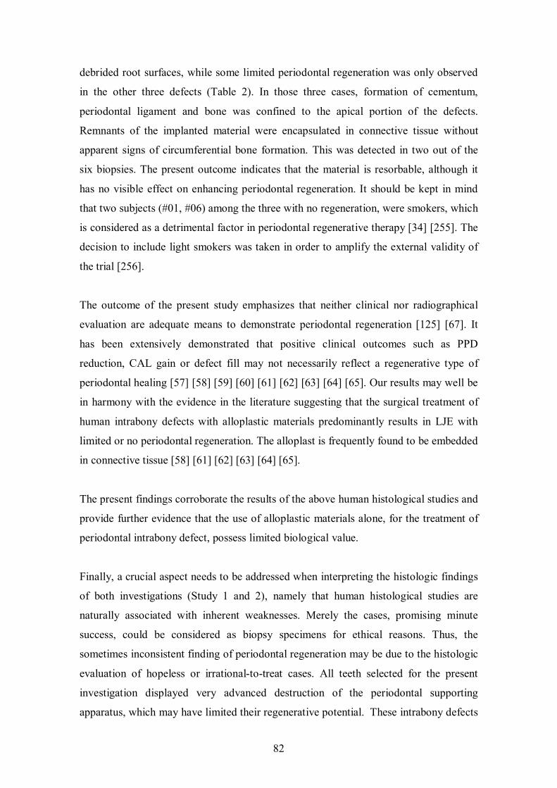
82
debrided root surfaces, while some limited periodontal regeneration was only observed
in the other three defects (Table 2). In those three cases, formation of cementum,
periodontal ligament and bone was confined to the apical portion of the defects.
Remnants of the implanted material were encapsulated in connective tissue without
apparent signs of circumferential bone formation. This was detected in two out of the
six biopsies. The present outcome indicates that the material is resorbable, although it
has no visible effect on enhancing periodontal regeneration. It should be kept in mind
that two subjects (#01, #06) among the three with no regeneration, were smokers, which
is considered as a detrimental factor in periodontal regenerative therapy [34] [255]. The
decision to include light smokers was taken in order to amplify the external validity of
the trial [256].
The outcome of the present study emphasizes that neither clinical nor radiographical
evaluation are adequate means to demonstrate periodontal regeneration [125] [67]. It
has been extensively demonstrated that positive clinical outcomes such as PPD
reduction, CAL gain or defect fill may not necessarily reflect a regenerative type of
periodontal healing [57] [58] [59] [60] [61] [62] [63] [64] [65]. Our results may well be
in harmony with the evidence in the literature suggesting that the surgical treatment of
human intrabony defects with alloplastic materials predominantly results in LJE with
limited or no periodontal regeneration. The alloplast is frequently found to be embedded
in connective tissue [58] [61] [62] [63] [64] [65].
The present findings corroborate the results of the above human histological studies and
provide further evidence that the use of alloplastic materials alone, for the treatment of
periodontal intrabony defect, possess limited biological value.
Finally, a crucial aspect needs to be addressed when interpreting the histologic findings
of both investigations (Study 1 and 2), namely that human histological studies are
naturally associated with inherent weaknesses. Merely the cases, promising minute
success, could be considered as biopsy specimens for ethical reasons. Thus, the
sometimes inconsistent finding of periodontal regeneration may be due to the histologic
evaluation of hopeless or irrational-to-treat cases. All teeth selected for the present
investigation displayed very advanced destruction of the periodontal supporting
apparatus, which may have limited their regenerative potential. These intrabony defects
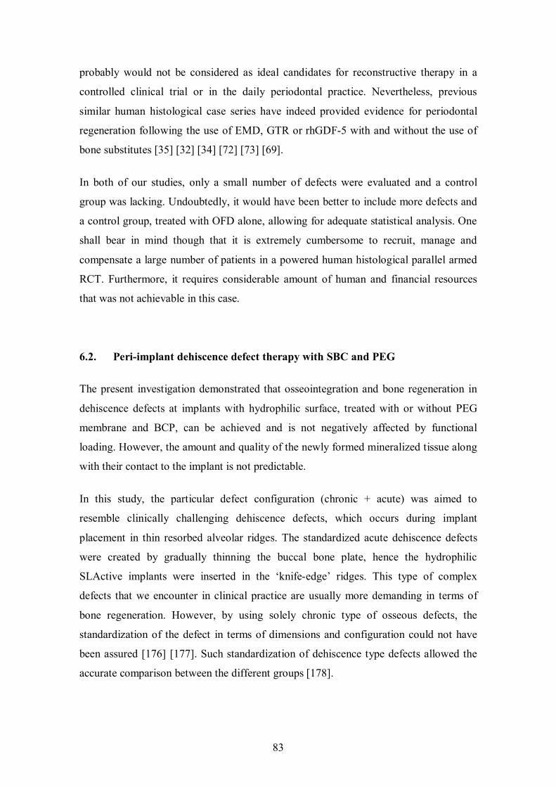
83
probably would not be considered as ideal candidates for reconstructive therapy in a
controlled clinical trial or in the daily periodontal practice. Nevertheless, previous
similar human histological case series have indeed provided evidence for periodontal
regeneration following the use of EMD, GTR or rhGDF-5 with and without the use of
bone substitutes [35] [32] [34] [72] [73] [69].
In both of our studies, only a small number of defects were evaluated and a control
group was lacking. Undoubtedly, it would have been better to include more defects and
a control group, treated with OFD alone, allowing for adequate statistical analysis. One
shall bear in mind though that it is extremely cumbersome to recruit, manage and
compensate a large number of patients in a powered human histological parallel armed
RCT. Furthermore, it requires considerable amount of human and financial resources
that was not achievable in this case.
6.2. Peri-implant dehiscence defect therapy with SBC and PEG
The present investigation demonstrated that osseointegration and bone regeneration in
dehiscence defects at implants with hydrophilic surface, treated with or without PEG
membrane and BCP, can be achieved and is not negatively affected by functional
loading. However, the amount and quality of the newly formed mineralized tissue along
with their contact to the implant is not predictable.
In this study, the particular defect configuration (chronic + acute) was aimed to
resemble clinically challenging dehiscence defects, which occurs during implant
placement in thin resorbed alveolar ridges. The standardized acute dehiscence defects
were created by gradually thinning the buccal bone plate, hence the hydrophilic
SLActive implants were inserted in the ‘knife-edge’ ridges. This type of complex
defects that we encounter in clinical practice are usually more demanding in terms of
bone regeneration. However, by using solely chronic type of osseous defects, the
standardization of the defect in terms of dimensions and configuration could not have
been assured [176] [177]. Such standardization of dehiscence type defects allowed the
accurate comparison between the different groups [178].

84
Hydrophilic implant surface
This recently developed chemically modified hydrophilic implant surface (SLActive),
which represents high free surface energy, has been shown to influence pro-osteogenic
and pro-angiogenic gene expression, to enhance production of local growth factors and
osteoblast activity and to accelerate osteogenic differentiation and mineralization
compared to smooth surface, conventional SLA surface or dual acid-etched surface with
a calcium phosphate modification [179] [180] [181] [182] [183] [184] [170] [185]
[186].
In the present investigation, histological observation of the SLActive implant inserted in
pristine sites showed mature lamellar bone in contact with the implant surface.
Furthermore, this study demonstrates the stability of the established osseointegration
after short-term functional loading at SLActive implants. The successful
osseointegration of SLActive titanium surfaces is in line with previous findings from
histomorphometric studies reporting promoted healing and enhanced bone apposition
even in circular peri-implant defects and dehiscence-type defects in comparison with
non-hydrophilic implant surface [180] [187] [188] [189] [184].
Irrespective of loading, osseointegration in the dehiscence defect seemed to be
supported by the presence of the SLActive titanium surface in situations where no
graft/GBR was applied. A thin layer of bone was observed in direct contact with the
implant surface in the defect area and new bone formation proceeded coronally, along
the SLActive implant surface. Furthermore, bone regeneration had taken place to a
significant extent within the defect. However, the defect was in no case completely
filled and a number of threads were always exposed at the most coronal point of the
implant, demonstrating that the chosen model was indeed of critical size.
It has to be highlighted that the loaded control group of dehiscence defect, where no
material was placed onto the exposed buccal side of the implants (N), presented the
highest percentage of bone-to-implant contact (BIC%: 64.1 ± 12.8) and the smallest
remaining dehiscence i.e. the lowest distance between the most coronal BIC to the
implant shoulder (S-BIC: 1.93 ± 0.69 mm) among all other groups. This data indicates
that the modified hydrophilic SLActive implant surface per se may have an
osteoconductive effect that improves osteogenic response.

85
Biphasic calcium phosphate bone substitute
In the present investigation, when a synthetic bone graft was used alone, or combined
with GBR osseointegration was observed along the implant surface. However, while
bone formation around the graft particles could be observed in the apical portion, in a
number of specimens, a variable amount of particles were encapsulated by soft tissue in
the coronal portion of the defect. This is in agreement with previous studies where
xenografts have been used in combination with GBR [190] [147] [172] or when
synthetic bone substitute was used [191] [192]. Thus, again, the positive effect in bone-
to-implant contact may be attributed to the SLActive surface rather than the present
alloplastic bone replacement material.
In addition, it should be noted that the seemingly inferior bone promoting property of
the present BCP for peri-implant dehiscence defects, demonstrated histologically, could
not be detected by merely clinical and radiographic observation [193]. This finding is in
harmony with the result of the experiment of the treatment of periodontal defects,
presented in this thesis, namely that the positive clinical outcome following use of the
same BCP material does not necessarily supported by histological evidence [73].
Polyethylene Glycol-Based Hydrogel Membrane
One of the main aims of the present study was to test the novel polyethylene glycol
hydrogel membrane. It is activated by mixing the two liquid components and is applied
in a gel state. Thus, the membrane has limited space-maintaining ability by itself and
was designed by the manufacturer to be placed in combination with a bone substitute.
Previous experimental studies have demonstrated that this type of membrane is cell-
occlusive [165] and has positive results on bone regeneration around implants inserted
in jaw bone as well in calvaria defects [167] [168] [169] [170] [166].
In the present investigation group T2 (PEG membrane and BCP) exhibited the largest
bone surface area (BS) in terms of new bone formation, followed by T1 and N (T2:
13.18 ± 5.85, T1: 9.69 ± 4.11, N: 8.81 ± 4.48 mm2). These differences were not
statistically significant. However, in group T2, soft tissue was frequently interposed
between the newly formed bone and the implant surface, irrespective of the functional
loading. This was reflected in a lower mean bone-to-implant contact (BIC%) compared
with groups N and T1, although differences were not statistically significant (T2:
33.9 ± 26.5, T1: 54.3 ± 27.1, N: 52.3 ± 20.9). Furthermore, the greatest distance
86
between the implant shoulder to the first BIC (S-BIC), i.e. the greatest residual vertical
defect alongside the buccal surface of the implant was also evident in this group (T2:
3.71 ± 1.83, T1: 2.43 ± 1.74, N: 2.67 ± 1.40, P: 2.50 ± 2.17 mm). According to these
unexpected results, not only the other test group, but both controls performed superior
compared to the PEG group. A possible explanation of the limitation of this barrier may
reside in its poor flexibility and elasticity after the membrane set following
polymerization. In the present study, the PEG membrane was extended 2–3 mm around
the bone defect and covered completely the coronal part of the implant. In a number of
implants mesio-distal rupture of the membrane above the occlusal part of the implant
was observed after polymerization was completed. When this occurred, the membrane
was removed and re-applied. Even though all animals were fed by soft diet, this may
have not prevented the rupture of the membrane above the implant after suturing of the
flap above the membrane or during the early healing phase. It can be speculated that this
has allowed soft tissue proliferation between the bone substitute and the implant
surface, preventing the functional attachment formation between the implant and the
newly formed hard tissue. These results might have an impact on possible clinical
application of this PEG membrane indicating that the application should be limited on
buccal surface avoiding complete coverage (i.e. occlusal surface) of the implant to avoid
rupture.
Significant differences were observed in the mean residual bone substitute surface
between groups T1 and T2. This value was several times higher in T1(4,94 ± 2.81 mm2)
compared with that in T2 (0.71 ± 0.80 mm2). This increased resorption/dispersion of the
BCP particles when combined with GBR was also reported in previous studies. In an
experimental study in dogs, Jung et al. observed the absence of BCP particles in 2/5
defects at six months [168], and Schwarz et al. showed a decrease in the mean graft
surface between two and eight weeks [170]. It has been speculated that this could be
correlated with the hydrolytic disruption of the PEG membrane, causing dispersion of
the BCP [170]. Polyethylene glycol hydrogels degrade by hydrolysis and this process
may take several weeks and is accompanied by an increase in the PEG volume of 30–
50% [165]. It is not clear, if the increase in volume could exert pressure on the graft
causing dislocation of its particles. However, in the present study, after five months of
healing, the graft particles were reduced in number and size when the PEG membrane
was used. In this treatment group, the small BCP particles were surrounded by

87
mineralized tissue. These observations may indicate an increased resorption rate of the
graft when the PEG membrane was used, rather than the dispersion of the particles.
Loading
It has been demonstrated that GBR could lead predictably in significant amounts of
bone formation when used prior to implant placement [136], or for treatment of
dehiscence/fenestration defects around implants [194] [127]. It has been suggested that
if the newly formed bone is not functionally loaded, then some bone resorption in height
might take place, once the protection of the secluded space created by a membrane is
removed [171] [172]. In the present study, where the regenerated bone was subjected to
functional loading for a short period, no significant differences were observed between
loaded and non-loaded implants in any of the examined parameters. However, in groups
P, N and T1, there was a clear tendency for increased bone regeneration (BIC%, S-BIC)
under functional loading which, however, never reached statistical significance. An
opposite trend was observed in group T2 (GBR). This group showed lower BIC% and
higher S-BIC and S-NBC at loaded sites. It could be suggested that these observations
are in agreement with previous studies in the literature where marginal bone loss around
implants after GBR had been observed [173] [195] [196].
This could also imply that in this study, the regenerated bone did not result in the
expected BIC in the PEG group and this may lead to bone resorption after a period.
These results emphasise not only the use of barrier membranes with appropriate
occlusive properties but also the importance on the surgical approach (membrane
application/ location) in order to avoid interference with the healing.
Clinical versus histological data
The present experiment provided some clinical as well as histological data on bone
regeneration. The distance between the newly formed tissue and the implant shoulder
was measured clinically prior to abutment connection and histologically after study
termination. According to clinical measurements, T2 performed most favorably with the
smallest remaining dehiscence along the implant surface (ROI) compared to T1 and N
(T2: 2.10, T1: 2.55, N: 3.35 mm). However, histological evidence failed to support this
clinical observation with T2 demonstrating the least favorable outcome (T2:
3.71 ± 1.83, T1: 2.43 ± 1.74, N: 2.67 ± 1.40 mm). This result corroborates that merely

88
clinical assessment of novel materials (i.e. without histological evaluation) used for
GBR procedures may result in delusive conclusions.
6.3. Current options for the treatment of extraction sockets by ARP
Key findings
This systematic review has demonstrated that different ARP techniques cannot totally
eliminate post extraction alveolar ridge resorption nor predictably promote new bone
formation. However, the reduction in ridge width and height following ARP may be less
than that which occurs following natural socket healing. The clinical data suggest that
the horizontal ridge contraction was most successfully limited in the two studies
applying GBR without additional bone grafts [213] [214], whereas the vertical
shrinkage was most efficiently limited by employing GBR with additional bone graft
[210] [212] .
Strengths of the review
The present systematic review was limited to randomised controlled trials, controlled
clinical trials and prospective cohort studies with a control group of empty untreated
sockets. Furthermore, the inclusion criteria of our systematic review were based on the
fact that the clinical merit of applying the different ARP techniques could only be
validated, if the clinical as well as histological outcomes, following the application of a
technique, are superior to that of unassisted socket healing.
In comparison to the previous systematic reviews [219] [223] the present review has
evaluated the histological characteristics of the alveolar socket healing with or without
ARP. The amount and the quality of the newly formed osseous tissues in the socket area
are essential, especially when the justification of ARP is to facilitate the placement of a
dental implant in the position of a previously extracted tooth. It is doubtful, whether an
ARP technique should be claimed successful if it only preserves the external contour of
the AR, but the newly formed tissue is of not superior or even inferior quality and
quantity (percentage of matured trabecular bone) to what is normally achieved
following extraction.

89
Finally, the quality of the included studies has also been meticulously assessed in this
review. Such a quality evaluation of the retrieved data is essential to estimate the source
and magnitude of potential bias that may lead to delusive conclusions.
Strength of evidence – risk of bias
The quality assessment of the included studies in this systematic review revealed that
none of the trials have qualified for a low risk of bias category. Ten out of the 14 studies
presented with high risk of bias, thus their results must be evaluated with caution. The
lack of clear reporting of research methodology elements, such as adequate
randomization and concealment and/or masking of the therapist and the examiner were
among the primary reasons for the high risk of bias [241]. We did not contact authors
for clarification of unclear methodology. Therefore, it is possible that actual study
conduct was better than that reported in the publication. Statistical considerations played
important role as well, since appropriate analytical statistics was completed and reported
merely in one study [237]. Power calculation was conducted in three trials only [210]
[212] [232], nevertheless the reported data were insufficient to determine the validity of
the calculation.
Dimensional changes and histological characteristics
Sufficient ridge width and height have been considered as one of the key requirements
for successful implant therapy and for the establishment of an aesthetically pleasing
emergence profile at fixed partial dentures [128] [133] [242]. Therefore, the alteration in
oro-facial (horizontal) and apico-coronal (vertical) AR dimensions was selected as the
primary outcome of the present review. Direct intrasurgical measurements on the AR at
re-entry are considered as the most precise method to evaluate the bone volume changes
following ARP. It is desirable though to establish and validate a surrogate measure that
avoids the need for re-entry surgery, while providing the clinician with a reliable
measure. Two dimensional radiographs, such as IOPA or orthopantomogram, are not
ideal to estimate the three dimensional changes of the AR [243]. Also, measurements of
the alveolar mucosa level or study casts incorporate not only the alveolar bone, but also
the overlaying soft tissue. For these reasons only studies performing clinical or three
dimensional radiographic evaluation of hard tissue were included in this review. Cone-
beam computerised tomography (CBCT) appears to offer a valid technique to assess
alveolar ridge changes, with newer models greatly reducing radiation exposure [244].
90
However, a prerequisite of this technique would be some type of standardisation, so that
the captured image is always being taken from exactly identical positions [245]. None
of the two included radiographic studies reported on such standardisation [215] [236].
For the interpretation of the results we attempted to cluster the studies in respect to the
type of intervention.
Unassisted sockets
In the present review the mean reduction of the AR width of the untreated sites varied
between 2.6±2.3 and 4.6±0.3 mm and the mean reduction of the AR height was between
0.8±1.6 and 3.6±1.5 mm after one to nine months of healing. This corroborates the
result of a previous clinical study which indicated that 95% of AR reduction should be
expected after three months of extraction [131]. Furthermore, it is in agreement with a
recent systematic review, which reported that the average reduction of the AR width
seemed to be higher (3.87 mm), than the reduction in AR height (1.67 mm) [200].
Even though both AR width and height present resorption, histologically, new bone
formation up to a variable extent was also observed in most studies as result of
unassisted socket healing [209] [215] [210] [212] [232] [235] [236] [238]. In addition,
a large area was occupied by bone marrow [210] [209] [238], such as reported in
preclinical studies [14] [203] [45]. Only a single study reported on connective tissue fill
and lack of mature bone in the control sites [233].
Bone grafts and substitutes
Effective grafting procedures for bone augmentation have been associated with the
osteoconductive, osteoinductive or osteogenetic properties of the graft [45] [246] [247].
This led to the assumption that the placement of these materials in the extraction socket
may accelerate new bone formation by the above biological properties and may also
reduce AR resorption by stabilising the blood clot, providing a scaffold and external
source of minerals and/or collagen [248] [14] [202] [249]. The placement of DBBM
with collagen in fresh extraction sockets resulted in limited reduction of the AR
dimensions, although delayed initial socket healing in terms of new bone formation was
also observed [14] [202]. Human studies reported similar unfavourable histological
observations when DFDBA was employed for ARP [205] [206].
91
In the present review of human experiments, two out of three studies reported that
socket grafting with autologous bone marrow [237] or alloplastic material [232] have
significantly limited the reduction of the AR width compared to the unassisted socket
healing. Three out of five studies reported that reduction of the resorption in AR height
was significant [232] [236] [237], while the ridge height was even increased in one
study, where sockets were grafted with polymer sponge [209]. We should emphasise
though that since the graft material (DBBM) in a radiographic study possessed
radiopaque characteristic [236], the alteration of the AR contour on the CBCT image
should be interpreted with caution.
Based on the histological evaluation of these studies, the above AR dimensional
changes were not necessarily accompanied by higher amount of new bone formation in
the socket, since the quality of newly formed tissue in the ARP sites was comparable to
that in the control sites. Furthermore, the sockets were occupied by a mixture of new
bone and connective tissue which in many occasions was surrounding the graft particles
[207] [210] [236].
GBR (membrane alone or in combination with bone substitute)
The conception of guided bone and tissue regeneration [136] was translated to ARP
procedures in order to exclude epithelial cells from the extraction socket by the use of
barrier membrane in four studies of the present review [210] [212] [213] [214].
a) GBR with membrane alone
ARP with GBR resulted in statistically significantly less resorption in ridge width and
height compared to unassisted socket healing, regardless of the type of membrane [213]
[214]. It should be noted that in one study [213]. in three out of 10 cases, the exposed
non-resorbable e-PTFE barrier had to be removed prematurely, highlighting the
importance of sufficient soft tissue closure and timing of removal of the barrier. The
outcomes in these three cases were similar to that in the control sites. Where healing
was uncompromised, a statistically significant difference was found after six months in
width and height changes in favour of the ARP group.
b) GBR with membrane and bone substitute
ARP resulted in statistically significantly less resorption in width [210] [212] and height
[212] in comparison to unassisted socket healing. The histological evaluation of the

92
GBR procedures in the included studies demonstrated new bone formation, but the
presence of graft particles was also evident in both studies, embedded either in newly
formed bone [210] or in connective tissue [212]. This is in agreement with a recent
trial, where a collagen membrane in combination with DBBM or a biphasic bone
substitute was used for ARP [192] [188].
Biological active materials
The potential benefit of biological active molecules were investigated in periodontal
and bone regeneration through fostering the proliferation and differentiation of different
mesenchymal cells in various preclinical models [250] [251]. The safety and feasibility
of rhBMP-2 on human ARP or ridge augmentation was evaluated and shown to be safe
in a bi-center clinical study [226]. Dimensional changes of the alveolar ridge were
measured on CBCT scans in an RCT [215]. Treatment with rhBMP-2 resulted in an
increase in ridge width, which was statistically significantly greater than in the controls.
However, this observation needs to be interpreted in light of the surprise finding of an
increase in ridge width of the untreated controls. This was a unique observation amongst
the studies that we reviewed. Histologically, no comparison between ARP and controls
sites was reported.
The human histological findings of the included papers of the present review were
generally found to be comparable to preclinical studies [248] [14] [202] [252] [203].
There are a number of aspects to be considered in the interpretation of the results.
Firstly, it has to be kept in mind that whilst the biopsies of the animal model incorporate
the cross section of the whole AR, the biopsy retrieval at human studies is limited to a
trephine core sample of part of the former socket. This location may not necessarily
coincide with the exact position of the previous extraction, thus making interpretation of
the results challenging. Furthermore, the differentiation between apical, mid and
coronal, as well as the central and lateral aspects of the biopsies was not always
apparent.
Another important parameter when considering a histological overview of the studies
was the variation in healing time. Due to the nature of post-extraction healing, the direct
comparison of the new tissue formation in studies between one and nine months of
healing could be misleading. This was highlighted in three studies which did not make a
93
distinction between the variable healing times within the groups, ranging from 2.5
months to nine months [207] [210] [233]. It must also be noted that the single study,
which completed and reported appropriate statistical methodology [237], did not
observe statistically significant difference between the test and control biopsies.
Furthermore, small sample sizes in the majority of the studies may also limit the
generalisability of the histological findings.
Merely two studies found statistically significant histological differences in new bone
formation favouring the test group [210] [232]. It is hard to draw generalizable
conclusions from these two experiments, since the test groups differed in many respects
compared with each other, including different technique (bone substitute only [232] vs.
GBR+graft [210] ), different material (MGCSH [232] vs. porcine bone with collagen
membrane [210] ), different flap management (flapless, no primary closure [232] vs.
mucoperiosteal flap, primary closure [210] ), different healing time (3 months [232] vs.
7-9 months [210] ). One common feature was that both groups limited their intervention
to sockets with four intact walls. It is noteworthy that all three studies that included
intact socket walls only, reported statistically significant differences both on AR width
and height in favour of ARP [232] [210] [237], while only one [215] out of two studies
[215] [209] with initial buccal bone loss reported similar significant difference between
test and control. Hence, socket morphology could be an important predicator of
improved ARP. The need for ARP in such sockets, in terms of future clinical
success/implant placement needs further investigation.
Other factors affecting interpretation of the findings
Flap management
All studies reporting statistically significant inter-group differences in both horizontal
and vertical clinical measurements achieved either primary flap closure [210] [213]
[214] [237], or did not detach the periosteum in a flapless procedure [232].
Furthermore, none of the studies without primary closure demonstrated statistically
significant differences between test and control in terms of both horizontal and vertical
clinical measurements [209] [212] [234] . Therefore, both achieving and maintaining
the epithelial seal above the socket may be essential to improve ARP. Further
corroboration of this concept was suggested where e-PTFE barriers were prematurely

94
exposed. The healing of these three exposed cases demonstrated no statistically
significant differences compared to the control sites [213].
Healing time
The optimal timing of re-entry following ARP is determined by the implant insertion.
The volume of the AR is gradually decreasing, while the quality of the newly formed
tissue is gradually increasing during the post extraction remodelling [131] [198].
Therefore, the implant placement should be considered as early as possible, but as late
as necessary, in order to maintain AR volume, but also to achieve complete epithelial
seal with some extent of osseous fill. The healing periods of the trials in the present
review varied considerably (one to nine months). Hence, the interpretation of the results
is difficult due to the heterogeneity present in the included studies.
Antimicrobials
Improvement of clinical parameters was demonstrated as a result of regular rinsing with
chlorhexidine following tooth extraction [253]. Subjects of the included trials in the
present review were prescribed various types of antibiotics and instructed to rinse with
chlorhexidine for two to three weeks. Thus, no conclusion could be drawn on the
necessity or benefit of employment of antibiotics/antimicrobials following ARP.
Smoking
Smoking is associated with delayed wound and socket healing and increased reduction
in post extraction alveolar width [254] [255]. Three trials in this review included
smokers [210] [212] [233] and the half of the studies did not report on smoking as an
exclusion factor, thus any conclusions about the impact of this well-recognised risk
factor for impaired healing are difficult to draw.
Periodontal treatment/health
Four studies included patients whose periodontal treatment was carried out prior to the
ARP [210] [209] [214] [238]. ARP resulted in statistically significant difference
between tests and controls in clinical [210] [214] and in histological parameters [210].
In addition, in the studies where periodontitis was present, but periodontal treatment
was not reported, no statistically significant histological differences were demonstrated
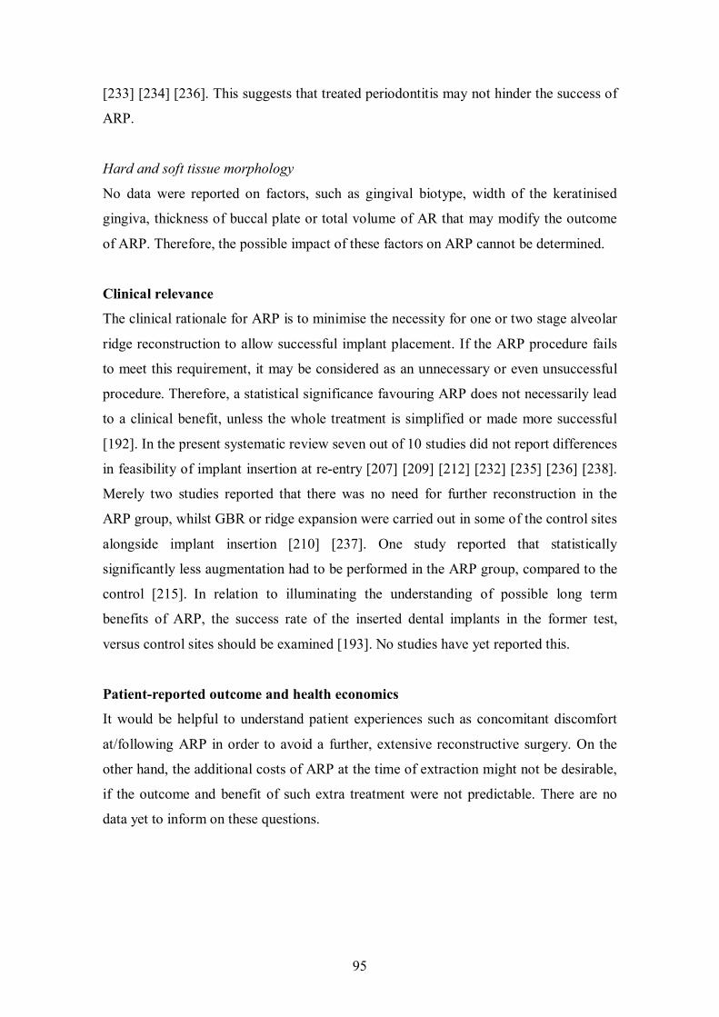
95
[233] [234] [236]. This suggests that treated periodontitis may not hinder the success of
ARP.
Hard and soft tissue morphology
No data were reported on factors, such as gingival biotype, width of the keratinised
gingiva, thickness of buccal plate or total volume of AR that may modify the outcome
of ARP. Therefore, the possible impact of these factors on ARP cannot be determined.
Clinical relevance
The clinical rationale for ARP is to minimise the necessity for one or two stage alveolar
ridge reconstruction to allow successful implant placement. If the ARP procedure fails
to meet this requirement, it may be considered as an unnecessary or even unsuccessful
procedure. Therefore, a statistical significance favouring ARP does not necessarily lead
to a clinical benefit, unless the whole treatment is simplified or made more successful
[192]. In the present systematic review seven out of 10 studies did not report differences
in feasibility of implant insertion at re-entry [207] [209] [212] [232] [235] [236] [238].
Merely two studies reported that there was no need for further reconstruction in the
ARP group, whilst GBR or ridge expansion were carried out in some of the control sites
alongside implant insertion [210] [237]. One study reported that statistically
significantly less augmentation had to be performed in the ARP group, compared to the
control [215]. In relation to illuminating the understanding of possible long term
benefits of ARP, the success rate of the inserted dental implants in the former test,
versus control sites should be examined [193]. No studies have yet reported this.
Patient-reported outcome and health economics
It would be helpful to understand patient experiences such as concomitant discomfort
at/following ARP in order to avoid a further, extensive reconstructive surgery. On the
other hand, the additional costs of ARP at the time of extraction might not be desirable,
if the outcome and benefit of such extra treatment were not predictable. There are no
data yet to inform on these questions.

96
6.4. Overall considerations
Why did we select these materials for our experiments?
Stabilization of blood clot and fibrin matrix in the wound healing process, scaffolding
for osteoblasts during new mineralized tissue formation as well as a ‘buttress’ of the
space maintaining materials, as well as an external source of collagen and/or minerals
believed to support tissue regeneration [66]. For these purpose ABP as adjunct to
GTR/GBR is still considered as the gold standard in GBR procedures. However,
autogenous tissues are always associated with their drawbacks, i.e. their harvest usually
requires a second surgical site (increased surgical trauma and donor site morbidity),
presents with limited volume, resorbs rapidly that may result in decrease in volume. In
order to overcome these shortcomings, the emphasis was placed on alloplastic bone
substitute materials. Various alloplasts have then been available and have desperately
been promoted by their manufacturers. In many cases though, a sound clinical and
histological evidence seemed (seems) to be lacking behind these novel materials. In
other words, the products of some (small) companies could earlier be discovered on the
shelves of their suppliers, than on to the pages of scientific journals.
The recently developed materials that we aimed to investigate histologically, appeared
to bear with very promising properties, in terms of new tissue formation and resorption,
according to previous in vitro or in vivo investigations. In addition, their manufacturers
are known for prioritizing the scientific evidence before launching a new material to the
market. Therefore, both BCP, nano-HA and PEG seemed to be worth to examine in
these different, but representative settings.
Why these study designs?
We aimed at scrutinizing the performance of these novel materials in reconstructive
periodontology and related fields. The ultimate evidence of a material or method being
‘regenerative’ is based on human histology, or where inappropriate or less feasible,
histological proof of randomized controlled preclinical trial [67] [68]. Therefore, the
effect of the novel BCP combined with EMD or the nano-HA alone on periodontal
intrabony pockets were investigated in human, single arm, clinical and histological case
series. The same BCP with the novel, innovative PEG membrane was designed to

97
improve bone regeneration in case of dehiscence defects around dental implants. Due to
the extremely challenging nature of performing human histology in such cases,
randomized, controlled preclinical trial was designed. To the best of our knowledge,
these were the first time to test the above materials in such settings in periodontology
and implant dentistry. In contrast to this, broad variety of materials has been indeed
tested for ARP procedures. Therefore, instead of conducting yet another human clinical
and histological trial, a systematic review of these procedures and materials was carried
out. To the best of our knowledge, no such review that systematically investigates not
only the clinical, but also the histological healing characteristic of the result of ARP
procedures compared to unassisted socket healing, alongside quality assessment of the
included trials, have yet been published.
Clinical ‘success’ vs. histological evidence
All the selected and investigated novel materials have been used successfully in
previous clinical trials and they all have shown improvements in clinical parameters.
The use of BCP + EMD in advanced periodontal defects resulted in significant PPD
reduction, CAL gain and defect fill at six month in a multicenter study [119]. These
results were comparable to that of their controls (EMD). In addition, these favourable
clinical results were corroborated by a recent long-term RCT [120]. In accordance with
these findings, the clinical results of our experiment (Study 1) were similarly beneficial,
with mean PPD reduction of 3.3 ± 1.4 mm and mean CAL gain of 3.0 ± 1.6 mm. In
spite of these promising clinical data, histology has revealed none to limited periodontal
regeneration i.e. 0.69 ± 0.67 mm mean new cementum and 0.16 ± 0.25 mm mean bone
formation. One third of the biopsies displayed LJE instead. One may consider that this
result looks inferior to that achieved by EMD only in a similar human histological
investigation i.e. formation of mean 1.87 ± 1.17 mm new cementum and 0.78 ± 0.97
mm new bone [36]. In addition, in our study the additional use of BCP resulted in soft
tissue encapsulation.
Study 2 has actually led to similar conclusions. Application of nano-HA for the surgical
treatment of intrabony periodontal defect in randomized controlled clinical trials
resulted in significant PPD reduction and CAL gain [124] [106] [105]. Furthermore, the
result in the test group (nano-HA + OFD) appeared to be significantly better than that in
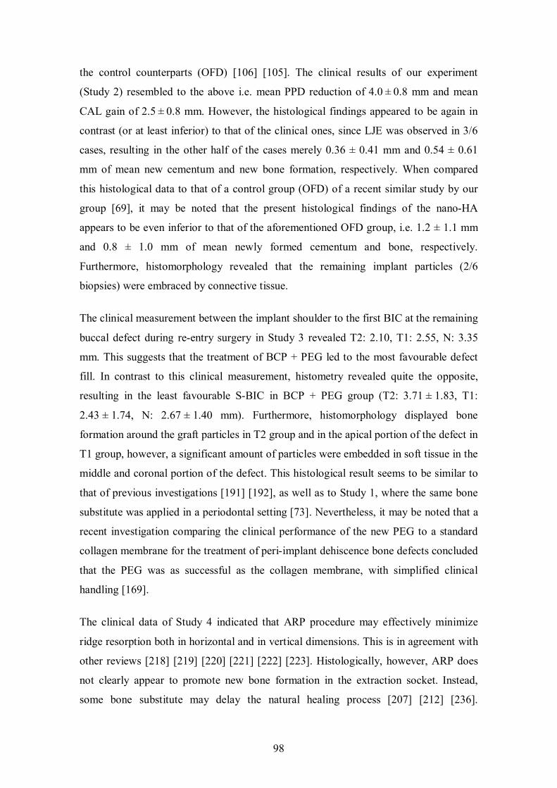
98
the control counterparts (OFD) [106] [105]. The clinical results of our experiment
(Study 2) resembled to the above i.e. mean PPD reduction of 4.0 ± 0.8 mm and mean
CAL gain of 2.5 ± 0.8 mm. However, the histological findings appeared to be again in
contrast (or at least inferior) to that of the clinical ones, since LJE was observed in 3/6
cases, resulting in the other half of the cases merely 0.36 ± 0.41 mm and 0.54 ± 0.61
mm of mean new cementum and new bone formation, respectively. When compared
this histological data to that of a control group (OFD) of a recent similar study by our
group [69], it may be noted that the present histological findings of the nano-HA
appears to be even inferior to that of the aforementioned OFD group, i.e. 1.2 ± 1.1 mm
and 0.8 ± 1.0 mm of mean newly formed cementum and bone, respectively.
Furthermore, histomorphology revealed that the remaining implant particles (2/6
biopsies) were embraced by connective tissue.
The clinical measurement between the implant shoulder to the first BIC at the remaining
buccal defect during re-entry surgery in Study 3 revealed T2: 2.10, T1: 2.55, N: 3.35
mm. This suggests that the treatment of BCP + PEG led to the most favourable defect
fill. In contrast to this clinical measurement, histometry revealed quite the opposite,
resulting in the least favourable S-BIC in BCP + PEG group (T2: 3.71 ± 1.83, T1:
2.43 ± 1.74, N: 2.67 ± 1.40 mm). Furthermore, histomorphology displayed bone
formation around the graft particles in T2 group and in the apical portion of the defect in
T1 group, however, a significant amount of particles were embedded in soft tissue in the
middle and coronal portion of the defect. This histological result seems to be similar to
that of previous investigations [191] [192], as well as to Study 1, where the same bone
substitute was applied in a periodontal setting [73]. Nevertheless, it may be noted that a
recent investigation comparing the clinical performance of the new PEG to a standard
collagen membrane for the treatment of peri-implant dehiscence bone defects concluded
that the PEG was as successful as the collagen membrane, with simplified clinical
handling [169].
The clinical data of Study 4 indicated that ARP procedure may effectively minimize
ridge resorption both in horizontal and in vertical dimensions. This is in agreement with
other reviews [218] [219] [220] [221] [222] [223]. Histologically, however, ARP does
not clearly appear to promote new bone formation in the extraction socket. Instead,
some bone substitute may delay the natural healing process [207] [212] [236].

99
Moreover, our results reflected various degree of new bone formation in both test and
control sockets. Statistically significantly more trabecular bone formation in the test
groups was reported merely in 2 of 11studies [232] [210]. In addition, in one study,
more vital bone was detected in the control sockets [212].
The results of the investigations in the present thesis indicate that clinical success may
not necessarily be routinely reflected by histology.
Recommendations for further research
• Randomised controlled trials on adequately powered sample sizes are needed
where unassisted socket healing serves as the negative control.
• Appropriate follow-up periods are required. Ideally, this should reflect implant
insertion protocols, such as six weeks (Type 2), three to four months (Type 3) or >6
months (Type 4) placement following extraction.
• Clinical studies should be designed to perform not only clinical (quantitative),
but also histological (qualitative) assessment.
• The role of additional factors like smoking, reason for extraction, tooth location,
initial buccal plate thickness, flap reflection and closure, antimicrobial regime should
also be investigated.
• Comparative studies should also be designed in order to identify the most
successful treatment options.
• It may be beneficial to seek for a cell occlusive barrier membrane that does not
require extensive soft tissue and muco-gingival junction mobilization for flap
approximation.
• Necessity of re-augmentation at implant placement should be investigated.
• Survival and success rates of implants, placed in former ARP sites should be
evaluated.
• Outcome evaluation should ideally incorporate patient’s preference, quality of
life, as well as treatment economy.

100
7. CONCLUSIONS
Within the limitations of each study the following conclusions could be drawn:
1. The combination of EMD and BCP for the surgical treatment of human
periodontal intrabony defects is safe and well tolerated; could lead to
improvement of clinical periodontal parameters; could result in meagre
formation of new cementum with associated PDL and in none or minimal new
bone formation. Therefore, this combination does not seem to possess additional
benefit for the regenerative periodontal treatment. (Study 1)
2. The use of nano-HA for the surgical treatment of human periodontal intrabony
defects is safe and well tolerated; could lead to improvement of clinical
periodontal parameters; could result in the formation of minuscule amount of
new cementum with associated PDL and little amount of new bone. Therefore,
the use of nano-HA seems to have limited potential to promote periodontal
regeneration. (Study 2)
3. For simultaneous augmentation of critical size, porcine, peri-implant dehiscence
defects, PEG membrane is safe and well tolerated; shows appropriate occlusive
property, hence may be effective in bone formation. However, the fragile
property of the polymerised material could lead to early rupture of the
membrane, which could have a negative impact on the whole healing process,
particularly on the bone-to-implant contact. (Study 3)
4. BCP alone does not result in predictable new bone formation in such defects.
(Study 3)
5. The hydrophilic implant surface (SLActive) per se could support peri-implant
new bone formation in these defects. (Study 3)
6. The short-term loading of SLActive implants inserted in augmented dehiscence
defects does not have a significant negative influence on osseointegration and
new bone formation. (Study 3)
7. The postextracion resorption of the AR cannot be totally prevented by ARP, but
some of the ARP techniques can limit dimensional changes of the AR. However,
ARP could be associated with increased incidence of adverse events. (Study 4)
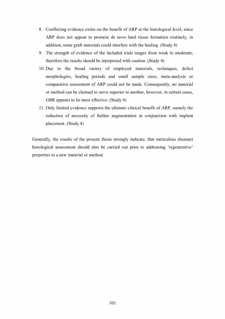
101
8. Conflicting evidence exists on the benefit of ARP at the histological level, since
ARP does not appear to promote de novo hard tissue formation routinely, in
addition, some graft materials could interfere with the healing. (Study 4)
9. The strength of evidence of the included trials ranges from weak to moderate,
therefore the results should be interpreted with caution. (Study 4)
10. Due to the broad variety of employed materials, techniques, defect
morphologies, healing periods and small sample sizes, meta-analysis or
comparative assessment of ARP could not be made. Consequently, no material
or method can be claimed to serve superior to another, however, in certain cases,
GBR appears to be most effective. (Study 4)
11. Only limited evidence supports the ultimate clinical benefit of ARP, namely the
reduction of necessity of further augmentation in conjunction with implant
placement. (Study 4)
Generally, the results of the present thesis strongly indicate, that meticulous (human)
histological assessment should also be carried out prior to addressing ‘regenerative’
properties to a new material or method.

102
8. SUMMARY
Several ‘regenerative’ methods and materials have been claimed as successful in the
field of reconstructive periodontology, implantology and related fields. However, they
do not always appear to promote the formation of new bone, periodontal tissue or
functional attachment, according to the only reliable histological evidences. The aim of
the studies included the present thesis was to investigate histologically some
representative materials and methods, which had shown favourable clinical outcomes
already. In the first scenario we have examined two novel materials used in
periodontology. The investigations demonstrated that the combination of a biphasic
calcium phosphate (Straumann BoneCeramic®; BCP) and enamel matrix derivative
(Emdogain®; EMD) (Study 1) or an unsintered, nanocrystalline, phase-pure
hydroxyapatite (Ostim®; nano-HA) alone (Study 2) for surgical treatment of human
intrabony periodontal defect is safe and well tolerated. Both treatments improved the
investigated clinical parameters, however, they resulted in none to meagre new
cementum and bone formation histologically. In the second scenario we examined the
regenerative potential of BCP with a novel polyethylene glycol-based hydrogel barrier
membrane (Membragel®; PEG) in the field of reconstructive implant dentistry (Study
3). Since it is not feasible to conduct a human histological study due to obvious ethical
considerations, we assessed the regeneration of a critical size, peri-implant dehiscence
defects on Göttingen minipigs. The investigation demonstrated that osseointegration
around hydrophilic dental implants (SLActive®), treated with or without BCP and PEG,
can be achieved and is not negatively affected by functional loading. However, the
amount and quality of the newly formed hard tissue, as well as their attachment to the
implant were not predictably achieved by the applied materials. In the third scenario we
investigated whether or not the bone loss, associated with tooth extraction, could be
prevented, hence to limit the need for peri-implant bone augmentation. Therefore, a
systematic review was conducted in order to obtain the highest level of histological and
clinical evidence (Study 4). Our results demonstrated that different alveolar ridge
preservation (ARP) techniques could limit post extraction shrinkage in width and
height, however the natural resorption cannot be totally eliminated. In addition, ARP
does not predictably promote new bone formation histologically. The results of the
thesis strongly indicate that meticulous (human) histological assessment should also be
carried out prior to addressing ‘regenerative’ properties to a new material or method.
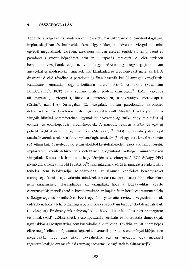
103
9. ÖSSZEFOGLALÁS
Többféle anyagokat és módszereket neveztek már sikeresnek a parodontológiában,
implantológiában és határterületeiken. Ugyanakkor, a szövettani vizsgálatok mint
egyedül megbízhatók tükrében, ezek nem minden esetben segítik elő az új csont és
parodontális szövet képződését, sem az új tapadás létrejöttét. A jelen tézisben
bemutatott vizsgálatok célja az volt, hogy szövettanilag megvizsgáljunk olyan
anyagokat és módszereket, amelyek már klinikailag jó eredményeket mutattak fel. A
disszertáció első részében a parodontológiában használt két új anyagot vizsgáltunk.
Kutatásunk bemutatta, hogy a kétfázisú kalcium foszfát csontpótló (Straumann
BoneCeramic®; BCP) és a zománc mátrix protein (Emdogain®; EMD) együttes
alkalmazása (1. vizsgálat), illetve a szinterezetlen, nanokristályos hidroxilapatit
(Ostim®; nano-HA) önmagában (2. vizsgálat), humán parodontális intraosszer
defektusok sebészi kezelésére biztonságos és jól tolerált. Mindkét kezelés javította a
vizsgált klinikai paramétereket, ugyanakkor szövettanilag nulla, vagy minimális új
cement- és csontképződést eredményeztek. A második részben a BCP és egy új
polietilén-glikol alapú hidrogél membrán (Membragel®; PEG) regeneratív potenciálját
tanulmányoztuk a rekonstruktív implantológia területén (3. vizsgálat) . Mivel itt humán
szövettani kutatás nyilvánvaló etikai okokból kivitelezhetetlen, ezért a kritikus méretű,
implantátum körüli dehiszcencia defektusok gyógyulását Göttingen minisertéseken
vizsgáltuk. Kutatásunk bemutatta, hogy létrejön osszeointegráció BCP és/vagy PEG
membránnal kezelt hidrofil (SLActive®) implantátumok körül és mindezt a funkcionális
terhelés nem befolyásolja. Mindazonáltal az újonnan képződött keményszövet
mennyisége és minősége, valamint mindezek tapadása az implantátum felszínéhez előre
nem kiszámítható. Harmadízben azt vizsgáltuk, hogy a fogeltávolítást követő
csontpusztulás megelőzhető-e, következésképp az implantátum körüli csontaugmentáció
szükségessége csökkenthető-e. Ezért egy ún. systematic review-t végeztünk annak
érdekében, hogy a lehető legmagasabb klinikai és szövettani bizonyítékot demonstráljuk
(4. vizsgálat). Eredményeink bebizonyították, hogy a különféle állcsongerinc-megtartó
technikák (ARP) csökkenthetik a csontpusztulás vertikális és horizontális dimenzióját,
ugyanakkor a csontpusztulás nem küszöbölhető ki teljesen. Továbbá az ARP nem képes
előre megjósolhatóan új csontot képezni szövettanilag. A tézis eredményei kifejezetten
megerősítik, hogy csak akkor nevezhetünk egy új anyagot, vagy módszert
regeneratívnak,ha ezt megfelelő (humán) szövettani vizsgálatok is alátámasztják.
104
10. REFERENCES
1) Papapanou P, Lindhe J. Epidemiology of periodontal diseases. In: Lindhe J, Lang
NP, Karring T (editors), Clinical periodontology and implant dentistry. Fifth
edition. Blackwell Publishing Ltd., Oxford, UK. 2008: 129-179.
2) Hermann P, Gera I, Borbély J, Fejérdy P, Madléna M. (2009) Periodontal health
of an adult population in Hungary: findings of a national survey. J Clin
Periodontol, 36(6): 449-57.
3) Axelsson P, Nyström B, Lindhe J. (2004) The long-term effect of a plaque control
program on tooth mortality, caries and periodontal disease in adults. Results after
30 years of maintenance. J Clin Periodontol, 31(9): 749-57.
4) Linghorne WJ & O'Connell DC. (1950) Studies in the regeneration and
reattachment of supporting structures of the teeth; soft tissue reattachment. J Dent
Res, 29(4): 419-28.
5) Wikesjö UM & Selvig KA. (1999) Periodontal wound healing and regeneration.
Periodontology 2000, 19:21-39. Review.
6) Melcher AH. (1976) On the repair potential of periodontal tissues. J Periodontol,
47(5): 256-60. Review.
7) Dahlin C, Linde A, Gottlow J, Nyman S. (1988) Healing of bone defects by
guided tissue regeneration. Plast Reconstr Surg, 81(5): 672-6.
8) Gottlow J, Nyman S, Karring T, Lindhe J. (1984) New attachment formation as
the result of controlled tissue regeneration. J Clin Periodontol, 11(8): 494-503.
9) Gottlow J, Nyman S, Lindhe J, Karring T, Wennström J. (1986) New attachment
formation in the human periodontium by guided tissue regeneration. Case reports.
J Clin Periodontol, 13(6): 604-16.
10) Nyman S, Lindhe J, Karring T, Rylander H. (1982) New attachment following
surgical treatment of human periodontal disease. J Clin Periodontol, 9(4): 290-6.
11) Polimeni G, Albandar JM, Wikesjö UM. (2005) Prognostic factors for alveolar
regeneration: effect of space provision. J Clin Periodontol, 32(9): 951-4.
12) Susin C, Wikesjö UM. (2013) Regenerative periodontal therapy: 30 years of
lessons learned and unlearned. Periodontol 2000, 62(1): 232-42.
13) Sculean A, Nikolidakis D, Schwarz F. (2008) Regeneration of periodontal tissues:
combinations of barrier membranes and grafting materials - biological foundation
105
and preclinical evidence: a systematic review. J Clin Periodontol, 35(8 Suppl):
106-16. Review.
14) Araújo M, Linder E, Lindhe J. (2009) Effect of a xenograft on early bone
formation in extraction sockets: an experimental study in dog. Clin Oral Implants
Res, 20(1): 1-6.
15) Stavropoulos A, Kostopoulos L, Nyengaard JR, Karring T. (2003) Deproteinized
bovine bone (Bio-Oss) and bioactive glass (Biogran) arrest bone formation when
used as an adjunct to guided tissue regeneration (GTR): an experimental study in
the rat. J Clin Periodontol, 30(7): 636-43.
16) Trombelli L, Lee MB, Promsudthi A, Guglielmoni PG, Wikesjö UM. (1999)
Periodontal repair in dogs: histologic observations of guided tissue regeneration
with a prostaglandin E1 analog/methacrylate composite. J Clin Periodontol, 26(6):
381-7.
17) Bosshardt DD, Sculean A. (2009) Does periodontal tissue regeneration really
work? Periodontol 2000, 51:208-19.
18) Hammarström L. (1997) Enamel matrix, cementum development and
regeneration. J Clin Periodontol, 24(9 Pt 2): 658-68. Review.
19) Hammarström L, Heijl L, Gestrelius S. (1997) Periodontal regeneration in a
buccal dehiscence model in monkeys after application of enamel matrix proteins. J
Clin Periodontol, 24(9 Pt 2): 669-77.
20) Heijl L, Heden G, Svärdström G, Ostgren A. (1997) Enamel matrix derivative
(EMDOGAIN) in the treatment of intrabony periodontal defects. J Clin
Periodontol, 24(9 Pt 2): 705-14.
21) Sculean A, Windisch P, Döri F, Keglevich T, Molnár B, Gera I. (2007) Emdogain
in regenerative periodontal therapy. A review of the literature. Fogorv Sz, 100(5):
220-32, 211-9.
22) Cattaneo V, Rota C, Silvestri M, et al. (2003) Effect of enamel matrix derivative
on human periodontal fibroblasts: Proliferation, morphology and root surface
colonization. An in vitro study. J Periodontal Res, 38:568-574.
23) Haase HR, Bartold PM. (2001) Enamel matrix derivative induces matrix synthesis
by cultured human periodontal ligament cells. J Periodontol, 72:341-348.
24) Lyngstadaas SP, Lundberg E, Ekdahl H, Andersson C, Gestrelius S. Autocrine.
(2001) growth factors in human periodontal ligament cells cultured on enamel
matrix derivative. J Clin Periodontol, 28:181-188.

106
25) Van der Pauw MT, Van den Bos T, Everts V, Beertsen W. (2000) Enamel matrix-
derived protein stimulates attachment of periodontal ligament fibroblasts and
enhances alkaline phosphatase activity and transforming growth factor β1 release
of periodontal ligament and gingival fibroblasts. J Periodontol, 71:31-43.
26) Arweiler NB, Auschill TM, Donos N, Sculean A. (2002) Antibacterial effect of an
enamel matrix protein derivative on in vivo dental plaque vitality. Clin Oral
Investig, 6:205-209.
27) Kawase T, Okuda K, Yoshie H, Burns DM. (2000) Cytostatic action of enamel
matrix derivative (EMDOGAIN) on human oral squamous cell carcinoma-derived
SCC25 epithelial cells. J Periodontal Res, 35:291-300.
28) Sculean A, Auschill TM, Donos N, Brecx M, Arweiler N. (2001) Effect of an
enamel matrix derivative (Emdogain) on ex vivo dental plaque vitality. J Clin
Periodontol, 28:1074-1078.
29) Amin HD, Olsen I, Knowles JC, Donos N. (2012) Differential effect of
amelogenin peptides on osteogenic differentiation in vitro: identification of
possible new drugs for bone repair and regeneration. Tissue Eng Part A, 18(11-
12): 1193-202.
30) Amin HD, Olsen I, Knowles JC, Dard M, Donos N. (2013) Effects of enamel
matrix proteins on multi-lineage differentiation of periodontal ligament cells in
vitro. Acta Biomater, 9(1): 4796-805.
31) Grandin HM, Gemperli AC, Dard M. (2012) Enamel matrix derivative: a review
of cellular effects in vitro and a model of molecular arrangement and functioning.
Tissue Eng Part B Rev, 18(3): 181-202.
32) Sculean A, Chiantella GC, Windisch P, Donos N. (2000) Clinical and histologic
evaluation of human intrabony defects treated with an enamel matrix protein
derivative (Emdogain). Int J Periodontics Restorative Dent, 20: 374-381.
33) Sculean A, Chiantella GC, Windisch P, Gera I, Reich E. (2002) Clinical
evaluation of an enamel matrix protein derivative (Emdogain) combined with a
bovine-derived xenograft (Bio-Oss) for the treatment of intrabony periodontal
defects in humans. Int J Periodontics Restorative Dent, 22(3): 259-67.
34) Sculean A, Windisch P, Keglevich T, Chiantella GC, Gera I, Donos N. (2003)
Clinical and histologic evaluation of human intrabony defects treated with an
enamel matrix protein derivative combined with a bovine-derived xenograft. Int J
Periodontics Restorative Dent, 23(1): 47-55.

107
35) Sculean A, Windisch P, Keglevich T, Gera I. (2005) Clinical and histologic
evaluation of an enamel matrix protein derivative combined with a bioactive glass
for the treatment of intrabony periodontal defects in humans. Int J Periodontics
Restorative Dent, 25(2):139-47.
36) Windisch P, Sculean A, Klein F, Tóth V, Gera I, Reich E, Eickholz P. (2002)
Comparison of clinical, radiographic and histometric measurements following
treatment with guided tissue regeneration or enamel matrix proteins in human
periodontal defects. J Periodontol, 73: 409-417.
37) Esposito M, Grusovin MG, Papanikolaou N, Coulthard P, Worthington HV.
(2009) Enamel matrix derivative (Emdogain®) for periodontal tissue regeneration
in intrabony defects. Cochrane Database Syst Rev, 7;(4): CD003875.
38) Guida L, Annunziata M, Belardo S, Farina R, Scabbia A, Trombelli L. (2007)
Effect of autogenous cortical bone particulate in conjunction with enamel matrix
derivative in the treatment of periodontal intraosseous defects. J Periodontol,
78(2): 231-8.
39) Lekovic V, Camargo PM, Weinlaender M, Nedic M, Aleksic Z, Kenney EB.
(2000) A comparison between enamel matrix proteins used alone or in
combination with bovine porous bone mineral in the treatment of intrabony
periodontal defects in humans. J Periodontol, 71(7): 1110-6.
40) Gurinsky BS, Mills MP, Mellonig JT. (2004) Clinical evaluation of demineralized
freeze-dried bone allograft and enamel matrix derivative versus enamel matrix
derivative alone for the treatment of periodontal osseous defects in humans. J
Periodontol, 75(10): 1309-18.
41) Sculean A, Pietruska M, Schwarz F, Willershausen B, Arweiler NB, Auschill TM.
(2005) Healing of human intrabony defects following regenerative periodontal
therapy with an enamel matrix protein derivative alone or combined with a
bioactive glass. A controlled clinical study. J Clin Periodontol, 32(1): 111-7.
42) Cochran DL, Jones A, Heijl L, Mellonig JT, Schoolfield J, King GN. (2003)
Periodontal regeneration with a combination of enamel matrix proteins and
autogenous bone grafting. J Periodontol, 74(9): 1269-81.
43) Bowers GM, Chadroff B, Carnevale R, Mellonig J, Corio R, Emerson J, Stevens
M, Romberg E. (1989) Histologic evaluation of new attachment apparatus in
humans. Part I J Periodontol 60:675–682.
108
44) Brunsvold, M.A. & Mellonig, J.T. (1993) Bone grafts and periodontal
regeneration. Periodontology 2000 1, 80-91.
45) Dragoo MR & Sullivan HC. (1973) A clinical and histological evaluation of
autogenous iliac bone grafts in humans. I. Wound healing 2 to 8 months. J
Periodontol 45:599–613.
46) Hiatt WH, Schallhorn RG, Aaronian AJ. (1978) The induction of new bone and
cementum formation. IV. Microscopic examination of the periodontium following
human bone and marrow allograft, autograft and non-grafted periodontal
regenerative procedures. J Periodontol 49:495–512.
47) Nevins ML, Camelo M, Lynch SE, Schenk RK, Nevins M. (2003) Evaluation of
periodontal regeneration following grafting intrabony defects with Bio-Oss
Collagen: a human histologic report. Int J Periodontics Restorative Dent 23:9–17.
48) Mellonig JT. (2000) Human histologic evaluation of a bovine derived bone
xenograftin the treatment of periodontal osseous defects. Int J Periodontics
Restorative Dent 20, 18-29.
49) Buck BE, Resnick L, Shah SM, Malinin TI. (1990) Human immunodeficiency
virus cultured from bone. Implications for transplantation. Clin Orthop 251:249–
253.
50) Nemzek JA, Arnoczky SP, Swenson CL. (1996) Retroviral transmission in bone
allotransplantation. The effects of tissue processing. Clin Orthop 324:275–282.
51) Barbé, G., Sculean, A. & Chiantella, G.C. (2001) Vertical reconstruction of
horizontal periodontal defect using a bioactive glass: a case report with a 30
months follow up. Journal de Parodontologie et d`Implantologie Orale 20, 51-60.
52) Bowen, J., Mellonig, J., Gray, J. & Towle, H. (1989) Comparison of decalcified
freeze-dried bone allograft and porous hydroxyapatite in human periodontal
osseous defects. J Periodontol 60, 647-544.
53) Gera I, Döri F, Keglevich T, Anton S, Szilágyi E, Windisch P. (2002) Experience
with the clinical use of beta-tri-calcium phosphate (Cerasorb) as a bone
replacement graft material in human periodontal osseous defects. Fogorv Sz,
95(4): 143-7.
54) Meffert, R., Thomas, J., Hamilton, K. & Browstein, C. (1985) Hydroxyapatite as
an alloplastic graft in the treatment of human periodontal osseous defects. J
Periodontol 56, 63-73.

109
55) Trombelli L, Heitz-Mayfield LJA, Needleman I, Moles D, Scabbia A. (2002) A
systematic review of graft materials and biological agents for periodontal
intraosseous defects. J Clin Periodontol 29(suppl 3):117–135.
56) Yukna RA (1993) Synthetic bone grafts in periodontics. Periodontol 2000(1):92–
99
57) Froum SJ. Kushner L. Scopp IW. (1982) Stahl SS. Human clinical and histologic
responses to Durapatite implants in intraosseous lesions. Case reports. J
Periodontol, 53(12): 719-25.
58) Froum S, Stahl SS (1987) Human intraosseous healing response to the placement
of tricalcium phosphate ceramic implants. II. 13 to 18 months. J Periodontol
58:103–109.
59) Galgut PN. Waite IM. Brookshaw JD. Kingston CP. (1992) A 4-year controlled
clinical study into the use of a ceramic hydroxylapatite implant material for the
treatment of periodontal bone defects. J Clin Periodontol, 19(8): 570-7.
60) Nery EB, LeGeros RZ, Lynch KL, Lee K. (1992) Tissue response to biphasic
calcium phosphate ceramic with different ratios of HA/ßTCP in periodontal
osseous defects. J Periodontol, 78:2216-2225.
61) Nevins ML, Camelo M, Nevins M, King CJ, Oringer RJ, Schenk RK, Fiorellini
JP. (2000) Human histologic evaluation of bioactive ceramic in the treatment of
periodontal defects. Int J Periodont Rest Dent, 20:458–467.
62) Saffar JL, Colombier ML, Detienville R. (1990) Bone formation in tricalcium
phosphate-filled periodontal intrabony lesions. Histologic observations in humans.
J Periodontol, 61:209–216.
63) Stahl S, Froum S. (1986) Histologic evaluation of human intraosseous healing
responses to the placement of tricalcium phosphate ceramic implants. I. Three to
eight months. J Periodontol 57:211–217.
64) Stahl SS, Froum S. (1987) Histologic and clinical responses to porous
hydroxylapatite implants in human periodontal defects. Three to twelve months
postimplantation. J Periodontol, 58:689–695.
65) Stavropoulos A, Windisch P, Szendröi-Kiss D, Rosta P, Gera I, Sculean A. (2010)
Clinical and histologic evaluation of granular Beta-tricalcium phosphate for the
treatment of human intrabony periodontal defects: a report on five cases. J
Periodontol, 81(2):325-34.

110
66) Sculean A, Nikolidakis D, Chiantella GC, Schwarz F and Stavropoulos A.
Combination strategies to enhance regenerative periodontal surgeries. In: Sculean
A (editor), Periodontal Regenerative Therapy. Quintessence Publishing Co. Ltd.,
New Maiden, UK. 2010:147-157.
67) Proceedings of the World Workshop in Periodontics. The American Academy of
Periodontology. Ann Periodontol 1996;1:618-670.
68) Karring T. (2000) Regenerative periodontal therapy. J Int Acad Periodontol, 2(4):
101-9. Review.
69) Stavropoulos A, Windisch P, Gera I, Capsius B, Sculean A, Wikesjö UME. (2011)
A phase IIa randomized controlled clinical and histological pilot study evaluating
rh GDF-5/β- TCP for periodontal regeneration. J Clin Periodontal, 38:(11) pp.
1044-1054.
70) Windisch P, Stavropoulos A, Molnár B, Szendröi-Kiss D, Szilágyi E, Rosta P,
Horváth A, Capsius B, Wikesjö UM, Sculean A. (2012) A phase IIa randomized
controlled pilot study evaluating the safety and clinical outcomes following the
use of rhGDF-5/β-TCP in regenerative periodontal therapy. Clin Oral Investig,
16:(4) 1181-1189.
71) Sculean A, Windisch P, Chiantella G. (2004) C. Human histologic evaluation of
an intrabony defect treated with enamel matrix derivative, xenograft, and GTR. Int
J Periodontics Restorative Dent, 24:(4) pp. 326-333. (2004).
72) Stavropoulos A, Chiantella G, Costa D, Steigmann M, Windisch P, Sculean,
(2011) A Clinical and histologic evaluation of a granular bovine bone biomaterial
used as an adjunct to GTR with a bioresorbable bovine pericardium collagen
membrane in the treatment of intrabony defects. J Peridontol, 82:(3) pp. 462-470.
73) Sculean A, Windisch P, Szendroi-Kiss D, Horvath A, Rosta P, Becker J, Gera I,
Schwarz F. (2008) Clinical and histologic evaluation of an enamel matrix
derivative combined with a biphasic calcium phosphate for the treatment of
human intrabony periodontal defects. J Periodontol, 79:(10) pp. 1991-1999.
74) Horvath A, Stavropoulos A, Windisch P, Lukacs L, Gera I, Sculean A. (2013)
Histological evaluation of human intrabony periodontal defects treated with an
unsinterednanocrystalline hydroxyapatite paste. Clin Oral Investig 17:(2) pp. 423-
430.
111
75) Zambon R, Mardas N, Horvath A, Petrie A, Dard M, Donos N. The effect of
loading in regenerated bone in dehiscence defects following a combined approach
of bone grafting and GBR. Clin Oral Implants Res 23:(5) pp. 591-601. (2012).
76) Horváth A, Mardas N, Mezzomo LA, Needleman IG, Donos N. (2013) Alveolar
ridge preservation. A systematic review Clin Oral Investig 17:(2) pp. 341-363.
77) Döri F, Arweiler N, Gera I, Sculean A. (2005) Clinical evaluation of an enamel
matrix protein derivative combined with either a natural bone mineral or beta-
tricalcium phosphate. J Periodontol, 76:2236-2243.
78) Jensen SS, Yeo A, Dard M, Hunziker E, Schenk R, Buser D. (2007) Evaluation of
a novel biphasic calcium phosphate in standardized bone defects: A histologic and
histomorphometric study in the mandibles of minipigs. Clin Oral Implants Res,
18: 752-760.
79) Schwarz F, Herten M, Ferrari D, Wieland M, Schmitz L, Engelhardt E, Becker J.
(2007) Guided bone regeneration at dehiscence-type defects: using biphasic
hydroxyapatite + beta tricalcium phosphate (Bone Ceramic) or a collagen-coated
natural bone mineral (BioOss Collagen): An immunohistochemical study in dogs.
Int J Oral Maxillofac Surg, 36:1198-1206.
80) Mangano C, Piattelli A, Perrotti V, Iezzi G. (2008) Dense hydroxyapatite inserted
into postextraction sockets: a histologic and histomorphometric 20-year case
report. J Periodontol, 79(5):929-33.
81) Doi Y, Iwanaga H, Shibutani T, Moriwaki Y, Iwayama J. (1999) Osteoclastic
responses to various calcium phosphates in cell cultures Biomed Mater Res, 47,
424–433.
82) Rauschmann MA, Wichelhaus TA, Stirnal V, Dingeldein E, Zichner L, Schnettler
R, Alt V. (2005) Nanocrystalline hydroxyapatite and calcium sulphate as
biodegradable composite carrier material for local delivery of antibiotics in bone
infections. Biomaterials, 26:2677-2684.
83) Kilian O, Wenisch S, Alt V, Lauer M, Fuhrmann R, Dingeldein E, Jonuleit T,
Schnettler R and Franke RP. (2007) Effects of platelet factors on biodegradation
and osteogenesis in metaphyseal defects filled with nanoparticular hydroxyapatite-
-an experimental study in minipigs. Growth Factors. 25(3): 191-201.
84) Tadic D & Epple M. (2004) thorough physicochemical characterisation of 14
calcium phosphate-based bone substitution materials in comparison to natural
bone. Biomaterials. 25(6): 987-94.
112
85) Thorwarth M. Schultze-Mosgau S. Kessler P. Wiltfang J. Schlegel KA. (2005)
Bone regeneration in osseous defects using a resorbable nanoparticular
hydroxyapatite. Int J Oral Maxillofac Surg. 63(11): 1626-33.
86) Kasaj A. Willershausen B. Reichert C. Rohrig B. Smeets R. Schmidt M. (2008)
Ability of nanocrystalline hydroxyapatite paste to promote human periodontal
ligament cell proliferation. Journal of Oral Science, 50(3): 279-85.
87) Kasaj A. Willershausen B. Reichert C. Gortan-Kasaj A. Zafiropoulos GG. (2008)
Schmidt M. Human periodontal fibroblast response to a nanostructured
hydroxyapatite bone replacement graft in vitro. Arch Oral Biol, 53(7): 683-9.
88) Kasaj A, Willershausen B, Junker R, Stratul SI, Schmidt M. (2012) Human
periodontal ligament fibroblasts stimulated by nanocrystalline hydroxyapatite
paste or enamel matrix derivative. An in vitro assessment of PDL attachment,
migration, and proliferation. Clin Oral Investig, 16(3): 745-54.
89) Sun W, Chu C, Wang J, Zhao H. (2007) Comparison of periodontal ligament cells
responses to dense and nanophase hydroxyapatite. J Mater Science-Mater in Med,
18(5): 677-83.
90) Busenlechner D. Tangl S. Mair B. Fugger G. Gruber R. Redl H. Watzek G. (2008)
Simultaneous in vivo comparison of bone substitutes in a guided bone
regeneration model. Biomaterials, 29(22): 3195-200.
91) Kilian O, Alt V, Heiss C, Jonuleit T, Dingeldein E, Flesch I, Fidorra U, Wenisch
S, Schnettler R. (2005) New blood vessel formation and expression of VEGF
receptors after implantation of platelet growth factor-enriched biodegradable
nanocrystalline hydroxyapatite. Growth Factors, 23(2): 125-33.
92) Kilian O, Wenisch S, Karnati S, Baumgart-Vogt E, Hild A, Fuhrmann R, Jonuleit
T, Dingeldein E, Schnettler R, Franke RP. (2008) Observations on the
microvasculature of bone defects filled with biodegradable nanoparticulate
hydroxyapatite. Biomaterials, 29(24-25): 3429-37.
93) Laschke MW. Witt K. Pohlemann T. Menger MD. (2007) Injectable
nanocrystalline hydroxyapatite paste for bone substitution: in vivo analysis of
biocompatibility and vascularization. Journal of Biomedical Materials Research.
Part B, Applied Biomaterials. 82(2): 494-505.
94) Huber FX, McArthur N, Hillmeier J, Kock HJ, Baier M, Diwo M, Berger I, and
Meeder PJ. (2006) Void filling of tibia compression fracture zones using a novel
resorbable nanocrystalline hydroxyapatite paste in combination with a

113
hydroxyapatite ceramic core: first clinical results. Archives of Orthopaedic &
Trauma Surgery, 126(8): 533-40.
95) Huber FX, J. Hillmeier, L. Herzog, N. McArthur, H. J. Kock, and P. J. Meeder.
(2006) Open reduction and palmar plate-osteosynthesis in combination with a
nanocrystalline hydroxyapatite spacer in the treatment of comminuted fractures of
the distal radius. Journal of Hand Surgery - British Volume, 31(3): 298-303.
96) Huber FX, O. Belyaev, J. Hillmeier, H. J. Kock, C. Huber, P. J. Meeder, and I.
Berger. (2006) First histological observations on the incorporation of a novel
nanocrystalline hydroxyapatite paste OSTIM in human cancellous bone. BMC
Musculoskeletal Disorders, 7:50.
97) Huber FX, J. Hillmeier, H. J. Kock, N. McArthur, C. Huber, M. Diwo, M. Baier,
and P. J. Meeder. (2008) [Filling of metaphyseal defects with nanocrystalline
hydroxyapatite (Ostim) for fractures of the radius]. [German]. Zentralblatt fur
Chirurgie, 133(6): 577-81.
98) Bezrukov VM. Grigor'iants LA. Zuev VP. Pankratov AS. (1998) [The surgical
treatment of jaw cysts using hydroxyapatite with an ultrahigh degree of
dispersity]. [Russian] Stomatologiia. 77(1): 31-5.
99) Canuto RA, Pol R, Martinasso G, Muzio G, Gallesio G, Mozzati M. (2011)
Hydroxyapatite paste Ostim(®) , without elevation of full-thickness flaps,
improves alveolar healing stimulating BMP- and VEGF-mediated signal
pathways: an experimental study in humans. Clin Oral Implants Res. 2013;24
Suppl A100:42-8.
100) Gerlach KL, Niehues D. (2007) Die Behandlung der Kieferzysten mit einem
neuartigen nanopartikulären Hydroxylapatit. Mund Kiefer GesichtsChir, 11:131–
137.
101) Smeets R. Grosjean MB. Jelitte G. Heiland M. Kasaj A. Riediger D. Yildirim M.
(2008) Spiekermann H. Maciejewski Hydroxyapatite bone substitute (Ostim) in
sinus floor elevation. Maxillary sinus floor augmentation: bone regeneration by
means of a nanocrystalline in-phase hydroxyapatite (Ostim)]. [French, German]
Schweizer Monatsschrift fur Zahnmedizin, 118(3): 203-12.
102) F. Schwarz, K. Bieling, T. Latz, E. Nuesry, and J. Becker. (2006) Healing of
intrabony peri-implantitis defects following application of a nanocrystalline
hydroxyapatite (Ostim) or a bovine-derived xenograft (Bio-Oss) in combination
114
with a collagen membrane (Bio-Gide). A case series. J Clin Periodont, 33(7): 491-
9.
103) Strietzel FP. Reichart PA. Graf HL. (2007) Lateral alveolar ridge augmentation
using a synthetic nano-crystalline hydroxyapatite bone substitution material
(Ostim): preliminary clinical and histological results. Clin Oral Implants Res,
18(6): 743-51.
104) Zuev VP. Dmitrieva LA. Pankratov AS. Filatova NA. (1996) [The comparative
characteristics of stimulators of reparative osteogenesis in the treatment of
periodontal diseases]. [Russian] Stomatologiia, 75(5): 31-4.
105) Kasaj A. Rohrig B. Zafiropoulos GG. Willershausen B. (2008) Clinical evaluation
of nanocrystalline hydroxyapatite paste in the treatment of human periodontal
bony defects--a randomized controlled clinical trial: 6-month results. J
Periodontol, 79(3): 394-400.
106) Heinz B, Kasaj A, Teich M, Jepsen S (2010) Clinical effects of nanocrystalline
hydroxyapatite paste in the treatment of intrabony periodontal defects: a
randomized controlled clinical study. Clin Oral Investig, 14:525–531.
107) Löe H. (1967) The Gingival Index, the Plaque Index and the Retention Index
system. J Periodontol, 38:610-616.
108) O`Leary TJ, Drake RB, Naylor JE (1972) The plaque control record. J Periodontol
43:38.
109) Lang NP, Joss A, Orsanic T, Gusberti FA, Siegrist BE. (1986) Bleeding on
probing. A predictor for the progression of periodontal disease? J Clin
Periodontol, 13:590–596
110) Sewerin I. (1990) Device for serial intraoral radiography with controlled
projection angles. Tandlaegebladet 94:613–617.
111) Blomlöf JPS, Blomlöf LB, Lindskog SF. (1996) Smear removal and collagen
exposure after non-surgical root planing followed by etching with EDTA gel
preparation. J Periodontol, 67:841-845.
112) Windisch P, Szendroi-Kiss D, Horvath A, Suba Z, Gera I, Sculean A. (2008)
Reconstructive periodontal therapy with simultaneous ridge augmentation.A
clinical and histological case series report. Clin Oral Invest, 12:(3) pp. 257-264.
113) Bosshardt DD, Sculean A, Windisch P, Pjetursson BE, Lang NP. (2005) Effects of
enamel matrix proteins on tissue formation along the roots of human teeth. J
Periodontal Res, 40(2): 158-67.
115
114) Mellonig JT. (1999) Enamel matrix derivative for periodontal reconstructive
surgery: Technique and clinical and histologic case report. Int J Periodontics
Restorative Dent, 19:8-19.
115) Sculean A, Windisch P, Chiantella GC, Donos N, Brecx M, Reich E. (2001)
Treatment of intrabony defects with enamel matrix proteins and guided tissue
regeneration. A prospective controlled clinical study. J Clin Periodontol, 28:397-
403.
116) Silvestri M, Ricci G, Rasperini G, Sartori S, Cattaneo V. (2000) Comparison of
treatments of intrabony defects with enamel matrix derivative, guided tissue
regeneration with a nonresorbable membrane and Widman modified flap. A pilot
study. J Clin Periodontol, 27:603-610.
117) Tonetti MS, Lang NP, Cortellini P, Suvan JE, Adriaens P, Dubravec D, Fonzar A,
Fourmousis I, Mayfield L, Rossi R, Silvestri M, Tiedemann C, Topoll H,
Vangsted T, Wallkamm B. (2002) Enamel matrix proteins in the regenerative
therapy of deep intrabony defects. J Clin Periodontol, 29(4): 317-25.
118) Zafiropoulos GGK, Hoffmann O, Kasaj A, Willershausen B, Weiss O, Van Dyke
TE. (2007) Treatment of intrabony defects using guided tissue regeneration and
autogenous spongiosa alone or combined with hydroxyapatite/β-tricalcium
phosphate bone substitute or bovine derived xenograft. J Periodontol, 78:2216-
2225.
119) Jepsen S, Topoll H, Rengers H, Heinz B, Teich M, Hoffmann T, Al-Machot E,
Meyle J, Jervøe-Storm PM. (2008) Clinical outcomes after treatment of intra-bony
defects with an EMD/synthetic bone graft or EMD alone: a multicentre
randomized-controlled clinical trial. J Clin Periodontol, 35(5): 420-8.
120) Pietruska M, Pietruski J, Nagy K, Brecx M, Arweiler NB, Sculean A. (2012)
Four-year results following treatment of intrabony periodontal defects with an
enamel matrix derivative alone or combined with a biphasic calcium phosphate.
Clin Oral Investig, 16(4): 1191-7.
121) Jensen SS, Chen B, Bornstein MM, Bosshardt DD, Buser D. (2011) Effect of
enamel matrix derivative and parathyroid hormone on bone formation in
standardized osseous defects: an experimental study in minipigs. J Periodontol,
82(8): 1197-205.

116
122) Piattelli A, Scarano A, Mangano C. (1996) Clinical and histologic aspects of
biphasic calcium phosphate ceramic (BCP) used in connection with implant
placement. Biomaterials, 17:1767-1770.
123) Sager M, Ferrari D, Wieland M, Dard M, Becker J, Schwarz F. (2012)
Immunohistochemical characterization of wound healing at two different bone
graft substitutes. Int J Oral Maxillofac Surg, 41(5): 657-66.
124) Chitsazi MT, Shirmohammadi A, Faramarzie M, Pourabbas R, Rostamzadeh An.
(2011) A clinical comparison of nano-crystalline hydroxyapatite (Ostim) and
autogenous bone graft in the treatment of periodontal intrabony defects. Med Oral
Patol Oral Cir Buccal, 16(3): e448-53.
125) Caton JG Greenstein GG. (1993) Factors related to periodontal regeneration.
Periodontol 2000, 1:9–15.
126) Adell, R., Lekholm, U., Rockler, B. & Brånemark, P. I. (1981) A 15-year study of
osseointegrated implants in the treatment of the edentulous jaw. International
Journal of Oral Surgery 10, 387–416.
127) Donos N, Mardas N, Chadha V. (2008) Clinical outcomes of implants following
lateral bone augmentation: systematic assessment of available options (barrier
membranes, bone grafts, split osteotomy). J Clin Periodontol, 35 (Suppl. 8): 173–
202.
128) Albrektsson, T., Brånemark, P.-I, Hansson, H.-A. & Lindström, J. (1981)
Osseointegrated Titanium Implants: Requirements for Ensuring a Long-Lasting,
Direct Bone-to-Implant Anchorage in Man. Acta Orthopaedica, 52: 155-170.
129) Amler, M.H. (1969) The time sequence of tissue regeneration in human extraction
wounds. Oral Surgery, Oral Medicine and Oral Pathology 27:309–318.
130) Araújo, M.G. & Lindhe, J. (2005) Dimensional ridge alterations following tooth
extraction. An experimental study in the dog. J Clin Periodontol, 32:212–218.
131) Schropp, L., Wenzel, A., Kostopoulos, L. & Karring, T. (2003) Bone healing and
soft tissue contour changes following single-tooth extraction: a clinical and
radiographic 12-month prospective study. International Journal of Periodontics &
Restorative Dentistry, 23: 313–323.
132) Joób FA. Kerekes F. Koppány F. Pfeifer B. Divinyi T. (2007) Changes in the
indications for oral surgical implants based on statistical analysis. [Hungarian]
Fogorvosi Szemle, 100(3):103-7.

117
133) Belser UC, Buser D, Hess D, Schmid B, Bernard JP, Lang NP. (1998) Aesthetic
implant restorations in partially edentulous patients—A critical appraisal.
Periodontol 2000, 17:132–150.
134) Chiapasco, M., Zaniboni, M. & Boisco, M. (2006) Augmentation procedures for
the rehabilitation of deficient edentulous ridges with oral implants. Clin Oral
Implants Res, 17(Suppl 2): 136–159.
135) Clementini M, Morlupi A, Canullo L, Agrestini C, Barlattani A. (2012) Success
rate of dental implants inserted in horizontal and vertical guided bone regenerated
areas: a systematic review. Int J Oral Maxillofac Surg, 41(7): 847-52.
136) Retzepi M, Donos N. (2010) Guided Bone Regeneration: biological principle and
therapeutic applications. Clin Oral Implants Res, 21(6): 567-76.
137) Buser, D., Dula, K., Belser, U. C., Hirt, H. P. & Berthold, H. (1995) Localized
ridge augmentation using guided bone regeneration. II. Surgical procedure in the
mandible. International Journal of Periodontics and Restorative Dentistry 15, 10–
29.
138) Buser, D., Dula, K., Hirt, H. P. & Schenk, R. K. (1996) Lateral ridge
augmentation using autografts and barrier membranes: a clinical study with 40
partially edentulous patients. J Oral Maxillofac Surgery 54, 420–432.
139) Becker, W., Becker, B. E., Handlesman, M., Celletti, R., Ochsenbein, C.,
Hardwick, R. & Langer, B. (1990) Bone formation at dehisced dental implant sites
treated with implant augmentation material: a pilot study in dogs. International
Journal of Periodontics and Restorative Dentistry 10, 92–101.
140) Buser D. Chen ST. Weber HP. Belser UC. (2008) Early implant placement
following single-tooth extraction in the esthetic zone: biologic rationale and
surgical procedures. International Journal of Periodontics & Restorative Dentistry,
28(5): 441-51.
141) Buser D, Wittneben J, Bornstein MM, Grütter L, Chappuis V, Belser UC. (2011)
Stability of contour augmentation and esthetic outcomes of implant-supported
single crowns in the esthetic zone: 3-year results of a prospective study with early
implant placement postextraction. J Periodontol, 82(3): 342-9.
142) Dahlin, C., Sennerby, L., Lekholm, U., Linde, A. & Nyman, S. (1989) Generation
of new bone around titanium implants using a membrane technique: an
experimental study in rabbits. Int J Oral Maxillofac Implants, 4, 19–25.

118
143) Hämmerle CH, Jung R, Feloutzis A. (2002) A systematic review of the survival of
implants in bone sites augmented with barrier membranes (guided bone
regeneration) in partially edentulous patients. Journal of Clinical Periodontology
29, (Suppl. 3) 226-231.
144) Buser D, Bragger U, Lang NP, Nyman S. (1990) Regeneration and enlargement of
jaw bone using guided tissue regeneration. Clin Oral Implants Res, 1(1): 22-32.
145) Dahlin C. Linde A. Gottlow J. Nyman S. (1988) Healing of bone defects by
guided tissue regeneration. Plastic & Reconstructive Surgery, 81(5): 672-6.
146) Dahlin C. Gottlow J. Linde A. Nyman S. (1990) Healing of maxillary and
mandibular bone defects using a membrane technique. An experimental study in
monkeys. Scandinavian Journal of Plastic & Reconstructive Surgery & Hand
Surgery, 24(1): 13-9.
147) Donos, N., Lang, N.P., Karoussis, I.K., Bosshardt, D., Tonetti, M. & Kostopoulos,
L. (2004) Effect of GBR in combination with deproteinized bovine bone mineral
and/or enamel matrix proteins on the healing of critical-size defects. Clin Oral
Implants Res, 15: 101–111.
148) Buser, D., Dula, K., Belser, U., Hirt, H.P. & Berthold, H. (1993) Localized ridge
augmentation using guided bone regeneration. 1. Surgical procedure in the
maxilla. The International Journal of Periodontics & Restorative Dentistry, 13:
29–45.
149) Donos, N., Kostopoulos, L. & Karring, T. (2002) Alveolar ridge augmentation by
combining autogenous mandibular bone grafts and non-resorbable membranes.
Clin Oral Implants Res, 13: 185–191.
150) Donos, N., Kostopoulos, L. & Karring, T. (2002) Augmentation of the rat jaw
with autogeneic cortico-cancellous bone grafts and guided tissue regeneration.
Clin Oral Implants Res, 13: 192–202.
151) Nevins M, Mellonig JT. (1992) Enhancement of the damaged edentulous ridge to
receive dental implants: a combination of allograft and the GORE-TEX
membrane. Int J Periodontics Restorative Dent, 12(2):96-111.
152) Urban IA, Jovanovic SA, Lozada JL. Vertical ridge augmentation using guided
bone regeneration (GBR) in three clinical scenarios prior to implant placement: a
retrospective study of 35 patients 12 to 72 months after loading. Int J Oral
Maxillofac Implants, 2009 May-Jun;24(3):502-10.

119
153) Jovanovic SA, Spiekermann H, Richter EJ. (1992) Bone regeneration around
titanium dental implants in dehisced defect sites: a clinical study. Int J Oral
Maxillofac Implants, 7(2):233-45.
154) Simion, M., Trisi, P. & Piattelli, A. (1994) Vertical ridge augmentation using a
membrane technique associated with osseointegrated implants. The International
Journal of Periodontics & Restorative Dentistry, 14: 496–511.
155) Zitzmann NU, Schärer P, Marinello CP. (1999) Factors influencing the success of
GBR. Smoking, timing of implant placement, implant location, bone quality and
provisional restoration. J Clin Periodontol, 26: 673–682.
156) Lorenzoni, M., Pertl, C., Keil, C. & Wegscheider, W.A. (1998) Treatment of peri-
implant defects with guided bone regeneration: A comparative clinical study with
various membranes and bone grafts. Int J Oral Maxillofac Implants, 13: 639–646.
157) Urban IA. Nagursky H. Lozada JL. (2011) Horizontal ridge augmentation with a
resorbable membrane and particulated autogenous bone with or without anorganic
bovine bone-derived mineral: a prospective case series in 22 patients. Int J Oral
Maxillofac Implants, 26(2): 404-14.
158) Urban IA. Nagursky H. Lozada JL. Nagy K. (2013) Horizontal ridge
augmentation with a collagen membrane and a combination of particulated
autogenous bone and anorganic bovine bone-derived mineral: a prospective case
series in 25 patients. International Journal of Periodontics & Restorative Dentistry.
33(3): 299-307.
159) Zitzmann, N.U., Naef, R. & Schärer, P. (1997) Resorbable versus nonresorbable
membranes in combination with bio-oss for guided bone regeneration. Int J Oral
Maxillofac Implants, 12: 844–852.
160) Hämmerle, C.H. & Jung, R.E. (2003) Bone augmentation by means of barrier
membranes. Periodontology 2000, 33: 36–53.
161) Moore, J.W. & Brekke, J.H. (1990) Foreign body giant cell reaction related to
placement of tetracycline-treated polylactic acid: Report of 18 cases. Journal of
Oral and Maxillofacial Surgery: Official Journal of the American Association of
Oral and Maxillofacial Surgeons, 48: 808–812.
162) Mettler, L., Audebert, A., Lehmann-Willenbrock, E., Schive, K. & Jacobs, V.R.
(2003) Prospective clinical trial of SprayGel as a barrier to adhesion formation: an
interim analysis. J Am Assoc Gynecol Laparosc, 10: 339–344.

120
163) Pang, S.N.J. (1993) Final report on the safety assessment of polyethylene glycols
(PEGs) -6, -8, -32, -75, -150, -14M, -20M. Journal of the American College of
Toxicology, 12: 429–456.
164) Herten, M., Jung, R.E., Ferrari, D., Rothamel, D., Golubovic, V., Molenberg, A.,
Hämmerle, C.H., Becker, J. & Schwarz, F. (2008) Biodegradation of different
synthetic hydrogels made of polyethylene glycol hydrogel/rgd-peptide
modifications: An immunohistochemical study in rats. Clin Oral Implants Res, 20:
116–125.
165) Wechsler, S., Fehr, D., Molenberg, A., Raeber, G., Schense, J.C. & Weber, F.E.
(2008) A novel, tissue occlusive poly(ethylene glycol) hydrogel material. J
Biomedical Materials Res Part A, 85: 285–292.
166) Thoma, D.S., Halg, G.A., Dard, M.M., Seibl, R., Hammerle, C.H. & Jung, R.E.
(2009) Evaluation of a new biodegradable membrane to prevent gingival ingrowth
into mandibular bone defects in minipigs. Clin Oral Implants Res, 20: 7–16.
167) Jung, R.E., Zwahlen, R., Weber, F.E., Molenberg, A., van Lenthe, G.H. &
Hammerle, C.H. (2006) Evaluation of an in situ formed synthetic hydrogel as a
biodegradable membrane for guided bone regeneration. Clin Oral Implants Res,
17: 426–433.
168) Jung, R.E., Lecloux, G., Rompen, E., Ramel, C.F., Buser, D. & Hammerle, C.
(2009) A feasibility study evaluating an in situ formed synthetic biodegradable
membrane for guided bone regeneration in dogs. Clin oral implants res, 20: 151–
161.
169) Jung, R.E., Hälg, G., Thoma, D.S. & Hämmerle, C. (2009) A randomized,
controlled clinical trial to evaluate a new membrane for guided bone regeneration
around dental implants. Clin Oral Implants Res, 20: 162–168.
170) Schwarz, F., Jung, R.E., Fienitz, T., Wieland, M., Becker, J. & Sager, M. (2010)
Impact of guided bone regeneration and defect dimension on wound healing at
chemically modified hydrophilic titanium surfaces: an experimental study in dogs.
Journal of Clinical Periodontology, 37: 474–485.
171) Buser, D., Ruskin, J., Higginbottom, F., Hardwick, R., Dahlin, C. & Schenk, R.K.
(1995) Osseointegration of titanium implants in bone regenerated in membrane-
protected defects: a histologic study in the canine mandible. Int J Oral Maxillofac
Implants. 1995;10(6):666-81.
121
172) Donos, N., Kostopoulos, L., Tonetti, M. & Karring, T. (2005) Long-term stability
of autogenous bone grafts following combined application with guided bone
regeneration. Clin Oral Implants Res, 16: 133–139.
173) Becker, W., Lekholm, U., Dahlin, C., Becker, B.E. & Donath, K. (1994) The
effect of clinical loading on bone regenerated by GTAM barriers: A study in dogs.
INT J ORAL MAXILLOFAC IMPLANTS, 9:305–3139: 305–313.
174) Donos N, Horvath A, Dedi KD, Mezzomo L, Mardas N. (2012) The role of
immediate provisional restorations on bone level implants: A randomised,
controlled clinical trial. J Clin Periodontol, s13:50.
175) Donath, K. & Breuner, G. (1982) A method for the study of undecalcified bones
and teeth with attached soft tissues. The säge-schliff (sawing and grinding)
technique. Journal of Oral Pathology, 11: 318–326.
176) Wikesjö, U.M.E. & Nilvéus, R. (1991) Periodontal repair in dogs Healing patterns
in large circumferential periodontal defects. J Clin Peridontol, 18: 49–59.
177) Wikesjö, U.M. & Selvig, K.A. (1999). Periodontal wound healing and
regeneration. Periodontology 2000, 19: 21–39.
178) Polimeni, G., Koo, K-T., Qahash, M., Xiropaidis, A.V., Albandar, J.M. &
Wikesjö, U.M.E. (2004) Prognostic factors for alveolar regeneration: effect of a
space-providing biomaterial on guided tissue regeneration. J Clin Periodontol, 31:
725–729.
179) Bornstein, M., Valderrama, P., Jones, A.A., Wilson, T.G., Seibl, R. & Cochran,
D.L. (2008) Bone apposition around two different sandblasted and acid-etched
titanium implant surfaces: A histomorphometric study in canine mandibles. Clin
Oral Implants Res, 19: 233–241.
180) Buser, D., Broggini, N., Wieland, M., Schenk, R.K., Denzer, A.J., Cochran, D.L.,
Hoffmann, B., Lussi, A. & Steinemann, S.G. (2004) Enhanced bone apposition to
a chemically modified sla titanium surface. J of Dental Res, 83: 529–533.
181) Donos, N., Hamlet, S., Lang, N.P., Salvi, G., Huynh-Ba, G., Bosshardt, D. &
Ivanovski, S. (2011) Gene expression profile of osseointegration of a hydrophilic
compared with a hydrophobic microrough implant surface. Clin Oral Implants
Res, 22: 365–372.
182) Ivanovski S, Hamlet S, Salvi GE, Huynh-Ba G, Bosshardt DD, Lang NP, Donos
N. (2011) Transcriptional profiling of osseointegration in humans. Clin Oral
Implants Res, 22(4):373-81.

122
183) Lee SB, Retzepi M, Petrie A, Hakimi AR, Schwarz F, Donos N. (2013) The effect
of diabetes on bone formation following application of the GBR principle with the
use of titanium domes. Clin Oral Implants Res, 24(1):28-35.
184) Schwarz, F., Sager, M., Kadelka, I., Ferrari, D. & Becker, J. (2010) Influence of
titanium implant surface characteristics on bone regeneration in dehiscence-type
defects: An experimental study in dogs. J Clin Periodontol, 37: 466–473.
185) Wall I, Donos N, Carlqvist K, Jones F, Brett P. (2009) Modified titanium surfaces
promote accelerated osteogenic differentiation of mesenchymal stromal cells in
vitro. Bone, 45(1):17-26.
186) G. Zhao, Z. Schwartz, M. Wieland, F. Rupp, J. Geis-Gerstorfer, D. L. Cochran, B.
D. Boyan. (2005) High surface energy of SLActive implants enhances cell
response to titanium substrate microstructure J. Biomed. Mater. Res, A, 74A: 49–
58
187) Lang NP, Salvi GE, Huynh-Ba G, Ivanovski S, Donos N, Bosshardt DD. (2011)
Early osseointegration to hydrophilic and hydrophobic implant surfaces in
humans. Clin Oral Implants Res, 22(4):349-56.
188) Mardas N, Schwarz F, Petrie A, Hakimi AR, Donos N. (2011) The effect of
SLActive surface in guided bone formation in osteoporotic-like conditions. Clin
Oral Implants Res, 22(4):406-15.
189) Oates, T., Valderrama, P., Bischof, M., Nedir, R., Jones, A., Simpson, J.,
Toutenburg, H. & Cochran, D.L. (2007) Enhanced implant stability with a
chemically modified SLA surface: a randomized pilot study. Int J Oral Maxillofac
Implants, 22: 755–760.
190) Araújo, M.G., Sonohara, M., Hayacibara, R., Cardaropoli, G. & Lindhe, J. (2002)
Lateral ridge augmentation by the use of grafts comprised of autologous bone or a
biomaterial. An experiment in the dog. J Clin Periodontol, 29: 1122–1131.
191) Jensen, S.S., Broggini, N., Weibrich, G., Hjörting-Hansen, E., Schenk, R. &
Buser, D. (2005) Bone regeneration in standardized bone defects with autografts
or bone substitutes in combination with platelet concentrate: a histologic and
histomorphometric study in the mandibles of minipigs. Int, 20: 703–712.
192) Mardas, N., Chadha, V. & Donos, N. (2010) Alveolar ridge preservation with
guided bone regeneration and a synthetic bone substitute or a bovine-derived
xenograft: a randomized, controlled clinical trial. Clin Oral Implants Res, 21: 688–
698.
123
193) Patel K, Mardas N, Donos N. (2013) Radiographic and clinical outcomes of
implants placed in ridge preserved sites: a 12-month post-loading follow-up. Clin
Oral Implants Res, 24(6):599-605.
194) Chiapasco, M. & Zaniboni, M. (2009) Clinical outcomes of gbr procedures to
correct peri-implant dehiscences and fenestrations: A systematic review. Clin Oral
Implants Res, 20(Suppl 4): 113–123.
195) Becker, W., Dahlin, C., Lekholm, U., Bergstrom, C., Van Steenberghe, D.,
Higuchi, K. & Becker, B.E. (1999) Five-year evaluation of implants placed at
extraction and with dehiscences and fenestration defects augmented with e-PTFE
membranes: Results from a prospective multicenter study. Clinical Implant
Dentistry and Related Research, 1: 27–32.
196) Zitzmann, N.U., Schärer, P. & Marinello, C.P. (2001) Long-term results of
implants treated with guided bone regeneration: A 5-year prospective study. Int J
Oral Maxillofac Implants, 16: 355–366.
197) Boyne PJ. (1966) Osseous repair of the postextraction alveolus in man. Oral Surg
Oral Med Oral Pathol, 21:805-813.
198) Cardaropoli G, Araújo M & Lindhe J. (2003) Dynamics of bone tissue formation
in tooth extraction sites. An experimental study in dogs. J Clin Periodontol
30:809–818
199) Claflin RS. (1936) Healing of disturbed and undisturbed extraction wounds. J Am
Dent Assoc, 23:945-959.
200) Van der Weijden F, Dell’Acqua F & Slot DE. (2009) Alveolar bone dimensional
changes of post-extraction sockets in humans: a systematic review. J Clin
Periodontol, 36:1048–1058.
201) Mardas N, D'Aiuto F, Mezzomo L, Arzoumanidi M, Donos N. (2011)
Radiographic alveolar bone changes following ridge preservation with two
different biomaterials. Clin Oral Implants Res, 22(4):416-23.
202) Araújo MG & Lindhe J. (2009) Ridge preservation with the use of Bio-Oss®
collagen: a 6-month study in the dog. Clin Oral Implants Res, 20:433–440.
203) Fickl S, Zuhr O, Wachtel H, Bolz W & Huerzeler MB. (2008) Hard tissue
alterations after socket preservation: an experimental study in the beagle dog. Clin
Oral Implants Res, 19:1111-1118.

124
204) Fickl S, Zuhr O, Wachtel H, Kebschull M & Hurzeler MB. (2009) Hard tissue
alterations after socket preservation with additional buccal overbuilding: a study in
the beagle dog. J Clin Periodontol, 36:898-904.
205) Becker W, Becker BE and Caffesse R. (1994) A Comparison of Demineralized
Freeze-Dried Bone and Autologous Bone to Induce Bone Formation in Human
Extraction Sockets. J Periodontol, 65:1128-1133.
206) Becker W, Urist M, Becker BE, Jackson W, Parry DA, Bartold M, Vincenzzi G et
al. (1996) Clinical and histologic observations of sites implanted with intraoral
autologous bone grafts or allografts. 15 human case reports. J Periodontol, 67:
1025–1033.
207) Froum S, Cho SC, Rosenberg E, Rohrer M, Tarnow D. (2002) Histological
comparison of healing extraction sockets implanted with bioactive glass or
demineralized freeze-dried bone allograft: a pilot study. J Periodontol, 73:94-102.
208) Artzi Z, Tal H & Dayan D. (2000) Porous bovine bone mineral in healing of
human extraction sockets. Part 1: Histomorphometric evaluations at 9 months. J
Periodontol, 71:1015-1023.
209) Serino G, Biancu S, Iezzi G, Piattelli A. (2003) Ridge preservation following tooth
extraction using a polylactide and polyglycolide sponge as space filler: a clinical
and histological study in humans. Clin Oral Implants Res, 14(5):651-8.
210) Barone A, Aldini NN, Fini M, Giardino R, Calvo Guirado JL, Covani U. (2008)
Xenograft versus extraction alone for ridge preservation after tooth removal: a
clinical and histomorphometric study. J Periodontol, 79:1370-7.
211) Horváth A, Windisch P & Gera I. (2010) Post-extraction site management by a
novel nanoporous polytetrafluoroethylene barrier. Clin Oral Implants Res,
21(10):1121.
212) Iasella JM, Greenwell H, Miller RL, Hill M, Drisko C, Bohra AA et al. (2003)
Ridge preservation with freeze-dried bone allograft and a collagen membrane
compared to extraction alone for implant site development: a clinical and
histologic study in humans. J Periodontol, 74:990-9.
213) Lekovic V, Kenney EB, Weinlaender M, Han T, Klokkevold P, Nedic M et al.
(1997) A bone regenerative approach to alveolar ridge maintenance following
tooth extraction. Report of 10 cases. J Periodontol, 68:563-70.
125
214) Lekovic V, Camargo PM, Klokkevold PR, Weinlaender M, Kenney EB,
Dimitrijevic B et al. (1998) Preservation of alveolar bone in extraction sockets
using bioabsorbable membranes. J Periodontol, 69:1044-1049.
215) Fiorellini JP, Howell TH, Cochran D, Malmquist J, Lilly LC, Spagnoli D et al.
(2005) Randomized study evaluating recombinant human bone morphogenetic
protein-2 for extraction socket augmentation. J Periodontol, 76:605-13.
216) Jung RE, Siegenthaler DW, Hammerle CH. Postextraction tissue management: a
soft tissue punch technique. International Journal of Periodontics & Restorative
Dentistry, 24(6):545-53, 2004.
217) Heberer S, Al Chawaf B, Hildebrand D, Nelson JJ, Nelson K (2008)
Histomorphometric analysis of extraction sockets augmented with Bio-Oss
Collagen after a 6-week healing period: a prospective study. Clin Oral Implants
Res 12:1219-1225.
218) Darby I, Chen S & De Poi R. (2008) Ridge preservation: what is it and when
should it be considered. Aust Dent J, 53:11–21.
219) Darby I, Chen ST & Buser D. (2009) Ridge preservation techniques for implant
therapy. Int J Oral Maxillofac Implants, 24:260-271.
220) Fiorellini JP & Nevins ML. (2003) Localized ridge augmentation/preservation. A
systematic review. Ann Periodontol, 8:321-7.
221) Fugazzotto PA. (2005) Treatment Options Following Single-Rooted Tooth
Removal: A Literature Review and Proposed Hierarchy of Treatment Selection. J
Periodontol, 76:821-831.
222) John V, De Poi R. & Blanchard S. (2007) Socket preservation as a precursor of
future implant placement: review of the literature and case reports. Compend Cont
Edu Dent, 28:646-53.
223) Ten Heggeler JMAG, Slot DE, Van der Weijden GA. (2011) Effect of socket
preservation therapies following tooth extraction in non-molar regions in humans:
a systematic review. Clin Oral Impl Res, 22:779–788.
224) Bianchi J, Fiorellini JP, Howell TH, Sekler J, Curtin H, Nevins ML et al. (2004)
Measuring the efficacy of rhBMP-2 to regenerate bone: a radiographic study using
a commercially available software program. Int J Periodontics Rest Dent, 24:579-
87.

126
225) Cranin AN, Ronen E, Shpuntoff R, Tobin G, Dibling JB. (1998) Hydroxylapatite
(H/A) particulate versus cones as post-extraction implants in humans. Parts I & II.
J Biomed Mater Res, 22:1165-80.
226) Howell TH, Fiorellini J, Jones A, Alder M, Nummikoski P, Lazaro M et al. (1997)
A feasibility study evaluating rhBMP-2/absorbable collagen sponge device for
local alveolar ridge preservation or augmentation. Int J Periodontics Rest Dent,
17:124-39.
227) Schepers EJ, Ducheyne P, Barbier L, Schepers S. (1993) Bioactive glass particles
of narrow size range: a new material for the repair of bone defects. Implant Dent
2:151-6.
228) Needleman IG. (2002) A guide to systematic reviews. J Clin Periodontol, 29:6-9.
229) Needleman I, Suvan J, Moles DR. & Pimlott J. (2005) A systematic review of
professional mechanical plaque removal for prevention of periodontal diseases. J
Clin Periodontology, 32:229–282.
230) Needleman I, Worthington HV, Giedrys-Leeper E. & Tucker R. (2006) Guided
tissue regeneration for periodontal infra-bony defects. Cochrane Database Syst
Rev, (2):CD001724.
231) Ong CT, Ivanovski S, Needleman IG, Retzepi M, Moles DR, Tonetti MS &
Donos N. (2008) Systematic review of implant outcomes in treated periodontitis
subjects. J Clin Periodontol, 35:438-462.
232) Aimetti M, Romano F, Griga FB, Godio L. (2009) Clinical and histologic healing
of human extraction sockets filled with calcium sulfate. International Int J Oral
Maxillofac Implants, 24(5):902-9.
233) Anitua E. (1999) Plasma rich in growth factors: preliminary results of use in the
preparation of future sites for implants. International Int J Oral Maxillofac
Implants, 14:529-35.
234) Camargo PM, Lekovic V, Weinlaender M, Klokkevold PR, Kenney EB,
Dimitrijevic B et al. (2000) Influence of bioactive glass on changes in alveolar
process dimensions after exodontia. Oral Surg Oral Med Oral Pathol Oral Radiol
Endod, 90:581-6
235) Guarnieri R, Pecora G, Fini M, Aldini NN, Giardino R, Orsini G et al. (2004)
Medical grade calcium sulfate hemihydrate in healing of human extraction
sockets: clinical and histological observations at 3 months. J Periodontol, 75:902-
8.

127
236) Nevins M, Camelo M, De Paoli S, Friedland B, Schenk RK, Parma-Benfenati S et
al. (2006) A study of the fate of the buccal wall of extraction sockets of teeth with
prominent roots. Int J Periodontics Restorative Dent, 26:19-29.
237) Pelegrine AA, da Costa CES, Correa MEP, Marques JFC Jr. (2010) Clinical and
histomorphometric evaluation of extraction sockets treated with an autologous
bone marrow graft. Clin Oral Implants Res, 21:535–542.
238) Serino G, Rao W, Iezzi G, Piattelli A. (2008) Polylactide and polyglycolide
sponge used in human extraction sockets: bone formation following 3 months
after its application. Clin Oral Implants Res, 19:26-31.
239) Landis JR & Koch GG. (1977) An application of hierarchical kappa-type statistics
in the assessment of majority agreement among multiple observers. Biometrics,
33(2):363-74.
240) Sclar AG. (1999) Preserving alveolar ridge anatomy following tooth removal in
conjunction with immediate implant placement. The Bio-Col technique. Atlas
Oral Maxillofac Surg Clin North Am, 7(2):39-59.
241) Fenwick J, Needleman I & Moles D. (2008) The effect of allocation concealment
and examiner blinding on outcome magnitude in periodontology. J Clin
Periodontol, 35: 775-782.
242) Buser D, von Arx T, ten Bruggenkate C & Weingart D. (2000) Basic surgical
principles with ITI implants. Clin Oral Implants Res, 11:59-68.
243) Garg AK, Vicari A. (1995) Radiographic modalities for diagnosis and treatment
planning in implant dentistry. Implant Soc, 5:7-11.
244) Quirynen M, Lamoral Y, Dekeyser C, Peene P, van Steenberghe D, Bonte J, Baert
AL. (1990) CT scan standard reconstruction technique for reliable jaw bone
volume determination. Int J Oral Maxillofac Implants, 5:384-9.
245) Economopoulos TL, Asvestas PA, Matsopoulos GK, Molnár B, Windisch P.
(2012) Volumetric difference evaluation of registered three-dimensional
preoperative and postoperative CT dental data. Dentomaxillofac Radiol, 41:328-
39.
246) Evian CI, Rosenberg ES, Coslet JG & Com H. (1982) The osteogenic activity of
bone removed from healing extraction sockets in humans. J Periodontol, 53:81-5.
247) Zitzmann NU, Schärer P, Marinello CP, Schüpbach P & Berglundh T. (2001)
Alveolar ridge augmentation with Bio-Oss: a histologic study in humans. Int J
Periodontics Restorative Dent, 21:289–295.
128
248) Araújo M, Linder E, Wennstrom J & Lindhe J. (2008) The influence of Bio-Oss
Collagen on healing of an extraction socket: an experimental study in the dog. Int
J Periodontics Restorative Dent, 28:123-135.
249) Brkovic BMB, Prasad HS, Rohrer MD, Konandreas G, Agrogiannis G, Antunovic
D & Sándor GKB. (2012) Beta-tricalcium phosphate/type I collagen cones with or
without a barrier membrane in human extraction socket healing: clinical,
histologic, histomorphometric and immunohistochemical evaluation. Clin Oral
Invest, 16:581–590.
250) Wikesjö UM, Xiropaidis AV, Thomson RC, Cook AD, Selvig KA & Hardwick
WR. (2003) Periodontal repair in dogs: rhBMP-2 significantly enhances bone
formation under provisions for guided tissue regeneration. J Periodontol, 30:705-
714.
251) Wikesjö UM, Qahash M, Thomson RC, Cook AD, Rohrer MD, Wozney JM &
Hardwick WR. (2004) rhBMP-2 significantly enhances guided bone regeneration.
Clin Oral Implants Res, 15:194-204.
252) De Coster P, Browaeys H, De Bruyn H. (2011) Healing of Extraction Sockets
Filled with BoneCeramic(R) Prior to Implant Placement: Preliminary Histological
Findings. Clin Implant Dent Relat Res, 13:34-45.
253) Lang NP, Schild U & Brägger U. (1994) Effect of chlorhexidine (0.12%) rinses on
periodontal tissue healing after tooth extraction. (I). Clinical parameters. J Clin
Periodontol, 21:415-421.
254) Saldanha JB, Casati MZ, Neto FH, Sallum EA & Nociti FH Jr. (2006) Smoking
may affect the alveolar process dimensions and radiographic bone density in
maxillary extraction sites: a prospective study in humans. J Oral Maxillofac Surg,
64:1359-1365.
255) Tonetti MS. (1998) Cigarette smoking and periodontal diseases: etiology and
management of disease. [Review] Ann Periodontol. 3(1):88-101.
256) Needleman I, Moles DR, Worthington H. (2005) Evidence-based periodontology,
systematic reviews and research quality. Periodontol 2000, 37:12-28.
129
11. LIST OF PUBLICATIONS (IF: 20.193)
Thesis-related publications
1) Horváth A, Stavropoulos A, Windisch P, Lukács L, Gera I, Sculean A. Histological
evaluation of human intrabony periodontal defects treated with an unsintered
nanocrystalline hydroxyapatite paste. Clin Oral Invest, 2013;2:423-30. IF: 2.200
2) Horváth A, Mardas N, Mezzomo LA, Needleman IG, Donos N. Alveolar Ridge
Preservation. A Systematic Review. Clin Oral Invest, 2013;2:341-63. IF: 2.200
3) Zambon R, Mardas N, Horvath A, Petrie A, Dard M, Donos N. The effect of
loading in regenerated bone in dehiscence defects following a combined
approach of bone grafting and GBR. Clin Oral Impl Res. 2012;23:591–601. IF:
3.433
4) Windisch P, Stavropoulos A, Molnár B, Szendröi-Kiss D, Szilágyi E, Rosta P,
Horváth A, Capsius B, Wikesjö UME and Sculean A. A phase IIa randomized
controlled pilot study evaluating the safety and clinical outcomes following the
use of rhGDF-5/β-TCP in regenerative periodontal therapy. Clin Oral Invest
2012;4:1181-1189. IF: 2.200
5) Sculean A, Windisch P, Szendröi-Kiss D, Horváth A, Rosta P, Becker J, Gera I,
Schwarz F. Clinical and Histologic Evaluation of an Enamel Matrix Derivative
Combined with a Biphasic Calcium Phosphate for the Treatment of Human
Intrabony Periodontal Defects. J Periodontol 2008;79:(10) 1991-1999. IF: 1.961
6) Windisch P, Szendrői-Kiss D, Horváth A, Suba Zs, Gera I, Sculean A.
Reconstructive periodontal therapy with simultaneous ridge augmentation. A
clinical and histological case series report. Clin Oral Invest 2008;3:257-264. IF:
1.953
7) Dőri F, Huszár T, Nikolidakis D, Tihanyi D, Horváth A, Arweiler NB, Gera I,
Sculean A. Effect of Platelet Rich Plasma on the Healing of Intrabony Defects

130
Treated with a β-Tricalcium Phosphate and Expanded Polytetrafluoroethylene
Membranes. J Periodontol 2008;79(4):660-669. IF: 1.961
Non-thesis-related publications
1) Gresz V, Horvath A, Gera I, Nielsen S, Zelles T. Immunolocalization of AQP5 in
resting and stimulated normal labial glands and in Sjögren's syndrome. Oral Diseases
2014; doi: 10.1111/odi.12239. IF: 2.377
2) Horváth A, Gera I. Salvage of a tooth with necrotised periodontium, caused by
endodontic use of radiosurgery. Long term results. (Rádiósebészeti technikával
gyökérkezelt fog körül kialakult parodontális nekrózis kezelése és a fog megmentése.
Hosszú távú eredmények) Fogorvosi Szemle 2013;106(2):71-77.
3) Vouros ID, Kalpidis CDR, Horváth A, Petrie A, Donos N. Systematic Assessment of
Clinical Outcomes in Bone-Level and Tissue-Level Endosseous Dental Implants. Int J
Oral Maxillofac Implants 2012;6:1359–1374. IF: 1.908
4) Papp Zs, Horváth A, Gera I. Surgical corrections of Miller I-II recessions
combined with abrasion. (A fognyaki kopással kombinált Miller I-II ínyrecessio
sebészi korrekciós lehetőségei: irodalmi összefoglaló) Magyar Fogorvos
2012;21(6):266-269.
5) Gera I, Kiss D, Tihanyi D, Horváth A, Rosta P. Clinical comparison of the
plaque removal effect of two different Oral-B toothbrush. (Két különböző típusú
Oral-B fogkefe interdentális plakkeltávolító hatásának klinikai vizsgálata)
Magyar Fogorvos 2004; 13 (5):248-252.

131
12. ACKNOWLEDGEMENTS
The present thesis was conceived through the collaboration of the following five
departments: Department of Periodontology, Semmelweis University, Budapest,
Hungary; Unit of Periodontology, UCL Eastman Dental Institute, London, UK;
Department of Periodontology, University of Berne, Switzerland; Department of
Periodontology and Oral Gerontology, Royal Dental College, University of Aarhus,
Denmark; Department of Oral Surgery, Heinrich Heine University, Düsseldorf,
Germany. Therefore, I would like to thank the immense support to the heads and the
staff of these departments.
Especially, I would like to express my sincere gratitude to:
- Professor István Gera (Head of Department of Periodontology, Semmelweis
University), my consultant, for his invaluable advices, resources and support by all
means throughout the whole period of my PhD studies and far beyond;
- Professor Péter Windisch (Deputy Head of Department of Periodontology,
Semmelweis University) for his priceless and continuous surgical guidance and
enthusiastic inspiration for innovative and biological thinking in the past and future;
- Professor Nikos Donos (Head of Unit of Periodontology, UCL Eastman Dental
Institute) for opening resources, tremendous scientific stimulation, alongside moral
and practical support during my years in London and thereafter;
- Professor Anton Sculean (Head of Department of Periodontology, University of
Berne) for his invaluable encouragement, constructive suggestions and enormous
scientific support during all these years;
- Professor Andreas Stavropoulos (Head of Department of Periodontology, Malmö
University, previously in University of Aarhus) and Professor Frank Schwarz
(Deputy Head of Department of Oral Surgery, Heinrich Heine University,
Düsseldorf) for their renowned professionality in histological processing and
evaluation;
- Professor Ian G Needleman (UCL Eastman Dental Institute) for placing the sound,
irremovable cornerstone of evidence based approach under all my scientific
considerations;
- Dr Nikos Mardas (UCL Eastman Dental Institute) for his practical thinking,
constant guidance and reinforcement in protocol and manuscript preparation;

132
- Professor Gábor Varga (Semmelweis University) for the moral and academic
support, recommendations and confidence in my PhD studies;
- Associate Professors Ferenc Dőri, Tibor Keglevich, György Kövesi and Erika
Benedek (Semmelweis University) for having always open doors to help;
- Dr Dóra Szendrői-Kiss, Dr Bálint Molnár, Dr Péter Rosta, Dr Dóra Tihanyi, Dr
Emese Szilágyi, Dr László Lukács (Semmelweis University) for their surgical
assistance, data collection, as well as patient recruitment and care;
- Dr Luis André Mezzomo, Dr Riccardo Zambon, Dr Aviva Petrie (UCL Eastman
Dental Institute) for their unlimited selfless support and statistical evaluation;
- Dr Michael Hotze, Dr Marcel Obrecht, Dr Michel Dard (Straumann) and Dr
Marianne Gräfin Schmettow (Heraeus Kulzer) for their indispensable
technological backing with regard to the study materials for smooth running the
trials;
- Mr Alastair Lomax (formerly UCL Eastman Dental Institute) for proofreading the
thesis;
- the Secretariat of School of PhD Studies (Semmelweis University), especially Ms
Emőke Márton and Ms Krisztina Tölgyesi-Lovász, for their priceless constant
technical assistance.
Last, but not least I would like to thank my wife, parents and friends for their enormous
patience and stamina during the past years.