the new gastroenterologist · tigue, and ongoing melena. he had no significant prior medi-cal...
TRANSCRIPT

NEW GASTROENTEROLOGISTINSIGHTS FOR FELLOWS & YOUNG GIs A Quarterly Supplement to GI & Hepatology News | Fall 2015
The
8 Finance
Repaying Your Student Loans:
Tips for Making Smart Decisions
25 Postfellowship
Pathways
Consider a Career in the
Biopharmaceutical Industry
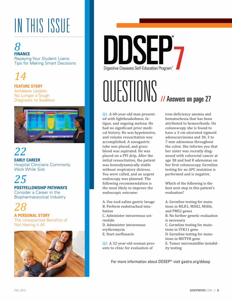
Achalasia
Update
No Longer a Tough
Diagnosis to Swallow
14

2 // THE NEW GASTROENTEROLOGIST: INSIGHTS FOR FELLOWS & YOUNG GIS FALL 2015
LetterF R O M T H E E D I T O R
Bryson W. Katona is an instructor of medicine in the division of gastroenterology at the University of Pennsylvania.
Dear Colleagues,
In recent years the field of acha-
lasia has advanced dramatically,
with better understanding of the
pathophysiology, improvement in
disease classification with the use of
high-resolution esophageal manom-
etry, and the development of novel
therapeutic approaches. In this fall
issue of The New Gastroenterologist,
Rena Yadlapati and John E. Pandolfi-
no from Northwestern University
provide a fantastic overview of the
current state of achalasia, addressing
epidemiology, pathophysiology, diag-
nostic criteria, and management.
In this issue’s section on postfel-
lowship pathways, Douglas S. Levine
from Shire provides a useful perspec-
tive on a career in the biopharmaceu-
tical industry and the opportunities
for gastroenterologists in this field.
Also in this issue is an enlightening
piece from Anne Peery, at the Uni-
versity of North Carolina, Chapel Hill,
in which she discusses work-life bal-
ance. Additionally, we provide cov-
erage of a recently published study
that highlights the dilemma that phy-
sicians often face when dealing with
their own illnesses. And, to address a
topic that is constantly on the minds
of most young career gastroenterol-
ogists, there is an informative over-
view of the basics of managing and
repaying student loans, as well as
many other features that I hope you
will find both interesting and useful.
Please download our app, which
is available free on iTunes, Google
Play, and Amazon, or read our online
edition, which is accessible through
www.gastro.org or www.gihepnews.
com. If you have any feedback about
The New Gastroenterologist, or have
ideas or contributions for future
issues, please e-mail me at bryson.
[email protected] or Erin Dub-
nansky at [email protected].
Sincerely,
Bryson W. Katona, M.D., Ph.D.
Editor in Chief
Editor in Chief
Bryson W. Katona, M.D., Ph.D.
AGA Institute Staff
Vice President of Publications
Erin C. Dubnansky
Editorial Assistant
Ryan A. Farrell
Frontline Medical News Staff
Editor
Lora T. McGlade
Senior Designers
Michael Hyde
Dolly Johnson
Production Manager
Rebecca Slebodnik
VP/Group Publisher:
Director, FMC Society Partners
Mark Branca
CEO, Frontline Medical
Communications
Alan J. Imhoff
Copyright © 2015 Frontline Medical
Communications Inc.
All rights reserved. No part of this publication
may be reproduced or transmitted in any form,
by any means, without prior written permission
of the Publisher. Frontline Medical Communi-
cations Inc. will not assume responsibility for
damages, loss, or claims of any kind arising from
or related to the information contained in this
publication, including any claims related to the
products, drugs, or services mentioned herein.
ON THE COVER
Dr. John E. Pandol�no and Dr. Rena
Yadlapati of Northwestern University,
Chicago.
Photo courtesy Dr. Rena Yadlapati
FALL 2015 GIHEPNEWS.COM // 3
8FINANCE
Repaying Your Student Loans: Tips for Making Smart Decisions
14FEATURE STORY
Achalasia Update:No Longer a Tough Diagnosis to Swallow
22EARLY CAREER
Hospital Clinicians CommonlyWork While Sick
25POSTFELLOWSHIP PATHWAYS
Consider a Career in theBiopharmaceutical Industry
28A PERSONAL STORY
The Unexpected Benets of Not Having it All
IN TH IS ISSUE
QUESTIONS // Answers on page 27
Q1: A 60-year-old man present-
ed with lightheadedness, fa-
tigue, and ongoing melena. He
had no significant prior medi-
cal history. He was hypotensive,
and volume resuscitation was
accomplished. A nasogastric
tube was placed, and gross
blood was aspirated. He was
placed on a PPI drip. After the
initial resuscitation, the patient
was hemodynamically stable
without respiratory distress.
You were called, and an urgent
endoscopy was planned. The
following recommendation is
the most likely to improve the
endoscopic outcome:
A. Use iced-saline gastric lavage
B. Perform endotracheal intu-
bation
C. Administer intravenous oct-
reotide
D. Administer intravenous
erythromycin
E. Start norfloxacin
Q2: A 32-year-old woman pres-
ents to clinic for evaluation of
iron-deficiency anemia and
hematochezia that has been
attributed to hemorrhoids. On
colonoscopy she is found to
have a 3-cm ulcerated sigmoid
adenocarcinoma and 30, 3 to
7-mm adenomas throughout
the colon. She informs you that
her sister was recently diag-
nosed with colorectal cancer at
age 38 and had 8 adenomas on
her first colonoscopy. Germline
testing for an APC mutation is
performed and is negative.
Which of the following is the
best next step in this patient’s
evaluation?
A. Germline testing for muta-
tions in MLH1, MSH2, MSH6,
and PMS2 genes
B. No further genetic evaluation
is necessary
C. Germline testing for muta-
tions in STK11 gene
D. Germline testing for muta-
tions in MUTYH gene
E. Tumor microsatellite instabil-
ity testing
For more information about DDSEP© visit gastro.org/ddsep
4 // THE NEW GASTROENTEROLOGIST: INSIGHTS FOR FELLOWS & YOUNG GIS FALL 2015EW GASTROENTEROLOGIST: INSIGHTS FORNE R
AGA OUTLOOK
NOV 10, 2015GI Boards
NOV 13-15, 201520th Annual Endoscopic Ultrasonography Live 2015
This course offers gastroenterologists, endoscopists,
surgeons, and nurses an opportunity to review the
indications and techniques used for EUS, fine-needle
aspiration, and injection therapy.
Chicago, IL
DEC 10-12, 20152015 Advances in Inflammatory Bowel Diseases, Crohn’s &
Colitis Foundation’s Clinical & Research Conference
Orlando, FL
FEB 5-6, 2016Women’s Leadership Conference —
Experienced Track & Early Career Track
Apply to participate in the premier leadership development
event that is tailor-made for women gastroenterologists.
Irving, TX
MAR 11-12. 2016AGA-AASLD Academic Skills Workshop
This jointly sponsored workshop is designed to
equip junior faculty and fellows with essential information
to help them navigate and succeed in the highly
competitive field of medical academia.
Phoenix, AZ
MAY 21-24, 2016Digestive Disease Week® (DDW)
DDW® is the premier meeting for the GI professional.
Every year it attracts approximately 15,000 physicians,
researchers, and academics from around the world who
desire to stay up-to-date in the field.
San Diego, CA
Digestive Disease Week® (DDW) Abstracts
Deadline: December 1, 2015
AGA-Rome Foundation Functional GI and
Motility Disorders Pilot Research Award
Deadline: January 15, 2016
AGA-Elsevier Pilot Research Award
Deadline: January 15, 2016
AGA-Elsevier Gut Microbiome Pilot Research Award
Deadline: January 15, 2016
AGA-Caroline Craig Augustyn & Damian Augustyn
Award in Digestive Cancer
Deadline: January 29, 2016
AGA-Covidien Research & Development
Pilot Award in Technology
Deadline: January 29, 2016
AGA Investing in the Future Student Research
Fellowship
Deadline: February 5, 2016
AGA-Eli & Edythe Broad Student Research
Fellowship(s)
Deadline: February 12, 2016
AGA/AGA-GRG Fellow Travel and
Abstract of the Year Awards
Deadline: February 26, 2016
AGA-Moti L. & Kamla Rustgi International
Travel Awards
Deadline: February 26, 2016
AGA Student Abstract Prizes
Deadline: March 4, 2016
AGA Outlook
For more information about upcoming events and awards deadlines, please visit www.gastro.org
Upcoming
Events
Awards Application
Deadlines

FALL 2015 GIHEPNEWS.COM // 5
CLINICAL CHALLENGES
AND IMAGES
What’s Your Diagnosis?
By Giovanni De Petris, M.D., Alexandra Corominas Cishek, M.D., and Ivana Dzeletovic, M.D.
Severe diarrhea following bone marrow transplantation is not always caused by GVHD
Published previously in Gastroenterology (2014;146:e5-6)
A35-year-old man com-
plained of persistent di-
arrhea 40 days after bone
marrow transplant. Esoph-
agogastroduodenoscopy
(EGD) and biopsies showed
graft-versus-host disease (GVHD)
grade IV (of Lerner) and gastric ulcers
with cytomegalovirus (CMV) infection.
Biopsies from the colonoscopy showed
GVHD (histologically compatible with
grade II of Lerner). The patient was
treated and showed improvement of
his symptoms but diarrhea persisted.
Follow-up EGD presented diffuse
improvement of the erythema in stom-
ach and duodenum; the colonoscopy
was normal. The pathology in each site
showed no evidence of GVHD or CMV,
and regenerative changes of the muco-
sa. Four days later, worsening of symp-
toms occurred despite treatment, with
severe diarrhea (4 L/d), intermittently
bloody, and mild abdominal pain.
The laboratory results were hemo-
globin, 10 g/dL; white blood cell count,
2,000 cells/microL; platelets, 60,000/
microL; blood urea nitrogen/creatinine,
normal; mild electrolyte abnormalities;
lactate dehydrogenase, 450 U/L; and
blood film, pancytopenia, no circulating
lymphoma cells, no schistocytes. Colo-
noscopy (Figures A and B) with the le-
sions depicted present throughout the
colon and the colon biopsies histology
(Figures C and D) are shown. n
What is this condition?
Dr. De Petris and Dr. Corominas Cishek
are in the Department of Pathology,
Mayo Clinic in Arizona, Scottsdale;
and Dr. Dzeletovic is in the Depart-
ment of Gastroenterology, Mayo Clinic
in Arizona, Scottsdale.
See The Answer on page 30
A B
C D

6 // THE NEW GASTROENTEROLOGIST: INSIGHTS FOR FELLOWS & YOUNG GIS FALL 2015
AGA NEWS
News from the AGA
AGA Proposes
Eliminating Secure
Exam for MOC
Frustrated by a maintenance of certifica-
tion process that didn’t improve patient
care, AGA convened a task force to pro-
pose an ideal pathway for recertification.
The AGA proposal, unveiled online this
August in both Gastroenterology and
Clinical Gastroenterology and Hepatol-
ogy, eliminates the high-stakes exam-
ination and replaces it with active and
adaptive learning self-directed modules
that allow for continuous feedback, and
are based solidly on learning theory.
Read the full proposal, Bridging
the G-APP: Continuous Professional
Development for Gastroenterologists:
Replacing MOC with a Model for Life-
long Learning and Accountability, at
http://www.gastrojournal.org/article/
S0016-5085(15)01177-4/pdf, and the
editorial, An Alternative to MOC?, at
http://www.gastrojournal.org/article/
S0016-5085(15)01178-6/pdf (log-in
required). The proposal will be avail-
able in the November print issues of
Gastroenterology and Clinical Gastroen-
terology and Hepatology.
Provide feedback to AGA on our sur-
vey page (www.surveymonkey.com/s/
gappfeedback).
“There is now a greater emphasis
than ever before on disease path-
ways, clinical guidelines, and quality
improvement, making it important
for physicians to remain current with
newer recommendations and practice
standards,” said Dr. Michael Camilleri,
President, AGA Institute. “Maintaining
certification should be a process of ac-
tive learning, not high-stakes testing.”
Three things to know about AGA’s Al-
ternate Recertification Pathway:
• Individual self-assessment path-
ways allow physicians to achieve a high
level of competency in one or more
areas, while maintaining a general level
of competency in other areas.
• Individualized self-assessment ac-
tivities provide constant feedback and
opportunities for learning and replace
the high-stakes exam now required ev-
ery 10 years.
• Physicians get credit for activities
they are already doing in practice, re-
search, or teaching.
For more information, watch a quick
video introduction (https://www.
youtube.com/watch?v=5hV70RlxP3Y)
by Dr. Suzanne Rose, MSEd, AGAF,
Education and Training Councillor on
the AGA Institute Governing Board. We
do not expect the process to change
overnight, but we’re getting the con-
versation started in a substantial,
meaningful way. AGA supports contin-
uous education and professional devel-
opment that enhances patient care.
Thanks to AGA members who served
on the task force:
Suzanne Rose, M.D., MSEd, AGAF
Brijen J. Shah, M.D.
Jane Onken, M.D., MHS, AGAF
Arthur J. DeCross, M.D., AGAF
Maura H. Davis
Rajeev Jain, M.D., AGAF
Lawrence S. Kim, M.D., AGAF
Kim Persley, M.D.
Sheryl A. Pfeil, M.D., AGAF
Lori N. Marks, Ph.D. n
AGA Fights for Fair
Colonoscopy Reimbursement
Earlier this summer, CMS proposed drastic cuts to the 2016
Medicare physician reimbursement rates for colonoscopy
and other lower GI endoscopy procedures. AGA, in coordi-
nation with ACG and ASGE, is fighting for fair and accurate
reimbursement for all lower endoscopy procedures, includ-
ing colonoscopy.
Some good news – we have the support of some important
members of Congress. Representatives Donald Payne Jr. (D-
NJ) and Leonard Lance (R-NJ) have asked their colleagues in
the U.S. House of Representatives to join them in expressing
concern over two key issues:
• Recently proposed Medicare payment cuts to colonoscopy.
• Impact of the cuts on access to colorectal cancer screen-
ing, especially in light of recent gains made in access to
this life-saving procedure.
AGA members have been critical in this fight. More than 550
members participated in our poll on colonoscopy pay cuts;
the results of which were presented to CMS by AGA, ACG, and
ASGE during a meeting in July. We also garnered the support
of more than 300 gastroenterologists who reached out to CMS
about how these cuts will affect their patients and practice. We
thank you for your help on this important issue.
We expect the final rule to be released later this month.
Stay tuned to your email and AGA eDigest for continuous up-
dates on this important matter. n

FALL 2015 GIHEPNEWS.COM // 7
AGA NEWS
Workshop Provides Insight on
Building a Career in Academic
Medicine
Trainees and junior faculty are encouraged to submit an
application for an opportunity to attend the AGA-AASLD
Academic Skills Workshop, taking place March 11 and 12,
2016, in Phoenix, AZ. This is a chance to get insight from
accomplished academicians on what it takes to successfully
shape a career in academic medicine. Not only will partic-
ipants be able to better understand academic processes,
they’ll also develop the skills necessary to help position
themselves for future success.
The workshop will address topics such as:
• Your first academic job – Learn how changes in health care
reimbursement are impacting academic medicine and discover
how to manage personal and workplace expectations.
• Academic medicine: tracks and pathways – Learn about
available opportunities and strategies that can lead to future
promotions.
• How to be a successful mentee – Find out how to get the
most out of your mentor-mentee relationship to help with
achieving short- and long-term goals.
• Writing and presentation skills – Acquire tips and strat-
egies for writing grants and preparing, editing, and submit-
ting manuscripts.
• Career development: strategy and funding – Get infor-
mation on early career funding opportunities, including
grants available for young investigators.
Forty candidates will be selected to participate in the
workshop and all interested candidates must be a member
of either AGA or AASLD. Women and underrepresented mi-
norities are strongly encouraged to apply. Applications are
due no later than Monday, Oct. 26, 2015.
Learn more about the AGA-AASLD Academic Skills Work-
shop at http://www.gastro.org/in-person/2016/3/11/
aga-aasld-academic-skills-workshop and apply today. n
AGA Guidelines
Patients Can
Understand
To help patients better under-
stand the latest clinical informa-
tion presented in AGA guidelines,
AGA has started creating patient
guideline summaries that:
• Provide information for
patients on the clinical issue ad-
dressed by the guideline.
• Explain the recommenda-
tions and their impact on patient
care.
• Pose helpful questions for
patients to ask their gastroenter-
ologists related to the guideline.
The patient guideline summa-
ries support physicians in their
efforts to effectively communi-
cate important information to
their patients and empower the
patient and physician to work
together to make the most in-
formed and appropriate care
decisions possible. As they are
published, the patient guideline
summaries will be available in
Gastroenterology and on the AGA
website (http://www.gastro.org/
patient-care/patient-center).
Current patient summaries in-
clude:
• Crohn’s disease drugs.
• HBV reactivation.
• IBS drugs.
• Pancreatic cysts.
AGA has a rigorous guideline
development process that devel-
ops focused, actionable clinical
recommendations based on in-
depth reviews of all available
evidence.
To supplement the robust
portfolio of guidelines, each
new guideline is supplemented
by a clinical decision support
tool, an illustrated algorithm
based on the evidence present-
ed in the technical review, and
a patient summary. These tools
can be used at the point of clini-
cal care to help with rapid deci-
sion making. n
©WAVEBREAKMEDIA/THINKSTOCK.COM

8 // THE NEW GASTROENTEROLOGIST: INSIGHTS FOR FELLOWS & YOUNG GIS FALL 2015
FINANCE:
STUDENT LOANS
Repaying Your Student Loans:
Tips for Making Smart DecisionsBy Jay M. Weinberg, CLU, ChFC, and Aaron Braunstein
Mr. Weinberg and Mr. Braunstein are nancial planners with Atlantic Pension Planning Corp., whose practices primarily assist physicians and dentists.
Mr. Weinberg can be reached at [email protected]; Mr. Braunstein at [email protected].
©W
AV
EB
RE
AK
ME
DIA
L
TD/T
HIN
KS
TO
CK.C
OM
FALL 2015 GIHEPNEWS.COM // 9
FINANCE:
STUDENT LOANS
Student loans are an import-
ant part of financial planning for physicians and therefore require proper management. In this article we will lay out several key considerations
of student loans that will guide you toward smart decisions for your fi-nancial future.
What is the difference between
federal loans and private loans?
Typically, federal loans have more flexible repayment options and po-
tential forgiveness. However, many private loan refinance options now offer significantly lower interest rates than existing federal loans.
Is the interest paid on student
loans tax deductible?
If you are single and your modified adjusted gross income is less than $80,000 or you are married and earn less than $160,000 combined, all or some of your interest paid – with a cap of $2,500 – is deductible. These figures can change annually, so it is best to refer to IRS publication 970 for the most up-to-date information.
How does the interest rate on
a student loan impact your
strategy towards paying it off?
Naturally, it is always advantageous to pay off the higher interest rate loans before the lower interest rate loans. However, there are additional factors to consider when developing a plan to pay off your loans:
• Are the loans federal or private?
• Are you planning to be eligible for a forgiveness program? • What is your overall loan balance?• Do you expect your income to in-
crease over time or could it have ma-
jor fluctuations?
Are all federal loans the same?
No. When you took out the loan, what the loan was for, and what your financial need was at the time
of the loan will dictate the type of government loan that you have. The three common federal loan types are Stafford, Perkins, and Plus. For more information about these types of loans, visit www.studentloans.gov/myDirectLoan/index.action.
What are the most common
federal repayment strategies?
• Income Based Repayment (IBR) – Generally, 15% of your discretionary income.• Pay As You Earn (PAYE) – 10% of your discretionary income. • Standard Repayment – You have a maximum of 10 years to pay off the outstanding principal and interest of the loan. • Extended Repayment – You have a maximum of 25 years to pay off the
outstanding principal and interest of the loan.
Some considerations to keep in mind about these repayment strate-
gies include:• There are no prepayment penalties
for government loans; therefore, you have the ability to pay “extra” money toward these loans to pay them off sooner.• As your income increases, so will the monthly payments on your IBR and PAYE. However, the monthly pay-
ment on your IBR and PAYE will nev-
er exceed your Standard Repayment.• Not all borrowers are eligible for PAYE currently; however, by the end of 2015 we expect that all borrowers
with “Direct Loans” will be eligible. Direct loans are those made directly by the U.S. Department of Education. PAYE is the most advantageous plan if you expect to be eligible for various forgiveness programs, as this option will require the lowest out-of-pocket payment.
What is Public Service Loan
Forgiveness (PSLF)?
In October 2007, the PSLF program was introduced by the U.S. Govern-
ment under the College Cost Reduc-
tion and Access Act of 2007 (CCRAA). This program offered forgiveness to individuals that make 120 qualifying payments on “Direct Loans.” There are many government or not-for-profit institutions that are eligible, but we highly recommended that
you verify, in advance, that your cur-
rent employer fits the qualifications. There is a lot of uncertainty related to caps on forgiveness amounts and eligibility requirements surrounding PSLF; therefore, it is important that you keep up to date on proposed
changes coming from our govern-
ment.
Is it better to pay the minimum on
loans or aggressively pay them off?
If you are hoping to be eligible for a forgiveness program, there is no rea-
son to “overpay” now on your loans only to have the balance forgiven at some point in the future. On the con-
trary, if a forgiveness program is not
There are no prepayment penalties for government
loans; therefore, you have the ability to pay “extra”
money toward these loans to pay them off sooner.

10 // THE NEW GASTROENTEROLOGIST: INSIGHTS FOR FELLOWS & YOUNG GIS FALL 2015
FINANCE:
STUDENT LOANS
an option, the interest rates on your loans will dictate how aggressively you should pay them off.
What is a private loan re�nance
and does it make sense?
In the past few years, several private (nongovernment) lenders have en-
tered into the student loan refinance market and offer repayment terms
that are far more favorable than the 6%, 7%, or 8% interest rates at-
tached to government loans.A private loan refinance is not for ev-
erybody. If you expect to be eligible for one of the many forgiveness programs, it may not be for you. However, if you have high-interest-rate government (or private) loans and are not eligible for any forgiveness programs, you should absolutely consider the pros and cons of refinancing all or some of your ex-
isting loans. Interest rates on private refinances can be as low as 2%, but are typically 3%-6%. You have the choice of a variable interest rate or a fixed interest rate. Repayment terms are generally from 5 to 20 years.
We have received extremely favor-
able feedback about two private lend-
ers in particular: SOFI (Social Finance) and DRB (Darien Rowayton Bank). If you are contemplating a refinance, we encourage you to contact both of these lenders and compare offerings. There are links at the end of this arti-
cle that offer bonuses if you refinance with either of these companies.
Should I consolidate
my federal loans?
Prior to 2006, it was advantageous to consolidate federal loans because it significantly lowered interest rates. Since then, the federal consolidation landscape has changed. If you do a federal consolidation today, the in-
terest rates on all of the consolidated loans are averaged together. Chances are that engaging in a federal consoli-dation will not save you money; it will simply make record keeping easier. If
you do a consolidation and extend the payment period of the loan, you may actually pay more money in interest over time compared to if you did not consolidate. It is important to know that a consolidation is one of the ways to get out of “default” and get back into a current repayment plan.
Can I re�nance some, but
not all, of my loans?
Yes, you can selectively pick which loans you want to “leave alone” and which loans you want to refinance or consolidate.
If I get into a bind with my
loans, what should I do?
We always recommended that you contact the servicer of your loan to find out what your options are. If you come upon a financial hardship, there may be some relief available but you need to ask for help in order to re-
ceive it. During a medical residency, forbearance is an option on your fed-
eral loans. In fellowship, deferment is an option for your federal loans.
Should I pay off my loans
before purchasing a home?
There is no “right” answer to this ques-
tion; however, there are certain factors that you need to take into account when weighing the options. What is the interest rate on the student loans?
Are you eligible for a tax deduction of $2,500 for student loan interest? What interest rate would the home mortgage be? What tax bracket are you currently in and do you see yourself being in over the next 5 or 10 years? How will the student loans be counted toward your debt-to-income ratios to qualify for a mortgage approval?
Most people would rather borrow as much money as they can at 3% or 4% (tax deductible) toward the purchase of a home and use current savings and cash flow to pay off high-interest-rate student loans (few or no tax benefits). No two situations
are the same and we recommend that you weigh your options and de-
cide which plan best suits your goals.
Developing a plan for either pay-
ing off your student loans or making yourself eligible for a forgiveness program are important steps toward the makings of a bright financial future. There are many resources available to borrowers and taking a proactive approach toward managing your student loans will certainly ben-
efit your situation over time. n
Resources:
https://studentloans.gov/myDirectLoan/index.
action – Federal Student Aid – U.S. Dept. of
Education
https://www.aamc.org/advocacy/med-
ed/79048/student_loan_repayment.html –
American Association of Medical Colleges
http://www.direct.ed.gov/calc.html – Loan Cal-
culators and Interest Rates
http://pgpresents.com/ – Student Loan Con-
sulting – Paul Garrard
http://sofi.com/AtlanticPensionPhysicians –
SOFI – Link to refinance and receive $200 wel-
come bonus
https://student.drbank.com/?provider=jwapp
– DRB Education Refinance – Link to refinance
and receive $200 referral bonus
©W
AV
EB
RE
AK
ME
DIA
L
TD/T
HIN
KS
TO
CK.C
OM

FALL 2015 GIHEPNEWS.COM // 11
SNAPSHOTS FROM THE
AGA JOURNALS
Snapshots from the AGA Journals
Dr. Stuart Gordon is professor of medicine at Wayne State University School of Medicine and director of the Division of Hepatology at Henry Ford Health Systems, Detroit. He has no relevant con�icts of interest.
The investigators from the
Drug-Induced Liver Injury
Network (DILIN) report a
previously unrecognized phe-
nomenon: Patients who receive
a single dose of an IV cephalo-
sporin prior to an operative procedure
may develop jaundice and biochemical
cholestasis 1-3 weeks later. Remark-
ably, every single patient in this case
series also presented with pruritus,
suggesting an allergic reaction. Nearly
half of the patients reported a previous
“drug allergy” (although it is uncertain
whether this was disclosed before their
procedures), and some were in fact
penicillin allergic, so likely should not
have ever received cefazolin.
Some of the late-onset cases de-
scribed in this series could have justi-
fied a misdiagnosis of “postoperative
cholestasis” or have led to a fishing
expedition for various “zebra” diagno-
ses. What is instructive in this report
is that, for the most part, neither the
patients nor the doctors evaluating
their unexplained hepatitis ever sus-
pected that an antibiotic had even been
given. This observation highlights the
fact that often medications that are
used just once in the surgical suite will
then disappear from a patient’s med-
ications list and are often difficult to
subsequently identify in the electronic
medical record.
Cefazolin is the workhorse for pre-
operative prophylaxis in cardiac and
orthopedic surgery, and most oper-
ations involving skin, such as plastic
surgery. Such antibiotic prophylaxis is
generally used very appropriately and
according to evidence-based clinical
guidelines, and it is a closely monitored
and audited quality indicator at hos-
pitals and surgical centers. The use of
intravenous cefazolin as preoperative
prophylaxis will likely not be dimin-
ished by these reports, but this case
series again emphasizes the need to
avoid cephalosporins among patients
who report previous beta-lactam aller-
gies. Early recognition of this culprit in
cases of unexplained cholestatic hepa-
titis, especially in patients who recently
underwent operative procedures, may
obviate hospitalization. n
Key clinical point: A single dose
of cefazolin can cause drug-in-
duced liver injury (DILI), and the
agent is implicated more often
than previously thought.
Major finding: Cefazolin ranked
sixth among causes of DILI, and
signs and symptoms began 1-3
weeks after initial exposure.
Data source: Registry-based study
of 1,212 cases of DILI.
Disclosures: The study was fund-
ed by the National Institute of
Diabetes and Digestive and Kidney
Diseases, the National Institutes of
Health, the National Cancer Insti-
tute, and by Clinical and Transla-
tional Science Award grants. The
investigators reported having no
conflicts of interest.
Commentary
Cefazolin Ranks Sixth as Cause of Drug-Induced Liver Injury
July Clinical Gastroenteology and Hepatology (doi: 10.1016/j.cgh.2015.01.010]

12 // THE NEW GASTROENTEROLOGIST: INSIGHTS FOR FELLOWS & YOUNG GIS FALL 2015
SNAPSHOTS FROM THE
AGA JOURNALS
Dr. Matthew J. DiMagno is in the division of gastroenter-ology and hepatology, department of internal medicine, University of Michigan, Ann Arbor. He serves as chair of the American Gastroenterological Association Institute Council Section on Pancreatic Disorders. He declared no relevant �nancial con icts of interest.
The study of acute pancreatitis
(AP) is economically and sci-
entifically essential because
acute pancreatitis is the most
common reason for hospital-
ization among patients with
GI diseases, consumes considerable
resources, and is treated primarily
with supportive measures. The pilot
study by Dr. Vege and his colleagues
reports that pentoxifylline treatment
is safe for patients with severe acute
pancreatitis and is associated with a
promising reduction in ICU utilization
and duration in patients requiring a
hospital stay > 4 days.
This study not only is provocative
but also raises the hypothesis-gener-
ating question of how pentoxifylline
might exert a salutary effect with-
out reducing blood tumor necrosis
factor–alpha levels (or IL-6, IL-8, or
C-reactive protein levels). The authors
ascribe this discordance to the tim-
ing of administering pentoxifylline
and to potential TNF-alpha indepen-
dent effects. Biologically, pancreatic
TNF-alpha levels increase within the
first 30-60 minutes of onset of acute
pancreatitis (Am. J. Surg. 1998;175:76-
83). In experimental AP, pentoxifylline
ameliorates severity, but data are con-
flicting about whether prophylactic or
delayed (Surgery 1996;120:515-21)
antagonism of TNF-alpha signaling is
more protective. Clinically relevant
data suggest that prophylactic admin-
istration of pentoxifylline does not
prevent postendoscopic retrograde
cholangiopancreatography pancreatitis
(Gastrointest. Endosc. 2007;66:513-8),
but nonprophylactic administration
of pentoxifylline improves short-
term survival in alcoholic hepatitis
without significantly reducing blood
TNF-alpha levels (Gastroenterology
2000;119:1637-48). Hence, pentoxi-
fylline appears to ameliorate AP and
alcoholic hepatitis through TNF-alpha
independent signaling, conceivably by
targeting the microcirculation, as de-
scribed for patients with claudication
(Angiology 1994;45:339-45).
Future studies might test this hy-
pothesis by determining whether
pentoxifylline blunts increases in del-
eterious vascular factors (for example,
angiopoietin-2) [Am. J. Gastroenterol.
2010;105:2287-92; J. Am. Coll. Surg.
2014;218:26-32; Am. J. Gastroenter-
ol. 2011;106:1859-61]) and reduces
vascular complications that correlate
with the need for ICU care and more
severe AP. n
Key clinical point: Pentoxifylline
topped placebo among patients
with severe acute pancreatitis.
Major finding: The pentoxifylline
treatment group had significantly
fewer hospitalizations requiring
more than 4 days (14% vs. 57% for
the placebo group; P = .046) and a
significantly shorter median dura-
tion of stay in the ICU (P = .03).
Data source: A single-center, ran-
domized placebo-controlled trial of
28 patients with predicted severe
acute pancreatitis.
Disclosures: A scholarly opportuni-
ty award from the Mayo Clinic sup-
ported the work. The investigators
reported having no relevant finan-
cial conflicts of interest.
Commentary
Pentoxifylline Beat Placebo in Acute Pancreatitis Trial
August Gastroenterology (doi:10.1053/j.gastro.2015.04.019)
FALL 2015 GIHEPNEWS.COM // 13
SNAPSHOTS FROM THE
AGA JOURNALS
Dr. Larissa V. Furtado and Dr. Jeremy P. Segal are both assistant professors and assistant directors of the division of genomic and molecular pathology in the de-partment of pathology at the University of Chicago Medical Center. Neither has any con�icts of interest.
As the oncology field advanc-
es toward implementation
of personalized medicine
programs, molecular and ge-
nomic analysis of circulating
tumor DNA (ctDNA) rep-
resents a promising approach for di-
agnosis, prognosis, therapy selection,
and minimal residual disease monitor-
ing of a wide array of malignancies.
With the purpose of assessing the
utility of extracellular tumor DNA as
a potential biomarker for hepatocel-
lular carcinoma (HCC), Dr. Ono and
colleagues analyzed serum ctDNA from
46 HCC patients using quantitative PCR
assays for somatic rearrangements un-
covered by whole-genome sequencing
of their primary tumors.
For the seven patients with detectable
ctDNA in preoperative serum, the inci-
dence of recurrence and extrahepatic
metastasis within 2 years following
hepatectomy were significantly worse
than in the ctDNA-negative group, al-
though no significant difference in the
cumulative survival rate was observed
between these patients. The ctDNA
positivity also was found to be an in-
dependent predictor of microscopic
vascular invasion of the portal vein, and
it correlated with larger tumor size and
higher alpha-fetoprotein and des-gam-
ma-carboxy prothrombin levels.
In addition, the investigators demon-
strated that transcatheter arterial
chemoembolization enriched ctDNA
levels in cell-free DNA in blood, and
that serum ctDNA levels were in-
creased with disease progression and
reflected response to treatments. The
diagnosis of HCC is currently based on
imaging and/or biopsies. Even though
there are no well-established biomark-
ers for early detection and monitoring
of HCC at present, the data presented
here indicate the potential utility of
personalized ctDNA testing for individ-
ualized management of hepatocellular
carcinoma patients. n
Key clinical point: The presence
of circulating tumor (ct) DNA indi-
cated progression of hepatocellular
carcinoma.
Major finding: Among seven pa-
tients who tested positive for ctDNA
before undergoing surgical resec-
tion, six developed recurrent HCC
and four developed extrahepatic
metastases.
Data source: Real-time quantitative
PCR analysis of serum samples from
46 patients with HCC who under-
went hepatectomy or liver trans-
plantation.
Disclosures: The study was funded
by the government of Japan, the
RIKEN President’s Fund, the Prin-
cess Takamatsu Cancer Research
Fund, and the Takeda Science Foun-
dation. The investigators declared
no competing interests.
Commentary
Circulating Tumor DNA Marked Progressive Liver Cancer
September Cellular and Molecular Gastroenterology and Hepatology (doi: 10.1016/j.jcmgh.2015.06.009)
14 // THE NEW GASTROENTEROLOGIST: INSIGHTS FOR FELLOWS & YOUNG GIS FALL 2015
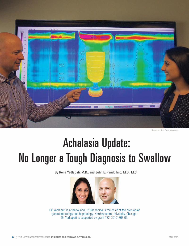
Achalasia Update:
No Longer a Tough Diagnosis to SwallowBy Rena Yadlapati, M.D., and John E. Pandolno, M.D., M.S.
Dr. Yadlapati is a fellow and Dr. Pandol no is the chief of the division of gastroenterology and hepatology, Northwestern University, Chicago.
Dr. Yadlapati is supported by grant T32 DK101363-02.
COURTESY DR. RENA YADLAPATI
FALL 2015 GIHEPNEWS.COM // 15
ACHALASIA UPDATE
Historical perspective
The medical field first rec-
ognized achalasia in 1674
when Sir Thomas Willis de-
scribed the use of a carved
whalebone with a sponge
at the distal end as management for
his patient with liquid dysphagia, at
the time referred to as cardiospasm.1
In the late 1920s, this disease entity
was renamed achalasia, a term de-
rived from the Greek khalasis, mean-
ing “not loosening or relaxing.”2,3
Over the past century, the diagnostic
understanding and management
strategies for achalasia have signifi-
cantly evolved.
Epidemiology and pathophysiology
Achalasia presents at an estimated
annual incidence of 1.6 in 100,000
and a prevalence of 10.8 in 100,000
without a racial or gender predilec-
tion.4 While its peak incidence occurs
between the ages of 30 and 60, acha-
lasia can present at any age.5 A defin-
ing feature of achalasia is impaired
deglutitive relaxation of the lower
esophageal sphincter (LES), consid-
ered to be a result of the functional
loss of the myenteric plexus. While
the precise cause of neuronal degen-
eration is unknown, it is thought to
be multifactorial, involving an auto-
immune process triggered by an in-
dolent viral infection in conjunction
with a genetically susceptible host.6
Normally, the postganglionic neu-
rons use nitric oxide and vasoactive
intestinal polypeptide as inhibitory
neurotransmitters. In the setting of
myenteric degeneration there exists
an imbalance between excitatory and
inhibitory neurons, with the unop-
posed cholinergic stimulation result-
ing in impaired LES relaxation and
distal esophageal dysmotility.7 Infec-
tion by Trypanosoma cruzi, known
as Chagas disease, can also manifest
with achalasia in addition to other
features of widespread myenteric
plexus destruction, including mega-
colon, heart disease, and neurologic
disorders.5,6,8
Clinical presentationThe hallmark symptom of achalasia,
present in 90% of cases, is a progres-
sive constant dysphagia of both solids
and liquids. Other common esoph-
ageal symptoms include heartburn,
noncardiac chest pain, regurgitation,
odynophagia, and epigastric pain.9,10
Additionally, patients with achalasia
can present with cough, asthma, and
chronic aspiration. Over time, patients
learn to adapt to their dysphagia by
modifying their dietary habits and
becoming “slow eaters.” Unintentional
weight loss may not present until the
end stage of the disease.2,6
Diagnosis
Diagnosing achalasia can be chal-
lenging, and patients often expe-
rience symptoms for years before
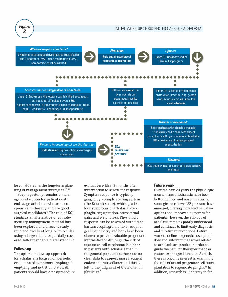
obtaining a diagnosis.9 An important
initial step is distinguishing esopha-
geal from oropharyngeal dysphagia
through a careful history and evalu-
ation of swallowing by observing the
patient drink water. In cases of sus-
pected esophageal dysphagia, struc-
tural mechanical obstruction should
be ruled out first via upper gastro-
intestinal endoscopy or radiologic
imaging. Endoscopic features sup-
portive of achalasia include a dilated
or tortuous esophagus, bolus impac-
tions with or without fluid pooling
in the esophagus, and resistance to
intubation of the esophagogastric
junction (EGJ).2,5 Esophageal inflam-
matory changes or ulcerations sec-
ondary to stasis, pill esophagitis, or
Candida esophagitis can also be seen
on endoscopy.5 To minimize aspira-
tion, patients should restrict oral in-
take for a minimum of 8 hours prior
to endoscopy; in addition, many cen-
ters recommend a liquid diet for 48
hours and a soft diet the week prior
to endoscopy in cases of suspected
achalasia to help clear the contents
of the esophagus. During endoscopy,
vigorous suctioning of retained fluid
in the esophagus should be per-
formed upon entry of the scope. The
classic “bird’s beak” appearance, of
the distal esophagus, a dilated con-
trast-filled esophagus, “corkscrew”
or “rosary bead” appearance and
absent peristalsis can be seen on
barium esophagram.2,6 While often
nondiagnostic, barium esophagram
is particularly helpful in cases with
equivocal esophageal manometry
An important initial step is distinguishing esophageal from oropharyngeal
dysphagia through a careful history and evaluation of swallowing by observing
the patient drink water.

16 // THE NEW GASTROENTEROLOGIST: INSIGHTS FOR FELLOWS & YOUNG GIS FALL 2015
ACHALASIA UPDATE
findings and to assess for late-stage
achalasia changes that have treat-
ment implications.5
Esophageal manometry is the gold
standard for diagnosing and clas-
sifying achalasia; all patients with
suspected achalasia who do not have
evidence of a mechanical obstruction
should undergo esophageal motility
testing.5 The landmark development
of esophageal pressure topography
(EPT) in the 1990s has transformed
esophageal manometry and the mod-
ern approach to achalasia.11-16 The
integrated relaxation pressure (IRP),
calculated as the mean of 4 seconds
of maximal EGJ relaxation following
pharyngeal contraction, is a metric
to quantify deglutitive EGJ relaxation
and has been instrumental in estab-
lishing diagnostic criteria for acha-
lasia.17-19 While a defined diagnostic
threshold continues to be debated,
a median IRP greater than 15 mm
Hg is concerning for an EGJ outflow
disorder. In addition, a classifica-
tion scheme of esophageal motility
disorders utilizing EPT metrics (the
Chicago classification) has refined
the conventional diagnosis of classic
achalasia into three clinically rele-
vant phenotypes (achalasia types I,
II, and III); these phenotypes portend
prognostic and therapeutic implica-
tions, and support the natural history
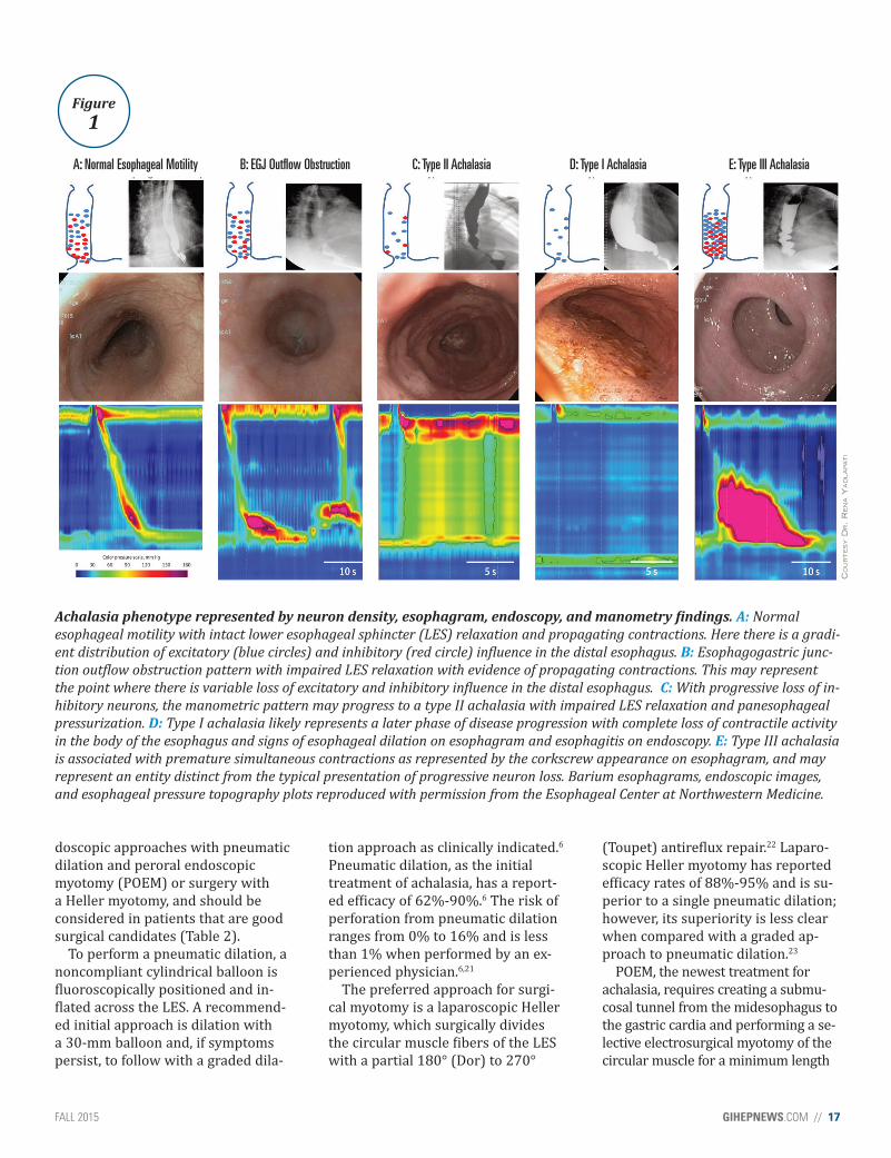
of achalasia (Table 1).17-20
Type II achalasia, associated
with impaired LES relaxation and
pan-esophageal pressurization,
results from a progressive loss of
inhibitory neurons. Complete loss of
contractile activity in the esophageal
body is seen in type I achalasia and is
considered to represent a later stage
of disease progression.2,20 Cases of
impaired LES relaxation with normal
or impaired peristalsis are referred
to as EGJ outflow obstruction (EGJ-
OO), and this may represent an early
or incomplete variant of achalasia
resulting from a variable loss of
excitatory and inhibitory influence.
Considered to be a distinct entity,
type III achalasia is associated with
premature simultaneous contractions
and does not follow the typical pre-
sentation of progressive neuron loss
(Figure 1).20
In cases of suspected achalasia, it
is imperative to evaluate for pseudo-
achalasia from infiltrative tumors or
secondary achalasia from extrinsic
processes such as a tight fundoplica-
tion or laparoscopic adjustable gastric
banding.5 Endoscopy with careful
evaluation of the gastric cardia in ret-
roflexion and/or barium esophagram
is required; and when the suspicion
of pseudoachalasia is high, endoscop-
ic ultrasound and/or computerized
tomography is recommended.6 Addi-
tionally, achalasia should be considered
in patients with refractory reflux, par-
ticularly prior to antireflux surgery, as
heartburn and regurgitation can be the
only presenting symptoms of achalasia;
in these patients, esophageal manome-
try should be performed (Figure 2).9,10
Treatment Curative therapies for achalasia do
not exist. The primary management
goals hinge on early diagnosis and
treatment in order to relieve symp-
toms, improve esophageal emptying,
and prevent late complications. First-
line definitive therapies include en-
CLASSIFICATION OF ESOPHAGEAL MOTILITY DISORDERS WITH IMPAIRED EGJ RELAXATION
Table
1
Considerations
• Severe dilation is associated with poor treatment response
• Best treatment response
• Worst treatment response• May bene�t from management directed at spasm
• Rule out mechanical obstruction with EUS/CT
Conceptual model
of natural history
• Late-stage• Complete loss of inhibitory neurons
• Progressive loss of inhibitory neurons
• May represent a distinct entity unrelated to loss of inhibitory neurons
• Early-stage• Variable loss of excitatory and inhibitory in�uence
Features on
manometry
• 100% absent peristalsis• No signi�cant esophageal pressurization
• 100% absent peristalsis• ≥20% of swallows with panesophageal pressurization to > 30 mmHg
• ≥20% of swallows with premature spastic contractions (distal latency < 4.5 s)
• Normal or impaired peristalsis
Phenotype according to
Chicago classication
Achalasia, Type I
Achalasia, Type II
Achalasia, Type III
Functional EGJ Out�ow Obstruction
Esophagogastric junction, EGJ; Endoscopic ultrasound, EUS; Computerized tomography, CT.
Achalasia should
be considered in
patients with refractory
re�ux, particularly prior
to antire�ux surgery.
FALL 2015 GIHEPNEWS.COM // 17
doscopic approaches with pneumatic
dilation and peroral endoscopic
myotomy (POEM) or surgery with
a Heller myotomy, and should be
considered in patients that are good
surgical candidates (Table 2).
To perform a pneumatic dilation, a
noncompliant cylindrical balloon is
fluoroscopically positioned and in-
flated across the LES. A recommend-
ed initial approach is dilation with
a 30-mm balloon and, if symptoms
persist, to follow with a graded dila-
tion approach as clinically indicated.6
Pneumatic dilation, as the initial
treatment of achalasia, has a report-
ed efficacy of 62%-90%.6 The risk of
perforation from pneumatic dilation
ranges from 0% to 16% and is less
than 1% when performed by an ex-
perienced physician.6,21
The preferred approach for surgi-
cal myotomy is a laparoscopic Heller
myotomy, which surgically divides
the circular muscle fibers of the LES
with a partial 180° (Dor) to 270°
(Toupet) antireflux repair.22 Laparo-
scopic Heller myotomy has reported
efficacy rates of 88%-95% and is su-
perior to a single pneumatic dilation;
however, its superiority is less clear
when compared with a graded ap-
proach to pneumatic dilation.23
POEM, the newest treatment for
achalasia, requires creating a submu-
cosal tunnel from the midesophagus to
the gastric cardia and performing a se-
lective electrosurgical myotomy of the
circular muscle for a minimum length
Figure
1
Achalasia phenotype represented by neuron density, esophagram, endoscopy, and manometry findings. A: Normal
esophageal motility with intact lower esophageal sphincter (LES) relaxation and propagating contractions. Here there is a gradi-
ent distribution of excitatory (blue circles) and inhibitory (red circle) influence in the distal esophagus. B: Esophagogastric junc-
tion outflow obstruction pattern with impaired LES relaxation with evidence of propagating contractions. This may represent
the point where there is variable loss of excitatory and inhibitory influence in the distal esophagus. C: With progressive loss of in-
hibitory neurons, the manometric pattern may progress to a type II achalasia with impaired LES relaxation and panesophageal
pressurization. D: Type I achalasia likely represents a later phase of disease progression with complete loss of contractile activity
in the body of the esophagus and signs of esophageal dilation on esophagram and esophagitis on endoscopy. E: Type III achalasia
is associated with premature simultaneous contractions as represented by the corkscrew appearance on esophagram, and may
represent an entity distinct from the typical presentation of progressive neuron loss. Barium esophagrams, endoscopic images,
and esophageal pressure topography plots reproduced with permission from the Esophageal Center at Northwestern Medicine.
A: Normal Esophageal Motility B: EGJ Out�ow Obstruction C: Type II Achalasia D: Type I Achalasia E: Type III Achalasia
CO
UR
TE
SY D
R. R
EN
A Y
AD
LA
PA
TI

18 // THE NEW GASTROENTEROLOGIST: INSIGHTS FOR FELLOWS & YOUNG GIS FALL 2015
of 6 cm up the esophagus and 2 cm
distal to the squamocolumnar junction
onto the gastric cardia.24 Initial success
rates of POEM in prospective cohorts
of patients with achalasia have been
greater than 90%.24-26 There have been
no randomized trials comparing POEM
to laparoscopic myotomy or pneumatic
dilation and its long-term efficacy re-
mains to be established.
Medical therapy and botulinum
toxin should be reserved for patients
who are poor candidates for definitive
treatment. Calcium channel blockers
and oral nitrates may be useful as
short-acting temporizing treatments,
given their prompt reduction of LES
pressure but are poor long-term treat-
ment options due to their limiting ad-
verse effects and inability to prevent
disease progression.27 For patients
intolerant or nonresponsive to these
agents, sildenafil is an alternative op-
tion; however, long-term data examin-
ing outcomes and safety are lacking.28
Botulinum toxin injection into the
LES muscle is a treatment option for
achalasia based on its ability to block
acetylcholine release. Because of the
temporary effect of botulinum tox-
in, it has limited efficacy beyond 12
months of administration with a high
rate of relapse requiring retreatment.
Repeated botulinum toxin treatments
can make subsequent Heller myoto-
my more challenging and this should
MANAGEMENT OPTIONS FOR ACHALASIATable
2
Procedure setting
Endoscopy laboratory with �uoroscopy; Moderate sedation/-MAC; Procedure time 30 mins; Observation time 4-6h
OR; General anesthesia; Procedure time 90 mins: Hospital stay 1-2d
OR; endoscopy laboratory; General anesthesia; Procedure time 90 mins; Overnight observation
N/A
Endoscopy laboratory; Moderate sedation/-MAC; Procedure time < 30 mins; Observation time 60 mins
Procedure description
Fluoroscopic balloon dilation across the LES with noncompli-ant, cylindrical balloon Recommended: Initially perform 30-mm balloon dilation, followed with a graded approach as indicated
Typically laparoscopic Heller myotomy with an antire�ux repair
Creation of an endoscopic submucosal tunnel from mid-esophagus to gastric cardia with selective myotomy
Nifedipine SL 10-30mg 30-45 mins before meal, Isosorbide dinitrate SL 5-10mg 15 mins before meal,Sildena�l 25-50mg with each meal
100 units of Botulinum toxin injected 1 cm proximal to SCJ in 4 radially dispersed aliquots with sclerotherapy needle during endoscopy
Treatment strategy
Pneumatic Dilation
Surgical Myotomy
Peroral Endoscopic Myotomy (POEM)
Oral Medical Therapy
Botulinum Toxin
Patients
Good Surgical Candidates
Poor Surgical Candidates Or Unwilling To Have Surgery
Lower esophageal sphincter, LES; monitored anesthesia care, MAC; operating room, OR; squamocolumnar junction, SCJ
Outcomes
62%-90% ef�cacy; 2-5 years durability (Increased ef�cacy & durability with graded approach)
88%-95% ef�cacy; 5-10 year durability
Initial success rates > 90%; long-term ef�cacy unknown
Short-acting temporizing effects
Up to 66% effective for up to 6 months; often require repeat injections within 12 months
Procedural issues
0 -16% perforation risk; Does not in�uence response to subsequent myotomy
Risk of post-surgical re�ux or dysphagia (dependent on surgical approach)
Post-procedural complications and issues unknown
Often have limiting side effects
Overall safe; Repeated injections make subsequent myotomy challenging
ACHALASIA UPDATE
FALL 2015 GIHEPNEWS.COM // 19
be considered in the long-term plan-
ning of management strategies.29,30
Esophagectomy remains a man-
agement option for patients with
end-stage achalasia who are unre-
sponsive to therapy and are good
surgical candidates.5 The role of EGJ
stents as an alternative or comple-
mentary management method has
been explored and a recent study
reported excellent long-term results
using a large-diameter partially cov-
ered self-expandable metal stent.31,32
Follow-up
The optimal follow-up approach
for achalasia is focused on periodic
evaluation of symptoms, esophageal
emptying, and nutrition status. All
patients should have a postprocedure
evaluation within 3 months after
intervention to assess for response.
Symptom response is typically
gauged by a simple scoring system
(the Eckardt score), which grades
four symptoms of achalasia: dys-
phagia, regurgitation, retrosternal
pain, and weight loss. Physiologic
response can be assessed with timed
barium esophagram and/or esopha-
geal manometry and both have been
shown to provide valuable prognostic
information.2,6 Although the risk of
squamous cell carcinoma is higher
in patients with achalasia than in
the general population, there are no
clear data to support more frequent
endoscopic surveillance and this is
left to the judgment of the individual
physician.5
Future work
Over the past 20 years the physiologic
mechanisms of achalasia have been
better defined and novel treatment
strategies to relieve LES pressure have
emerged, offering increased palliative
options and improved outcomes for
patients. However, the etiology of
achalasia remains poorly understood
and continues to limit early diagnosis
and curative interventions. Future
work to delineate genetic susceptibil-
ities and autoimmune factors related
to achalasia are needed in order to
guide the path for therapies that can
restore esophageal function. As such,
there is ongoing interest in examining
the role of neural progenitor cell trans-
plantation to regenerate ganglia.33 In
addition, research is underway to fur-
INITIAL WORK-UP OF SUSPECTED CASES OF ACHALASIAFigure
2
When to suspect achalasia?
Symptoms of esophageal dysphagia to liquids/solids
(90%), heartburn (75%), bland regurgitation (45%),
non-cardiac chest pain (20%)
First step:
Rule out an esophageal
mechanical obstruction
Options:
Upper GI Endoscopy and/or
Barium Esophagram
Features that are suggestive of achalasia:
Upper GI Endoscopy: dilated/tortuous uid lled esophagus,
retained food, difcult to traverse EGJ
Barium Esophagram: dilated/contrast lled esophagus, “bird’s
beak,” “corkscrew” appearance, absent peristalsis
If these are normal this
does not rule out
esophageal motility
disorder or achalasia
If there is evidence of mechanical
obstruction (stricture, ring, gastric
band, extrinsic compression) this
is not achalasia
Evaluate for esophageal motility disorder:
Gold standard: High-resolution esophageal
manometry
Normal or Deceased:
Not consistent with classic achalasia.
*Achalasia can be seen with absent
peristalsis in setting of a normal or borderline
IRP or evidence of panesophageal
pressurization
Elevated:
EGJ outow obstruction or achalasia is likely,
see Table 1
EGJrelaxationpressure

20 // THE NEW GASTROENTEROLOGIST: INSIGHTS FOR FELLOWS & YOUNG GIS FALL 2015
ACHALASIA UPDATE
ther characterize the profile of autoan-
tibodies implicated with achalasia. To
date, studies support an organ-specific
autoimmune process with cytotoxic T
cells as possibly the principal effectors
of myenteric degeneration; future
research exploring the role of immu-
nomodulator therapies in the manage-
ment of achalasia is of interest.7
Continued investigation into the
physiologic nuances of the spectrum
of achalasia in order to facilitate ear-
lier detection and intervention is im-
perative. For instance, the functional
lumen imaging probe (FLIP) is a nov-
el diagnostic tool that measures EGJ
distensibility and recent studies have
demonstrated that intraprocedural
EGJ distensibility measurements with
FLIP are predictive of postoperative
symptomatic outcomes.34 FLIP pro-
vides uniquely valuable information
about esophageal compliance and its
role as a predictive and prognostic
tool for achalasia is an area of ongo-
ing research.34
Finally, future work is needed to
streamline personalized high-qual-
ity patient-centered approaches
for achalasia. As such, randomized
controlled trials comparing the long-
term efficacy, cost-effectiveness, and
patient-centered outcomes of cur-
rent and future treatment strategies
should be a research priority. n
References
1. T W. Pharmaceutice Rationalis Sive Diatribe
de Medicamentorum Operationibus in Human
Corpore. London, England: Hagae Comitis;
1674.
2. Pandolfino J.E., Gawron A.J. JAMA.
2015;313:1841-52.
3. Hurst A.F., Rowlands R.P. Proc. R Soc. Med.
1924;17(Clin Sect):45-6.
4. Sadowski D.C., et al. Neurogastroenterol. Mo-
til. 2010;22:e256-61.
5. Vaezi M.F., et al. Am. J. Gastroenterol.
2013;108:1238-49; quiz 50.
6. Pandolfino J.E., Kahrilas P.J. Clin. Gastroenter-
ol. Hepatol. 2013;11:887-97.
7. Kraichely R.E., et al. Dig. Dis. Sci.
2010;55:307-11.
8. de Oliveira R.B., et al. Am. J. Gastroenterol.
1995;90:1119-24.
9. Richter J.E. Clin. Gastroenterol. Hepatol.
2011;9:1010-1.
10. Kessing B.F., et al. Clin. Gastroenterol. Hepa-
tol. 2011;9:1020-4.
11. Gyawali C.P. Neurogastroenterol. Motil.
2012;24 Suppl 1:2-4. Epub 2012/01/25.
12. Gyawali C.P, et al. Neurogastroenterol. Motil.
2013;25:99-133. Epub 2013/01/23.
13. Carlson D.A., Pandolfino J.E. Gastroenterol.
Clin. North Am. 2013;42:1-15.
14. Fox M., et al. Neurogastroenterol. Motil.
2004;16:533-42. Epub 2004/10/27.
15. Fox M.R., Bredenoord A.J. Gut. 2008;57:405-
23. Epub 2007/09/27.
16. Fox M.R., et al. Dis. Esophagus. 2014. Epub
2014/09/05.
17. Bredenoord A.J., et al. Neurogastroen-
terol. Motil. 2012;24 Suppl 1:57-65. Epub
2012/02/22.
18. Kahrilas P.J., et al. Neurogastroenterol. Motil.
2015;27(2):160-74.
19. Roman S., et al. Gastrointest. Endosc. Clin. N.
Am. 2014;24:545-61. Epub 2014/09/14.
20. Pandolfino J.E., et al.. Gastroenterology
2008;135:1526-33.
21. Lynch K.L., et al. Am. J. Gastroenterol.
2012;107:1817-25.
22. Kurian A.A., et al. JAMA Surg. 2013;148:85-
90.
23. Farhoomand K., et al. Clin. Gastroenterol.
Hepatol. 2004;2:389-94. Epub 2004/05/01.
24. Inoue H., et al. Endoscopy. 2010;42:265-71.
Epub 2010/04/01.
25. Inoue H., Kudo S.E. Nihon Rinsho.
2010;68:1749-52. Epub 2010/09/18.
26. Hungness E.S., et al. J. Gastrointest. Surg.
2013;17:228-35. Epub 2012/10/12.
27. Gelfond M., et al. Gastroenterology
1982;83:963-9.
28. Bortolotti M., et al. Gastroenterology
2000;118:253-7.
29. Annese V., et al. Gut. 2000;46:597-600.
30. Vaezi M.F., et al. Gut. 1999;44:231-9.
31. Zhao J.G., et al. Eur Radiol. 2009;19:1973-80.
32. Sioulas A.D., et al. World J. Gastrointest. En-
dosc. 2015;7:45-52.
33. Wagner J.P., et al. Surg. Res. 2014;192:27-33.
34. Teitelbaum E.N., et al. Surg. Endosc.
2015;29:522-8.
Dr. John E. Pandolfino and Dr. Rena Yadlapati
CO
UR
TE
SY D
R. R
EN
A Y
AD
LA
PA
TI
Research Funding Opportunity
2750-050FND_15-1
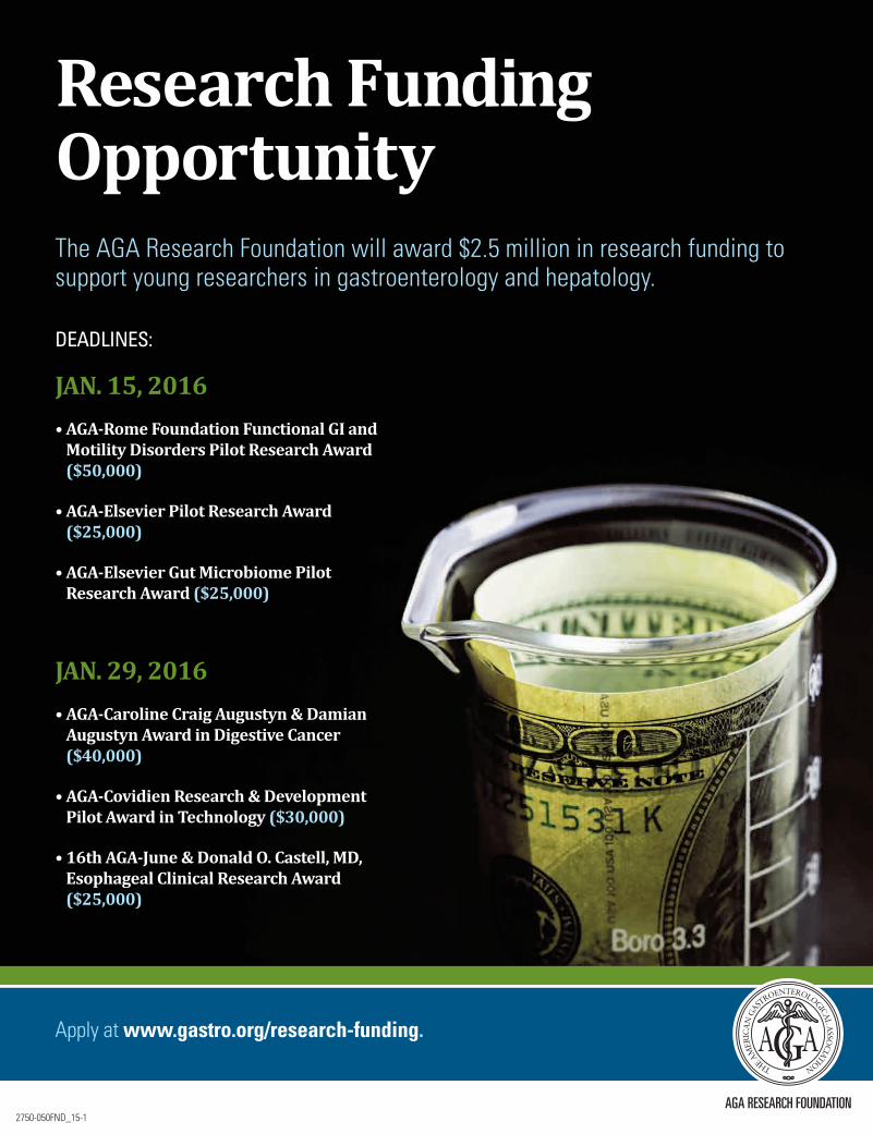
The AGA Research Foundation will award $2.5 million in research funding to support young researchers in gastroenterology and hepatology.
DEADLINES:
JAN. 15, 2016
• AGA-Rome Foundation Functional GI and
Motility Disorders Pilot Research Award
($50,000)
• AGA-Elsevier Pilot Research Award
($25,000)
• AGA-Elsevier Gut Microbiome Pilot
Research Award ($25,000)
JAN. 29, 2016
• AGA-Caroline Craig Augustyn & Damian
Augustyn Award in Digestive Cancer
($40,000)
• AGA-Covidien Research & Development
Pilot Award in Technology ($30,000)
• 16th AGA-June & Donald O. Castell, MD,
Esophageal Clinical Research Award
($25,000)
Apply at www.gastro.org/research-funding.

22 // THE NEW GASTROENTEROLOGIST: INSIGHTS FOR FELLOWS & YOUNG GIS FALL 2015
Hospital Clinicians
Commonly Work
While Sick By Tara Haelle // Frontline Medical News
©WAVEBREAKMEDIA LTD/THINKSTOCK.COM

FALL 2015 GIHEPNEWS.COM // 23
EARLY CAREER:
WORKING WHILE SICK
The vast majority of doctors and
other trained medical profession-
als at a hospital went to work
while sick within the past year,
even though they realized the
risk that decision places on pa-
tients, according to a recent study.
In fact, almost 1 in 10 hospital cli-
nicians worked while sick at least five
times in the past year, primarily because
of staffing concerns or not wanting
to let colleagues down, reported Julia
Szymczak, Ph.D., and her associates
at the Children’s Hospital of Philadel-
phia (JAMA Pediatr. 2015 July 6 [doi:
10.1001/jamapediatrics.2015.0684]).
“A combination of closed- and open-end-
ed questions illustrated that the decision
to work while sick was shaped by sys-
tems-level and sociocultural factors that
interacted to cause our respondents to
work while symptomatic, despite recog-
nizing that this choice may put patients
and colleagues at risk,” the authors wrote.
Of 929 surveys sent out, 538 clinicians
completed them, which included 280 of
459 physicians (61%) and 256 of 470
advanced-practice clinicians (54.5%).
The advanced-practice clinicians included
registered nurses, physician assistants,
clinical nurse specialists, registered nurse
anesthetists, and certified nurse midwives.
Of those who responded, 15.7% worked in
intensive care, 13.1% in surgery, 12.5% in
general pediatrics, and 44.8% in another
pediatric subspecialty.
Although 95.3% of respondents believed
working while sick put patients at risk,
83.1% reported having done so at least
once in the past year. Further, that propor-
tion included 52% of all respondents who
reported coming to work sick twice in the
past year and 9.3% who worked while ill
at least five times in the past year.
Nearly a third of respondents said they
would work even if they had diarrhea
(30%), while 16% said they would work
with a fever, and 55.6% would work with
acute respiratory symptoms, including
cough, congestion, rhinorrhea, and sore
throat.
But doctors were more likely than oth-
W H Y D O H O S P I T A L D O C T O R S G O T O W O R K S I C K ?IN THEIR OWN WORDS ►
Attending physician, pediatric subspecialty: “Our division does not have any systems in place to support physicians calling out sick. So on the occasions where I have called out sick, it was a disaster: Patients were not called to reschedule, phone calls were not for-warded, etc.”
Attending physician, emergency department: “There is an un-spoken understanding that you probably should be on your deathbed if you are calling in sick. It inconveniences colleagues, is compli-cated to pay back shifts, and makes me look bad to do so (like I am weak or something). It is much, much less stressful to suck it up and come in sick than call out.”
Attending physician, ICU/critical care: “There is no reliable mechanism to have another person cross-cover on short notice. Ev-eryone is busy. If a person is not on service, he/she is usually doing something that is not easily disrupted. I feel extremely guilty about needing someone else to cover me due to an illness.” n
From JAMA Pediatr. 2015 July 6 (doi:10.1001/jamapediat-
rics.2015.0684). Quotes are from a cross-sectional, anonymous
survey administered to 459 attending physicians and 470 ad-
vanced-practice clinicians.
24 // THE NEW GASTROENTEROLOGIST: INSIGHTS FOR FELLOWS & YOUNG GIS FALL 2015
EARLY CAREER:
WORKING WHILE SICK
er professionals to say they would go to work
with these symptoms: 38.9% of doctors would
work despite diarrhea, compared with 19.9%
of advanced-practice clinicians. Doctors and ad-
vanced-practice clinicians would also work with
acute respiratory symptoms (60% vs. 50.8%, re-
spectively), a fever only (21.8% vs. 9.8%), and fe-
ver and chills with body aches (18.6% vs. 10.9%,
all P < .03).
Nearly every respondent (98.7%) said they
worked despite being sick because they did
not want to let their colleagues down, just as
almost all of them worried the hospital would
not have enough staff (94.9%) or that they
would let their patients down (92.5%).
Smaller majorities of respondents also worked
because others also work while sick (65%),
worried their colleagues would ostracize them
(64%) if they didn’t work, were concerned about
their patients’ continuity of care (63.8%), had
unsupportive leadership (56.2%), or believed
they could not be easily replaced (52.6%).
Among the 316 respondents who filled in addi-
tional reasons, 64.9% said they had a very hard
time finding someone to cover their shift, 61.1%
described a strong cultural norm to work unless
extremely sick, and 57% expressed uncertainty
about what is considered “too sick to work.”
The Centers for Disease Control and Preven-
tion funded the research. The authors reported
no disclosures. n
W H E N H E A L T H C A R E W O R K E R S A R E S I C K , F I R S T D O N O H A R M
For centuries, a guiding principle for health care workers has been primum non nocere, or �rst do no harm. However, health care workers do exactly that when they work with patients
while ill themselves with contagious infections. Even common but untreatable infectious like enterovirus and respiratory syncytial virus can prove deadly to immunocompromised patients.
The propensity to work while ill is in�uenced by cultural trends. In past years, many ill physicians worked even to the point of receiving intravenous �uids while on the job; working while sick was regarded as a badge of courage. Dr. Szymczak and colleagues identi�ed as an issue the absence of an effec-tive sick relief system that has suf�cient �exibility to “staff up” during high rates of health care worker illness. Sick relief sys-tems and policies need to be clear regarding when health care workers should stay away from work, how patient coverage will be ensured, and the availability of and access to paid sick leave.
Determining what constitutes being too sick to work is compli-cated and lacks a suf�cient evidence base. Using a system that bases work restrictions on the presence of key symptoms may add clarity and enable health care workers to recognize when they need to stay home.
Creating a safer and more equitable system of sick leave for health care workers requires a culture change in many institu-tions to decrease the stigma – internal and external – associated with health care worker illness. Identifying solutions to prioritize patient safety must factor in workforce demands and variability in patient census and emphasize �exibility. Strong administrative and physician leadership and creativity are essential to support appropriate sick leave and ensure adequate staf�ng. Hospital leadership must ensure that the culture supports a paid sick leave policy that is adequate and nonpunitive. n
These comments are selected from an accompanying edi-
torial (JAMA Pediatr. 2015 July 6 [doi:10.1001/jamapedi-
atrics.2015.0994]), written by Dr. Jeffrey R. Starke of the
department of pediatrics at Baylor College of Medicine in
Houston, and Dr. Mary Anne Jackson of the division of infectious
diseases at Children’s Mercy Hospital, University of Missouri–
Kansas City. Dr. Starke and Dr. Jackson reported no disclosures.
Doctors were more likely than
other professionals to say they
would go to work with these
symptoms: diarrhea, fever, and
acute respiratory symptoms
including cough, rhinorrhea,
and sore throat.
FALL 2015 GIHEPNEWS.COM // 25
POSTFELLOWSHIP PATHWAYS:
BIOPHARMACEUTICAL CAREER
Postfellowship Pathways: Consider a
Career in the Biopharmaceutical IndustryBy Douglas S. Levine, M.D., FACG
What is your day-to-day
life like in the biophar-
maceutical industry?
My role is like an aca-
demic division head: I
create and implement
strategies; manage personnel and bud-
gets; engage in research, publications,
and training; and report to a depart-
ment head. I work with colleagues
with different specialized industry
expertise, in different medical special-
ties, inside and outside of departments
such as Medical Affairs: Research and
Development, Commercial, Regulatory,
Information Technology, Legal, Com-
pliance, Human Resources, Finance,
Program Management, Corporate
Communications, and Corporate Devel-
opment. Our activities align with com-
pany goals such as ensuring the safe
and appropriate use of products, help-
ing address patients’ unmet medical
needs, exploring new treatments for
research and development, and spon-
soring patient and professional educa-
tion. Leadership and communication
drive progress through the creation
of plan documents and reports, team
meetings, tele- and video conferences,
emails, phone calls, and web-based
applications. These activities often
lead to collaborative engagements
with external stakeholders including
content matter experts, members of
professional societies, patient advocacy
groups, other biopharmaceutical com-
panies, and health care professionals at
medical scientific congresses.
What do you enjoy most about
working in this industry?
In a word – people. It’s about patients
and the different individuals I work
with both in my company and the
health care sector more broadly. These
relationships enable the growth and
accomplishment essential to any ca-
reer in which one hopes to excel. It is
satisfying to achieve expertise in intel-
lectual disciplines and – for a medical
subspecialty – to continue to apply it
in patient care, research, writing, and
consultation. With the need for medical
input into multidisciplinary activities
that constitute product development
and commercialization, I have the op-
portunity to assist multifunctional and
multicultural global teams in actualiz-
ing complex projects. Because of this
multifaceted engagement, there are
myriad opportunities for interdepart-
mental and international collaboration
in addition to interacting with business
clients. With time and experience, one
can work as manager and leader as
well as assume teaching, coaching, and
mentoring responsibilities to support
the professional growth of colleagues –
things I especially enjoy.
What led you to pursue a career
in this �eld?
Early in training, I developed an
interest in the potential benefits of
Dr. Levine was a fellow and faculty member in the division of gastroenterology at the Univer-
sity of Washington School of Medicine, Seattle. He has worked in clinical research, medical
affairs, and management roles at AstraZeneca, Ironwood Pharmaceuticals, and Shire, where
he is currently Head of Medical Affairs, Gastroenterology, and Internal Medicine (dolevine@
shire.com). He is an employee and shareholder of Shire. The opinions expressed in this article
are those of Dr. Levine and not those of Shire.
26 // THE NEW GASTROENTEROLOGIST: INSIGHTS FOR FELLOWS & YOUNG GIS FALL 2015
POSTFELLOWSHIP PATHWAYS:
BIOPHARMACEUTICAL CAREER
a drug for inflammatory bowel dis-
eases. As an internist who depended
on prescribing medications to treat
patients, I became enthusiastic about
the possible novel uses of drugs and
the insights they might provide into
disease processes. Next, I joined a
team to investigate GI somatic ge-
netic abnormalities as predictors of
progression to cancer. This transla-
tional research involved collabora-
tion with basic and applied scientists
and clinicians, and yielded findings
possible only with integrated, multi-
disciplinary approaches; all of which
are intrinsic to the team-based cul-
ture of industry. I cared for patients
with Crohn’s disease and ulcerative
colitis, most of whom were refractory
to standard therapies. This prompt-
ed me to enlist as an investigator in
industry-sponsored clinical trials to
help create therapeutic innovations
and potentially provide benefits to
eligible patients. These experiences
have led me to recognize and under-
stand the fundamental role of indus-
try in drug development.
What types of industry
opportunities are available
for gastroenterologists?
Industry roles are available that
match both interests and experience.
Medical affairs has field roles (e.g.,
medical science liaison) focused on
engaging expert health care pro-
fessionals as well as headquarters
roles to oversee the accuracy of
promotional materials, respond to
questions from prescribers, and
support research and education
initiatives pertinent to products and
approved treatment indications.
Clinical research involves designing
trials in different phases of drug
development, monitoring studies
in progress, and analyzing and re-
porting results. Pharmacovigilance
involves monitoring, assessing, and
reporting adverse events with use
of drugs in clinical trials or during
the postmarketing period. Medical
device companies may attract gas-
troenterologists interested in devel-
oping diagnostic and treatment tools
such as endoscopic devices. Phy-
sicians with basic science training
may qualify for bench science roles
in early clinical development, non-
clinical research, pharmacology, and
toxicology. Physicians with business
training may qualify for commercial
marketing, analytics, operational,
health economic roles, or business
leadership positions.
Are there opportunities
to still see patients?
Many companies permit physicians
time for patient care apart from their
day-to-day responsibilities and there
are policies in place to address limits
on service and malpractice coverage.
Physicians new to the industry are
allowed to continue patient care in
order to sustain skills should they
choose to return to full-time practice.
Some do this to enhance their capa-
bilities in their industry roles, while
others view the activity as distract-
ing from company responsibilities.
Employees within the industry are
aware of the legal and regulatory
boundaries between individual pa-
tient-physician relationships and
company activities that necessitate
medical professional tradeoffs.
Instead of directly engaging in in-
dividual patient care, industry physi-
cians indirectly participate via drug
development that can help patients
and provide prescription options for
clinicians.
When is the best time
to look for a job in the
biopharmaceutical industry?
There is no one “best time”! Phy-
sicians differ in interests, training,
experience, goals, and career paths.
Companies vary in the types of jobs
offered and availability of entry-lev-
el positions versus roles for which
experience is required. Postfellow-
ship experience in research and
clinical practice is favored by many
companies but physicians can en-
ter the industry immediately after
training as well as at mid- and end
of career.
It is more common for applicants
to move from academic positions
than from clinical practices. Identify-
ing the best time to apply is depen-
dent upon what you learn about the
industry and individual companies.
Understanding the industry as a
whole is important but – as with
any job – you should be selective in
choosing which companies to target.
This research will allow you to find
the kinds of opportunities available
and help determine the best time to
begin your career transition.
What can be done during
fellowship/early career to prepare
for a career in industry?
During this professional stage, you
should focus on current activities in
gastroenterology. Your value to any
company is a knowledge of GI, spe-
cial clinical and research topics, in-
vestigational methods, clinical skills,
and real-world experience. Industry
work is team based. Gain experience
in working with other physicians,
nursing professionals, PhD research-
ers, and administrators. Capabilities
in computer applications may not be
essential but basic proficiency will
cushion your entry into an industry
that relies on digital information
management. Learn about industry
activities through various organiza-
tions such as the American Associ-
ation of Pharmaceutical Scientists,
Biotechnology Industry Organiza-
tion, and Pharmaceutical Research-
ers and Manufacturers of America.
Seek out industry members who
attend medical congresses and talk
with faculty at your institution who
participate in industry-sponsored
trials. Review online listings of com-

FALL 2015 GIHEPNEWS.COM // 27
POSTFELLOWSHIP PATHWAYS:
BIOPHARMACEUTICAL CAREER
panies and investigate websites to
learn what products are marketed
and/or are in clinical development.
Learn about the accomplishments,
policies, philosophies, and culture
of different companies so you can
differentiate them and determine
which may be the right fit.
In summary, biopharmaceutical
companies provide career opportu-
nities for gastroenterologists that
advance patient care and are pro-
fessionally satisfying. The variety of
job roles and large number of com-
panies, which differ in size, scope,
and culture, allow for mutually pro-
ductive matches among physician
scientists. If you want to contribute
your medical skills to the part of
the health care sector that drives
creation of drug products to address
unmet medical needs, consider a
career in the biopharmaceutical in-
dustry. n
ANSWERS // From page 3
Q1: ANSWER: D
CRITIQUE
This patient has no medical comorbidities, and based on
epidemiological studies will most likely have a peptic ulcer.
He had a bloody NG aspirate, which may limit endoscopic
visualization. After stabilization, the first priority is to en-
sure that timely endoscopy can be performed in an effec-
tive manner. The patient has no comorbidities to suggest a
variceal bleed, therefore intravenous octreotide (Choice C)
and prophylactic antibiotics (Choice E) would be inappro-
priate. The patient does not demonstrate altered mental
status, hemodynamic or respiratory instability, and there-
fore endotracheal intubation (Choice B) is not necessary.
Iced-saline gastric lavage is without demonstrated benefit,
and it may be harmful by inducing ischemic mucosal injury
to previously noninvolved areas of gastric mucosa (Choice
A). Intravenous erythromycin prior to endoscopy has been
reported to clear the stomach and improve endoscopic
examination. Furthermore, erythromycin was found to
be cost effective with an increase in quality-adjusted life-
years.
References
1. Carbonell, N., Poulwels. L., Boelley, P.Y., et al. Erythromycin infusion prior
to endoscopy for acute upper gastrointestinal bleeding: a randomized, con-
trolled trial. Am. J. Gastroenterol. 2006;101:1211-5.
2. Winstead, N.S., Wilcox, C.M.. Erythromycin prior to endoscopy for acute
upper gastrointestinal hemorrhage: a cost-effectiveness analysis. Aliment.
Pharmacol. Ther. 2007;15:1371-7.
Q2: ANSWER: D
CRITIQUE
Multiple colorectal adenomas may be attributable to the
autosomal dominant polyposis syndrome, familial adeno-
matous polyposis (FAP), due to germline mutations in the
APC gene, or secondary to mutations in the base excision
repair gene MUTYH. MUTYH-associated polyposis (MAP) is
an autosomal recessive trait characterized by the presence
of adenomatous polyposis and an increased risk of colo-
rectal cancer. Germline mutations in MLH1, MSH2, MSH6,
PMS2 genes would be indicative of Lynch syndrome. Lynch
syndrome is characterized by early onset of CRC and a
predisposition to cancers of the endometrium, ovary, stom-
ach, small bowel, urinary tract, and brain. Individuals with
Lynch syndrome do not develop a large number of polyps
as seen in MAP and FAP. Immunohistochemical analysis
and microsatellite instability testing of the sigmoid ade-
nocarcinoma would be indicated if the diagnosis of Lynch
syndrome were being considered. BMPR1A, SMAD4 mu-
tations are associated with juvenile polyposis syndrome
(JPS) that is characterized by the development of multiple
juvenile polyps and not adenomas. STK11 gene mutations
are associated with Peutz Jeghers syndrome (PJS). Individ-
uals with PJS do not present with multiple adenomas but
instead have hamartomatous polyps.
Reference
1. American Gastroenterological Association medical position state-
ment: hereditary colorectal cancer and genetic testing. Gastroenterology
2001;121:195.
28 // THE NEW GASTROENTEROLOGIST: INSIGHTS FOR FELLOWS & YOUNG GIS FALL 2015
A PERSONAL STORY
A Personal Story:
The Unexpected Benets of Not Having It AllBy Anne Peery, M.D., MSCR
Dr. Peery is an assistant professor of medicine at the University of North Carolina, Chapel Hill.
It felt awkward being asked to
write an article on work-life bal-
ance for young gastroenterolo-
gists. My life is rarely balanced. I
owed three papers to the mentor
who suggested I take this on. My
husband and I were getting ready to
move. We had been eating a lot of
take-out at home, burritos and burg-
ers, and my 13-month-old son had
learned that he really liked French
fries. Wordless, he would hold out
his hand and ask for them with a
grabbing motion. I had been mean-
ing to go to the gym more – doesn’t
every work-life balance author tell
you to make time for exercise? I
thought I might need to hire a per-
sonal trainer just to get this article
written. “I’ll do it,” I told the editor,
“but only if we can push the dead-
line back to August.”
When I started my fellowship in
gastroenterology, I barely gave a
thought to work-life balance. I want-
ed all of the research and clinical
opportunities I had been offered as
well as the people who ran the place
to think they had made a good choice
by offering those opportunities to
me. Even if there were days I might
have traded a 2 a.m. food impaction
patient for a good night’s sleep, or
wished for a lighter call schedule,
I knew that my fellowship was the
right choice for me.
Oddly, the lack of control over my
time as a fellow made my years of
training easier. I could not change
what rotation I was on, or how many
patients there would be, or how late
the day would go. Outside of clini-
cal decision making, I had only one
choice – gastroenterology or some-
thing else. I made that choice every
day for 4 years. Work-life balance
was often about remembering to eat
dinner and go to sleep at the end of
the day. After I had my first son, it
was about letting the baby sleep in
bed beside me while I wrote consult
notes.
I think most fellows are able to
work at this level because we know
that training is temporary. People
sometimes compare fellowship to a
marathon. At the end of a marathon,
of course, there is a finish line and
you get to stop running. As a trainee,
the finish line of fellowship was al-
ways clear. After that, the future de-
tails of my life could be worked out.
Once I had graduated, however,
I found that the work-life balance
I promised myself was more chal-
lenging than I had imagined. A lot
of work-life balance articles offer
lists of things to do, and I have made
good efforts at many of them. I made
time to go the gym. I went out with
friends. I scheduled quality time with
my family and sometimes I would get
a good night’s sleep. I investigated
different schemes for time man-
agement to make sure I was being
efficient. I made lists and I stayed or-
ganized and I met with my mentors.
These were all good things to do but
none of these things ever made my
life feel in balance.
The problem seemed to be that
there was always something else of
value that I could have been doing
instead of what I was actually doing.

FALL 2015 GIHEPNEWS.COM // 29
A PERSONAL STORY
Yes, I might be at the gym but should
I have been revising a grant appli-
cation instead? Yes, I was revising a
paper for submission but should I
have really been spending time with
my son who seemed irritable that
morning? When I was in training,
the few hours under my control
offered a limited array of choices.
After graduation, I was surrounded
with fantastic opportunities but
I found myself out of practice at
choosing between them. Even worse,
I found myself out of practice at
accepting the costs and benefits of
each choice that I made.
This was a problem that no work-
life balance checklist ever solved.
Instead, I had to learn to think about
things differently. After a decade of
doing the clinical and research work
assigned to me, I had to relearn to
make my own decisions and accept
responsibility for those choices.
After 2 years, I am finally succeed-
ing at a different kind of intellectual
discipline. If I am with my kids, I
know it is important to enjoy that
time instead of thinking about the
paper I meant to write last week.
Even more importantly, if I am
tucked into my office and finally
writing the paper, I know that I need
to accept that my husband and fan-
tastic preschool are taking good care
of my kids. I can look at a picture
of them and smile (there are lots
of these pictures on my computer)
but then I need to let myself enjoy
writing.
All of us in medicine are fortunate.
We get to do complex, intellectually
satisfying work that helps people. I
consider myself even more fortunate
to have supportive colleagues who
value my family and a supportive
physician husband who values my ca-
reer. At the end of the day, however,
all the support in the world will not
help you feel balanced if you focus on
everything you have to do instead of
the thing you are actually doing. Tak-
ing responsibility for my choices has
helped me be more present in every
part of my life. It has been the hard-
est part of becoming an attending but
it has given me a much better sense
of who I am.
So by all means, make time to
exercise. Go out with friends. Meet
with your mentors. Everything the
other articles tell you may be help-
ful. But also, be sure to appreciate
your life and your career as they are
happening. Be sure to accept and
embrace the sometimes-difficult
decisions you have to make about
time management. You will never
find some simple trick for getting
everything done. After all, it’s “ev-
erything.” Instead, enjoy and commit
to the things you take on. In a long
and full life in medicine and the
world, that will almost assuredly be
enough. nDr. Anne Peery and her two young sons at home.
CO
UR
TE
SY D
R. A
NN
E P
EE
RY
If I am with my kids, I
know it is important to
enjoy that time instead
of thinking about the
paper I meant to write
last week.

30 // THE NEW GASTROENTEROLOGIST: INSIGHTS FOR FELLOWS & YOUNG GIS FALL 2015
CLINICAL CHALLENGES
AND IMAGES
The Answer
This is intestinal transplant–associated microangi-
opathy (ITAM), a life-threatening complication of
allogeneic bone marrow transplantation. Trans-
plant-associated microangiopathy can be systemic
(characterized by hemolysis with schistocytes,
thrombocytopenia, renal and neurologic dysfunc-
tion, and elevated lactate dehydrogenase) or isolated to
the intestine.1 The incidence is unknown, but the con-
dition is likely underrecognized. Risk factors for ITAM
are intestinal GVHD, CMV colitis, and treatment with
calcineurin inhibitors. Figures A and B show the discrete
ulcers scattered along the entire colon and small whitish
plaques (thin arrows in Figure A). The latter was a mani-
festation of pseudolipomatosis (innocuous small air bub-
bles in the mucosa).
Pathology is shown in Figures C and D. Pathology is the
gold standard for diagnosis: Microangiopathy (thick ar-
rows), crypt loss, and platelet thrombi (thin arrows) are
present. Apoptotic bodies in the crypts can be seen (verti-
cal arrows in D).
This condition has to be distinguished from GVHD: The
therapy offers few good options — intensified immuno-
suppression/calcineurin inhibitors are discontinued, an
approach opposite of the treatment for GVHD.2
Recombinant thrombomodulin can be tried. Prognosis
is poor as underlined by mortality reported between 30%
and 57% of cases.
Tapering of immunosuppression in the patient proved
efficacious. He was discharged 6 days later with consider-
able reduction of volume of stools. n
References
1. Pettit, A.R., Clark, R.E. Thrombotic microangiopathy following bone mar-
row transplantation. Bone Marrow Transplant. 1994;14:495-504.
2. Nishida, T., Hamaguchi, M., Hirabayashi, N. et al. Intestinal thrombotic mi-
croangiopathy after allogeneic bone marrow transplantation: a clinical im-
itator of acute enteric graft-versus-host disease. Bone Marrow Transplant.
2004;33:1143-50.
From What’s Your Diagnosis? on page 5
A B
C D
©2015 Braintree Laboratories, Inc. 16-00590 March 2015
BRIEF SUMMARY: Before prescribing, please see full Prescribing Information and Medication Guide for SUPREP® Bowel Prep Kit (sodium sulfate, potassium sulfate and magnesium sulfate) Oral Solution. INDICATIONS AND USAGE: An osmotic laxative indicated for cleansing of the colon as a preparation for colonoscopy in adults. CONTRAINDICATIONS: Use is contraindicated in the following conditions: gastrointestinal (GI) obstruction, bowel perforation, toxic colitis and toxic megacolon, gastric retention, ileus, known allergies to components of the kit. WARNINGS AND PRECAUTIONS: SUPREP Bowel Prep Kit is an osmotic laxative indicated for cleansing of the colon as a preparation for colonoscopy in adults. Use is contraindicated in the following conditions: gastrointestinal (GI) obstruction, bowel perforation, toxic colitis and toxic megacolon, gastric retention, ileus, known allergies to components of the kit. Use caution when prescribing for patients with a history of seizures, arrhythmias, impaired gag reflex, regurgitation or aspiration, severe active ulcerative colitis, impaired renal function or patients taking medications that may affect renal function or electrolytes. Pre-dose and post-colonoscopy ECG’s should be considered in patients at increased risk of serious cardiac arrhythmias. Use can cause temporary elevations in uric acid. Uric acid fluctuations in patients with gout may precipitate an acute flare. Administration of osmotic laxative products may produce mucosal aphthous ulcerations, and there have been reports of more serious cases of ischemic colitis requiring hospitalization. Patients with impaired water handling who experience severe vomiting should be closely monitored including measurement of electrolytes. Advise all patients to hydrate adequately before, during, and after use. Each bottle must be diluted with water to a final volume of 16 ounces and ingestion of additional water as recommended is important to patient tolerance. Pregnancy: Pregnancy Category C. Animal reproduction studies have not been conducted. It is not known whether this product can cause fetal harm or can affect reproductive capacity. Pediatric Use: Safety and effectiveness in pediatric patients has not been established. Geriatric Use: Of the 375 patients who took SUPREP Bowel Prep Kit in clinical trials, 94 (25%) were 65 years of age or older, while 25 (7%) were 75 years of age or older. No overall differences in safety or effectiveness of SUPREP Bowel Prep Kit administered as a split-dose (2-day) regimen were observed between geriatric patients and younger patients. DRUG INTERACTIONS: Oral medication administered within one hour of the start of administration of SUPREP may not be absorbed completely. ADVERSE REACTIONS: Most common adverse reactions (>2%) are overall discomfort, abdominal distention, abdominal pain, nausea, vomiting and headache. Oral Administration: Split-Dose (Two-Day) Regimen: Early in the evening prior to the colonoscopy: Pour the contents of one bottle of SUPREP Bowel Prep Kit into the mixing container provided. Fill the container with water to the 16 ounce fill line, and drink the entire amount. Drink two additional containers filled to the 16 ounce line with water over the next hour. Consume only a light breakfast or have only clear liquids on the day before colonoscopy. Day of Colonoscopy (10 to 12 hours after the evening dose): Pour the contents of the second SUPREP Bowel Prep Kit into the mixing container provided. Fill the container with water to the 16 ounce fill line, and drink the entire amount. Drink two additional containers filled to the 16 ounce line with water over the next hour. Complete all SUPREP Bowel Prep Kit and required water at least two hours prior to colonoscopy. Consume only clear liquids until after the colonoscopy. STORAGE: Store at 20°-25°C (68°-77°F). Excursions permitted between 15°-30°C (59°-86°F). Rx only. Distributed by Braintree Laboratories, Inc. Braintree, MA 02185
IMPORTANT SAFETY INFORMATION
SUPREP® Bowel Prep Kit (sodium sulfate, potassium sulfate and magnesium sulfate) Oral Solution is an osmotic laxative indicated for cleansing of the colon as a preparation for colonoscopy in adults. Most common adverse reactions (>2%) are overall discomfort, abdominal distention, abdominal pain, nausea, vomiting and headache.
Use is contraindicated in the following conditions: gastrointestinal (GI) obstruction, bowel perforation, toxic colitis and toxic megacolon, gastric retention, ileus, known allergies to components of the kit. Use caution when prescribing for patients with a history of seizures, arrhythmias, impaired gag reflex, regurgitation or aspiration, severe active ulcerative colitis, impaired renal function or patients taking medications that may affect renal function or electrolytes. Use can cause temporary elevations in uric acid. Uric acid fluctuations in patients with gout may precipitate an acute flare. Administration of osmotic laxative products may produce mucosal aphthous ulcerations, and there have been reports of more serious cases of ischemic colitis requiring hospitalization. Patients with impaired water handling who experience severe vomiting should be closely monitored including measurement of electrolytes. Advise all patients to hydrate adequately before, during, and after use. Each bottle must be diluted with water to a final volume of 16 ounces and ingestion of additional water as recommended is important to patient tolerance.
For additional information, please call 1-800-874-6756 or visit www.suprepkit.com

A CLEAN SWEEP
#1 MOST PRESCRIBED, BRANDED BOWEL PREP KIT1
©2015 Braintree Laboratories, Inc. 16-00590 March 2015
*This clinical trial was not included in the product labeling. †Standard 4-Liter Prep [sulfate-free PEG electrolyte lavage solution]. ‡Based on investigator grading. §Statistically signifi cant difference. References: 1. IMS Health, NPA Weekly, March 2015. 2. Rex DK, Di Palma JA, Rodriguez R, McGowan J, Cleveland M. A randomized clinical study comparing reduced-volume oral sulfate solution with standard 4-liter sulfate-free electrolyte lavage solution as preparation for colonoscopy. Gastrointest Endosc. 2010;72(2):328-336. 3. Rex DK, Schoenfeld PS, Cohen J, et al. Quality indicators for colonoscopy. Gastrointest Endosc. 2015;81(1):31-53. 4. SUPREP Bowel Prep Kit [package insert]. Braintree, MA: Braintree Laboratories, Inc; 2012.
EFFECTIVE RESULTS IN ALL COLON SEGMENTS
>90% no residual stool in all colon segments compared
to Standard 4-Liter Prep2*†‡
· These results were statistically signifi cant in the cecum (P=.010)2*§
· Signifi cantly more subjects in SUPREP® group had no residual
fl uid in 4 out of 5 colon segments2*‡
Help meet Gastroenterology Quality Improvement Consortium (GIQuIC) benchmarks for 85% quality cleansing3 with the split-dose effi cacy of SUPREP ® Bowel Prep Kit.4