the physics of lasers as applied to medicine sandy mosse medical physics, uclh national medical...
TRANSCRIPT

The physics of lasers as applied to medicine
Sandy Mosse
Medical Physics, UCLH
National Medical Laser Centre, UCL

Light
Amplification by
Stimulated
Emission of
Radiation
• Lasers– basic principles– common types
• Tissue optics & treatments– scattering & absorption– modes of interaction
• Safety
Contents

Spontaneous Emission
EnergyLevels Photon

Stimulated Emission and Absorption

Population inversion

Lasers
mirrors create illusion of a long tube
But diode laser “Collimation”
Low gain (say 10% per metre) requires long tube

Power / Energy
• Power (Watts) = rate at which energy is delivered
• Energy (Joules) = amount of energy delivered
• 1 Watt = 1 Joule per second
• Energy = power x time, J = Wt

Some lasers in medical useCO2 10.6 micronEr:YAG 2.94 mironHo:YAG 2.1 micronNd:YAG 1064 nmAr 488/514 nmKr 647 nmCu vapour 511/578 nm Dye eg 630/675 nmDiode eg 600 / 905 nmExcimer > 350 nm

Boxes

Laser/Tissue interaction
RefractionScatter Absorption


Laser penetration depths

Photothermal - low power density
Coagulation: Thermal denaturation of proteins at T > 42.5 oC At 60 oC, soft tissue is coagulated in 1 second Disruption of tri-helical structure of collagen Tissue whitening due to increased optical scattering Tissue shrinkage Thrombus formation Haemostasis

Mesenteric vessel photothermally coagulated by a 60W Nd:YAG laser

Photothermal - high power density. Ablation

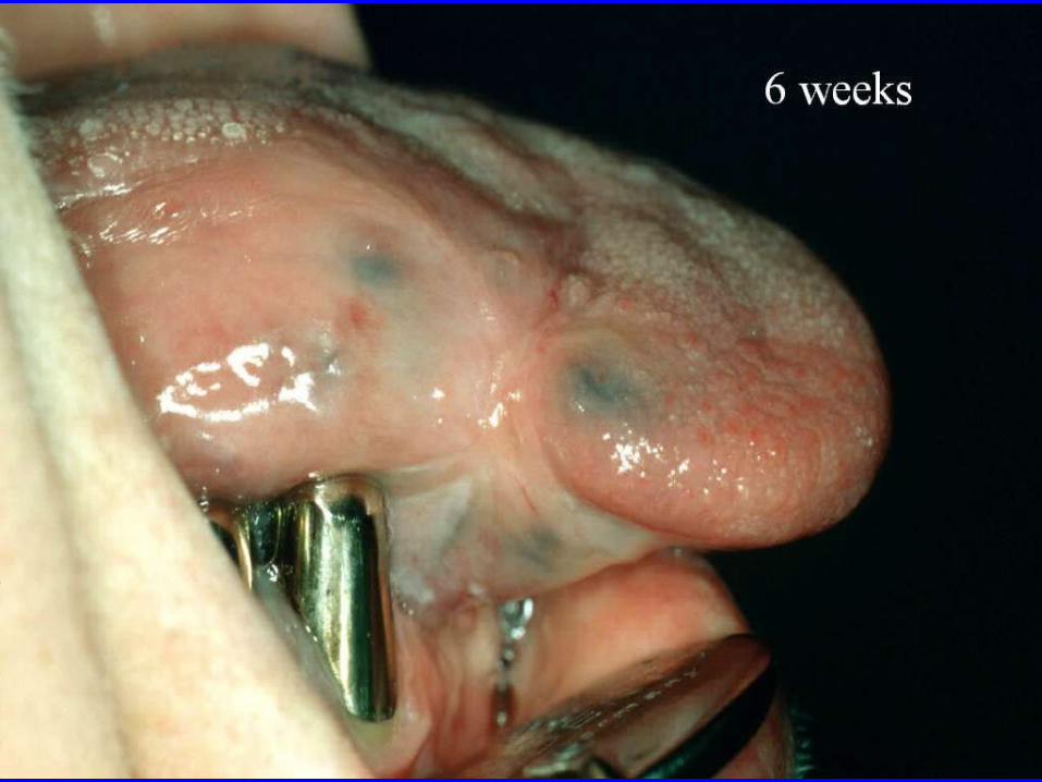
Tongue before & after CO2

Charring: Carbonisation of residual cellular components c. 4000C, tissue blackens and smokes 5000C, tissue burns and evaporates Hyperthermia: Cell death caused by long exposure to T > 42.50C Welding: Thermal bonding of vessels and nerves.

Effect of power density on tissueP
ower
Den
sity
W/c
m2
Time (s)

Photoablative effect ofUV excimer laser light
Binding Energyapprox. 3.5 eV
PhotonEnergy
approx. 6.4 eV
Approx. 0.25 mCorneal Tissue
Excimer Beam
Corneal Tissue
Excimer Beam

Precision of the Excimer Laser

LASIK for Myopic ablation
Cornea
DesiredCorneal surface
Tissue to be removed
LLaseraser AAsssistedsisted
IIn n SiSitutu KKeratomileusiseratomileusis

LASIK in MyopiaLASIK in Myopia


LASIK in MiopiaLASIK in Miopia

LASIK in MyopiaLASIK in Myopia

Plasma formed by a focused Q-switch laser

Laser fragmentation of a kidney stone

Pulse dye laser fragmentation of a kidney stone

Photodynamic Therapy - PDT
Taken from http://omlc.ogi.edu

Dougherty’s pet cat

Modern times

Photosensitising Drugs
• Based on porphyrins or similar structures
• Absorbed photon energy is transferred to form singlet oxygen
• 1O2 highly reactive, reacts within ~ 20 nm
• Sufficient cell damage causes cell death
• Should concentrate in the tumour

Kills, rather than annihilates, cells.
• Direct photodamage: 1-2 log kill of tumour cells1. Treatment needs 6 - 8 log
• Most cells die as a result of ~– Damage to microvasculature – Inflamatory reaction– Immune response

AdvantagesThe alternatives;~
Surgery
Chemotherapy is systemic
Radiation serious side effects. Once only.
PDT kills cells but does less damage to collagenous tissue structures, and normal cells will repopulate these structures.
PDT is potentially a low-cost minimally invasive localised treatment.
PDT is repeatable, unlike radiation therapy.
Allows wide area shallow treatment

Disadvantages
• Light sensitive patient
• Poorly defined volume of treatment
• Access for light
• Variable potency

Light sensitivity

Achilles’ heal


Laser safety
• Many types of laser – Local safety rules - written for each laser in
each location.
• Laser light is just light,nothing magic, but may be HIGHLY COLLIMATED – Travel a long way without diverging – Focus to a tiny spot - high power density

Laser hazards
• Exposure >100s photochemical hazard
• <100s and >100s thermal hazard
• Nanosecond photoacoustic shock
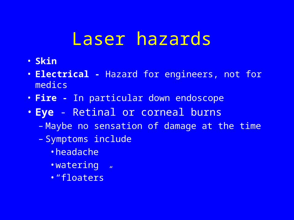
Laser hazards • Skin• Electrical - Hazard for engineers, not for medics• Fire - In particular down endoscope
• Eye - Retinal or corneal burns– Maybe no sensation of damage at the time– Symptoms include
• headache• watering• “floaters”

Eye
Laser (visible & NIR)

Electromagnetic SpectrumW
avel
engt
h (m
)
Gamma rays10-13
10-10
10-6
103
10-9
100
10-2
X rays
Ultra violet
Infra red
Radio frequency
UHFVHF
RadarFM radio
Broadcast
Power lines
Microwaves
Angstrom (Å)nano (nm)
micro (m)
kilo (Km)
centi (cm)1400 nm
700 nm
400 nm
visible
near IR

Ultraviolet RadiationUV-B and UV-C are absorbed by thecornea, over-exposure resulting inphotochemically induced photokeratitis(snow-blindness, arc-eye or welder's flash)and conjunctivitis.
UV-A (315-400 nm) is absorbed in thelens, over-exposure resulting in thephotochemical formation of an area ofopacity i.e. a cataract.

Eye - visible (400-700 nm) light
• Focussed to a point - intensified x100,000
• Blink protects but takes ~ 1/4 second
• Safe limit = 1mW, – bright London sunlight ½ mW/mm2
• Fovea only 3-4% of area of retina

Eye - Invisible light
• NIR transmitted to the retina. No deliberate focussing but, relaxed eye will focus anyway.
• IR (>1400 nm) absorbed by cornea, lens etc.– glass blower’s or furnace man’s cataract
• NO BLINK RESPONSE

Damage to the RetinaThe thin highly absorbing layer makes the retina particularlysensitive to damage from burns.
Burn to the peripheral retina peripheral blind spot Burn to fovea loss of central vision (major vision loss) Burn on exit of optic nerve total or partial blindness Haemorrhage & debris in the vitreous humour
permanent impairment of varying degree
Photochemical damage is possible but less common.
The blink reflex, effective for 400 nm< < 700 nm, limitsthe maximum retinal exposure time to 0.25 s.

Eye Injury
Nd:YAG (1064 nm) - permanent damage. Victim saw white flash, heard a click, then immediately a dark spot in visual field

Eye Injury
Lasers which destroy retinal tissue can cause haemorrhaging into the vitreous - this will eventually clear but the retinal damage is permanent

Safety goggles
CHECK THEY ARE FOR THE CORRECT WAVELENGTH
Usually 4-6 O.D. (1 OD = 1 log reduction = 10% transmission)

Fumes & debris
• Plume of smoke during laser ablation– may contain viruses etc.
• Always use a fume extractor as close as possible to the treatment area