the spectral sensitivit of y calliphora maggots
TRANSCRIPT
. Exp. Biol. (1961), 38, 237-248 2 3 7
ih 7 text-figures
Printed in Great Britain
THE SPECTRAL SENSITIVITY OFCALLIPHORA MAGGOTS
BY PRISCILLA H. STRANGE
Visual Research Division, Ophthalmological Research Unit {Medical Research Council),Institute of Ophthalmology, Judd Street, London, W.C. 1
(Received 23 September i960)
If fly larvae are put on a table by a window they immediately begin to travel awayfrom the light. Turn the table round, and the maggots change direction so as to movedirectly away from the light again. They travel by a 'tacking' process—the head endis extended to right and left alternately, and each time the rear end is drawn after it.The animal moves on a straight course if this process is symmetrical; should a move-ment of the head bring it into a region of higher illumination or other undesirablecondition the head is withdrawn, and a second step made to the opposite side, turningthe animal round.
Placed between two opposing lights, the maggots will travel towards the weaker, andthe direction of travel may be reversed by increasing the intensity of the weaker lightuntil it, in turn, is the brighter. In the present work this reaction is used to equatethe luminosity of lights of different wavelengths to a standard, and hence to draw aspectral sensitivity curve.
Bolwig (1946) performed a series of detailed experiments, behavioural and micro-anatomical, on the larva of the related Musca domestica, and says: ' The search for thelight-sensitive cells has resulted in rinding some cells at the bottom of a pair of pockets(directed forward) in the anterior end of the pharyngeal skeleton; these cells mustundoubtedly be regarded as the light-sensitive cells.'
The gulf which separates the maggot from the adult fly, with its compound eyesand array of ocelli, is crossed in the 10 days or so of pupation. Ellsworth (1933)had thought that the tips of the maxillary lobes of the fleshfly larva were its lightreceptors, and that they must degenerate at metamorphosis without taking part in theformation of the compound eyes; this is not the case in larvae with lateral ocelli.Bolwig's finding, that these maxillary papillae are chemoreceptors in the houseflylarva, means that the compound eye may evolve from the rudimentary lateral larvalstructure. It would be interesting to know if any light-sensitive pigment used by themaggot is retained through metamorphosis, or whether the emancipated fly hasreplaced the whole chemical basis of its larval light sense.
Much attention has been paid to the spectral sensitivity of adult blowflies, usingbehavioural methods (Schneider, 1956) and electrophysiological means (e.g. Walther& Dodt, 1959; Autrum, 1955). Apart from the work of Mast (1917) on unidentifiedblowfly maggots, and Bolwig (1946) on housefly larvae, however, there appears tobe no information about spectral sensitivity of larval Diptera.
The wealth of information about adult flies appears rather inconsistent at firstsight. Comparison of the various spectral sensitivity curves, which are illustrated in
238 P. H. STRANGE
Figs. 6 and 7, is reserved for the discussion; it will then be possible to include thepresent results, and to try to show an emerging pattern of dependence on intensityand other experimental conditions.
APPARATUS
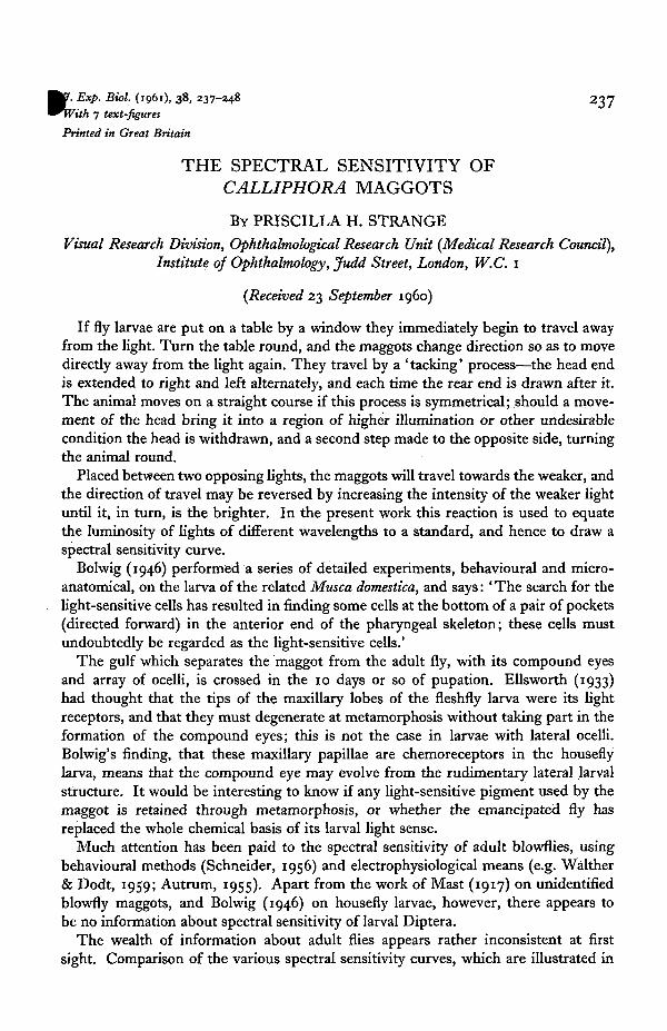
A metal bridge was made, covered with black adhesive plastic film. The platformof the bridge was 12 in. long by 4 in. wide, and the sides were turned up to form1 \ in. high walls to prevent the maggots from straying. The ends of the platform wereturned down to form sloping ramps 4 in. high, each leading down into a steep-walleddish to collect the maggots. Fig. 1 illustrates the experimental arrangement.
oU
Bridge^ TifiSlldllllx1
Dish
Monochromaticlight
Black box '
Fig. 1. A diagrammatic longitudinal section through the apparatus.
The bridge and the two dishes were placed symmetrically in a black box 17 in.long. In the end-walls of the box, on a level with the platform, were 2 cm. squareholes filled with flashed-opal glass, illuminated respectively by the two light sources.Behind one opal square was a calibrated monochromatic light source. This consistedof a 32 V., 100 W. Philips projector lamp, S, fan-cooled. Two pieces of Chance glassabsorbed heat from the lamp, an achromatic lens combination of focal length i£ in.collimated the beam, which passed through a 2 cm. square Balzer interference filter, A(type B, 40 % transmission, the width of the band of wavelengths being about 10 m/xat the 20 % transmission level), and through neutral density filters to the opal square.The wavelengths used in all the experiments were 402, 417, 442, 462, 489, 499, 513,531. 554> 579 anc* 602 m/x. At the lower intensity level (level B) filters at 362, 392, 621and 640 mjx were included. A filter at 470 mfi was added to the later series, at thehigher intensity level (level A).
Suitable screening masks were provided, and the opal square was inspected frominside the black box before each run to confirm that stray light was excluded.
Behind the second opal square, at the other end of the bridge, was the standardlight source, which consisted of a 12 V., 6 W. car headlamp bulb, S', another pieceof opal glass to diffuse the light, and a neutral density filter. (Both incandescent lampswere supplied by 50 cyc./sec. a.c, using suitable transformers.)
The standard light, without its neutral filter, gave a reading of 0-3 f.c. at the middleof the bridge, measured with a Holophane photometer. Two values of neutral filterswere used, one (level A) giving approximately 001 f.c. at the middle of the bridge,and the other (level B) o-ooi f.c. (1 f.c. = 1076 lumens per square metre).
The spectral sensitivity of Calliphora maggots 239
The relative energy of the monochromatic source was found at each wavelength bya null method: the photo-current of a Mazda type 27 M1 photomultiplier, of knownrelative spectral sensitivity, was kept constant by the addition of neutral densityfilters and neutral wedges.
The photomultiplier was calibrated at the National Physical Laboratory, and thenominally neutral filters and wedges were calibrated by comparison with a set ofrotating sectors.
The same Ilford neutral density filters, in steps of approximately 0-25 log unit, werealso used for the experiments.
MAGGOTS
The Calliphora vomitoria maggots, used in most of the experiments, were sold as' gentles' for fishing. A sample of each batch was allowed to develop into flies, whichwere all identified as C. vomitoria.
The C. erythrocephala stock was kindly supplied by Prof. V. B. Wigglesworth.The C. vomitoria were bred on stable manure, C. erythrocephala on lean beef. In
both cases the animals were used after they had left their food, which was no longervisible in the alimentary tract.
METHOD
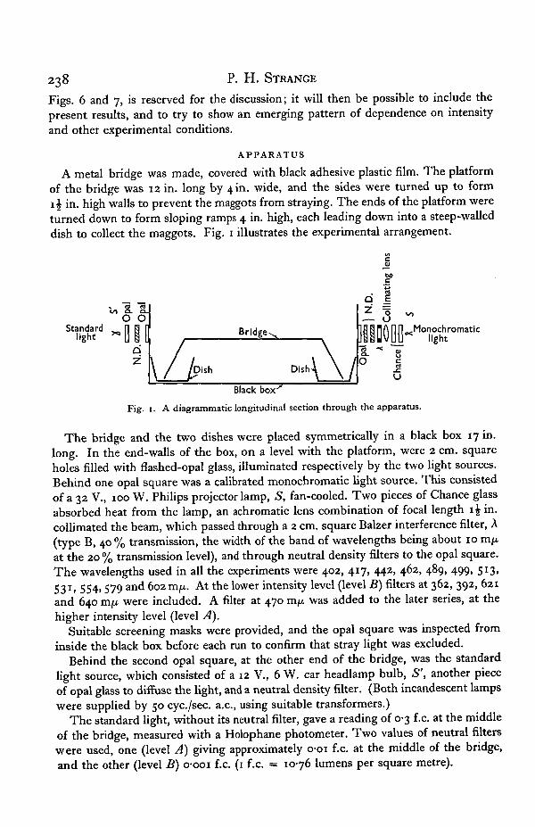
About forty maggots, dark-adapted for at least 2 hr., were placed in the middle of thebridge, using a wide funnel, in very faint light. A black cloth was spread over theouter box, and the two lights, one at each end of the bridge, were switched on simul-taneously. After 10 min., the lights were switched off, and any maggots and pupaeleft on the bridge were discarded. The maggots in each dish were counted, and thenumber which had travelled towards the standard light was expressed as a percentageof the total in both dishes. (With the standard light alone, at least 70 % travelled awayfrom the light.) Each interference filter was used in combination with a series of neutraldensity filters, at approx. 0-25 log unit intervals; according to the intensity of thismonochromatic light either more or less than half of the maggots went towards thestandard light. An intensity was found by interpolation at which the two lights wereequally effective, as in Fig. 2. The energy at each wavelength, matched like this to thesame standard, is the relative threshold on an equal energy basis. To get the relativequantum threshold, the energy needed at each wavelength, A, must be divided by themagnitude of a quantum of energy, which is proportional to i/A.
The reciprocal of the relative quantum threshold, expressed as a percentage of itsmaximum, is the percentage quantum sensitivity. This function B, plotted against A,may be considered a first approximation to the absorption spectrum of a hypo-thetical visual pigment.
RESULTS
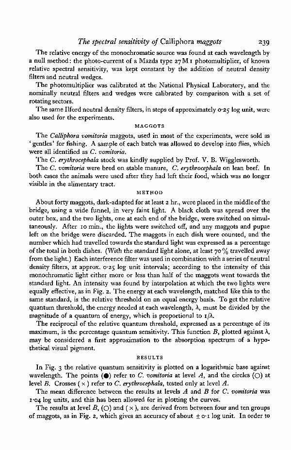
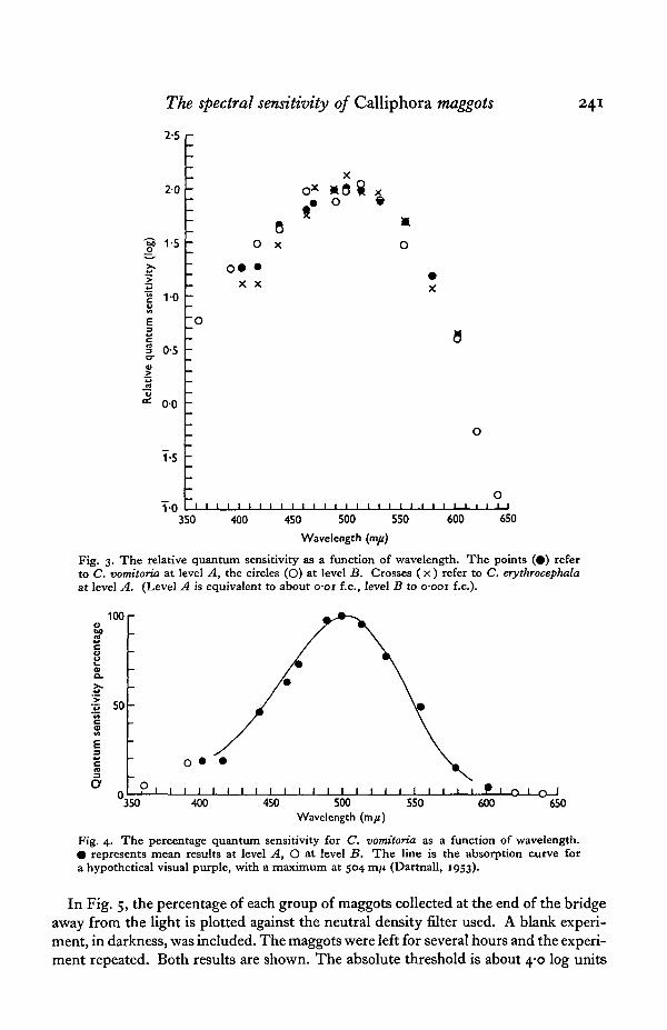
In Fig. 3 the relative quantum sensitivity is plotted on a logarithmic base againstwavelength. The points (#) refer to C. vomitoria at level A, and the circles (O) atlevel B. Crosses ( x ) refer to C. erythrocephala, tested only at level A.
The mean difference between the results at levels A and B for C. vomitoria was1-04 log units, and this has been allowed for in plotting the curves.
The results at level B, (O) and ( x), are derived from between four and ten groupsof maggots, as in Fig. 2, which gives an accuracy of about ± o-i log unit. In order to
240 P. H. STRANGE
find the maximum more exactly for C. vomitoria at level A at least 15 groups of mag-gots were used to determine each of the points around 500 m/x, and 32 groups forthe wavelength 499 rcifx. which was taken as 100% (2-0 on the logarithmic scale).The three sets of results are, within experimental accuracy, the same.
1 1-5 2Filter density (log)
Fig. 2. Results of tests done on one day at 499 m/t. Each point represents the result for agroup of maggots placed between the standard light and a given intensity of the monochromaticlight, determined by the neutral density filter shown in the abscissa. When the monochro-matic light is more effective, over 50 % travel towards the standard light.
The lower level of illumination, level B, was used in order to extend the wave-length range (362-640 m/x). At level A it was restricted (from 402 to 602 m/x) bythe energy of the monochromatic source, but this level had the advantage that themaggots could be used when their response to light was not at its strongest.
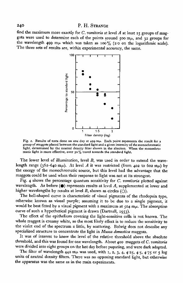
Fig. 4 shows the percentage quantum sensitivity for C. vomitoria plotted againstwavelength. As before (#) represents results at level A, supplemented at lower andhigher wavelengths by results at level B, shown as circles (O).
The bell-shaped curve is characteristic of visual pigments of the rhodopsin type,otherwise known as visual purple; assuming it to be due to a single pigment, itwould be best fitted by a visual pigment with a maximum at 504 m/x. The absorptioncurve of such a hypothetical pigment is drawn (Dartnall, 1953).
The effect of the epithelium covering the light-sensitive cells is not known. Thewhole maggot is creamy white, so the most likely effect is to reduce the sensitivity tothe violet end of the spectrum a little, by scattering. Bolwig does not describe anyspecialized structure to concentrate the light in Musca domestica maggots.
It was of interest to know the level of the relative threshold above the absolutethreshold, and this was found for one wavelength. About 400 maggots of C. vomitoriawere divided into eight groups on the last day before pupating, and were dark adapted.
The filter of wavelength 442 m/* was used, with 1, 2, 3, 4, 4-25, 4-5, 475 or 5 logunits of neutral density filters. There was no opposing standard light, but otherwisethe apparatus was the same as in the main experiments.
The spectral sensitivity of Calliphora maggots 241
20
1-5
1 0
0-5
00
1-5
1 0
O** *0$ x* ° *
8Ox O
X X
1 I i I I I Io
350 400 600 650450 500 550
Wavelength (m/t)
Fig. 3. The relative quantum sensitivity as a function of wavelength. The points (•) referto C. vomitoria at level A, the circles (O) at level B. Crosses ( x ) refer to C. erythrocephalaat level A. (Level A is equivalent to about o-oi f.c, level B to o-ooi f.c).
100o2
2.5-5 50cM
E
350 400 450 500Wavelength (m/i)
550 600-O—1—O—I
650
Fig. 4. The percentage quantum sensitivity for C. vomitoria as a function of wavelength.• represents mean results at level A, O at level B. T h e line is the absorption curve fora hypothetical visual purple, with a maximum at 504 ran (Dartnall, 1953).
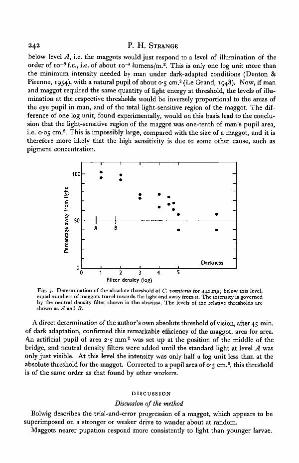
In Fig. 5, the percentage of each group of maggots collected at the end of the bridgeaway from the light is plotted against the neutral density filter used. A blank experi-ment, in darkness, was included. The maggots were left for several hours and the experi-ment repeated. Both results are shown. The absolute threshold is about 4-0 log units
242 P. H. STRANGE
below level A, i.e. the maggots would just respond to a level of illumination of theorder of icr6 f.c, i.e. of about icr5 lumens/m.2. This is only one log unit more thanthe minimum intensity needed by man under dark-adapted conditions (Denton &Pirenne, 1954), with a natural pupil of about 0-5 cm.2 (Le Grand, 1948). Now, if manand maggot required the same quantity of light energy at threshold, the levels of illu-mination at the respective thresholds would be inversely proportional to the areas ofthe eye pupil in man, and of the total light-sensitive region of the maggot. The dif-ference of one log unit, found experimentally, would on this basis lead to the conclu-sion that the light-sensitive region of the maggot was one-tenth of man's pupil area,i.e. 0-05 cm.2. This is impossibly large, compared with the size of a maggot, and it istherefore more likely that the high sensitivity is due to some other cause, such aspigment concentration.
1 2 3 4 5Filter density (log)
Fig. 5. Determination of the absolute threshold of C. vomitoria for 442 m/j; below this level,equal numbers of maggots travel towards the light and away from it. The intensity is governedby the neutral density filter shown in the abscissa. The levels of the relative thresholds areshown as A and B.
A direct determination of the author's own absolute threshold of vision, after 45 min.of dark adaptation, confirmed this remarkable efficiency of the maggot, area for area.An artificial pupil of area 2-5 mm.2 was set up at the position of the middle of thebridge, and neutral density filters were added until the standard light at level A wasonly just visible. At this level the intensity was only half a log unit less than at theabsolute threshold for the maggot. Corrected to a pupil area of 0-5 cm.2, this thresholdis of the same order as that found by other workers.
DISCUSSION
Discussion of the method
Bolwig describes the trial-and-error progression of a maggot, which appears to besuperimposed on a stronger or weaker drive to wander about at random.
Maggots nearer pupation respond more consistently to light than younger larvae.
The spectral sensitivity of Calliphora maggots 243
For example, the same light which made 70 % travel away, 2 days before pupation,caused 97 % to travel away on the last day. In each case the maggots had rested un-disturbed in the dark overnight. When maggots which had travelled to the light weregathered up, rested in the dark and tested separately, they divided in the same ratioas before; the group which had travelled away from the light did the same. Thissuggests that the photonegative behaviour is superimposed on random wandering,rather than that any of the maggots were photopositive.
The finding that older larvae are more responsive to light does not agree withPatten (1916). From a teleological point of view, it is in the interest of the maggotsto leave the shelter of the food and disperse, before finding a dark corner to pupate.Bolwig attributes the mechanism of the more accurate orientation of older larvae,relative to the light, to the increasing shielding of the light-sensitive organs from lightcoming from behind; he says that younger larvae soon become tired and stop respond-ing to light, as was also found in the present work.
The only effect of this lower response on the relative threshold determinationis to make the graph of the percentage travelling away from the monochromaticlight (e.g. Fig. 2) slope less steeply—it does not change the point of crossing the 50 %line. Mast's work on blowfly maggots was a relative threshold, while Bolwig'sthreshold for the housefly maggot suffers the disadvantage of being an absolutethreshold.
Certain precautions were taken with a knowledge of the habits of maggots. Ifthey were damp from their food, they were mixed with dry bran before dark adapta-tion, since a smooth vertical surface presents no obstacle to a wet maggot. The bridgewas cleaned with a damp cloth between experiments to remove the trails of the pre-vious group, which maggots tend to follow, and the bridge was levelled with a spirit-level to ensure symmetry.
The cooling fan alone, without the lights, was found to have no effect.Patten (1914) found that maggots of C. erythrocephala responded equally to a steady
light and to a light interrupted 30 times a second, provided that the total quantity oflight per second was the same. Since the Bunsen-Roscoe law is thus obeyed, it canmake no difference whether 50 c./s. alternating or direct current is used for the incandes-cent stimulus lamps.
Comparison of present results with previous spectral sensitivity curves
In order to compare the present results for Calliphora larvae with the findings ofother workers on muscid larvae and flies, the line of Fig. 5 is redrawn in each part ofFigs. 6 and 7, extended to 362 and 640 m x̂.
First, attention must be drawn to the simple nature of the maggot's spectral sensi-tivity curve—there is no sign of any maximum away from 504 m .̂, either in the ultra-violet or red parts of the spectrum.
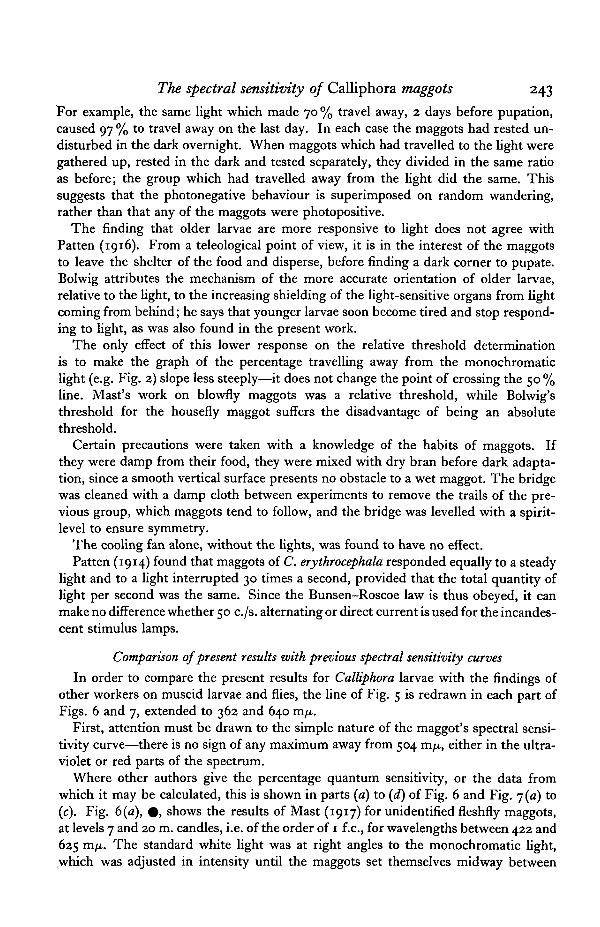
Where other authors give the percentage quantum sensitivity, or the data fromwhich it may be calculated, this is shown in parts (a) to (d) of Fig. 6 and Fig. 7 (a) to(c). Fig. 6 (a), # , shows the results of Mast (1917) for unidentified fleshfly maggots,at levels 7 and 20 m. candles, i.e. of the order of 1 f.c, for wavelengths between 422 and625 m/x. The standard white light was at right angles to the monochromatic light,which was adjusted in intensity until the maggots set themselves midway between
244 P. H. STRANGE
the two. The maximum is at 504 vcifj., the same as for the present work, but the curveis narrower.
Fig. 6(a), O, is the absolute quantum sensitivity for the housefly maggot for wave-lengths between 405 and 700 m/x, drawn from Bolwig (1946). The criterion here wasthe ability of maggots to re-orient themselves when the paper which they were on
(a)
Fig. 6. Percentage quantum sensitivities found by other workers for muscid flies and larvae.The present results for Calliphora larvae are repeated in each diagram (thick line), (a) Maggots;• , Mast, blowfly; O, Bolwig, housefly, (b) Schneider, C. erythrocephala. Adult (optokinetic).(c) Walther & Dodt, C. erythrocephala. Adult (electrophysiological). (d) Dormer & Kriszat.Adult fly (electrophysiological).
was turned through a right angle. The maggots were not all of the same age, describedas 'second and third instar larvae', which may have influenced the absolute thresholdmethod. (The peak at 520 m/u. is sharp compared with other spectral sensitivity curves.)Again, there is no return of sensitivity at the red end of the spectrum; the ultra-violetwas not investigated.
In Fig. 6(b) we come to the spectral sensitivity of adult flies, this time from^

The spectral sensitivity of Calliphora maggots 245
Schneider (1956), the only behavioural results for which we have the data in this form.This is an optokinetic method for C. erythrocephala, at a very low level of intensity,in the human scotopic region. The fly was glued to a glass capillary at the centre of aglass cylinder, outside which turned a concentric cylinder with vertical black stripes,illuminated by monochromatic light of wavelength between 425 and 570 m/x. Whenthe fly had enough light to see the stripes, it attempted to follow them. The maximumsensitivity was at 480 m/x.
Walther & Dodt (1959) used an electrophysiological method, and their results areshown in Fig. 6(c), for wavelengths between 290 and 675 m/x. This curve is the meanresult for 24 C. erythrocephala flies, all from the same stock. The criterion here was aconstant height of on-effect of the electroretinogram (e.r.g.) in response to 40 msec,flashes of light every 30 sec, for the intact fly. These workers found that the relativeheight of the secondary maximum at 630 m/x was dependent on intensity, while theultra-violet peak did not change with intensity in a regular way.
Donner & Kriszat (1949) also used the on-effect for the intact fly as an index ofthreshold between 440 and 640 m/x. The curve shown as Fig. 6 {a) is for an individualfly which showed the peak at 500 m/x most clearly. For others the sensitivity was stillrising at the violet end of their available spectrum. They found no apparent differencesbetween the species Musca domestica, Lucilia caesar, Calliphora vomitoria and Polleniarudis, and do not identify the fly to which this diagram refers. Adaptation increasedthe relative sensitivity to the violet end of the spectrum (420-40 m/x), the effect beinggreatest for green, then red, and then violet adapting lights.
Cameron (1939) found that houseflies were attracted by 366 m/x more than by anyother wavelength. Weiss, Soraci & McCoy (1941), also using a behavioural methodfor houseflies, found a maximum of response to an equal energy spectrum at 490 m/x,a minimum at 460 m/x, and the response was still rising at 360 m/x. Granit (1947,p. 297) mentions an electrophysiological experiment, in which the eye of the houseflywas found to be most sensitive to 490 m/x, sensitivity falling off rapidly on the long-wave side of the maximum. Granit extracted a 'very light-resistant carrot-colouredpigment' from the eye of the housefly. The only pigment bleached by light which hasso far been found in a fly was extracted by Bowness & Wolken (1959) from houseflyheads and had maximum absorption at 437 m/x.
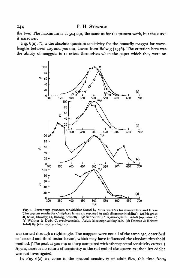
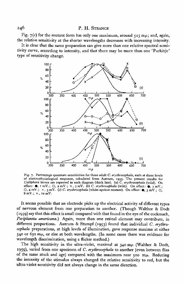
The work of Autrum (1955), using the on-effect of the e.r.g. as an index of sensi-tivity, may provide a key to this diversity. Autrum uses the isolated head of C.erythrocephala and determines the magnitude of electrical responses at each wave-length for an equal-quantum spectrum. He gives the results at a series of quantumlevels and, by replotting the response against quantum level, it is possible to find therelative quantum sensitivity at an arbitrary mV response level by interpolation. Thishas been done for three individual flies, at three levels of response, in increasing order,represented by # , O, and x in Fig. 7. Fig 7 (a) and (b) refer to two wild-typeC. erythrocephala, and y(c) to a white-apricot mutant of the same species. In Fig. 7(0)the graph at the lowest intensity, 0 , is of the same type as Fig. 6(c), and increasingintensity changes the wavelength of the peak around 500 m/x to about 530 m/x, atthe same time increasing the relative height of a peak at about 630 m/x. 7 (b) tells asimilar tale, even more dramatically, as the peak in the red completely dwarfs the restI at the highest intensity.
246 P. H. STRANGE
Fig. 7(c) for the mutant form has only one maximum, around 515 m/n; and, again,the relative sensitivity at the shorter wavelengths decreases with increasing intensity.
It is clear that the same preparation can give more than one relative spectral sensi-tivity curve, according to intensity, and that there may be more than one 'Purkinje'type of sensitivity change.
300 350 400 450 500 550 600 650 700
300 350 400 450 500 550 600 650 700
650 700
Fig. 7. Percentage quantum sensitivities for three adult C. erythrocephala, each at three levelsof electrophysiological response, calculated from Autrum, 1955. The present results forCalliphora larvae are repeated in each diagram (thick line), (a) C. erythrocephala (wild). Oneffect: • , 1 mV.; O, 2 mV.; x , 3 mV. (b) C. erythrocephala (wild). On effect: • , 1 mV.;O, 2mV.; x , 3 mV. (c) C. erythrocephala (white-apricot mutant). On effect: • , 3 mV.; O,8 mV.; x , 10 mV.
It seems possible that an electrode picks up the electrical activity of different typesof nervous element from one preparation to another. (Though Walther & Dodt(1959) say that this effect is small compared with that found in the eye of the cockroach,Periplaneta americana.) Again, more than one retinal element may contribute, indifferent proportions. Autrum & Stumpf (1953) found that individual C. erythro-cephala preparations, at high levels of illumination, gave response maxima at either540 or 630 m^, or else at both wavelengths. (In some cases there was evidence forwavelength discrimination, using a flicker method.)
The high sensitivity in the ultra-violet, maximal at 340 mfi (Walther & Dodt,I959)» varied from one specimen of C. erythrocephala to another (even between fliesof the same stock and age) compared with the maximum near 500 mfi. Reducingthe intensity of the stimulus always changed the relative sensitivity to red, but theultra-violet sensitivity did not always change in the same direction.

The spectral sensitivity of Calliphora maggots 247
Inspection of Figs. 6 and 7 shows that all these muscid flies have a maximum ofsensitivity near 500 m/x which is especially prominent at low levels of illumination.This may well be a continuance from the visual mechanism of the larval state, mostsensitive to 504 m/x. No light-sensitive pigment has yet been found to account forthis; the atypical pigment found in the housefly by Bowness & Wolken (1959), withits maximum absorption at 437 m/x, is not a likely candidate. The form of the spectralsensitivity curve of the maggot would suggest a visual purple (or rhodopsin) and thelow absolute threshold leads to the idea that such a pigment might be present inmeasurable concentration.
SUMMARY
1. The relative spectral sensitivity of larvae of Calliphora vomitoria and C. ery-throcephala has been determined, using the maggot's natural tendency to traveltowards the weaker of two opposing lights.
2. The response to light at each wavelength between 402 and 602 m/z. was thesame for the two species, within ± o-i log unit, and the results are well fitted by thebell-shaped curve characteristic of a (hypothetical) visual purple, maximal at 504 m/x.
3. Reduction of the intensity of the white standard light by one log unit did notchange the shape of the curve, and the extended wavelength range, between 362 and640 m/x, shows no sign of a subsidiary maximum of sensitivity. The levels of illumina-tion were io~2 and io~3 f.c, and the absolute threshold was found to be about io~6 f.c.
4. The spectral sensitivity found here is compared with the results of other workersfor muscid flies and larvae. It is concluded that one of the independent maxima ofsensitivity found in the flies is a continuance from the larval state.
I should like to thank Prof. V. B. Wigglesworth for the C. erythrocephala cultureused, and the many people who have helped me with their special knowledge andadvice.
REFERENCES
AUTRUM, H. (1955). Die spektrale Empfindlichkeit der Augenmutation white-apricot von Calliphoraerythrocephala. Biol. Zbl. 74, 515-24.
AUTRUM, H. & STUMPF, H. (1953). Elektrophysiologische Untersuchungen iiber das Farbensehen vonCalliphora. Z. vergl. Physiol. 35, 71-104.
BOLWIG, N. (1946). Senses and sense organs of the anterior end of the house-fly larvae. Vidensk. Medd.dansk. naturh. Foren. Kbh. 109, 80-219.
BOWNESS, J. M. & WOLKEN, J. J. (1959). A light-sensitive yellow pigment from the house-fly. J. gen.Physiol. 42, 779-92.
CAMERON, J. W. McB. (1939). Reactions of House-flies to light of different wave-lengths. Nature,Lond., 143, 208.
DARTNALL, H. J. A. (1953). The interpretation of spectral sensitivity curves. Brit. Med. Bull. 9, 24-30.DENTON, E. J. & PIRENNE, M. H. (1954). The absolute sensitivity and functional stability of the human
eye. J. Physiol. 123, 417-42.DONNER, K. O. & KRISZAT, G. (1949). Die elektrophysiologisch bestimmte Sensitivitats verteilung
des Fliegenauges im sichtbaren Spectrum. Ark. Zool. 42A, no. 14, 1—7.ELLSWORTH, J. K. (1933). The photoreceptive organs of a flesh-fly larva, Lucilia seratica (Meigen):
an experimental and anatomical study. Ann. Ent. Soc. Amer. 26, 203-15.GRANIT, R. (1947). Sensory Mechanisms of the Retina. Oxford University Press: Geoffrey Cumberlege.LE GRAND, Y. (1948). Optique Physiologique, 2. Lumi&re et couleurs. Paris: Editions de la Revue
d'Optique.MAST, S. O. (1917). The relation between spectral color, and stimulation in the lower organisms.
J. Exp. Zool. 22, 471-528.
248 P. H. STRANGE
PATTEN, B. M. (1914). A quantitative determination of the orienting reaction of the blowfly larva"{Calliphora erythrocephala Meigen). J. Exp. Zool. 17, 213-28.
PATTEN, B. M. (1916). Changes of the blowfly larva's photosensitivity with age. J. Exp. Zool. 20,S85-98.
SCHNEIDER, *G. (1956). Zur spektralen Empfindlichkeit des Komplexauges von Calliphora. Z. vergl.Physiol. 39, 1—20.
WALTHER, J. B. & DODT, E. (1959). Die Spektralsensitivitat von Insekten-Komplexaugen in Ultra-violett bis 290 m/x. Elektrophysiologische Messung in Calliphora und Periplaneta. Z. Naturforsch.146, 273-8.
WEISS, H. B., SORACI, F. A. & MCCOY, E. E., Jr. (1941). Additional notes on the behaviour of certaininsects to different wavelengths of light. J. N. Y. Ent. Soc. 49, 149-59.