the structure of the ovary and the formation of the corpus luteum
TRANSCRIPT
The Structure of the Ovary and the formationof the Corpus Luteum in Hoplodactylus
Maculatus, Gray.By
Mary M. M. Boyd, H.Se.,
With Plates 13-19 and 4 Text-figures.
CONTENTS.PA6E
INTRODUCTION . . . . . . . . . . 337MATERIAL AND METHODS . . . . . . . . 338
T H E OVARY . . . 339
The Follicular Epithelium 347TheTheca 349The Vitelline Membranes 350Follicular Atresia 353
DISCUSSION 354
T H E U T E R I N E OVUM 355
Nuclei in the Germinal Disk . . . . . . . 358T H E CORPUS L U T E U M 381
Stage 1 363Stage 2 365Stage 3 366Stage 4 367Stage 5 368Stage 6 369
DISCUSSION 370
INTRODUCTION.
THE investigations into the ovarian structure of the vivi-parous New Zealand Gecko, H o p l o d a c t y l u s m a c u l a t u sGray, recounted here, were prompted by the question of thepresence or absence of a shell membrane surrounding theuterine egg. No complete account of the structure of thereptilian ovary has appeared since that of Loyez in 1906;though Thing published a paper on the zona in turtles in 1918,and Lai described cytoplasmic inclusions in the eggs of certainIndian snakes in 1933. With the advances in our knowledge of

338 MARY M. M. BOYD
oogenesis and in histological technique since Loyez's paperappeared, it was hoped that further light might be thrown onsome of the problems of ovarian structure by another account.
The structure of the corpus luteum in Geckonidae has hithertonot been described. The accounts of this organ in variousscincid lizards indicate a variability in structure uncorrelatedeither with generic relationships or with breeding habits, i.e.oviparity or viviparity. It was therefore of interest to discoverwhether an examination of the corpus luteum in a differentfamily would assist in its classification and the determinationof its evolutionary history.
I wish to express my grateful thanks to Professor H. B. Kirk,Wellington, N.Z., for help and encouragement in the early partof the work, to Professor J. P. Hill, F.E.S., for Ms continuedhelp and criticism, to Dr. Gr. E. de Beer for the critical reading ofthis paper, and to Mr. M. J. D. White for advice on the ovarianchromosomes. I am also greatly indebted to Mr. 3?. J. Pittoekfor the photomicrographs in the plates, and to Mr. K. G.Eichardson for advice on technique.
MATERIAL AND METHODS.
Lizards were collected at intervals around the southern andsouth-western coasts of the North Island of New Zealand duringspring, summer, and autumn of two successive years. A numberwere kept in captivity during the winter, and ovaries withcorpora lutea obtained from them in the following spring andsummer. Owing to the irregularity of the breeding season theage of the corpus luteum is largely a matter of chance.
The following fixing fluids were used: Bouin, Zenkerformol,Zenker, Hemming, F.W.A., Maximow, and Eegaud. Some ofthe material was double-embedded in 20 per cent, celloidininfiltrated with paraffin wax, and cut at 3 micra. The remainderwas embedded in paraffin wax and cut at 5, 8, or 10 micra.
The stains used were Heidenhain's iron haematoxylin witheosin as counter-stain, Erlieh's haematoxylin, Feulgen's nuclealreaction for chromatin, Wilder's silver impregnation forreticular fibres, resorcin-fuchsin, Mallory triple, Crossmann-Mallory triple, Bensley-Cowdry acid fuchsin, and methyl green.
OVAEY OP HOPLODACTYLUS 839
For' lamp-brush' chromosomes, staining with Heidenhain's ironhaematoxylin for 24 hours after Memming-without-acetie, gavegood results.
THE OVARY.
The ovary of this lizard, as Loyez (1906) states to be the casein all Geckos, has only one small localized area of germinalepithelium where oogonia are produced. The amount of stromapresent is also not great, the bulk of the ovary being composedof the enlarging oocytes. Shortly after the breeding seasoneach year, the largest oocyte in each ovary, measuring about1 mm. in diameter, enlarges rapidly, and yolk-spheres appearin it. By the folio-wing spring its diameter is about 8 mm., andit is ready for ovulation. Thus only two eggs are ovulatedannually, one from each ovary.
The germinal epithelium is composed of a layer of cells about0-01 mm. in height, the limits of which are not clearly dis-cernible in fixed material (fig. 2, G.E., PL 13). Their oval nucleimeasure about 0-006 x 0-003 mm. in diameter, and contain onenucleolus. The oogonia usually sink below the surface beforeundergoing transformation into oocytes, and grow deeper asthey become larger, until they are surrounded by follicularepithelium (fig. 2, PL 13). Not infrequently, however, the ger-minal epithelium is interrupted by the presence of a smalloocyte in it; but the oocytes must migrate inwards before theybecome surrounded by follicular epithelium. Occasionallyseveral enlarging oocytes lie together; but usually each isseparated from its neighbours by cells of the germinal epitheliumwhich have migrated below the surface, or by stroma cells. Thispart of the ovary, where oogonia are developing and under-going transformation into oocytes, will be referred to as thegerminal bed.
The cell membrane of an oogonium which is enlarging toform an oocyte becomes very distinct. The cytoplasm stainsvery lightly, and a crowd of mitochondria in the form of short,straight, or bent rods of varying thickness form a crescent-shaped mass beside the nucleus in oocytes of about 0-02 mm.in diameter. Also beside the nucleus, among the mitochondria,is a centrosphere surrounded by clear cytoplasm, as Loyez and
NO. 326 Z

340 MARY M. M. BOYD
others have described. The nucleus is spherical and usuallyeccentric.
When the nucleus of the enlarging oogonium reaches 0-008mm. in diameter (cell about 0-01 mm.) the chromosomes haveassumed the form of blob-like bodies at the periphery (fig. 1,PL 13). The nucleolus is usually more or less central. Thiscondition gives place to the leptotene stage. Zygotene followsnormally, and, after the pachytene stage, in which the threadsdo not appear to be as definitely attached at one side as intypical cases, the diplotene stage is entered upon. By this timethe oocyte has reached a diameter of 0-024 mm., and thenucleus 0-014 mm. The chromosomes have become granulated,as typically occurs, and they stain more lightly than at pachy-tene (fig. 2, X., PL 13). The filaments then begin to assume theform of 'lamp-brush' chromosomes (fig. 2, PL 13), which ceaseto react to Feulgen's stain. At the same time the nucleolusenlarges and becomes vacuolated (fig. 2, Nl., PL 13), and severalsmall nucleoli appear in addition.
Loyez has described and figured lamp-brush chromosomesin several lizards, but failed to recognize this as the continuationof the diplotene stage. She considered that a 'reticulum'characteristic of the resting nucleus was formed after earlydiplotene, and gave rise to the lamp-brush chromosomes. Theposition of the chromosomes is unrelated to that of the nucleoli,and Loyez's observation that the position of the nucleoli hada definite relation to that of the 'reticulum' was probably dueto an artifact.
As growth continues a rearrangement of the lamp-brushchromosomes occurs, so that they are more or less evenly dis-tributed in the nucleus when the oocyte reaches about 0-07 mm.in diameter, and the nucleus 0-03 mm.
At the beginning of the diplotene stage the cytoplasm of theoocyte increases rapidly in quantity and density. As in anumber of ether vertebrates, the centrosphere disappears beforethe diplotene stage. The mitochondria scatter throughout theincreasing cytoplasm, although for a time a crescentic regionwhere they are most numerous is distinguishable by thenucleus.

OVARY OF HOPLODACTYLUS 341
In an oocyte of 0-08-0-1 mm., with a nucleus measuring0*035 mm. in diameter, all the space between the lamp-brashchromosomes is occupied by a very finely granular acidopMlnucleoplasm (fig. 2, Y., PL 13). The cytoplasm of oocytes ofthis size is becoming alveolar in character, and appears to eon-tain a loose network of fine twisted and branching threads,which becomes more and more marked with growth (figs. 2and 3, PL 13).
Small vacuoles begin to appear near the periphery of theoocyte of 045-0-2 mm. in diameter (fig. 8, PL 14). They areoccupied by small fat globules which Loyez states to be derivedfrom the cells of the follicular epithelium, which contain fat(fig. 6, PL 14). It travels into the oocyte through the tubularprolongations of the large epithelial cells. The periphery of theoocyte contains, also, small, irregular, dense patches, whichLoyez suggests to be due to substances, other than fat, passedin from the follicular epithelium. In addition, the mitochondriahave largely become aggregated at the periphery, althoughmany remain scattered throughout the cytoplasm.
Except for an increase in the number of small vacuolescontaining fat, no changes occur in the cytoplasm and its in-clusions until after the oocyte has reached 1 mm. in diameter;but a number of changes occur in the nucleus during this time.When the germinal vesicle grows beyond 0-035 mm. in diameter,the chromosomes are no longer distributed throughout it, butform a spherical group in the centre 0-035 mm. across. Thisis due to the chromosomes maintaining their earlier arrange-ment unaltered by the growth of the nucleus (fig. 3, PL 13).This feature is apparently peculiar to reptiles, since it has notbeen recorded in cyclostomes, amphibia, or birds. In each ofthese classes the lamp-brush chromosomes are spread through-out the nucleus.
In oocytes between 0-1 and 1*0 mm. in diameter there areseveral generations of nucleoli. Oocytes of 0-1-0-3 mm., withnuclei of 0-06-0-1 mm., contain up to five or six large vacuolatednucleoli situated among the chromosomes. The growth of thevacuoles, of which there are several in each nucleolus giving it amorula-like shape, causes an increase in the size of the nucleolus,

342 MARY M. M. BOYD
which may reach 0-014 mm. in diameter. As a result thewalls become extremely thin and almost achromatic. Thenucleoli then disappear by the bursting of the walls. This modeof disappearance of nucleoli has been described in oocytes ofT r i t o n by Lubosch (1902). The numbers of large, multi-vacuolated nucleoli are maintained and increased by the growthand vacuolation of small nucleoli which constantly appear inthe nucleus.
Some of the nucleoli at this period resemble those called byLoyez (1906) 'nucleoles triples' and 'nucleoles avec corpsaccessoires'. This appearance is due to the chance juxtapositionof one or more small nucleoli against a larger one, or to theviewing of one nucleolus partly over another. The eccentricposition of vacuoles accounts for the appearance of forms likethose Loyez figures with a dense crescentic thickening at oneside. No evidence of multiplication by budding as she describeshas been observed. All the nucleoli seem to appear first asminute bodies which grow and become vacuolated.
The large multi-vacuolated nucleoli have all disappearedfrom oocytes of 0-35 mm. in diameter. In oocytes of 0-3-0-6 mm.with nuclei of 0-1-0-2 mm., several nucleoli of about 0-004 mm.in diameter are present (fig. 3, Nl., PL 13). These nucleoli stainwell, in contrast with the multi-vacuolated ones, contain onlya single vacuole, and do not grow beyond 0-004 mm. or becomeachromatic. They usually lie near the border of the group ofchromosomes. The number of small or minute nucleoli presentin the nucleus is greater during this period than the last.
The larger vacuolated nucleoli all disappear from oocytesover 0-6 mm.; but there is no evidence of their being passedinto the cytoplasm as Loyez and others have described in thelower vertebrates. In oocytes of 0-6 mm. and over the smallnucleoli increase enormously in number. They are no longerconfined to the central part of the nucleus among the chromo-somes, although for a time they are absent at the periphery.
The lamp-brush chromosomes in oocytes of about 0-6 mm.in diameter become more closely arranged, so that the groupformed by them in the centre of the nucleus is only about0-030 mm. across. In oocytes of 0-8 mm. the lamp-brush
OVAEY OF HOPLODACTYLUS 843
chromosomes have become more compact, and condensationcontinues until, in oocytes of about 0-95 mm. in diameter withnuclei of 0*31 mm., the chromosomes have returned to the formof beaded threads (fig. 4, PL 13). They are still in the diplotenestage.
The small nucleoli are now scattered thickly throughout thenucleus. Those near the periphery tend to be larger than thosenear the centre (fig. 4, PI. 13).
In an oocyte of 1-1 mm- in diameter the bivalent chromo-
Chr
TEXT-EIG. I.
Nucleus of 1-35-mm. oocyte. Chr., chromosome group; Nl., nucleolus.X50.
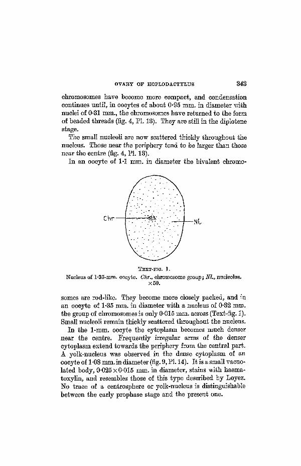
somes are rod-like. They become more closely packed, and n'nan ooeyte of 1-35 mm. in diameter with a nucleus of 0-32 mm.the group of chromosomes is only 0-015 mm. across (Text-fig. 1).Small nucleoli remain thickly scattered throughout the nucleus.
In the 1-mm. oocyte the cytoplasm becomes much densernear the centre. Frequently irregular arms of the densercytoplasm extend towards the periphery from the central part.A yolk-nucleus was observed in the dense cytoplasm of anoocyte of 1 -08 mm. in diameter (fig. 9, PI. 14). It is a small vacuo-lated body, 0-025 X 0-015 mm. in diameter, stains with haema-toxylin, and resembles those of this type described by Loyez.No trace of a centrosphere or yolk-nucleus is distinguishablebetween the early prophase stage and the present one.

344 MARY M. M. BOYD
Shortly after this, in oocytes of about 1-1 mm. in diameter,a zone of vaouoles occurs at a distance of about one-third of theradius from the periphery, outside the central dense cytoplasm.At the same time extremely minute, basophil yolk-spheresappear at the periphery, and extend in among the vacuoles.The spheres originate in minute vacuoles which they usuallyfill completely. The vacuoles forming the zone mentionedabove do not contain yolk-spheres. To them is probably duethe arrangement of the yolk in the ripe egg, between fairly largevacuoles. There is a gradual transition between the minuteyolk-spheres at the periphery and those among the vacuoles,which are larger and fewer in number. At the centre of theoocyte the dense cytoplasm is becoming markedly alveolar.The angular yolk elements observed by Loyez do not occurhere, and at this stage acidophil yolk elements are indis-tinguishable.
The vacuolated zone extends inwards, and the vacuoles be-come larger. In an oocyte of 1-5 mm. in diameter the zoneextends through the middle third of the egg. The yolk-spheresare now much more numerous; but away from the peripherythey are still scarcer and larger. The peripheral spheres varyin size from those just visible to those of 0-5 micra in diameter.Independently of size some are acidophil, and others, whichoccur more frequently, are basophil.
In the denser cytoplasm inside the zone of vacuoles, extremelyminute yolk-spheres, like those at the periphery in the 1-1-mm.oocyte, are now appearing in fair number. There is no transitionin size between these and the larger spheres in the vacuolatedzone.
In an oocyte of about 6 mm. in diameter, vitellogenesis isnearing completion. The large oocyte composes the greaterpart of the ovary, which shows a considerable increase in sizesince the close of the last gestation period. The germinal bed andsmaller oocytes form low protrusions on the cephalic side of the6-mm. oocyte. The germinal disk is situated at the opposite side,unlike the frog. It is nearly 2 mm. in diameter and its greatestdepth is 0-8 mm. At its margin the germinal disk is continuouswith a narrow layer of cytoplasm, containing fine yolk-spheres,

OVARY OF HOPLODACTYLUS 345
which lies immediately beneath the zona. Similar fine yolk-spheres are thickly scattered, without definite arrangement,in the germinal disk. The nucleus, which is excentric throughoutthe development of the oocyte, but remains some distance fromthe periphery even after vitellogenesis has begun, is nowsituated just above the deep border of the germinal disk. Thelatebral neck passes down from the germinal disk below thenucleus.
The nucleus is slightly kidney-shaped, but shows no increasein size over that of the 1-35-mm. oocyte at the beginning ofvitellogenesis. It measures about 0-37 X 0-26 mm. The rate ofincrease in the size of the nucleus is greater than that of theoocyte until the onset of the diplotene stage. Prom then untilthe oocyte reaches 1 -3 mm., at the beginning of vitellogenesis,the increase of the nucleus is approximately proportional tothat of the oocyte. Thereafter the oocyte increases enormously,while the nucleus remains stationary. The only change in thenucleus is the crowding of the bivalent chromosomes to forma small spheroidal group, measuring 0-006x0-012 mm., nearthe centre of the nucleus. The very numerous small nucleoliremain as before in the nucleoplasm.
The yolk, apart from that in the germinal disc and peripheralzone, is arranged around and between numerous vacuoles ofabout 0-03 mm. in diameter (fig. 13, PL 15). This arrangementis interrupted at intervals in most eggs, causing the yolk to bedivided into two or three zones (Text-fig. 2). Owing to theshrinkage of fixed material, these zones appear to be separatedby a thin layer where the yolk is sparsely scattered. The zonesare no doubt homologous with the alternating layers of fineand coarse yolk described by Sarasin (1883) for L a c e r t aag i l i s , and attributed by him to periods of greater activityin yolk-formation, governed by changes in nutrition, or otherfactors affecting the parent. The yolk-spheres vary from0-005-0-01 mm. in diameter.
The latebra consists of clear alveolar cytoplasm, containingnear the border many large vaeuoles similar in size and arrange-ment to those among the yolk (fig. 13, PL 15). It is pear-shaped,directed obliquely to the plane of the germinal disk, and lies

346 MAEY M. M. BOYD
somewhat excentrically near the germinal disk. The narrowerend, which is nearer to the germinal disk, is continuous with thelatebral neck, which follows a somewhat winding course, asSarasin observed in L a c e r t a , to the germinal disk. The
GD
LN
TEXT-FIG. 2.
Section through 6-mm. oocyte (diagrammatic). G.D., germinaldisk; L., latebra; L.N., latebral neck; Nu., nucleus; Z., zonein yolk. xlO.
latebral neck consists of cytoplasm which is markedly alveolar,and contains fine yolk-spheres that merge with the coarseryolk around them. Bordering the vacuoles near the marginof the latebra, and also between them, are minute yolk-spheres.There is a gradual transition of these minute spheres into thecoarser ones farther out, where the cytoplasm is not dis-cernible.

OVARY OF HOPIiODACTYMJS 847
The F o l l i c u l a r E p i t h e l i u m .The first indication of the formation of follicular epithelium
around the oocyte occurs when its diameter is about 0-024 mm.,corresponding with the onset of the diplotene stage in thenucleus. Cells of the germinal epithelium which have migratedinwards, and lie scattered among the enlarging oocytes, beginto surround the latter. Occasionally a degenerating nucleushas been observed in an oocyte of this size, containing also anapparently healthy nucleus. Whether this is due to the frag-mentation of a degenerating nucleus, or to the combinationof two oocytes and the subsequent degeneration of one nucleus,as Balfour (1878) affirms happens constantly in the dogfish, isobscure.
The follicular epithelium is almost complete when thediameter of the oocyte reaches 0-045 mm. The cells are roughlycubical and measure about 0-008 mm. in thickness. By thetime the oocyte is 0-07 mm. in diameter the follicular epitheliumis becoming double. The oocyte has moved to one side of thegerminal bed, always the same side, and is in contact with thebulk of the stroma on its inner side (fig. 2, PL 13). This positionobviates the destruction of the stroma and germinal bed duringthe subsequent enlargement of the oocyte.
Certain epithelial cells here and there now become enlarged.The follicular epithelium at this stage is very irregular incharacter, consisting in some places of a double layer of smallcells, in others of a single layer, and interrupted now and againby an enlarged cell (fig. 2, PL 13).
Loyez considers the changes undergone bj the enlargingfollicular epithelial cells to be similar to those in the develop-ment of the oocyte from the oogonium. In this lizard, however,there are important differences, at least in regard to the nucleus.The nuclei of the small follicle cells resemble those of the oogoniain the germinal epithelium. As they enlarge the ehromatinceases to react to Feulgen's stain; but no stage comparable withleptotene or zygotene occurs. The pre-meiotic phase of blob-like, contracted chromosomes at the periphery of the oogoniumabout to give rise to an oocyte is also absent from the enlarging

348 MARY M. M. BOYD
follicular cells. During the growth of the nucleus the nucleo-lus increases in size and becomes vacuolated, resembling thatof the oocyte shown in fig. 2, PI. 13. The nucleus grows to adiameter of about 0-02 mm.
A crowd of rod-shaped mitochondria appear in the cytoplasmof the enlarging epithelial cell, as in the young oocyte. Thecytoplasm stains better than it does in the transformingoogonium. The centrosphere, observed by Thing (1918) in theepithelial cells of turtles, is not distinguishable here. An oocyteof about 0-13 mm. in diameter is completely surrounded bya layer of enlarged cells, each about 0-03 mm. in diameter. Alayer of small cells remains immediately outside the egg mem-brane, the cells occupying the spaces between the protrudingends of the large cells. Another layer of small cells lies outsidethe larger ones, just beneath the theca. Loyez records theoccurrence of mitoses only in the inner layer of small cells; but inthis lizard they occur in the small cells of both the inner andouter layers. Division is more frequent in the inner layer, asit is from this layer that all the larger cells are derived.
The follicular epithelium of an oocyte of 0-3 mm. in diameterexhibits the typical character and arrangement found through-out the functional period of the epithelium. The large cellshave become flask-shaped, as Loyez has described, having theirlong axes directed radially and their broad ends outward(fig. 12, L.F., PI. 15). They measure about 0-20x0-03 mm. butmay be narrower and longer. The narrow end is prolongedto form a small tube which pierces the zona in a funnel-shapedopening, resembling those figured by Loyez (fig. 5, M.T.,PL 14). The knob-like enlargement of these tubes at the yolk-surface, which Thing observed in turtles, is absent. The nucleuslies near the outer end of the cell. It is unchanged, except foran increase in the size of the nucleolus (fig. 12, NL, PI. 15). Thewalls of the nucleolus become very thin, like those in oocytes of0-06-0-03 mm., and may burst, sometimes discharging a fewsmall nucleolus-like granules contained in the vacuoles, as Loyezobserved. There may be two or three vacuolated nucleoli,resulting from the growth and change of small ones. The cycleof changes which these nucleoli undergo in the oocyte does not

OVARY OF HOPLODACTYLUS 349
appear to occur usually in the epithelial cells. The cytoplasmnear the nucleus contains a crowd of mitochondria, and alsosmall fat-globules (fig. 6, F.G., PI. 14). Sometimes large cellsin the follicular epithelium of oocytes over 1 mm. in diameterbecome packed with fat-globules, the nucleus degenerates, andthe tubular connexion with the ooeyte is lost.
Small undifferentiated cells still lie between the swollen endsof the large epithelial cells, which occupy the meshes of a net-work of small cells when viewed in tangential section (fig. 12,S.F., PI. 15). Between the narrow inner ends of the large cellsis an irregular layer of intermediate cells derived from the innerlayer of small cells in the same way as the large cells (fig. 9, J.F.,PL 14). Their nuclei, which do not react with Feulgen's stain,contain a vacuolated nucleolus, while the cytoplasm containsmitochondria and some minute fat-globules. They are anintermediate stage in the formation of the large cells, and as thesurface of the oocyte increases they grow to become part of thelayer of large cells. Against the zona there remains an incom-plete layer of small cells, interrupted by the presence of inter-mediate cells. The follicular epithelium of this lizard is thusof the same type as that of the Geckos examined by Loyez.Cytoplasmic connexions between the follicle cells, as figured byEimer (1872) and also observed in turtles by Thing, occur here.They may branch, and in the case of the large follicle cellsthey may be comparatively coarse.
The follicular epithelium of an oocyte near the close ofvitellogenesis, measuring 6-8 mm. in diameter, consists, asa result of shrinkage of the larger cells, of a single layer of smallcells only (fig. 7, F.C., PI. 14). The cells have a shrivelled mar-gin, and the quantity of cytoplasm is small, with the consequentoccurrence of intercellular spaces. The oval nuclei of the cellsmeasure about 0-008 x 0-004 mm., and contain one or two smallnucleoli.
The Theca .The theca is derived from the stroma when the oocyte, sur-
rounded by follicular epithelium, leaves the germinal bed, asLoyez (1906) described. It consists of fibroblasts concentrically

350 MARY M. M. BOYD
arranged (fig. 12, T., PI. 15). Elastic fibres are numerous, andreticular fibres are present, in addition to collagen fibres. It isa simple layer, not differentiated into theca interna andexterna until late in the development of the oocyte.
The theca of the ripe or nearly ripe oocyte, 6-8 mm. indiameter, measures only about 0-05 mm. in thickness. Surround-ing the follieular epithelium is a membrana propria, distinctfrom the rest of the theca. It is composed of flattened fibro-blasts (fig. 7, M.P., PI. 14). Immediately outside this in someplaces, especially between capillaries, are cells arranged as asingle layer, having oval nuclei and no fibres among them(fig. 7, T.I., PI. 14). Although this layer is so discontinuous, thegaps being greater than the areas where it is present, it seemsnevertheless to constitute a rudimentary theca mterna com-parable with that of mammalia. The theca externa consists offlattened fibroblasts, and resembles the undifferentiated theca.Small capillaries run beneath the basement membrane, andlarge flattened vessels lie outside them (fig. 7, B.V., PI. 14).
The Vi te l l ine Membranes .As in other vertebrates, the egg is surrounded by an egg
membrane, which is derived from the primitive membrane ofthe transforming oogonium. Between this and the zonapellucida there is a striated layer (fig. 7, PI. 14). The eggmembrane is called by Loyez the internal membrane, while thezona pellucida is called vitelline membrane. Both Loyez (1906)and Thing (1918) designate the striated layer as the zonaradiata. Thing includes the striated layer with the dense outerlayer under the name zona pellucida.
In transverse section the striated layer appears to consistof thin, radially-directed, rod-like protuberances of the zonapellucida (fig. 7, Z.B., PL 14). Small spaces of varying widthare present between the rods, which thicken at their junctionwith the zona pellucida. In tangential section the striatedlayer resembles a layer of small vacuoles.
The zona pellucida is composed of a dense substance, appa-rently homogeneous, even in tangential section. It is piercedby prolongations of the large follieular epithelial cells, but does
OVABY OF HOPLODACTYLUS 351
not appear striated in the intervals, in fixed material. Loyezobserved radial striations in the zona pellucida of one or twoGeckos, however, and Thing found such striations in livingeggs of turtles. The striations, as Loyez suggests, are prob-ably produced bj the prolongations of follicular epithelialcells.
The zona pellucida first appears as a few irregular thickeningsbetween the egg membrane and the follicular epithelium of anoocyte of about 0-07 mm. in diameter, around which the single-layered follicular epithelium is complete (fig. 8, PL 13). Thethickenings follow the contour of the follicle cells, and. oftenjut out to a point between the ends of adjacent cells, as Thingobserved in turtles. There are, however, no intercellularchannels filled with intercellular substance as in turtles, andthe epithelial cells lie against the egg membrane before theformation of the zona. The inner borders of the thickenings aresmooth and follow the contour of the egg membrane. Theyhave the appearance of a homogeneous secretion.
The thickenings increase and join together as the follicle cellsmultiply and enlarge. In this way the zona becomes completeround an oocyte of about 0-15 mm. in diameter; but for sometime after this it is interrupted by the presence of small folliclecells of the inner layer engulfed in it at frequent intervals.These cells lie against the egg membrane. Some of themoccasionally sink into the oocyte, are isolated from the epithe-lium by the growth of the zona, and subsequently degenerate.Loyez observed small degenerating follicle cells in the cyto-plasm of the oocytes of other lizards.
Thing describes the structure of the zona as a primary net-work with a superimposed secondary network; but this cannotbe traced here. The apparently homogeneous cuticular sub-stance of the zona is, however, interrupted by prolongationsof the large follicle cells, and in addition there are a numberof minute pores in it, which may contain cytoplasmie connexionsfrom the smaller follicle cells. These do not show in the fixedmaterial, although the prolongations of the large cells aredistinct in tangential sections.
The striated layer is evident in an oocyte of about 0-15 mm.

352 MARY M. M. BOYD
as a very thin, clear layer between the zona pellucida and theegg membrane, in sections stained with Crossmann-Mallory triplestain, which stains the egg membrane purple and the zonapellucida green.
The zona of an oocyte of 0-3 mm. in diameter varies con-siderably in thickness, since the outer surface still follows theirregularities of the follicular epithelium; but it averages0-002 mm. Its inner face in transverse section presents ascalloped outline, due to its being perforated at intervals bythe tubular prolongations of the enlarged epithelial cells, asLoyez and Thing observed. It becomes very thin around theseperforations.
As the zona pellucida thickens it becomes denser, and theegg membrane also becomes stouter and stains more deeply.The zona pellucida of an oocyte of 0-7 mm. in diameter measures0-003 mm. and the striated layer 0-001 mm. in thickness. Theradial striations are now distinguishable in the latter. Theystain green like the zona pellucida with Crossmann-Mallorytriple stain, and are thus distinguished from cytoplasmicmaterial which stains pink or purple. The zona has lost itsscalloped outline, the perforations for the epithelial cell con-nexions with the oocyte being funnel-shaped (fig. 5, M.T.,PL 14). As Loyez pointed out, the narrower ends of the openingsare next to the epithelium. The egg membrane lines the funnel-shaped openings.
The zona pellucida increases to 0-004 mm. in thickness roundan oocyte of about 1 mm. in diameter; but the striated layershows no increase (fig. 12, PI. 15). Owing to refraction of lightthere appears to be a dark layer in the zona next to the striatedlayer in fig. 9, PI. 14. This is not a true distinction.
When the oocyte reaches 6 mm. in diameter the zonapellucida is reduced in thickness and measures just over0-001 mm., while the striated layer has greatly increased andmeasures 0f005 mm. (fig. 7, PL 14). The egg membrane remainson its inner surface, but is rather indefinite and has a granularappearance. It is more distinct in the uterine ovum. Theperforations for prolongations of the large epithelial cells areapparently lost. The reduction in thickness of the zona

OVARY OF HOPLODACTYLUS 853
pellucida as the egg ripens is in contrast with the behaviourin turtles, monotremes, and other vertebrates.
The formation and appearance of the zona pellucida in thislizard confirms the statement made by Thing, as a result of herresearches in turtles, that it is a cuticular structure. Thestriations of the striated layer are here clearly shown byCrossmann-Mallory triple staining to be composed of the samecuticular substance. They are, therefore, not cytoplasmie inorigin and cannot be prolongations of epithelial cells.
F o l l i e u l a r A t r e s i a .Two atretic follicles are present in only a single ovary among
fifteen examined. The cause of atresia in this case appears tobe that two oocytes have enlarged together, and considerablevitellogenesis has occurred in both. Normally only one oocyteattains maturity at any one time in the ovary. Loyez remarksupon the rarity of atretic follicles in the Gecko, P l a t y -d a c t y l u s m u r a l i s , in which she found none, thoughMingazzini (1893) observed atresia in P l a t y d a c t y l u sm a u r i t a n i c u s .
One atretic follicle is 3 mm. in diameter, and the other,4-5 mm., is irregular in shape. Atresia is more advanced in thelatter. In neither is there any trace of the germinal disk or thelatebra.
The theca of the 3 mm. follicle is thinner than that of a healthyooeyte of this size and contains few capillaries. The zona hascompletely disappeared. The follicular epithelium has givenrise to a festooned layer of giant cells with bulging free endsprotruding into the yolk. Their cytoplasm is extremelyalveolar, and the nuclei are irregular in shape and containvacuolated nucleoli. The cells contain masses of yolk-spheres,particularly round the margins, in various stages of dissolution.The normally basophil yolk-spheres become acidophil andvacuolated. In a few places fibroblasts from the theca havegrown in between the giant cells. No cells have yet migratedinto the yolk, but the spheres have amalgamated to formirregular vacuolated masses between the large vacuoles presentin normal yolk.
354 MARY M. M. BOYD
The theca of the larger follicle has become very thin, due,apparently, to the inward migration of fibroblasts to invest thegiant cells, and penetrate the yolk. Capillaries are small andvery few in number. The giant cells have become reduced tohalf their former size and form a layer two or three cells deepat the periphery of the egg. Their cytoplasm is denser thanbefore, owing partly to absorption of the broken-down yolk.Cell boundaries are often difficult to distinguish.
Both epithelial cells and fibroblasts from the theca havepenetrated into the deep yolk. Many of the large vacuoles arenow occupied by epithelial cells with extremely alveolarcytoplasm. Numbers of polymorphonuclear and mononuclearleucocytes are present in the masses of yolk near blood-vessels,which are beginning to penetrate into the yolk.
The course followed in regression in this species seems tobe first the disappearance of a distinctive theca, then theabsorption of the yolk by giant cells and later by leucocytesalso. Finally, the giant cells become reduced and disappear,the position of the atretic follicle being marked for a time byfibroblasts which grow in from the theca.
DISCUSSION.
The occurrence of a prolonged diplotene stage with theformation of 'lamp-brush' chromosomes during the growthperiod of the oocyte has previously been observed in all classesof vertebrates having large yolky eggs, with the exception ofreptilia. The description given above of this phenomenon in areptile therefore completes the h'st. That precisely similarnuclear changes occur in Scincidae and Anguidae is shown bythe figures given by Loyez (1906).
Among cyclostomes, Okkelberg (1921) described the ovary ofthe brook-lamprey, and states that the diplotene stage persiststhroughout the early part of the growth period and probably upto the time of maturation. He finds that the nucleus becomesvery large and the chromatin material may be scatteredthroughout its whole extent, and therefore it is difficult tofollow the history of the chromosomes during the growth period.There is little doubt from this description, and a comparison

OVAEY OF HOPLODAOTYIiTJS 355
of his figures 59, 60, and 62, that the chromosomes have as-sumed the lamp-brush form.
In Elasmobranchs Euckert (1892) described and figuredtypical lamp-brash chromosomes during the growth period ofthe oocyte.
King (1908) described and figured lamp-brush chromosomesin B u f o among Anura during the growth period of the oocyte,but did not recognize this as the diplotene stage. Garnoy andLebrun (1897) had previously observed lamp-brush chromo-somes in oocytes of B a n a, but mistook their origin.
In T r i t o n among Urodeles Koltzoff (1938) has given adetailed and well-illustrated account of the lamp-brush chromo-somes in the nucleus of the enlarging oocyte.
Crew (1933) noted the persistence of the diplotene stage andthe occurrence of lamp-brush chromosomes in the growingoocyte of the domestic fowl. Koltzoff has described this phasein the chick and the pigeon.
fflynn and Hill (1939), in their paper on the growth of theovarian ovum in the Monotreme, have designated as pseudo-chromosomes what are evidently lamp-brush chromosomes inthe diplotene stage.
The observations of Loyez and the present account show thatthe Lacertilia are distinguished from the other classes in thatthe lamp-brush chromosomes have condensed to bivalent rod-like chromosomes before the yolk is deposited, and for sometime before this they are confined to the more central part ofthe nucleus.
THE UTEBINE OVUM.
The ripe ovarian egg measures about 8 mm. in diameter;but in utero the ovum is oval in shape, due, no doubt, to thepressure of the muscular uterine wall which fits closely aboutthe soft egg membranes. It measures 9x7 mm.
Bounding the yolk is a very thin membrane which seems tobe the egg membrane (fig. 10, V.M., PL 15). It is much moredistinct than in the 6-mm. oocyte. Immediately outside it isa thin and very delicate striated layer which measures only0-001-0-002 mm. in thickness (fig. 10, Z.B., PL 15). Thisapparently represents the striated layer of the 6-mm. oocyte.
NO. 326 A a

356 MAEY M. M. BOYD
Attached to it is a dense membrane varying in thickness fromabout 0-003-0-01 mm. (fig. 11, PL 15). This is bilaminar. Theinner layer is slightly denser, thinner, and constant in thickness,measuring 0-002 mm. It is apparently the zona pellucida whichhas doubled its thickness in the 6-mm. oocyte. This inter-pretation is supported by the fact that its inner face showsminute denticulations, like the thickened terminations of thestriations of the striated layer in the large ovarian oocyte.
The outer part of the bilaminar membrane is formed by theshell membrane which varies in density over different parts ofthe egg. In the thicker places the density is less. It is eosino-phil, having the appearance of fine felted fibres embedded ina matrix, as Giersberg (1922) observed to be the appearanceof the shell membrane in other lizards. He has shown that noalbumin is secreted round the ova of lizards. Occasional smallcells are present between the shell membrane and the zona.They resemble follicular epithelial cells, and probably remainedattached to the zona at ovulation. Small globules of a secretionsimilar in appearance to the shell membrane are tangled hereand there in the cilia of the uterine epithelial cells.
Among the peripheral yolk-spheres, which are now onlyslightly smaller than the rest, is a little cytoplasm and somecoagulum. Vitellogenesis has ceased first, as it began, at theperiphery, for no minute yolk-spheres, like those still occurringin the latebra, are present here. The yolk, except at the peri-phery, is arranged between large vacuoles, as in the 6-mm.oocyte (fig. 13, PI. 15); but no zones of growth are distin-guishable. They may have been destroyed by the alterationin shape of the uterine ovum, and by the distortion due tosectioning; but Sarasin (1883) states that they vary in distinct-ness in different eggs.
The latebra, as in the 6-mm. oocyte, is pear-shaped, but itslong axis is now parallel to the plane of the germinal disk, sothat the neck traverses a curv,e of about 90° as it winds up tothe disk (fig. 13, L.N., PI. 15). The latebra lies anteriorly to thegerminal disk (in the parent) and towards the remote side ofthe egg. The displacement may be due to the change of shapeundergone by the uterine egg; but Sarasin often found the

OVARY OF HOPLODACTYMJS 357
latebra excentric in L a c e r t a ag i l i s . The character of thelatebra differs somewhat from that of the 6-mm. ovum. It hasa marked alveolar appearance, but is much denser than before,and resembles the cytoplasm of oocytes just before the onsetof vitellogenesis (fig. 13, PI. 15). There is a quantity of eoagulumscattered in the yolk, mainly at the periphery and near thelatebra, and this seems to be partly responsible for the increasein density of the latebra. The central part of the cytoplasmis no longer vacuolated, while the vacuoles near the border aresmaller than those of the 6-mm. ooeyte, and do not exceed0-005 mm. in diameter. The transition between the latebraand the yolk is therefore sharper than before, as is expectedafter the cessation of vitellogenesis. Fine yolk-spheres arepresent between the marginal vacuoles of the latebra; but thislayer is thin. The innermost spheres are acidophil and veryfine, while the rest are basophil. The latebra differs from that ofthe oviducal egg of L a c e r t a agi l is in that fine yolk-spheresare present throughout the latter.
The core of cytoplasm in the latebral neck is dense where itleaves the latebra, is about 0-8 mm. in diameter and is sur-rounded by fine yolk extending out for at least 0*5 mm. (fig.13, L.N., PI. 15). Both the density and the diameter of thecytoplasmie core diminish as it winds upwards. About half-wayup it is only 0-02 mm. across, and is very vacuolated and stainslightly. The diameter increases again as it approaches thegerminal disk.
The germinal disk lies dorsally in the uterus and is oval, asNicolas (1900) noted in Anguis f r ag i l i s . It measuresapproximately 2-3x2-7 mm. in diameter, while its greatestdepth is 0-8 mm. It extends downwards, plug-like, at its centre,as in O r n i t h o r h y n c u s , and into this 'axial plug' passesthe latebral neck. The core of clear cytoplasm in the latebralneck can be traced to the centre of the axial plug, where itdisappears. The cytoplasm of the germinal disk is closely anduniformly packed with very small yolk-spheres which passgradually into the coarser yolk at the lower side of the disk.It thus differs from that of Anguis f ragi l i s (Nicolas,1900) in containing no cytoplasm more or less clear of yolk-

358 MABY M. M. BOYD
spheres, except sometimes a thin fringe beneath the egg mem-branes.
Oppel (1892) observed at the periphery of eggs of Anguisf ragi l i s patches of cytoplasm containing fine yolk-spheres,resembling the germinal disk, but separated from it. Nicolasfound such patches in a few of his eggs. In the egg underdescription such a patch is present well round to the side ofthe egg. It is small, being only 0*2 mm. across. It seems to beof merely accidental occurrence, due to some irregularity ofvitellogenesis at that place, and of no significance.
Probably owing to abrasions of the surface in sectioning nopolar bodies were found.
Nuclei in t h e Germina l Disk.Approximately at the centre of the disk, and 0-06 mm.
beneath the surface, are the male and female pronuclei (fig. 11,PI. ). They lie in contact, one deeper than the other, thedeeper one slightly indented by the other and with their longaxes parallel to the surface. They are, therefore, in positionfor the appearance of the spindle of the first division. Theirarrangement is like that described by Nicolas at a certain stagein Anguis f r ag i l i s . The deeper one measures 0-015x0-008 mm. in diameter, and the other 0-013x0-01 mm. Bothare in the resting condition, being vesicular and having oneconspicuous deeply staining nueleolus. The nuclear membranesstain deeply and are well defined. It is impossible to distinguishthe male pronucleus from the female.
At the surface of the germinal disk, opposite the pronuclei,the entrance cone of the spermatozoon is plainly evident asa portion of dense cytoplasm containing no yolk-spheres. Asmall area of cytoplasm round the pronuclei is also free fromyolk-spheres (fig. 11, PL ). There is, however, no trace ofa centrosome or aster. The tail of the spermatozoon cannot beidentified; but its path is visible owing to the presence of somevacuoles, and the partial absence of yolk-spheres from it.Nicolas found that the track of the spermatozoon was oftenvacuolated. It has the form of a parabolic arc.
Six other nuclei are present in the germinal disk, three

OVARY OF HOPLODACTYLTJS 359
situated anteriorly and three posteriorly to the pronuclei(Text-fig. 3). They are supernumerary sperm nuclei, theentrance cones of which are usually visible nearby. Sometimesthe track of the spermatozoon is also distinguishable, in whichcase it is a parabolic arc. The following table gives the positionand measurements of these nuclei, lettered as in Text-fig. 3:
Nucleus.
ABCDEB1
Measure-ments
in mm.
0-12x0-010-12x0-0090-01x0-010-012x00090-009x0-0090-01x0008
Distancebelow
Surface.
nun.0 0 30-0340-0550-030050-036
Distance An-terior (4-) orPosterior (—)to Pronuclei.
mm.+0-95+0-4+0-34—0-2—0-35- 1 - 2 4
DistanceBight (+)orLeft(-)of Mid-line.
mm.-0-7+0-9-0-1+0-2- 0 - 4 5—0-75
Position ofEntrance Gone.
doubtfuloppositeopposite0-22 mm. to left0-5 mm. to right0-1 mm. anterior
TEXT-BIG. 3.
Diagram showing situation of the supernumerary sperm nuclei inthe germinal disk. P.N., male and female pronuclei; A.-F., super-numerary sperm nuclei, x 12.
These nuclei all closely resemble each other. They are in theresting state, being vesicular and containing one deeply-staining nucleolus (fig. 8, PI. ). Like the pronuclei, each issituated in a small area of dense cytoplasm free from yolk-spheres. Their long axes are parallel to the surface. In twocases a short length of the tail is distinguishable. In one, asmall, very dense portion of cytoplasm, possibly representingthe middle piece of the spermatozoon, lies against the nucleus,

360 MARY M. M. BOYD
and from the opposite side of this cytoplasm the tail extendsbetween yolk-spheres for about 0-02 mm. (Text-fig. 4, T.). Inthe other the tail lies partly in the clear area of cytoplasm roundthe nucleus, and curves out between the yolk-spheres. It canbe traced for about 0-04 mm. The tails are very fine and, asNicolas found, stain black with Heidenhain's haematoxylin.
Oppel and Nicolas have described the occurrence of similar
Nu
YS
1
®
4
TEXT-FIG.
• ; • • • •
) } • *
i ®
4.A supernumerary sperm nucleus, showing middle-piece (M.P.)
and sperm tail (T.). Nu, nucleus. YS, yolk-sphere, x 1,000.
supernumerary sperm nuclei in Anguis f rag i l i s , andNicolas observed the tail-piece beside a number of them. Inmost details those described here agree with the descriptionsgiven by Nicolas. Their long axes are parallel to the surface,none is quite so deep as the conjugating nuclei, and they containa 'reticulum'.
The degree of polyspermy is evidently variable. Nicolasrecords the presence in eggs of Anguis f ragi l i s of from oneto forty-six. Oppel found up to five in the eggs examined byhim. The smaller numbers occur more frequently. For thereason given by Nicolas, viz. that the sperm nuclei are all atthe same stage of transformation, the relative number isevidently not affected by the time elapsing, since they mustenter not successively, but more or less simultaneously.

OVARY OF HOPLODACTYLUS 361
THE CORPUS LTJTBUM.
Literature concerning the reptilian corpus Iuteum is ratherscanty, and no account of the corpus Iuteum in Geckonidae hasso far appeared. In 1893 Mingazzini (1893) published a paperon 'Corpi lutei veri e falsi dei Bettili', distinguishing corporalutea and atretic follicles. He showed, principally in Sepscha lc ides , that the follicular epithelium is not expelled atovulation, but remains within the follicle and takes part, to-gether with fibroblasts from the theca, in the formation of thecorpus Iuteum.
In 1903 Lucien described the formation of the corpus Iuteumin the viviparous lizards, Anguis fragil is and Sepscha l c ides , by the hypertrophy (but probably not mitotiedivision) of the follicular epithelium, and the invasion of theluteal tissue by connective tissue from the theca.
Hett, in 1924, gave a detailed account of the development ofthe corpus Iuteum from the follicular epithelium in the ovi-parous Lace r t a agil is . Connective tissue, containingcollagen fibres and supporting capillaries, was shown to pene-trate the luteal tissue. The presence of a well-developed corpusIuteum is therefore not dependent on viviparity.
Cunningham and Smart, in 1934, described corpora lutea inL a c e r t a v i r i d i s , Anguis f ragi l i s , and Zoot icav i v i p a r a in support of their theory that 'the true corpusIuteum, i.e. the persistence and development of the follicularcells within the ruptured follicle, with associated changes in thecells of the theca interna, is the consequence and result of theinternal development of the fertilized ovum in oviduct oruterus, or in the cavity of the ovary, and that in oviparous formsthe ruptured follicle at once begins to undergo reduction andabsorption'.
An examination of their material has shown, however, thatin the case of the oviparous L a c e r t a v i r i d i s , they describedand figured in their Text-fig. 3 and fig. 7, PI. 14, a supposedcorpus Iuteum which is none other than an atretie follicle, withthickened theca and a layer of giant cells derived from thefollicular epithelium. It contains a central cavity in which are

362 MAEY M. M. BOYD
yolk debris. They apparently failed to observe in the same ovarytwo well developed, typical corpora lutea containing strandsof ingrown fibroblasts and blood-vessels. Their stage 3 is afairly early one in the regression of the corpus luteum, markedby the enlargement of some nuclei preparatory to degeneration,as occurs in some other lizards and in Monotremes. Somesections of ovaries of L a c e r t a v i r i d i s belonging to Pro-fessor J. P. Hill show stages in the development of the corpusluteum by hypertrophy of the follicular epithelium, and confirmthe fact that it is as well formed in this oviparous lizard as inviviparous ones.
Weekes, also in 1934, published a comparative account ofcorpora lutea in various oviparous and viviparous Australianscinks and in L a c e r t a v iv ipa ra . She found in all theselizards corpora lutea more or less penetrated by connectivetissue from the theca, showing 'that luteal formation is to bealso associated with oviparity'. The degree of penetration byfibroblasts has no relation to viviparity, since in three vivi-parous scinks this was only superficial, while in three othersdefinite strands of connective tissue carry blood-vessels into theluteal tissue, and in one of these fibroblasts also penetratebetween the luteal cells. Moreover, 'ingrowths of fibroblastsamong luteal cells cannot be particularly associated withplacentation, as it occurs in the oviparous lizards Amphi -bo lu rus m u r i c a t u s and L a c e r t a agi l is (Hett, 1924).
Since in H o p l o d a c t y l u s only one egg is ovulatedannually from each ovary only one corpus luteum develops ineach. In the fresh ovary it appears in early stages as a creamy-white, flattened body, the opening through which the oocyteescaped being visible as a small, somewhat elongated depressionstained with clotted blood. It is easily distinguishable from themore translucent white young oocytes, and forms rather morethan half the volume of the ovary. At later stages its size isdiminished and its surface becomes rounded. The shape of thecorpus luteum in later stages varies considerably in individuals.The shape assumed is a matter of chance, being that which bestfits the depression between the oocytes and leaves the surfaceof the ovary as regular as possible.

OVARY OF HOPLODACTYLITS 868
The history of the corpus luteum is described below undersix arbitrary stages, numbered from 1-6.
S tage 1.—The youngest corpus luteum obtained relates toa uterine egg in which the male and female pronuclei are inapposition. It is flattened and measures 2-25 x 045 mm. indiameter. The egg ovulated measures 8 mm. in diameter. Theluteal tissue, including the central cavity, is 1-28 x0-45 mm, indiameter.
The theca is much thickened since ovulation, having increasedfrom 0-05 mm. to 0-25 mm. (cf. fig. 7, PL 14; fig. 14, T., PL 16).The increase is due, at least partly, to shrinkage; but there maybe some proliferation also. Weekes (1934) records the presenceof mitoses in the theca of lizards at an earlier stage in theformation of the corpus luteum, and O'Donoghue (1916)observed it in the Marsupial P h a s c o l a r c t o s e i n e r e u s .No mitotic figures were observed here; but a few strands ofspindle-shaped cells growing out from the damaged wall of thetheca, near the opening, indicate the occurrence of mitotic oramitotic cell division.
In the theca of the ripe follicle the fibroblasts are concen-trically arranged; but evidently as a result of the shrinkagefollowing ovulation they are now radially arranged. The innerborder of the theca is conspicuous owing to the presence ofnumerous deeply-staining fibroblast nuclei. Elastic fibres areparticularly numerous here, and both nuclei and fibres areconcentrically directed. Under low magnification this has theappearance of a basement membrane; but it is not a sharplydefined layer (figs. 14 and 15, T., PL 16). It is probably derivedfrom the membrana propria of the ripe follicle. Hett and Weekesdistinguish as theca interna the inner part of the theca, whichthey find to contain fewer fibres than the rest. Evidently it isnot comparable with the inner part of the theca in this case;and, since this part has nothing in common with the thecainterna in mammalia, it is not called so.
Large blood-vessels run in the outer part of the theca, whilesmall capillaries are very numerous in its inner border. Someof these project into the luteal tissue accompanied by fibro-blasts from the theca (fig. 16, C, PL 17). Fibroblasts have also

364 MARY M. M. BOYD
begun to grow in strands into the luteal tissue from the wholeinner surface of the. theca (fig. 15, F.B., PI. 16). These cells arederived not only from the inner border of the theca, but alsofrom the tissue beneath, cells of which break through and irruptthe inner border. Spaces occur between the ingrowing fibro-blasts just inside the theca, and thus the luteal tissue in manyparts is separated from the theca. The fibroblast nuclei oftenstain more deeply than the luteal nuclei and are smaller andelongated. Both elastic and collagen fibres penetrate the lutealtissue, and some reticular fibres also occur in it.
There is no definite evidence of the fate of the small patchesof cells which form a doubtful theca interna in the ripe follicle.Here and there cells with small spherical nuclei, averaging0-005 mm. in diameter, are situated singly or in small groupsin the bases of the ingrowing strands of fibroblasts, whichsurround them. It is probable that these represent the cellsof the theca interna, and they will be called so. They may havemultiplied after ovulation, but are still not very frequent, andtherefore cannot play an important part in the function of thecorpus luteum.
The luteal tissue has almost obliterated the flattened cavityof the burst follicle, except opposite the opening (fig. 14, PL 16).The cells are radially arranged, as in the marsupial corpusluteum, and consequently the tissue appears to be composed ofradiating strands of cells. The cytoplasm, which has increasedin bulk since ovulation, is granular and stains well. There area few intracellular spaces, and numerous radially compressedintercellular spaces (fig. 15, PI. 16). The epithelial nuclei of theripe follicle average 0-005 mm. in diameter, while the ovalluteal nuclei average 0-008 mm. (fig. 16, L.C., PI. 17). A few ofthese nuclei, irregularly distributed, stain deeply.
No mitoses were observed in the luteal cells; but divisionmay take place earlier, as Weekes observed mitoses in an earlierstage in A m p h i b o l u r u s m u r i c a t u s . The arrangement ofthe cells suggests that multiplication has occurred. All traceof the folding of the follicular epithelium after ovulation,observed in a corresponding stage in the corpus luteum ofanother lizard by Weekes, has been obliterated.

OVAEY OF HOPLODACTYLUS 365
The cavity of the corpus luteum contains degenerating bloodcorpuscles, some cell debris, and also occasional detachedhealthy-looking cells. Gatenby and Hill (1924), in their paperon the Monotreme corpus luteum, remark that these seem to beof no particular significance.
It is interesting that the ovum related to this corpus luteumis at exactly the same stage as those related to the earliestcorpora lutea obtained by Lucien (1903), viz. at fertilization;but it has reached a stage of development considerably laterthan that of his. Lucien found the follicular epithelium 'plisse,revenu sur lui-meme et decolle par endroits sur une etendueplus ou moins grande par des extravats sanguins'. The lutealcells described above have, however, altered considerably fromthe follieular cells of the ripe oocyte; and the thecal irruptionsindicate that more time has elapsed between ovulation andfertilization in this case than in those of Lucien. This corpusluteum is also at a later stage than the early ones described byWeekes which were related to eggs already segmented. It may,perhaps, indicate that ovulation occurs independently ofcopulation and consequent fertilization; but it may be merelythe result of some irregularity in behaviour.
S tage 2.—The next available stage of development of thecorpus luteum is one where the related egg contains an embryowith approximately thirty somites. It is more or less spherical,measuring 1-44 mm. in diameter, while the luteal tissue measures1*1 mm. The opening has narrowed considerably, and itsmargin is turned in and projects into the luteal tissue. Thecavity is now filled with luteal tissue except for a small spaceinside the opening. The whole organ is much more compactthan at stage 1, and stains more deeply. It has the appearanceof an actively secreting body.
The theca averages 0-17 mm. in thickness (fig. 16, T., PI. 17).The nuclei in it are smaller than before, and often irregular.The great majority stain intensely. The fibres have lost theirradial arrangement. The inner border of the theca is stilldistinctive and has not altered particularly. Capillaries arevery numerous; but they no longer project into the lutealtissue. Vacuoles occur frequently in their vicinity.
366 MARY M. M. BOYD
The theca interna cells have become more or less cubical andhave granular cytoplasm (fig. 17, T.I., PI. 17). Their nucleiare slightly larger than in stage 1.
A considerable change has taken place in the luteal tissue.The cells are more crowded and the nuclei are smaller and morevariable in size. The latter often stain a homogeneous darkblue in haematoxylin; but others do not stain quite so deeply,while a few here and there stain lightly. Frequently a nucleusis indented by a vacuole, or sometimes by several minute onesabout it. This is perhaps the most noticeable feature of thecorpus luteum at this stage (fig. 17, PL 17). The vacuoles areoccupied by a lipoid substance which blackens with osmiumtetroxide (fig. 21, PI. 19). It may contain secretions of physio-logical importance. The vacuoles are very numerous and mea-sure up to 0-007 mm. in diameter. In addition to the definitevacuoles there are numerous irregular intercellular spaces,traversed by fibroblasts, which give the tissue the appearanceof an irregular network.
The strands of fibroblasts growing in from the theca form,as it were, septa in the corpus luteum (figs. 16,17, PI. 17). Thelarger strands are surrounded by a kind of thin membrane ofcollagen, elastic, and even reticular fibres (fig. 18, PI. 18).Kbroblasts from the strands ramify among the individualluteal cells and form an investment round them (fig. 17, PI. 17).There is no connective tissue core, such as occurs in somelizards and Marsupials, in this corpus luteum. Blood-vesselsare absent from the luteal tissue.
S tage 3.—Corpora lutea at this stage are related to eggscontaining embryos 33 mm. in length from snout to tip of tail.The one described here is club-shaped in transverse section,though it appears rounded in situ in the ovary. It is 1-8 mm.in diameter and averages 0-6 mm. in thickness. The lutealtissue averages 1-7x0-5 mm. The opening is still obvious insections, but has been filled up to the surface level with lutealtissue in which the inturned margin of the opening is embedded.The thecal tissue has not grown over the cicatrix.
The theca has become markedly thinner, averaging only0-05 mm. in thickness. This reduction probable accounts for the
OVABT OF HOPLODACTYIiUS 367
concentric arrangement of many of the fibroblasts, since theyare somewhat stretched round the luteal tissue. The distinctiveinner border of the theca has disappeared, owing, no doubt, toirruption by the ingrowing fibroblasts. The thecal nuclei aremore or less flattened, and for the most part stain intensely;but, particularly at the inner side of the theca, many are under-going chromatolysis. Capillaries in the theca are somewhatsmaller and fewer than in earlier stages.
The theca interna cells remain few in number but the nucleihave increased in size, averaging 0-009 mm. in diameter. Theyare often bluntly triangular or oval in shape. The enlargementmay be preparatory to degeneration, or they may play somepart in the regression of the corpus luteum; but their functionis obscure. It would appear that they are too few in numberto effect any extensive changes. A coagulum is present amongthe fibroblasts just inside the theca where the theca interna cellsare situated. It may be a product of cytolysis in the theca, forhere and there small clumps of degenerating thecal cells projectslightly into the spaces among the fibroblasts. Weekes (1934)describes a similar coagulum in the scink Lygosoma quoy i .A few eosinophil and polymorphonuelear leucocytes have ap-peared in the corpus luteum, principally in these parts.
The luteal cytoplasm appears denser than in stage 2. Spheri-cal vacuoles containing a lipoid substance are still numerous;but a coagulum has appeared in many of the intercellularspaces. This is finely granular and differs from that among thefibroblasts. The luteal nuclei are less frequently indented byvacuoles at this stage. A few are pycnotic, and many stainrather deeply. Here and there nuclei occur which have regainedtheir size in stage 1; but the majority are only about half thatsize. There are no capillaries in the luteal tissue.
Secretion is clearly diminished at this stage, and part of thelipoid secretion present before has been removed. The reductionin the blood supply of the theca is an index of the reducedactivity of the organ.
S tage 4.—This stage is just a little later than the last, butthe corpus luteum shows further regression. It is oval in trans-verse section, rounded in surface view, and considerably smaller

368 MARY M. M. BOYD
than stage 3, measuring 0-8x0-54 mm. in diameter, while theluteal tissue measures 0-7 X 0-45 mm. The opening in the theeais not appreciably smaller than before, but is plugged withluteal cells now bounded externally by a single layer of fibro-blasts which appear to be derived from those in the lutealtissue rather than directly from the theca (fig. 20, O.P., PI. 18).
The theca averages 0-05 mm. in thickness, and therefore isnot much reduced since stage 3, except that it has contractedround the diminished luteal tissue without increasing in thick-ness (fig. 20, T., PL 18). A corpus luteum intermediate betweenthis one and stage 3 measures 1-08 X 0-72 mm. in diameter, andthe theca is also 0-05 mm. in thickness. The contraction of thetheca probably accounts for the fibres again showing irregulararrangement. The nuclei are, as a result, less flattened than instage 3. Their size is variable: some show an increase and stainlightly. The cytoplasm stains well, partly due, perhaps, to thepresence in it of the coagulum observed inside it at the laststage. Bosinophil leucocytes are more frequent than before,both in the thecal and luteal tissues. They are particularlynumerous among the fibroblasts at the bases of the septa.The large theca interna nuclei here stain only lightly (fig. 19,T.I., PI. 18). A further reduction has occurred in the size andnumber of the capillaries in the theca.
The luteal tissue resembles that of stage 3. The reductionof the intercellular spaces causes it to appear a little denser.The arrangement of the narrow spaces causes the cells to appearseparated into strands. Occasional nuclei are irregular, shrunken,and stain lightly. The most important change at this stage isthe appearance of a small ragged space in the middle of theluteal tissue (fig. 20, PI. 19). This is a result of degenerationof the cells, some of which are present in the space. Degenera-tion, therefore, sets in first at the centre, as in other Eeptiles(Oppel, 1892) and in Monotremes (Hill and Gatenby, 1926).
S tage 5.—This stage is from a female with intra-uterineeggs containing young very shortly before birth, measuringabout 40 mm. from snout to tip of tail. In this case the corpusluteum is kidney shaped, both in surface view and transversesection, and measures approximately 0-8x0-5 mm. in diameter

OVAEY OF HOPLODACTYLTJS 369
while the luteal tissue is 0-7X0-4 mm. in diameter. The thecaltissue has not grown over the cicatrix, the thecal opening beingplugged only by luteal tissue.
The theca is further reduced, averaging 0-04 mm. in thick-ness. The fibroblasts are again directed more or less concentri-cally (fig. 22, T., PI. 19). The tissue stains deeply, probablybecause it is permeated bj an eosinophil coagulum describedin stages 3 and 4. There are often spaces between the fibro-blasts, particularly in the outer part of the theca. Capillariesare small and few. Many of the nuclei are undergoing chroma-tolysis.
The number of fibroblasts among the luteal cells has increasedconsiderably. The luteal tissue is very vacuolated, both thesize (up to 0-012 mm.) and the number of the vacuoles havingincreased, giving the tissue the appearance of a network. Manyof the nuclei are irregular or even pycnotic. Chromatolysis isoccurring in others. The irregular ragged space near the middleof the tissue is still not large, but this is probably on account ofthe shrinkage of the organ around it.
S tage 6.—The final stage available was obtained from afemale some days after the birth of the young. Owing toindividual variations in the breeding season, the time elapsingsince parturition cannot be given more exactly. The corpusluteum measures 0-33 mm. in diameter. Although small, it iseasily distinguishable among the oocytes on account of itscharacteristic deep cream colour.
The boundary between the theca and luteal tissue is veryindefinite, owing to the general degeneration and the growthof fibroblasts into the luteal tissue (fig. 23, PI. 19). Where it isdistinguishable, the thickness of the theca is about 0-02 mm.Many of the nuclei have disappeared, and the fibres are appar-ently degenerating.
Many of the luteal nuclei have also disappeared, so that onlycomparatively few remain (fig. 23, PL 19). A few of these areundergoing chromatolysis, while others are enlarged to twicetheir former size and stain lightly. Still others are a littleshrunken or irregular. Large vacuoles have appeared in thecytoplasm, which stains very lightly and is markedly alveolar
370 MARY M. M. BOYD
in character. Cell limits are not distinguishable. The fibro-blasts have largely disappeared from among the luteal cells,but some of the larger strands remain. A coagulum, whichstains like collagen with Crossmann-Mallory triple stain andmay be derived from the degeneration of the fibres, occurs bothin the theca and in parts of the luteal tissue. There is no longera space in the middle of the luteal tissue.
Owing to the degenerate state of the theca the point of ruptureat ovulation is no longer distinguishable. It is evident from thecomparatively short time elapsing between stages 5 and 6 thatrapid regression is occurring, and the corpus luteum will shortlyhave disappeared completely.
DISCUSSION.
The corpora lutea in this lizard and in L a c e r t a v i v i p a r a(Weekes, 1934) are the only ones so far described in reptileswhere fibroblasts penetrate between individual lutein cells aswell as form septa. In L a c e r t a v i v i p a r a , however, blood-vessels grow in with the connective tissue. In the corpora luteaof the viviparous scinks Lygosoma quoy i , Lygosomaq u a d r i d i g i t a t u m , and Bgern ia w h i t e i , there is alsono penetration by blood-vessels; but 'there are only superficialingrowths of fibroblasts among the luteal cells and consequentlythere are no blood-vessels among the luteal cells'. This differ-ence indicates, however, that blood-vessels do not necessarilyaccompany the ingrowth of connective tissue septa. Thepenetration of fibroblasts is greater in H o p l o d a c t y l u s thanin L a c e r t a v i r id i s , where there are blood-vessels in theluteal tissue.
Another variation from most reptilian corpora lutea described,though probably of no significance, is the persistence of theopening in the follicle which becomes plugged only with lutealtissue, and is not grown over by the theca. This is interestingas the opening in Monotremes and Butheria also becomesplugged with luteal cells and covered by fibroblasts from thesepta.
Hett and Weekes found that the inner part of the thecawhere the capillaries lie contains fewer fibres than the remainder,

OVARY OP HOPLODACTYLUS 371
and designate it 'theca interna'. This distinction does notoccur in H o p l o d a c t y l u s ; and, since this use of the termdoes not correspond with that in mammals, it was thoughtwell to avoid it in reference to the inner part of the theca, andto apply it, as in mammals, to a very discontinuous layer ofcells without fibres just outside the membrana propria of theripe follicle. This designation brings the structure of the corpusluteum into line with that of the Monotreme (Hill and Gatenby,1926), where strikingly similar septa are formed by the ingrowthof fibroblasts from the theca externa. In the Monotreme theyirrupt the theca interna, just as they pass between the thecainterna cells where these are present in H o p l o d a c t y l u s .In both cases the theca externa provides, in addition, fibro-blasts which ramify among the individual lutein cells. Thelatter have a similar derivation, resulting from the hypertrophyand possibly, at the earliest stages, the mitotic division of thefollicular epithelium. Minor points of similarity are the thinningof the theca as development proceeds, the commencement ofregression at the centre first, and its accompaniment by theappearance of a coagulum. An important point of difference isthe absence of any lipoid secretion in the Monotreme. In thisrespect H o p l o d a c t y l u s agrees with the Butheria.
With regard to the rudimentary state of the theca interna, thecorpus luteum of H o p l o d a c t y l u s resembles more closelythat of the Marsupials (O'Donoghue, 1916) than the Mono-tremes; but O'Donoghue states that cells of both theca internaand theca externa grow in to form septa and a central core.The structure and appearance of the corpora lutea in the twocases are, however, strikingly similar, although this lizard lacksblood-vessels in the luteal tissue, and a core of connectivetissue.
From comparison it appears that notwithstanding the failureof blood-vessels to penetrate the luteal tissue, the corpusluteum in H o p l o d a c t y l u s reaches a higher stage of develop-ment than do those where the penetration of fibroblasts issuperficial, or where they are not interstitial but confined tosepta. Secretions can easily be passed in the intercellularspaces, which, owing to the strand-like arrangement of the
HO. 326 B b
372 MARY M. M. BOYD
luteal cells, resemble irregular intercellular channels, to therichly vascularized inner border of the theca.
At present it is quite impossible to predict the structure ofthe reptilian corpus luteum from a knowledge of that of anothermember of the genus, from the behaviour at the breedingseason (whether oviparous or viviparous), or from the structureof the placenta. The variations of structure which occur inthis lizard do not clarify correlation. It would appear that thecorpus luteum in reptiles has arisen independently in thevarious species, and independently of the presence or absenceof placentation. Since corpora lutea are formed in oviparousreptiles they may have appeared before the placenta, and laterhave acquired a function connected with placentation.
No work has yet been done on the physiology or nature ofthe secretions of the reptilian corpus luteum. It is, perhaps, notwithout significance that Weekes found that ' in the viviparousspecies atresia begins early in the gestation period and continuesuntil the birth of the young'; but in H o p l o d a c t y l u s noatretic follicles have been found in the ovaries of pregnantfemales. Instead, the ova do not enlarge but remain very smallduring the whole of the gestation period, i.e. during the lifeof the corpus luteum. In a paper to appear shortly (Boyd,'Proc. Zool. Soc.'), a description of placental changes in a non-pregnant uterus of a female H o p l o d a c t y l u s in which theother uterus is pregnant indicates that placentation in reptilesis under the influence of hormones, one or more of which mayoriginate from the corpus luteum.
SUMMAKY.
The structure of the ovary, including stages in the ripeningof the oocytes, is described. A prolonged diplotene stage with' lamp-brush' chromosomes is shown to occur in reptiles, as inother classes of vertebrates with large yolky eggs.
The striated layer of the egg membrane is shown to becomposed of the same cuticular substance as the zona pellucida.A follicular epithelium composed of three types of cells, laterreduced to a single layer of small cells, agreeing with Loyez'sobservations, is described.
OVARY OF HOPLODACTYLTJS 873
A discontinuous theca interna, comparable with that ofmammalia, is noted outside the membrana propria of the nearlyripe oocyte.
A thin, soft, fibrous shell membrane is formed round theuterine egg and polyspermy occurs. The latebra, and the maleand female pronuclei in apposition, are described.
The corpus luteum is shown to consist of luteal cells investedby fibroblasts from the theca externa. Septa of fibroblasts arealso present, but no blood-vessels. The theca is rich in capil-laries. The theca interna plays no part in the development ofthe corpus luteum. A lipoid secretion, which may be of physio-logical importance, is formed in it. It is compared with that inMonotremes and Marsupials.
EEFBEENCES.
Balfour, F. M., 1878.—"Structure and Development of Vertebrate Ovary",' Quart. Journ. Micr. Sci.', 18.
Carnoy and Lebrun, 1897.—"Vesieule Gerininative et Globules Polaireschez les Batraciens", 'Cellule', 12; 1898,14; 1899,16; 1900,17.
Crew, F. A. E., 1933.—"Non-disjunction in the Fowl", 'Proc. Roy. Soc.,Edin.', 53.
Cunningham, J. T., and Smart, W. A. M., 1934.—"Structure and Origin ofCorpora Lutea in Lower Vertebrata", 'Proc. Boy. Soc., Lond.', B, 116.
Eimer, Th., 1872.—"Unters. ii. d. Eier d. Reptilien", 'Arch. mifar. Anat.', 8.Flynn, T. T., and Hill, J . P., 1939.—"Development of Monotremes.
Part IV", 'Trans. Zool. Soc'Gatenby, J. B., and Hill, J. P., 1924.—"On an Ovum of Ornithorhyncus",
'Quart. Journ. Micr. Sci.', 68.Giersberg, H., 1922.—"Unters. u. Physiol. u. Histol. des Eileiters d.
Reptilien u. Vogel", 'Z. wiss. Zool.', 120.Hett, 1924.—"Corpus Luteum d.Laeertaagilis", 'Z.mikr.-anat.Forsch.',l.Hill, J. P., and Gatenby, J. B., 1926.—"Corpus Luteum of Monotremata",
'Proc Zool. Soc, Lond.', 47.King, H. T>., 1908.—"Oogenesia of Bufo", 'Journ. Morph.', 19.Koltzoff, N. K., 1938.—"Structure of the Chromosomes", 'Biol. Zh., Mosk.'Lai, 1935.—"Cytoplasmic Inclusions in Eggs of Indian Snakes", 'Quart.
Journ. Micr. Sei.', 76.Lebrun, 1901.—"Vesieule Germinative et Globules Polaires chez les
Batraciens", 'Cellule', 19; 1902, 20.Loyez, M.—1906.—"Rech. sur le Developpement Ovarien des (Eufs
Meroblastiques a Vitellus Nutritif Abondant", 'Arch. Anat. Micr.',8,1905-6.
374 MARY M. M. BOYD
Lubosch, W., 1902.—"Ueber die Nucleolarsubstanz des reifenden Trio-toneies nebst Betrachtungen fiber das Wesen dcr Eireifung", 'Jen Zeit.Naturwiss.', 37.
Lucien, 1903.—"Note Preliminaire sur les Premiers Phases de la Formationdes Corps Jaunes chez certains Reptiles", 'C. R. Soc. Biol.', 20.
Mingazzini, 1893.—"Corpi Lutei veri e falsi dei Rettili", 'Ricerca Lab.,Anat. Normale, R. Univ. Roma', 3.
Nicolas, 1900.—"Recherches sur l'Embryologie des Reptiles", 'Arch.Anat. Micr.', 3.
1903.—"Recherches sur l'Embryologie des Reptiles", 'C. R. Soc.Biol.', 55.
• 1904.—"Recherches sur l'Embryologie des Reptiles", 'Arch. Biol.',20.
O'Donoghue, C. H., 1916.—" On the Corpora Lutea and Interstitial Tissueof the Ovary in the Marsupialia", 'Quart. Journ. Micr. Sci.', 61.
Okkelburg, P., 1921.—"The Early History of the Germ Cells in the BrookLamprey, Entosphenus wilder (Gage)", 'Journ. Morph.', 35.
Oppel, 1892.—"Die Befruchtung des Reptilieneies", 'Arch. inkr. Anat.',39.
Ruckert, J., 1892.—"Zur Entwickelungsgeschichte des Ovarialeies beiSelachiern", 'Anat. Anz.', 7.
Sandes, F. P., 1903.—"The Corpus Luteum of Dasyurus viverrinus, withObservations on the Growth and Atrophy of the Graafian Follicle",'Proc. Linn. Soc. N.S.W.', 1903.
Sarasin, 1883.—'Reifung und Furchung des Reptilieneies'. Inaug.-Diss.Wurzburg, Weistaden 1883, bei Kreidel.
Thing, A., 1918.—"The Zona in Turtles", 'Amer. Journ. Anat.', 23.Weekes, H. C, 1934.—"The Corpus Luteum in certain Oviparous and
Viviparous Reptiles", 'Proc. Linn. Soc. N.S.W.', 59.
EXPLANATION OP PLATES 13-19.
LETTERING.
B.V., blood-vessel; C, capillary; Chr., chromosomes; Cy., cytoplasm;E.F., elastic fibres; F., fat-globules; F.B., fibroblast; F.C., follicle cells;O.E., germinal epithelium; I.F., intermediate follicle cells; L., lipoidsubstance; La., latebra; L.B.C., lamp-brush chromosomes; L.G., lutealcell; L.F., large follicle cell; L.N., latebral neck; M.P., membrana propria;M.T., opening of tubular prolongation of large follicle cell through zona;NL, nucleolus; Nu., nucleus; O.P., opening in theca; P., pigment; S.,fibroblast septum; S.F., small follicle cell; S.M., shell membrane; S.Z.,striate zone; T., theca; T.F., tubular prolongation of large follicle cell;T.I., theca interna cell; F., vacuole; V.M., vitelline membrane; Y.Nu.,yolk-nucleus; Y.8., yolk-sphere; Z., zona pellucida.

OVARY OF HOPLODACTYLUS 375
EXPLANATION OP PLATES 13-19.
PLATE 13.
Fig. 1.—Nucleus of 0-0I5-mm. oocyte in the germinal bed, showingthe blob-like chromosomes, x 950.
Fig. 2.—Nucleus of 0-025-mm. oocyte (left) in early diplotene stage,and of 0-09-mm. oocyte (right), showing vaeuolated nucleolus and 'lamp-brush' chromosomes, x 790.
Fig. 3.—Nucleus of 0-175-mm. oocyte, showing lamp-brush chromosomesand large nucleoli. x 630.
Fig. 4.—Nucleus of 0-6-mm. oocyte, showing lamp-brush chromosomescondensed to beaded threads, and numerous small nucleoli. X 560.
PLATE 14.
Fig. 5.—Follicle cells and zoma of a 1-mm. oocyte, showing the perfora-tion of the zona by tubes from the large follicle cells, x 825.
Fig. 6.—Part of the follicle and periphery of an 0-8-mm. oocyte, fixedin Maximow's fluid, to show fat. x 630.
Fig. 7.—A portion of the theca, follicle, and zona of a 6-mm. oocyte,showing theca interna cells, thin zona pellucida, and thick striated layer.X860.
Fig. 8.—A supernumerary sperm nucleus in the same uterine ovum.X 1,330.
Fig. 9.—The yolk-nucleus of a 1-1-mm. oocyte fixed in Maximow's fluid.X710.
PLATE 15.
Fig. 10.—Membranes of the uterine ovum, x 1,100.Fig. 11.—The male and female pronuclei in a uterine ovum, x 1,330.Fig. 12.—Follicle cells of a 1-25-mm. oocyte. x 825.Fig. 13.—The latebra in a uterine ovum. The winding latebral neck is
cut in three places. X 95.
PLATE 16.
Fig. 14.—A recently burst follicle, from which the uterine ovum describedwas liberated, in which a corpus luteum is developing. X 45.
Fig. 15.—A portion of fig. 14, more highly magnified, showing fibro-blasts among the luteal cells. X 825.
PLATE 17.
FIG. 16.—Portion of an older corpus luteum (stage 2) showing ingrowingsepta of fibroblasts. x 190.
Fig. 17.—Portion of fig. 16 under a higher power. X 710.Fig. 18.—A slightly older corpus luteum stained to show elastic fibres.
X80.

376 MAKY M. M. BOYD
PLATE 18.
Fig. 19.—Portion of fig. 20 more highly magnified to show theca internacells at the bases of the fibroblast septa. X 710.
Fig. 20.—A corpus luteum in an early stage of regression, showing thepresence of an irregular central cavity. The opening in the theca persists,plugged only by luteal tissue, x 120.
PLATE 19.
Fig. 21.—Portion of a corpus luteum of about the same age as that infig. 18, fixed in Maximow's fluid to show the lipoid secretion, x 320.
Fig. 22.—Portion of an older corpus luteum, showing the reduction ofthe theca. x 300.
Fig. 23.—Late stage in the regression of the corpus luteum, taken froma parent shortly after the birth of the young, x 210.
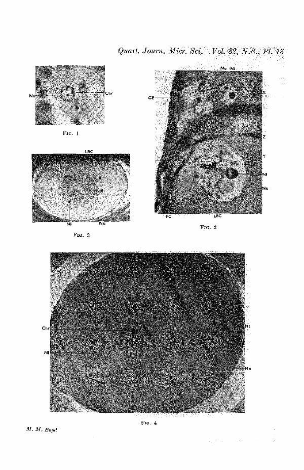
Quart. Journ. Micr. Sci. Vol. 82, N.S., PL 13
N u ~ "ChrGE-
FIG. 1
LBCr*>. '
f
Nl
FIG. 3
Chr
Nl
FC LBC
FIG. 2
f. M. BoydFIG. 4
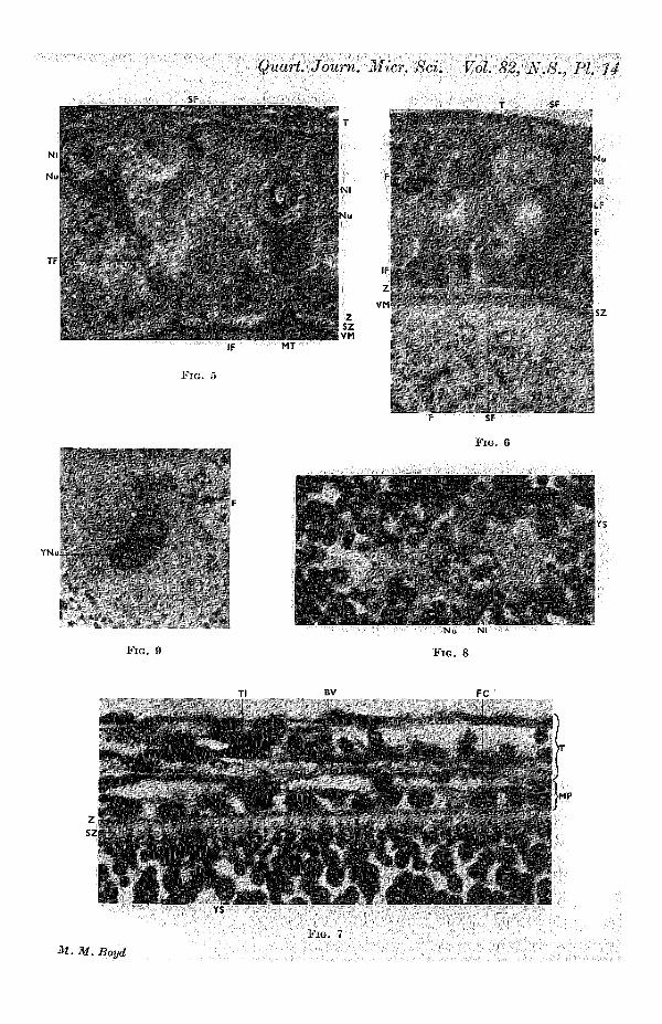
N l lNu
, , \ *
TF-
Quart. Journ. Micr. Sci. Vol 82, N.8., PI 14
FIG.
Nt
IF MT
J Z
' JVM
IF
Z
lu
II
F
YNu
FIG. 9
F SF
FIG. 6
A "v V-
•'?Nu
FIG. 8
Tl FC
i'iu.
5MP

Quart. Journ. Micr. Sci. Vol. 82, N.8., PL 15
- Z
If'

,-vaf
LC-
Quart. Journ. Micr. Sci. Vol. 82, N.S., PI 16
BV
FIG. 14
BV
FIG. 15
LC
M. M. Boyd

* * ' • •
Quart. Journ. Micr. Sci. Vol 82, N.8., PL 17
••••
Oft
fB
"-i TI
' i s - , «•*•
•-.* c
c .' *
F I G . 17
EF
LC
T
FIG. 16
M. M. Boyd
FIG. 18

Quart, Journ. Micr. Sci. Vol. 82, N.8., PL 18
LCII
I
"
FIG. 20
4 " ?!
;
> At "V- ','
FIG. 19
M. M. Boyd

Quart. Journ. Mies. Sci. Vol. 82, N.8., PL 19
FIG. 21
BV C
iJN^«g|s?" F3
FIG. 22
LC