the use of ultrasound for the diagnosis and treatment of ......the use of ultrasound for the...
TRANSCRIPT

The Use of Ultrasound for the Diagnosis and Treatment of the
Musculoskeletal System
St. Joseph’s Refresher Course
March 2020
Kenneth Iles, DC
John Finkenstadt, MD

ULTRASONOGRAPHY IN FAMILY MEDICINE
• Musculoskeletal – Diagnosis and Injectioneg: Carpal Tunnel; Shoulder Rotator Cuff; Knee Aspiration;
Ganglion Cysts; Dequervain’s Tenosynovitis
• Thyroid Nodules
• Enlarged Lymph Nodes
• Pericardial Effusion
• Abdominal Aortic Aneurysm
• Obstetrics

REFERENCES: ULTRASOUND IN FAMILY MEDICINE
1. Point of Care Ultrasound in General Practice: A Systematic Review
Ann Fam Med Jan/Feb 2019 vol. 17 no. 1 61-69.
2. Point of Care Ultrasonography in Family Medicine
Am Fam Physician. 2018 Aug 15;98(4):200-202.


THIS WORKSHOP WILL FOCUS ON 2 MAIN TOPICS:
DIAGNOSIS AND TREATMENT OF:
Carpal Tunnel Syndrome
Shoulder Rotator Cuff Tears

WHY ULTRASOUND?
•Portability
•Cost-Effective
•Patient Comfort
•High Resolution Imaging
•Real Time Dynamic Imaging

• Every patient can undergo Ultrasound
• No problem with hardware
• No problems with claustrophobia or need for sedation
• Probe can be placed over symptomatic area
• Color Doppler shows inflammation
• Better for differentiating fluid from solid areas
• Facilitates bilateral comparison
• Can be used to follow the long course of a structure such as a nerve
• Useful in guiding interventions
OTHER REASONS TO CONSIDER ULTRASOUND

LIMITATIONS OF ULTRASOUND IN
MUSCULOSKELETAL DIAGNOSIS
Intra-articular Pathology
Degenerative Change
Labral Tears
Fractures
Bone Tumors
Operator Dependent

REQUEST FOR VOLUNTEERS FOR DEMOS
Carpal Tunnel Evaluation
Shoulder Pain Evaluation

CARPAL TUNNEL SYNDROME
- Compression of the Median Nerve
beneath the
Flexor Retinaculum


Flexor
Retinaculum
Anatomy of Movement p. 151
Anatomy of Movement p. 151

Left Hand

Anatomy of
Movement p. 151
Carpal Tunnel
Left Hand
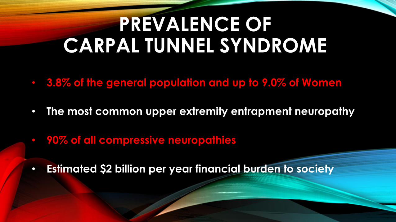
PREVALENCE OF CARPAL TUNNEL SYNDROME
• 3.8% of the general population and up to 9.0% of Women
• The most common upper extremity entrapment neuropathy
• 90% of all compressive neuropathies
• Estimated $2 billion per year financial burden to society

RISK FACTORS FOR
CARPAL TUNNEL SYNDROME
Obesity
Diabetes
Pregnancy
Hypothyroidism
Rheumatoid Arthritis

SYMPTOMS OF CARPAL TUNNEL SYNDROME
• Motor, Sensory and Autonomic Impairments
• Pain (especially at night) and Tingling
• Intrinsic Hand Weakness
• Reduced Grip Strength
• Alteration of Temperature Control

CARPAL TUNNEL SYNDROME PHYSICAL EXAM
• Tinel’s Sign
• Phalen’s Test
• Carpal Compression Test
• Pinch Grip (thumb and 5th finger)
• Sensory Examination – pin wheel


Phalen’s Test

Reverse Phalen’s Test

ULTRASOUND DIAGNOSIS OF
CARPAL TUNNEL SYNDROME
• CSAc(Cross Sectional Area crease) >10 mm²
• WFR(Wrist-Forearm Ratio) >1.2
• Enlarged Median N. proximal to the Carpal Tunnel inlet in longitudinal view(Notch Sign, or Dumbell Sign)
• Bowing of the Flexor Retinaculum at the Scaphoid-Pisiform level
• Distal flattening of the Median N. in the Carpal Tunnel
• Decreased mobility of Median N. on dynamic imaging

ULTRASOUND EVALUATION OF CTS
MSMPC DIAGNOSTIC PROTOCOL
- See Handout

Proximal
Carpal Tunnel
- Normal

Proximal
Carpal Tunnel
- Abnormal

THE ROLE OF ULTRASOUND IN THE DIAGNOSIS AND MANAGEMENT OF
CARPAL TUNNEL SYNDROME : A NEW PARADIGM
AUTHORS: MCDONAGH C, ALEXANDER M, KANE D.RHEUMATOLOGY (OXFORD), 2015 JAN;54(1):9-19.
NERVE CONDUCTION – SENSITIVITY > 85%
ULTRASOUND – SENSITIVITY UP TO 97.9%
SPECIFICITY WAS SIMILAR WITH BOTH PROCEDURES
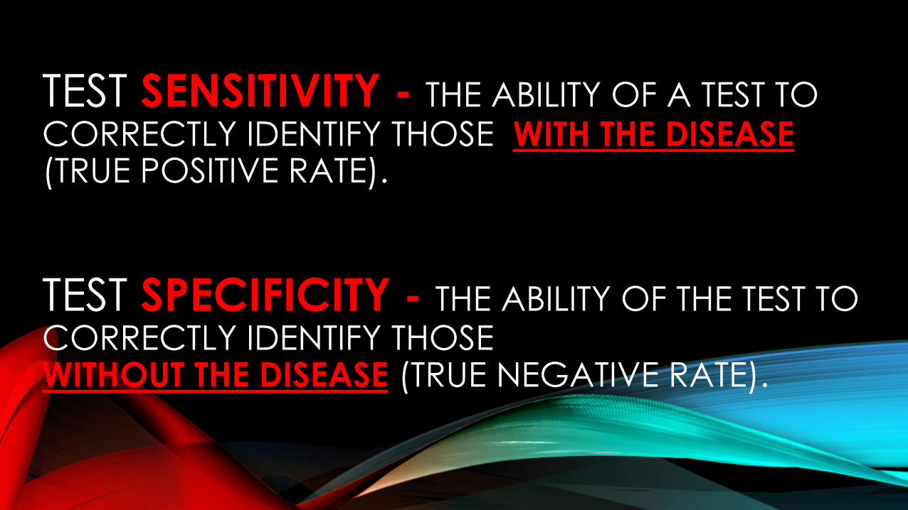
TEST SENSITIVITY - THE ABILITY OF A TEST TO CORRECTLY IDENTIFY THOSE WITH THE DISEASE(TRUE POSITIVE RATE).
TEST SPECIFICITY - THE ABILITY OF THE TEST TO CORRECTLY IDENTIFY THOSE WITHOUT THE DISEASE (TRUE NEGATIVE RATE).

DIAGNOSTIC ULTRASOUND FOR
CARPAL TUNNEL SYNDROME
Patient Demonstration

Steinbrocker and Neustadt p. 59
N.B. - Inject in line with the radial aspect of the 3rd and 4th fingers.
Carpal Tunnel
InjectionRight Hand
Flexor Carpi
Radialis
Palmaris
Longus

CANDIDATES FOR ULTRASOUND GUIDED
CARPAL TUNNEL HYDRODISSECTION
• Carpal Tunnel Surgical Failures
• Pregnant Women
• Poor Surgical Risks: eg. Elderly patient with multiple medical problems
• Reasonable Alternative for Conservative Treatment Failures
• Alternative for Patients Refusing Surgery

BENEFITS OF ULTRASOUND GUIDED
CARPAL TUNNEL HYDRODISSECTION
• Overall Safety
• Better than Non-Imaged Guided
• Accuracy of Medication Placement
• Ease of Performance
• Lower Level of Invasiveness than Surgery
• Lower Cost vs. Surgical Release
• Shorter Recovery Period
• Effectiveness

CARPAL TUNNEL HYDRODISSECTIONGoal – Decrease pressure on the median nerve from the flexor retinaculum and decrease inflammation of the median nerve and underlying flexor tendons
27G - 1 ½ inch needle - 8 ml of fluid with Kenalog - 40 (0.5 to 1.0 ml)
Ultrasound Guided – Transverse position of the probe
Local skin anesthesia optional
Maintain slight extension of the wrist
Supine position
Inject in distal direction
Inject along the Radial and Ulnar aspects of the nerve
Start approx. 1.0 cm proximal to the wrist crease and aim for radial aspect of the 3rd and 4th fingers
Almost horizontal injection – the needle travels parallel to the tendons and nerve
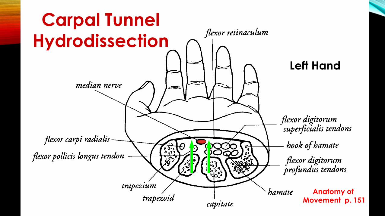
Anatomy of
Movement p. 151
Carpal Tunnel
Hydrodissection
Left Hand

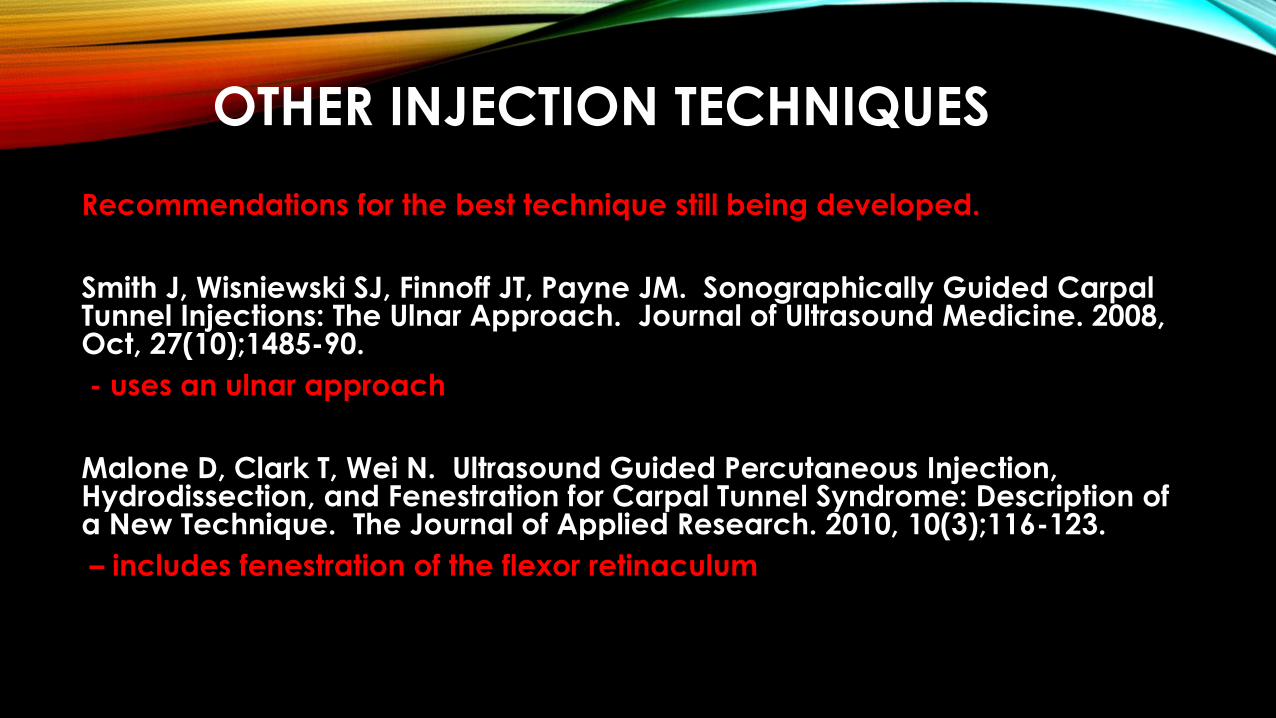
OTHER INJECTION TECHNIQUES
Recommendations for the best technique still being developed.
Smith J, Wisniewski SJ, Finnoff JT, Payne JM. Sonographically Guided Carpal Tunnel Injections: The Ulnar Approach. Journal of Ultrasound Medicine. 2008, Oct, 27(10);1485-90.
- uses an ulnar approach
Malone D, Clark T, Wei N. Ultrasound Guided Percutaneous Injection, Hydrodissection, and Fenestration for Carpal Tunnel Syndrome: Description of a New Technique. The Journal of Applied Research. 2010, 10(3);116-123.
– includes fenestration of the flexor retinaculum

CARPAL TUNNEL HYDRODISSECTION CASE STUDY
66 y.o. female – c/o recurrence of discomfort, numbness, tinging in the right hand.
S/P Carpal Tunnel Surgery on Right in 1999, Left 1996.
4/27/18: Dx Ultrasound on the Right consistent with CTS
Exam: mild tenderness, Phalen test equivocal, Pinch grip negative
5/17/18: Carpal Tunnel Hydrodissection – 40 mg Kenalog with D5W –6 ml total.
6/21/18: F/U – Significantly improved. Tingling and numbness much better. c/o Mild recurrence of symptoms with activity.
2/11/19: F/U – Remains improved. Essentially pain free. Occasional tingling only.

QUESTIONS
ABOUT
CARPAL TUNNEL SYNDROME ?
DIAGNOSTIC ULTRASOUND
OF THE
SHOULDER

Anterior
Anterior
Rotator Cuff – Right Shoulder
Humerus

Posterior
Anterior
Long Head
Short Head
Right Shoulder

Right Shoulder

Dr. James H. Cyriax, M.D., M.R.C.P.

Cyriax p.229
C5 Dermatome
SHOULDER PHYSICAL EXAM
DEMONSTRATION
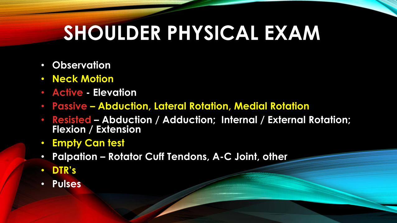
SHOULDER PHYSICAL EXAM
• Observation
• Neck Motion
• Active - Elevation
• Passive – Abduction, Lateral Rotation, Medial Rotation
• Resisted – Abduction / Adduction; Internal / External Rotation; Flexion / Extension
• Empty Can test
• Palpation – Rotator Cuff Tendons, A-C Joint, other
• DTR’s
• Pulses

PASSIVE MOVEMENTSCapsulitis – Impaired
Elevation and Lateral Rotation
Rotator Cuff Pathology – Impaired Elevation and normal Lateral Rotation
RESULTS OF PASSIVE TESTING

STRETCHING EXERCISES
The Essentials of Musculoskeletal Care p. 99

RESULTS OF RESISTED TESTING
Resisted ABDUCTION - SUPRASPINATUS
Resisted ADDUCTION - PECTORALIS MAJOR, LATISSIMUS DORSI
Resisted LATERAL ROTATION – INFRASPINATUS
Resisted MEDIAL ROTATION – SUBSCAPULARIS
Resisted ELBOW FLEXION – BICEPS
Resisted ELBOW EXTENSION - TRICEPS

PALPATION OF THE SUPRASPINATUS TENDON

Netter p. 400

PALPATION OF THE SUBSCAPULARIS TENDON

Cyriax p. 43
SUBSCAPULARISTENDON
Right Shoulder
Anterior View

PALPATION OF THE INFRASPINATUS TENDON

Cyriax p. 42
Infraspinatus
Tendon
Left Shoulder - Posterior View

ROTATOR CUFF PALPATION
•Supraspinatus – Hand in back pocket
•Subscapularis – Elbow flexed and arm externally rotated
• Infraspinatus – Hand on opposite shoulder
• Other areas: A-C Joint, Biceps Tendon, Capsule, Coracoid, Etc.

PALPATION OF THE A-C JOINT

ORIENTATION OF THE A-C JOINT
Neumann p.101
Right
Shoulder

ULTRASOUND EVALUATION OF THESHOULDER
MSMPC DIAGNOSTIC PROTOCOL
– See Handout

DIAGNOSTIC ULTRASOUND OF
THE SHOULDER
Patient Demonstration

Normal

PartialTear

Supraspinatus
Longitudinal View
Partial Tear

Supraspinatus
Transverse View
Partial Tear

Supraspinatus
Longitudinal View
Partial Tear

Supraspinatus
Transverse View
Partial Tear
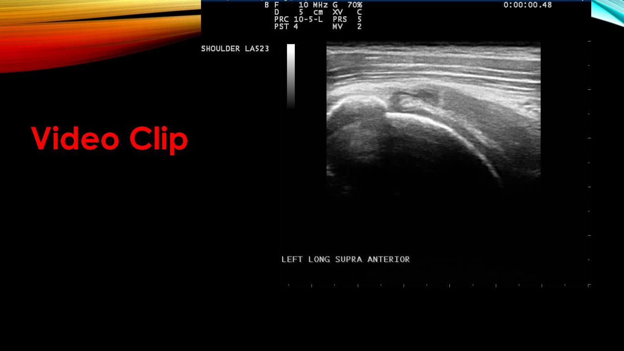
Video Clip

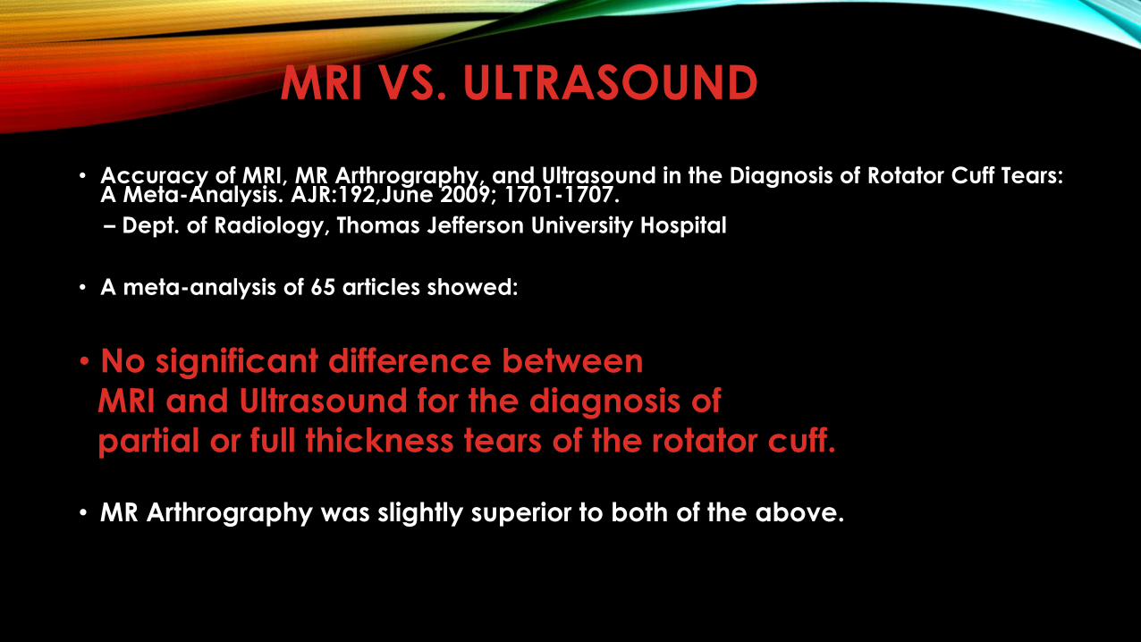
MRI VS. ULTRASOUND
• Accuracy of MRI, MR Arthrography, and Ultrasound in the Diagnosis of Rotator Cuff Tears: A Meta-Analysis. AJR:192,June 2009; 1701-1707.
– Dept. of Radiology, Thomas Jefferson University Hospital
• A meta-analysis of 65 articles showed:
• No significant difference between MRI and Ultrasound for the diagnosis of partial or full thickness tears of the rotator cuff.
• MR Arthrography was slightly superior to both of the above.

PLATELET RICH PLASMA (PRP)
PRP promotes natural healing mechanisms by release of growth factors and other bioactive substances.
PDGFs – Platelet Derived Growth Factors alpha and beta• Plays a role in cell differentiation and neovascularization
TGF – Transforming Growth Factors beta 1 and beta 2• Stimulates Tendon Differentiation and Formation of Collagen
EGF – Epithelial Growth Factor• Induces Fibroblast Proliferation
VEGF – Vascular Endothelial Growth Factor• Stimulates Neovascularization

PLATELET RICH PLASMA (PRP)- AUTOLOGOUS CONCENTRATION 0F PLATELETS OBTAINED BY WHOLE BLOOD CENTRIFUGATION
Mention Secondary Processing to remove rbc’s


PRP SHOULDER INDIVDUAL CASE STUDY61 yo accountant and avid weight lifter c/o R shoulder pain.
Hx of Right shoulder surgery. Several years of pain with activity.
Pain is primarily anterior.
Exam: Restriction to elevation – 170 degrees (180 left), lateral rotation 60 degrees.
Positive empty can test, Tender over the supraspinatus
Prolotherapy with aqueous testosterone: 12/28/16 and 5/9/17
– with a marginal response.
6/09/17: Dx Ultrasound– partial thickness tear supraspinatus, A-C joint degenerative changes, subscapularis tendinosis
PRP Right shoulder
7/07/17: F/U - Sore for 2 days, no improvement as yet
8/17/18: F/U –About 30% improved, restarted working out
9/08/17: F/U and Repeat Ultrasound –partial thickness tear supraspinatus. Continues to improve clinically.
4/18/18: Phone – Significantly improved, about 80% better.
2/04/19: Phone –Essentially pain free. Full workouts. Still limited tightness.

EFFICACY OF TREATING ROTATOR CUFF PATHOLOGY WITH PRP
• American Academy of Orthopaedic Surgeons: 2013 Poster Presentation
Study on rotator cuff tendinopathy without a full thickness tear
• 204 patients – 102 injected directly into tendon with PRP
- 102 controls injected into the subacromial space with steroid
• 1- year follow-up: PRP group had significantly better ROM
48 steroid treated patients and only 3 PRP treated patients required surgery

EFFICACY OF TREATING ROTATOR CUFF PATHOLOGY WITH PRP
• Multiple studies have shown mixed results.
• Our impression is that over 90% of patients have had positive results.
• We hope to have our case study results completed later this year.
• More research needed.

QUESTIONS ?
You are all invited to our office on a select Friday to witness our clinical use of ultrasound imaging.

The End
John Finkenstadt, M.D.
Kenneth Iles, D.C.
Madison-Irving Medical Center
475 Irving Avenue - Suite 402
Syracuse, N.Y. 13210
(315) 478-9710