therapeutic potential of chlorotoxin-like neurotoxin from the chinese
TRANSCRIPT
A
a(stgCaor©
K
BitAsabiq
caeg
0d
Neuroscience Letters 412 (2007) 62–67
Therapeutic potential of chlorotoxin-like neurotoxin from theChinese scorpion for human gliomas
Yue-Jun Fu a, Li-Tian Yin a, Ai-Hua Liang a,∗, Chao-Feng Zhang b, Wei Wang a,Bao-Feng Chai a, Jian-Yi Yang c, Xiao-Jun Fan a
a Key Laboratory of Chemical Biology and Molecular Engineering of Ministry of Education, Institute of Biotechnology,Shanxi University, Taiyuan 030006, PR China
b Key Laboratory of Chemical Biology and Molecular Engineering of Ministry of Education, Institute of Molecular Science,Shanxi University, Taiyuan 030006, PR China
c Department of Biology, Shanxi Medical University, Taiyuan 030001, PR China
Received 10 May 2006; received in revised form 12 September 2006; accepted 12 October 2006
bstract
Chlorotoxin, one of the key toxins in scorpion Leiurus quinquestriatus venom, has been shown to bind specifically to glioma cell surface asspecific chloride channel blocker. In this study, a purified, recombinant chlorotoxin-like peptide from the scorpion Buthus martensii Karsch
named rBmK CTa) was characterized by in vivo and in vitro studies. The results from cell proliferation assay with human glioma (SHG-44) cellshowed that rBmK CTa inhibits the growth of glioma cells in a dose-dependent manner, with an IC50 value of approximately 0.28 �M. Underhe same conditions, the IC50 value for normal astrocytes increased to 8 �M. This clearly indicated that rBmK CTa had specific toxicity againstlioma cells but not astrocytes. Results from whole-cell patch-clamp recording showed that chloride current in SHG-44 was inhibited by rBmK
Ta in a voltage-dependent manner and percent inhibitions for the blocking action of rBmK CTa (0.07 and 0.14 �M) on ICl was 17.64 ± 3.06%nd 55.86 ± 2.83%, respectively. Histological analysis of rBmK CTa treated mice showed that brain, leg muscle and cardiac muscle were the targetrgans of this toxin. These results suggest that rBmK CTa may have potential therapeutic application in clinical treatment of human glioma. Itepresents an approach for developing a novel therapeutic agent.2006 Elsevier Ireland Ltd. All rights reserved.
curren
sgvrBattegf
eywords: Chlorotoxin-like peptide; Human glioma cells (SHG-44); Chloride
uthus matensii Karsch is a widely distributed scorpion speciesn Asia. A variety of studies have been conducted to iden-ify and purify toxin peptides from this particular species [5].
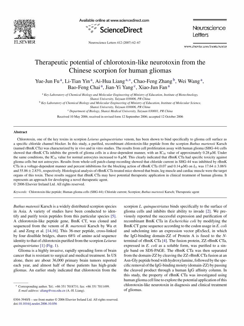
chlorotoxin-like peptide gene, BmK CT, was cloned andequenced from the venom of B. martensii Karsch by Wu etl. and Zeng et al. [14,16]. This 36-mer peptide, cross-linkedy four disulfide bridges, shares 68% of amino acid sequencedentity to that of chlorotoxin purified from the scorpion Leiurusuinquestriatus [1] (Fig. 1).
Glioma is a highly invasive, rapidly spreading form of brainancer that is resistant to surgical and medical treatment. In US
lone, there are about 36,000 primary brain tumors reportedach year, and almost half of these patients has high-gradeliomas. An earlier study indicated that chlorotoxin from the∗ Corresponding author. Tel.: +86 351 7018731; fax: +86 351 7011499.E-mail address: [email protected] (A.-H. Liang).
Actthco
304-3940/$ – see front matter © 2006 Elsevier Ireland Ltd. All rights reserved.oi:10.1016/j.neulet.2006.10.056
t; Scorpion; Buthus martensii Karsch; Therapeutic agent
corpion L. quinquestriatus binds specifically to the surface oflioma cells and inhibits their ability to invade [2]. We pre-iously reported the successful expression and purification ofecombinant BmK CTa in Escherichia coli by modifying themK CT gene sequence according to the codon usage in E. colind subcloning into an expression vector pExSecI, in whichhe IgG-binding domain-ZZ of Protein A is fused to the N-erminal of rBmK CTa [4]. The fusion protein, ZZ-rBmK CTa,xpressed in E. coli as a soluble form, was purified to a sin-le band on SDS-PAGE. The rBmK CTa was then separatedrom the domain-ZZ by cleaving the ZZ-rBmK CTa fusion at ansn-Gly peptide bond with hydroxylamine, followed by the spe-
ific removal of the IgG-binding moiety (domain-ZZ) by passinghe cleaved product through a human IgG affinity column. In
his study, the property of rBmK CTa was investigated usinguman glioma cell line to explore the potential application of thishlorotoxin-like neurotoxin in diagnosis and clinical treatmentf gliomas.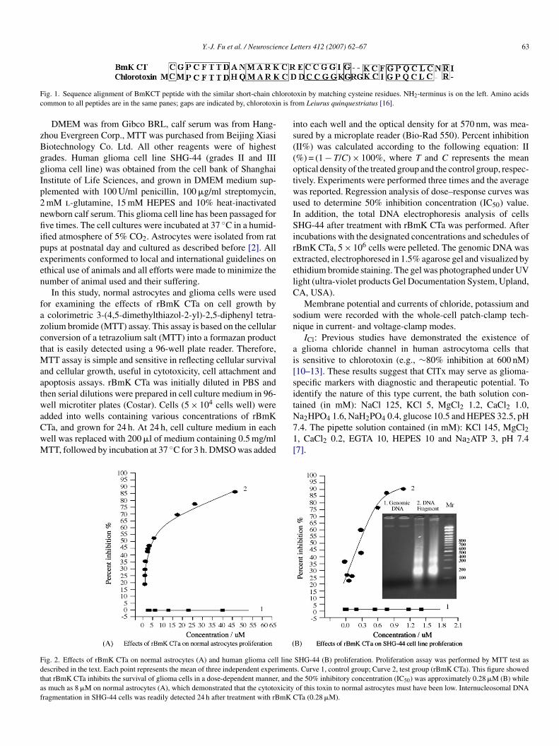
Y.-J. Fu et al. / Neuroscience Letters 412 (2007) 62–67 63
F lorotoc n is fr
zBggIp2nfiipeen
fazctMaatwaCwM
is((otwuISireelC
sn
ai[sit
Fdtaf
ig. 1. Sequence alignment of BmKCT peptide with the similar short-chain chommon to all peptides are in the same panes; gaps are indicated by, chlorotoxi
DMEM was from Gibco BRL, calf serum was from Hang-hou Evergreen Corp., MTT was purchased from Beijing Xiasiiotechnology Co. Ltd. All other reagents were of highestrades. Human glioma cell line SHG-44 (grades II and IIIlioma cell line) was obtained from the cell bank of Shanghainstitute of Life Sciences, and grown in DMEM medium sup-lemented with 100 U/ml penicillin, 100 �g/ml streptomycin,mM l-glutamine, 15 mM HEPES and 10% heat-inactivatedewborn calf serum. This glioma cell line has been passaged forve times. The cell cultures were incubated at 37 ◦C in a humid-
fied atmosphere of 5% CO2. Astrocytes were isolated from ratups at postnatal day and cultured as described before [2]. Allxperiments conformed to local and international guidelines onthical use of animals and all efforts were made to minimize theumber of animal used and their suffering.
In this study, normal astrocytes and glioma cells were usedor examining the effects of rBmK CTa on cell growth bycolorimetric 3-(4,5-dimethylthiazol-2-yl)-2,5-diphenyl tetra-
olium bromide (MTT) assay. This assay is based on the cellularonversion of a tetrazolium salt (MTT) into a formazan producthat is easily detected using a 96-well plate reader. Therefore,
TT assay is simple and sensitive in reflecting cellular survivalnd cellular growth, useful in cytotoxicity, cell attachment andpoptosis assays. rBmK CTa was initially diluted in PBS andhen serial dilutions were prepared in cell culture medium in 96-ell microtiter plates (Costar). Cells (5 × 10⁴ cells well) were
dded into wells containing various concentrations of rBmKTa, and grown for 24 h. At 24 h, cell culture medium in eachell was replaced with 200 �l of medium containing 0.5 mg/mlTT, followed by incubation at 37 ◦C for 3 h. DMSO was added
N71[
ig. 2. Effects of rBmK CTa on normal astrocytes (A) and human glioma cell lineescribed in the text. Each point represents the mean of three independent experimenthat rBmK CTa inhibits the survival of glioma cells in a dose-dependent manner, ands much as 8 �M on normal astrocytes (A), which demonstrated that the cytotoxicityragmentation in SHG-44 cells was readily detected 24 h after treatment with rBmK C
xin by matching cysteine residues. NH2-terminus is on the left. Amino acidsom Leiurus quinquestriatus [16].
nto each well and the optical density for at 570 nm, was mea-ured by a microplate reader (Bio-Rad 550). Percent inhibitionII%) was calculated according to the following equation: II%) = (1 − T/C) × 100%, where T and C represents the meanptical density of the treated group and the control group, respec-ively. Experiments were performed three times and the averageas reported. Regression analysis of dose–response curves wassed to determine 50% inhibition concentration (IC50) value.n addition, the total DNA electrophoresis analysis of cellsHG-44 after treatment with rBmK CTa was performed. After
ncubations with the designated concentrations and schedules ofBmK CTa, 5 × 10⁶ cells were pelleted. The genomic DNA wasxtracted, electrophoresed in 1.5% agarose gel and visualized bythidium bromide staining. The gel was photographed under UVight (ultra-violet products Gel Documentation System, Upland,A, USA).
Membrane potential and currents of chloride, potassium andodium were recorded with the whole-cell patch-clamp tech-ique in current- and voltage-clamp modes.
ICl: Previous studies have demonstrated the existence ofglioma chloride channel in human astrocytoma cells that
s sensitive to chlorotoxin (e.g., ∼80% inhibition at 600 nM)10–13]. These results suggest that ClTx may serve as glioma-pecific markers with diagnostic and therapeutic potential. Todentify the nature of this type current, the bath solution con-ained (in mM): NaCl 125, KCl 5, MgCl2 1.2, CaCl2 1.0,
a2HPO4 1.6, NaH2PO4 0.4, glucose 10.5 and HEPES 32.5, pH.4. The pipette solution contained (in mM): KCl 145, MgCl2, CaCl2 0.2, EGTA 10, HEPES 10 and Na2ATP 3, pH 7.47].SHG-44 (B) proliferation. Proliferation assay was performed by MTT test ass. Curve 1, control group; Curve 2, test group (rBmK CTa). This figure showedthe 50% inhibitory concentration (IC50) was approximately 0.28 �M (B) whileof this toxin to normal astrocytes must have been low. Internucleosomal DNATa (0.28 �M).
64 Y.-J. Fu et al. / Neuroscience Letters 412 (2007) 62–67
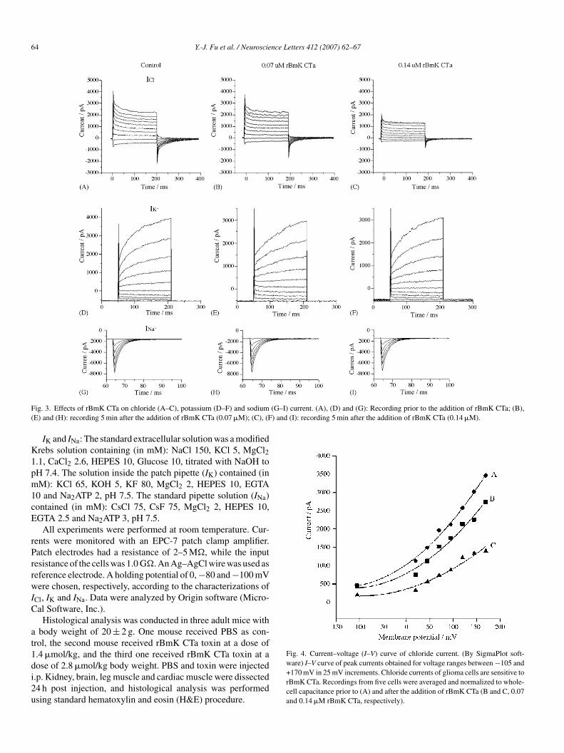
F (G–I) current. (A), (D) and (G): Recording prior to the addition of rBmK CTa; (B),( ) and (I): recording 5 min after the addition of rBmK CTa (0.14 �M).
K1pm1cE
rPrrwIC
at1di2u
Fig. 4. Current–voltage (I–V) curve of chloride current. (By SigmaPlot soft-
ig. 3. Effects of rBmK CTa on chloride (A–C), potassium (D–F) and sodiumE) and (H): recording 5 min after the addition of rBmK CTa (0.07 �M); (C), (F
IK and INa: The standard extracellular solution was a modifiedrebs solution containing (in mM): NaCl 150, KCl 5, MgCl2.1, CaCl2 2.6, HEPES 10, Glucose 10, titrated with NaOH toH 7.4. The solution inside the patch pipette (IK) contained (inM): KCl 65, KOH 5, KF 80, MgCl2 2, HEPES 10, EGTA
0 and Na2ATP 2, pH 7.5. The standard pipette solution (INa)ontained (in mM): CsCl 75, CsF 75, MgCl2 2, HEPES 10,GTA 2.5 and Na2ATP 3, pH 7.5.
All experiments were performed at room temperature. Cur-ents were monitored with an EPC-7 patch clamp amplifier.atch electrodes had a resistance of 2–5 M�, while the inputesistance of the cells was 1.0 G�. An Ag–AgCl wire was used aseference electrode. A holding potential of 0, −80 and −100 mVere chosen, respectively, according to the characterizations of
Cl, IK and INa. Data were analyzed by Origin software (Micro-al Software, Inc.).
Histological analysis was conducted in three adult mice withbody weight of 20 ± 2 g. One mouse received PBS as con-
rol, the second mouse received rBmK CTa toxin at a dose of.4 �mol/kg, and the third one received rBmK CTa toxin at a
ose of 2.8 �mol/kg body weight. PBS and toxin were injected.p. Kidney, brain, leg muscle and cardiac muscle were dissected4 h post injection, and histological analysis was performedsing standard hematoxylin and eosin (H&E) procedure.ware) I–V curve of peak currents obtained for voltage ranges between −105 and+170 mV in 25 mV increments. Chloride currents of glioma cells are sensitive torBmK CTa. Recordings from five cells were averaged and normalized to whole-cell capacitance prior to (A) and after the addition of rBmK CTa (B and C, 0.07and 0.14 �M rBmK CTa, respectively).
nce L
hsclnccF
ccccv
FmfmL2d
Y.-J. Fu et al. / Neuroscie
Several recent studies have suggested that chlorotoxin is aighly specific ligand for malignant human gliomas, withoutignificant binding to normal brain cells [2,6,9]. However, theytotoxicity and therapeutic characterization of the chlorotoxin-ike peptide from the Chinese scorpion B. martensii Karsch have
ot been studied. In this report, normal astrocytes and gliomaells (SHG-44) were used to examine the effects of rBmK CTa onell proliferation by a colorimetric MTT assay. The results (seeig. 2) showed that rBmK CTa inhibited the growth of gliomaacaD
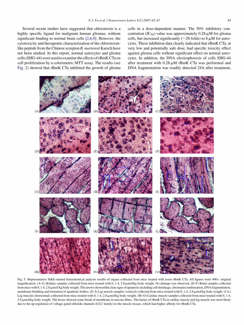
ig. 5. Representative H&E-stained histochemical analysis results of organs collecagnification. (A–C) Kidney samples collected from mice treated with 0, 1.4, 2.8 �m
rom mice with 0, 1.4, 2.8 �mol /kg body weight. The arrows showed the clear signs of aembrane bledding and formation of apoptotic bodies; (G–I) Leg muscle samples (veeg muscles (horizontal) collected from mice treated with 0, 1.4, 2.8 �mol/kg body w.8 �mol/kg body weight. The boxes showed some break of membrane in sarcous fibue to the up-regulation of voltage-gated chloride channels (CLC family) in the musc
etters 412 (2007) 62–67 65
ells in a dose-dependent manner. The 50% inhibitory con-entration (IC50) value was approximately 0.28 �M for gliomaells, but increased significantly (∼28-folds) to 8 �M for astro-ytes. These inhibition data clearly indicated that rBmK CTa, atery low and potentially safe dose, had specific toxicity effect
gainst glioma cells without significant effect on normal astro-ytes. In addition, the DNA electrophoresis of cells SHG-44fter treatment with 0.28 �M rBmK CTa was performed andNA fragmentation was readily detected 24 h after treatment,ted from mice treated with toxin rBmK CTa. All figures were 400× originalol/kg body weight. No damage was observed. (D–F) Brain samples collected
poptosis including cell shrinkage, chromatin condensation, DNA fragmentation,rtical) collected from mice treated with 0, 1.4, 2.8 �mol/kg body weight. (J–L)eight. (M–O) Cardiac muscle samples collected from mice treated with 0, 1.4,
res. The harms of rBmK CTa in cardiac muscle and leg muscle was most likelyle tissues, which had higher affinity for rBmK CTa.
6 nce L
wTsu
cc4dE1(iw5
cstiwruicn
ditmCbticpisitn
tcnodtscogclrt
gaiethied
cthricfiJaaop
TH
T
K
B
L
C
6 Y.-J. Fu et al. / Neuroscie
hich resulted in a ladder pattern—a sign of apoptosis (Fig. 2B).his is a novel aspect in the biological properties of the Chinesecorpion neurotoxin and an important step in elucidating thenderlying molecular mechanisms of its antitumor action.
In order to explore the mechanism of rBmK CTa, whole-ell patch-clamp recording analysis was performed with gliomasells. As shown in Fig. 3, chloride current of gliomas cells (SHG-4) was inhibited by rBmK CTa. On the other hand, rBmK CTaid not cause any inhibition for potassium and sodium currents.ffects of rBmK CTa at 0.07 and 0.14 �M (corresponding to/4 IC50 and 1/2 IC50, respectively) on chloride current–voltageI–V) relationship was examined between −105 and +170 mVn 25 mV increments. As shown in Fig. 4, the chloride currentas inhibited by 17.64 ± 3.06% at 0.07 �M rBmK CTa and5.86 ± 2.83% (n = 5) at 0.14 �M rBmK CTa.
In an earlier study, the inhibitory effect of glioma chlorideurrent was reversible after eluting the chamber with externalolution for 3 min. This was identical with the predicted func-ion (a short-chain glioma chloride channels blocker) based onts high sequence homology with chlorotoxin [15]. In this paper,hole-cell patch-clamp recording analysis showed that chlo-
ide current of gliomas cells (SHG-44) was observably inhibitednder control conditions in the presence of rBmK CTa, but thisnhibition was not presented in potassium current and sodiumurrent, which demonstrates that it was a glioma chloride chan-els blocker, but not potassium and sodium channel blocker.
To evaluate the safety of rBmK CTa, mice were given to twooses of toxin by intraperitoneal injection. Twenty-four postnjection, organs were examined for damages. Fig. 5 showshe histological analysis results of four organs collected from
ice treated with PBS (control), 1.4 and 2.8 �mol/kg of rBmKTa. Voltage-gated chloride channels have been implicated aseing important for cell proliferation and migration. Hence,hose voltage-gated chloride channels in normal cells may benhibited by rBmK CTa (a short-chain voltage-gated chloridehannel blocker) in regulating cell volume in the context of cellroliferation and migration. Moreover, hematoxylin–eosin stains a useful method for general histology. Basophilic nuclei are
tained “blue” with hematoxylin. And some signs of apoptosisn nuclei can be detected in this assay. Therefore, the followinghree parameters were measured for each organ: cell membrane,ucleus and cell interval (Table 1). Firstly, the results indicatedagac
able 1istological analysis of four organs after treated with the rBmK CTaa
issues rBmK CTa concentrations (�mol/kg body weight)
idney 1.42.8
rain 1.42.8
eg muscle 1.42.8
ardiac muscle 1.42.8
a The following three parameters were measured: cell membrane, nucleus and cell
etters 412 (2007) 62–67
here are different degrees of damage for brain, leg muscle andardiac muscle in toxin treated mice. On the other hand, there iso detectable damage for kidney. Thirdly, although penetrationf extrinsic proteins through intact blood–brain barrier is veryifficult, observed responses (or damage for brain) using thisoxin were promising. Two logic important features were pre-umed [2]: (i) rBmK CTa is a small, 36 amino acid, peptide thatan cross blood and tissue barriers, the effects of rBmK CTa werebserved within 1 day post injection. (ii) It can binds to brainlioma cells by the high affinity action power with voltage-gatedhloride channels. Fourthly, the damage in cardiac muscle andeg muscle caused by rBmK CTa was most likely due to the up-egulation of voltage-gated chloride channels (CLC family) inhese muscle tissues, which have higher affinity for rBmK CTa.
The voltage-gated channels are specifically upregulated inlioma membranes and endow glioma cells with an enhancedbility to transport Cl−. This may in turn facilitate rapid changesn cell size and shape as cells divide or invade through tortuousxtracellular brain spaces [8]. Based on the findings presented inhis study, it is clear that the chlorotoxin-like peptide rBmK CTaas specific affinity for binding to glioma cells and is capable ofnhibiting the chloride channel and proliferation. The inhibitoryffect of rBmK CTa on glioma cell proliferation is potentiallyue to the mechanism of apoptosis.
It is interesting to note that the toxin rBmK CTa, alone oronjugated with radioisotope, could be a potential agent forreating human gliomas. Along this line, the recent study [2]ad identified matrix metalloproteinase-2 (MMP-2) as a specificeceptor for chlorotoxin. MMP-2, a proteinase involved in tumornvasion, is specifically up-regulated in gliomas and related can-ers, but is not expressed in normal brain cells. Recent reportsrom TransMolecular, Inc. indicated that 131I-chlorotoxin is safen a phase 2 human clinical trial study (see news release onune 05, 2006 from www.transmolecular.com). Currently, were working on the generation antibodies against rBmK CTand the identification of specific receptors for toxin rBmK CTan glioma cells [3]. In our study, polycolonal antibodies to theurified toxin were raised in rats. Overlay assay and pull-down
ssay showed that this toxin specially binds to two proteins in thelioma cells with corresponding molecular weights of about 80nd 35 kDa. They may serve as candidate receptors or alternativeellular component for interaction with rBmK CTa.Histological parameters
Cell membrane Nucleus Cell interval
− − −− − −− − −+ + −+ − −++ − −− − −+ − −
interval. Degree of damage: −, non-significant; +, minor; ++, significant.
nce L
tac
A
UTto
R
[
[
[
[
[
[
Y.-J. Fu et al. / Neuroscie
In conclusion, the findings presented in this study are essen-ial for the further exploration of this peptide. It represents anpproach for developing a novel therapeutic agent and a potentiallinical treatment of human gliomas cancer.
cknowledgments
We thank Dr. Jin-an Jiao, vice-president of Hematech. Inc.,SA, for reading the manuscript and help us with the English.his work was supported by National Natural Science Founda-
ion of China (No. 30270204) and Natural Science Foundationf Shanxi Province (20051065).
eferences
[1] J.A. DeBin, J.E. Maggio, G.R. Strichartz, Purification and characterizationof chlorotoxin, a chloride channel ligand from the venom of the scorpion,Am. J. Physiol. 264 (1993) 361–369.
[2] J. Deshane, C.C. Garmer, H. Sontheimer, Chlorotoxin inhibits gliomacell invasion via matrix metalloproteinase-2, J. Biol. Chem. 278 (2003)4135–4144.
[3] Y.J. Fu, L.T. Yin, A.H. Liang, Polyclonal antibody against a recombi-nant chlorotoxin-like peptide from the Chinese scorpion and detection ofits putative receptors in human glioma cells, Biotechnol. Lett. 28 (2006)1439–1443.
[4] Y.J. Fu, L.T. Yin, W. Wang, B.F. Chai, A.H. Liang, Synthesis expressionand purification of a type of chlorotoxin-like peptide from the scorpion,
Buthus martensii Karsch, and its acute toxicity analysis, Biotechnol. Lett.27 (2005) 1597–1603.[5] C. Goudet, C.W. Chi, J. Tytagt, An overview of toxins and genes from thevenom of the Asian scorpion Buthus martensii Karsch, Toxicon 40 (2002)1239–1258.
[
etters 412 (2007) 62–67 67
[6] S.A. Lyons, J. O’Neal, H. Sontheimer, Chlorotoxin a scorpion-derived pep-tide, specifically binds to gliomas and tumors of neuroectodermal origin,Glia 39 (2002) 162–173.
[7] C. Maertens, L. Wei, J. Tytgat, G. Droogmans, B. Nilius, Chloro-toxin does not inhibit volume-regulated, calcium-activated and cyclicAMP-activated chloride channels, Br. J. Pharmacol. 129 (2000) 791–801.
[8] M.L. Olsen, S. Schade, S.A. Lyons, M.D. Amaral, H. Sontheimer, Expres-sion of voltage-gated chloride channels in human glioma cells, J. Neurosci.23 (2003) 5572–5582.
[9] L. Soroceanu, Y. Gillespie, M.B. Khazaeli, H. Sontheimer, Use of chloro-toxin for targeting of primary brain tumors, Cancer Res. 58 (1998)4871–4879.
10] N. Ullrich, A. Bordey, G.Y. Gillespie, H. Sontheimer, Expression ofvoltage-activated chloride currents in acute slices of human gliomas, Neu-roscience 83 (1998) 1161–1173.
11] N. Ullrich, G.Y. Gillespie, H. Sontheimer, Human astrocytoma cells expressa unique chloride current, Neuroreport 7 (1995) 343–347.
12] N. Ullrich, H. Sontheimer, Biophysical and pharmacological characteri-zation of chloride currents in human astrocytoma cells, Am. J. Phys. 39(1996) 1511–1521.
13] N. Ullrich, H. Sontheimer, Cell cycle-dependent expression of a glioma-specific chloride current: proposed link to cytoskeletal changes, Am. J.Phys. 42 (1997) 1290–1297.
14] J.J. Wu, D. Li, Z.D. Lan, C.W. Chi, The gene cloning and sequencingof Bm-12, a Chlorotoxin-like peptide from the scorpion Buthus martensiiKarsch, Toxicon 38 (2000) 661–668.
15] R. Yang, F. Peng, H. Liu, Z.J. Cao, W.X. Li, X. Mao, D.H. Jiang, Func-tional analysis of a gene encoding a chlorotoxin-like peptide derivedfrom scorpion toxin, Chin. J. Biochem. Mol. Biol. 21 (2005) 19–
23.16] X.C. Zeng, W.X. Li, S.Y. Zhu, F. Peng, Z.H. Zhu, K.L. Wu, F.H. Yang,Cloning and characterization of a cDNA sequence encoding the precursorof a chlorotoxin-like peptide from the Chinese scorpion Buthus martensiiKarsch, Toxicon 38 (2000) 1009–1014.