thermodynamic and kinetic analysis of an rna kissing interaction
TRANSCRIPT

Biophysical Journal Volume 102 March 2012 1097–1107 1097
Thermodynamic and Kinetic Analysis of an RNA Kissing Interactionand Its Resolution into an Extended Duplex
Nilshad Salim,6Rajan Lamichhane,6Rui Zhao, Tuhina Banerjee, Jane Philip, David Rueda,* and Andrew L. Feig*Department of Chemistry, Wayne State University, Detroit, Michigan
ABSTRACT Kissing hairpin interactions form when the loop residues of two hairpins have Watson-Crick complementarity. Ina unimolecular context, kissing interactions are important for tertiary folding and pseudoknot formation, whereas in a bimolecularcontext, they provide a basis for molecular recognition. In some cases, kissing complexes can be a prelude to strand displace-ment reactions where the two hairpins resolve to form a stable extended intermolecular duplex. The kinetics and thermody-namics of kissing-complex formation and their subsequent strand-displacement reactions are poorly understood. Here,biophysical techniques including isothermal titration calorimetry, surface plasmon resonance, and single-molecule fluorescencehave been employed to probe the factors that govern the stability of kissing complexes and their subsequent structural rear-rangements. We show that the general understanding of RNA duplex formation can be extended to kissing complexes butthat kissing complexes display an unusual level of stability relative to simple duplexes of the same sequence. These interactionsform and break many times at room temperature before becoming committed to a slow, irreversible forward transition to thestrand-displaced form. Furthermore, using smFRET we show that the primary difference between stable and labile kissingcomplexes is based almost completely on their off rates. Both stable and labile complexes form at the same rate within error,but less stable species dissociate rapidly, allowing us to understand how these complexes can help generate specificity alonga folding pathway or during a gene regulation event.
INTRODUCTION
In recent years, reports of riboregulation have becomecommonplace, dramatically changing our view of thecentral dogma and the way in which gene regulation iscontrolled (1). Regulation may occur cotranscriptionally,as in the case of most metabolite sensing riboswitches(2), or posttranscriptionally, as in the case of the smallbacterial regulatory RNAs (3) or eukaryotic microRNAs(4,5). Riboswitches are structures that allow small-moleculebinding to an aptamer domain to control a unimolecularrearrangement, allowing the RNA to adopt either a termina-tion or an antitermination structure, and hence, they affectthe transcription of downstream genes (2,6). Posttranscrip-tional regulation typically requires two RNAs to recognizeone another (an sRNA and its cognate mRNA target), some-times on their own, but more commonly with the assistanceof RNA-binding proteins (7,8). The resulting complex canthen be recognized and acted upon by cellular machinery.Stability of these RNA-RNA complexes must be finelytuned. If the initial contact yields a species that is too stable,selectivity is lost, since release of the noncognate partnerbecomes slow. Conversely, if the initial complex interactstoo weakly, its lifetime is too short to allow stable pairinginteractions to form and the benefits of colocalization arelost. Thus, the initial contact must fall within a relativelynarrow range of lifetimes to be beneficial.
Submitted August 15, 2011, and accepted for publication December 30,
2011.6Nilshad Salim and Rajan Lamichhane contributed equally to this work.
*Correspondence: [email protected] or [email protected]
Editor: Samuel Butcher.
� 2012 by the Biophysical Society
0006-3495/12/03/1097/11 $2.00
In the case of bacterial and viral systems, loop-loopcontacts (kissing interactions) are particularly prevalent inregulatory complexes. Kissing interactions occur whencomplementary sequences in apical loops of two hairpinscan form Watson-Crick basepairing (9). The stability ofthese kissing complexes is primarily based on loop comple-mentarity, but factors such as the orientation of the loops,internal loop structure, the loop-closing basepair, and thesequence of the stems adjacent to the loops are all knownto contribute to the stability of these interactions (10–12).Numerous examples of rearrangements are known thatdepend upon the initial formation of loop-loop interactions.Complexes between CopA and CopT RNAs and betweenRNAI and RNAII were among the earliest members ofthis class of RNAs to be studied (10,13). Both systemscontrol plasmid replication and copy number. In the biologyof human immunodeficiency virus (HIV), the dimerizationinitiation sequences (DISs) form kissing complexes andultimately resolve into an extended duplex that dimerizes theHIV genome for packaging into capsids (14–17). Despitethe prevalence of these interactions, information on the ther-modynamic and kinetic stabilities of these kissing hairpinsis relatively scarce (18).
Multistep structural rearrangements involving transientmetastable intermediates (i.e., kissing complexes that re-solve into long-lived, thermodynamically stable duplexes)help to balance the needs for stability and selectivity inbiological signaling networks (19). The paradigm for a kiss-ing complex that resolves to an extended duplex is HIV DIS,but several other systems are also known. As illustrated inFig. 1 A, the strand-displacement reaction can initiate
doi: 10.1016/j.bpj.2011.12.052

FIGURE 1 Structural rearrangements associated
with KC formation and the model hairpins used.
(A) Possible pathways for strand displacement
reactions. Strand displacement can be nucleated
by formation of a KC (a), by initiation at the
termini (b), or through complete unfolding of the
hairpins (c). (B) Schematic diagram of the hairpin
constructs used in this study: hairpins capable of
rearranging into ED complexes (left), and hairpin
sequences incapable of further rearrangement
(right).
1098 Salim et al.
through two possible mechanisms at modest tempera-tures: a), nucleation through the complementary hairpinloops, resulting in a kissing complex; or b), nucleationthrough 30/50 termini, resulting in formation of a zipper inter-mediate) (20,21). At elevated temperatures where smallamounts of the unfolded statemight be present, or in the pres-ence of helicases that can actively unfold the RNAs, an un-folding and annealing pathway (Fig. 1A, c) is also accessible.
In this study, we have used isothermal titration calorim-etry (ITC), single-molecule Forster resonance energy trans-fer (smFRET), and other biophysical methods to study thekinetic and thermodynamic properties of a series of RNAhairpins that can form kissing complexes and the subsequentrearrangement to an extended duplex—a classical strand-displacement reaction such as might occur during a ribore-
Biophysical Journal 102(5) 1097–1107
gulation event. For simplicity, in this study, all proteins havebeen removed from the system, although we recognize thatin certain cases, reactions such as these would be facilitatedby RNA chaperones such as Hfq or NCp7.
Our results show that the transition from the kissing inter-action to an extended duplex has a large kinetic barrier andthat in the absence of accessory components (such as theHfq protein), dissociation is favored over strand displace-ment except at very high RNA concentrations. Our resultsimply that recognition between two RNAs is often underkinetic control at the level of the RNA-RNA intermediates,such as a kissing interaction. We further compared the ther-modynamics of kissing complexes to those of simple RNAduplexes with identical sequences, showing comparableenergetic trends despite the differences in structural context.

RNA Kissing Interactions 1099
Overall our work describes a potential thermodynamic andkinetic model for kissing-complex formation and associatedstructural changes.
MATERIALS AND METHODS
Materials and methods that were used in this study can be found in the
Supporting Material.
RESULTS AND DISCUSSION
Design of RNA hairpins
In this study, kissing interactions have been probed betweenthe complementary loops of short hairpins to measure thethermodynamics for these processes. Transformation of thekissing complex (KC) into an extended-duplex (ED) con-formation was also measured to understand the energeticlandscape faced by regulatory interactions that rely on thistype of structural dynamics.
The hairpin constructs used in the study are shown inFig. 1 B. The parent kissing hairpins HP1 and HP2 derivefrom the E. coli DsrA-rpoS bulge-loop interaction thatoccurs during cold shock (22). This interaction initiatesa strand-displacement reaction in rpoS mRNA to activatetranslation in the presence of the chaperone protein Hfq.The kissing interaction between HP1 and HP2 has alsobeen extensively studied previously during the developmentof the laser-assisted single-molecule refolding (LASR)technique (23). The parent hairpins are complementary inboth their loop and stem regions, enabling them to forma KC and also to resolve into the ED state. HP3 derivesfrom HP1, but lacks complementarity with HP2 within thestem region, arresting the reaction at the level of the KC.To assess the effects of loop size and sequence on the overallenergetics, mutations were introduced as shown in Fig. 1 B.
Each of these hairpins was subjected to thermal meltinganalysis before biophysical analysis was initiated on thesesystems. The thermodynamic parameters for each hairpinare shown in Table S1 in the Supporting Material. Inaddition to the isolated hairpins, the bimolecular extendedduplexes of HP1::HP2 and HP1::HP2-A(3-5)C were alsoassayed. The melting temperatures (TM) of the hairpinswere independent of sample concentration, showing thatthe observed transitions were unimolecular and thus repre-sent the hairpins and not the dimeric forms of the molecules.
Thermodynamic analysis of kissing interactionsby ITC
Thermodynamics of the kissing interactions were probedusing ITC. ITC is a powerful method for measuring thethermodynamics of molecular interactions of biomolecules(24–27). Since calorimetry uses the heat of binding ratherthan hyperchromicity for detection, it is typically muchmore sensitive to kissing interactions than thermal melting
studies. In addition, it allows one to probe temperaturedependence of the reactions to look at their detailed thermalprofiles. Since the formation of stably folded structuresrequires the compensation of the negative charges of back-bone phosphates (whose electrostatic repulsion inhibitsclose packing), titrations were performed in the presenceof either 1 M NaCl or 10 mM MgCl2.
HP2 and HP3 can form up to seven basepairs within thekissing complex, of which four are AU pairs (Fig. 1 B,right). Native gel electrophoresis was used to confirm thatthese sequences folded into single stable hairpins withoutcontaminating duplex structures after annealing (Fig. S1).Optical melting studies could not detect the kissing inter-actions because of the small change in hyperchromicitybetween the free and bound states (data not shown), butthe interaction was clearly evident by ITC. A representativetitration for the binding of HP2 to HP3 with formation ofa KC is shown in Fig. 2 A. Thermodynamic parametersobtained for this interaction under various conditions aresummarized in Table S2.
Thermodynamics for the kissing interaction between HP2and HP3 measured in 1 M NaCl are plotted as a function oftemperature in Fig. 2 B, exemplifying the results. The heatof binding for the kissing interaction was measurable byITC between 10�C and 35�C but unmeasurable at 45�C,indicating a melting transition between 40�C and 45�C.As shown in Fig. 2 B, DG of the kissing interaction wasessentially unchanged over this range, but analysis of DHand TDS shows significant enthalpy-entropy compensa-tions, potentially originating from the reorganization ofhairpin loops as a function of temperature. Significant ion-dependent effects were observed for these reactions aswell, consistent with this interpretation. In 1 M NaCl, KCformation displayed a positive heat capacity change (DCP)of 0.8 5 0.2 kcal mol�1 K�1. DH was most favorable atlow temperatures. On the other hand, in 10 mM MgCl2the trends were reversed, yielding a DCP of �0.3 50.2 kcal mol�1 K�1. The reaction was entropically opposedat all temperatures, but more so at lower temperatures.Furthermore, the overall free energy for KC formationwas stabilized by ~1.0 kcal/mol in 10 mM MgCl2 relativeto 1 M NaCl (Table S2). ITC experiments cannot determinewhether the observed differences derive from changes inthe ground state (conformation of the loop nucleotides) orproduct state (altered coaxial stacking in the KC), althoughbased on the ion-dependent behavior, we believe loop con-firmation may be the more likely of the two explanations.
To analyze sequence effects on the energetics ofKCs, mutations were introduced within the loops. First,the central adenosine (A4) in HP2 was mutated to cytosine(HP2-A4C) together with its counterpart within HP3 to formHP3-U4G, changing the central A,U basepair of the kissinginteraction into a G,C that would presumably form a slightlymore stable KC. Thermodynamics were measured usingITC and are shown in Table S2. An extra stabilization
Biophysical Journal 102(5) 1097–1107
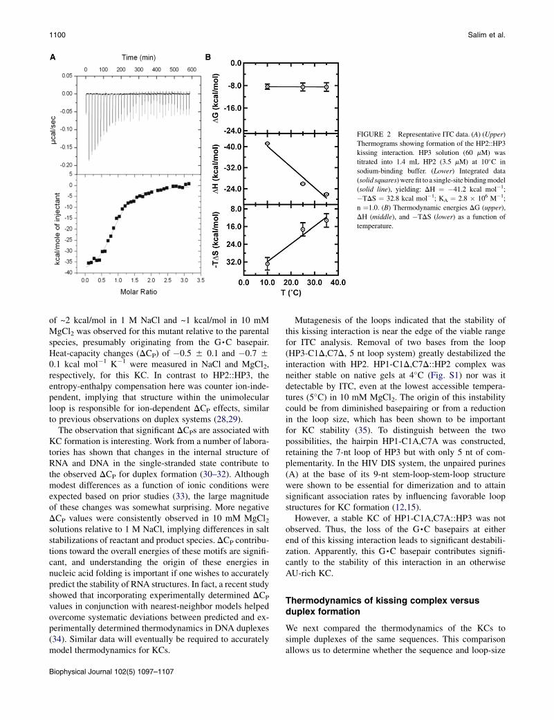
FIGURE 2 Representative ITC data. (A) (Upper)
Thermograms showing formation of the HP2::HP3
kissing interaction. HP3 solution (60 mM) was
titrated into 1.4 mL HP2 (3.5 mM) at 10�C in
sodium-binding buffer. (Lower) Integrated data
(solid squares)werefit toa single-site bindingmodel
(solid line), yielding: DH ¼ �41.2 kcal mol�1;
�TDS ¼ 32.8 kcal mol�1; KA ¼ 2.8 � 106 M�1;
n ¼1.0. (B) Thermodynamic energies DG (upper),
DH (middle), and �TDS (lower) as a function of
temperature.
1100 Salim et al.
of ~2 kcal/mol in 1 M NaCl and ~1 kcal/mol in 10 mMMgCl2 was observed for this mutant relative to the parentalspecies, presumably originating from the G,C basepair.Heat-capacity changes (DCP) of �0.5 5 0.1 and �0.7 50.1 kcal mol�1 K�1 were measured in NaCl and MgCl2,respectively, for this KC. In contrast to HP2::HP3, theentropy-enthalpy compensation here was counter ion-inde-pendent, implying that structure within the unimolecularloop is responsible for ion-dependent DCP effects, similarto previous observations on duplex systems (28,29).
The observation that significant DCPs are associated withKC formation is interesting. Work from a number of labora-tories has shown that changes in the internal structure ofRNA and DNA in the single-stranded state contribute tothe observed DCP for duplex formation (30–32). Althoughmodest differences as a function of ionic conditions wereexpected based on prior studies (33), the large magnitudeof these changes was somewhat surprising. More negativeDCP values were consistently observed in 10 mM MgCl2solutions relative to 1 M NaCl, implying differences in saltstabilizations of reactant and product species. DCP contribu-tions toward the overall energies of these motifs are signifi-cant, and understanding the origin of these energies innucleic acid folding is important if one wishes to accuratelypredict the stability of RNA structures. In fact, a recent studyshowed that incorporating experimentally determined DCP
values in conjunction with nearest-neighbor models helpedovercome systematic deviations between predicted and ex-perimentally determined thermodynamics in DNA duplexes(34). Similar data will eventually be required to accuratelymodel thermodynamics for KCs.
Biophysical Journal 102(5) 1097–1107
Mutagenesis of the loops indicated that the stability ofthis kissing interaction is near the edge of the viable rangefor ITC analysis. Removal of two bases from the loop(HP3-C1D,C7D, 5 nt loop system) greatly destabilized theinteraction with HP2. HP1-C1D,C7D::HP2 complex wasneither stable on native gels at 4�C (Fig. S1) nor was itdetectable by ITC, even at the lowest accessible tempera-tures (5�C) in 10 mM MgCl2. The origin of this instabilitycould be from diminished basepairing or from a reductionin the loop size, which has been shown to be importantfor KC stability (35). To distinguish between the twopossibilities, the hairpin HP1-C1A,C7A was constructed,retaining the 7-nt loop of HP3 but with only 5 nt of com-plementarity. In the HIV DIS system, the unpaired purines(A) at the base of its 9-nt stem-loop-stem-loop structurewere shown to be essential for dimerization and to attainsignificant association rates by influencing favorable loopstructures for KC formation (12,15).
However, a stable KC of HP1-C1A,C7A::HP3 was notobserved. Thus, the loss of the G,C basepairs at eitherend of this kissing interaction leads to significant destabili-zation. Apparently, this G,C basepair contributes signifi-cantly to the stability of this interaction in an otherwiseAU-rich KC.
Thermodynamics of kissing complex versusduplex formation
We next compared the thermodynamics of the KCs tosimple duplexes of the same sequences. This comparisonallows us to determine whether the sequence and loop-size

RNA Kissing Interactions 1101
effects within the kissing interaction originated from thestructural context of the hairpin loops or were inherent tosequences themselves. Thus, RNA2::RNA3 is equivalentto HP2::HP3, just removed from the constraint of the KChairpins. Thermodynamic values measured in 1 M NaClare provided in Table S3. Similar trends in stability wereobserved for duplexes and KCs of the identical sequence.Stable duplexes formed in the case of the wild-typesequence and the central GC mutant (A4C::U4G). Theduplex with terminal GA mismatches (HP2::HP3-C1A,C7A) was detected at 5�C in ITC, but the shortenedHP3 with 50 and 30 overhangs was not sufficiently stableto be observed. These results suggest that the inherentsequence- and loop-size effects observed in the DG of theKCs simply recapitulated stability issues of the underlyingduplexes.
Given the matching trends that were observed in theenergetics of KCs and duplexes, it is interesting to seehow similar these kissing interactions are to simple duplexesof the same sequence. Can one simply use a nearest neigh-bor (NN) formalism (derived from simple duplexes) topredict the energies of the kissing complexes? To test thispossibility, NN parameters (27,36,37) were used to calculatefree energies for RNA duplexes mentioned above and thencompared to the measured values for the relevant simpleduplexes and kissing interactions (Table S4). As expected,NN rules predicted simple duplex stability quite well forthese short RNAs with DDG298 < 1 kcal/mol. NN predic-tions fared less well with the kissing hairpins, where thekissing complexes were >2 kcal/mol more stable than pre-dicted, likely due to additional stacking interactions fromcoaxial alignment of the helices, electrostatic interactionsor possible metal binding events not accounted for in theNN model. Furthermore, for duplexes RNA2::RNA3-C1D,C7D and HP2::HP3-C1A,C7A free energies of ~�2.6and ~�4 kcal/mol were predicted by NN methods. Thiswas in good agreement with what was observed in duplexand kissing contexts where stable complexes were notdetectable by ITC. Of even more interest, these data putforth the notion that at least for these sample systems, KCstability can be approximated using a slight offset predictedby the NNmethod for the identical RNA duplex. Thermody-namic analysis on a more extensive collection of sequenceswill be required to establish a suitable correction value toaccount for the offset.
Several interesting characteristics in energetics betweenKCs and duplexes were revealed by ITC, however (TableS2 and Table S3). Enthalpic and entropic components forduplex and KC formation vary significantly. In the contextof a simple duplex, the energetics were more enthalpicallyfavorable and more entropically opposed relative to that ofthe related KC. This is in part due to the expenditure ofenergy required by one hairpin, in reorganizing the internalstructure of the loop, to interact with its complementaryhairpin. In duplex formation, the reorganization energy is
much smaller relative to KCs (28). On the other hand, theentropic penalty was greater in duplexes due to the higherloss in degrees of freedom from the random-coil state ofsingle-stranded RNAs relative to the structured hairpins.These observations are similar to what was observed foranother type of tertiary structure, the tetraloop-tetraloop re-ceptor interactions (39) and thus may be part of a moregeneral trend in the folding energetics of RNA tertiarystructures.
Duplex formation was also accompanied by a sizeableDCP, �0.4 kcal mol�1 K�1 for the parental sequence anda much larger �2.4 kcal mol�1 K�1 for the duplex withthe central GC pair (RNA2-A4C::RNA3-U4G). The�0.4 kcal mol�1 K�1 value is typical of simple duplexes,which have an average of �42 cal mol�1 K�1 bp�1 (40),whereas the larger sequence is more indicative of a specieswith a latent structure in the single-stranded state. Theobserved trends in DCP are consistent between the KCsand duplexes. These data suggest that thermodynamic prin-ciples that govern duplex formations can readily be ex-trapolated to KCs.
Thermodynamic analysis of strand-displacementreactions
In the experiments described above, hairpins could onlyform KCs and could not proceed further, because the stemswere noncomplementary. Next, the sequences were allowedto resolve into the ED conformation. Using ITC, HP1 wastitrated into HP2 to determine the energetics of the strand-displacement reaction as a function of temperature in either1 M NaCl or 10 mM MgCl2 (Table S5). Thermodynamicparameters for these reactions are quite different from thoseassociated with KC formation, indicating that the reactionproceeds rapidly to the ED state under these conditions.Similar to the case of KC formation, there is a significantDCP associated with these transitions and the magnitude ishighly dependent on the ionic strength of the solution. Usingthe approximation that DCP is temperature-independent,values of �1.1 5 0.1 and �1.8 5 0.1 kcal mol�1 K�1 in1 M NaCl and 10 mM MgCl2, respectively, were ob-served for the rearrangement. The magnitude of DCP
observed for ED formation is slightly more negativethan that seen for KC formation. The more negative heat-capacity change was once again associated with theMgCl2 reaction conditions.
As described in Fig. 1 A, multiple pathways might allowED formation from a pair of hairpins when the RNAs areself complementary. NMR and x-ray structures are availablefor both DIS KCs and ED formation produced by a numberof groups (41–43) using 23-mer RNAs. The loop-loop con-tacts in the KC were retained during the transition to the ED(44) and the propensity for ED formation decreased withincreasing RNA size and in the absence of Ncp7 (45,46),implying topological constrains faced during ED formation.
Biophysical Journal 102(5) 1097–1107
FIGURE 3 Kinetics of HP2::HP3 KC formation measured by SPR.
(A) Representative sensorgrams as a function of HP3 concentration (400–
800 nM), with surface-immobilized HP2 RNA monitored at 15�C. Associ-ation and dissociation data were fit into a Langmuir binding model. (B)
Eyring plot of HP2::HP3 association rates studied between 10�C and 40�C.
1102 Salim et al.
If ED formation were solely dependent on an intermediateKC, mutations that reduce or abolish loop complementarityshould prevent the reaction. To determine the effect ofdestabilizing the kissing interaction on strand displacement,all three adenosines in the loop of HP2 were substitutedwith cytosines (HP2-A(3-5)C). As expected, both nativegel analysis and ITC at 5�C failed to show the formationof a stable kissing interaction, but strand displacement stillproceeded rapidly to form the ED (Table S5). Since theseRNAs cannot kiss to any appreciable extent, the data indi-cate that formation of a stable KC is not required for stranddisplacement in the context of this model system, and thefraying pathway described in Fig. 1 A (pathway c) maydominate in the absence of the KC. This result shows thatthe fraying pathway can also be accessible to a certainextent during this type of structural transition. Calorimetryshowed relatively small changes in DG between KC andED states at these temperatures. The enthalpic and entropiccomponents of the ED state are, however, quite differentfrom those of the KC, with much greater enthalpic stabili-zation in the case of the ED. The kinetic behaviors of thetwo species are also dramatically different, as describedbelow.
This conclusion is similar to recent discussions on themechanism of strand displacement for HIV DIS. Therehas been much discussion regarding the proposal that theKC proceeds through a two-step rearrangement whereinthe first step involves pairing across the loop and the secondstep results in coaxial alignment of the three helical ele-ments (20,47). Although this latter step may be critical tothe stability of a KC, it likely inhibits the rapid expansionof the intermolecular basepairing at the expense of intramo-lecular interactions, as that pathway requires destacking ofthe helical elements at each step of the helix expansion.
Kinetic analysis of KC formation
To measure the kinetics of KC formation, surface plas-mon resonance (SPR) and smFRET were used. For SPR,50-biotinylated hairpins were bound to streptavidin-coatedsensor chips. The complementary hairpin was then intro-duced at varying concentrations to monitor the interactionin 10 mM MgCl2. Sensorgram traces were fit to a Langmuirbinding model to obtain association and dissociation ki-netics. A representative SPR sensorgram for HP2::HP3KC formation is shown in Fig. 3 A. Kinetic data obtainedfor HP2::HP3 and HP2-A4C::HP3-U4G KC formation arecollected in Table S6. Both wild-type and HP2-A4C::HP3-U4G associate with moderate on-rates that slightly increasewith temperature. On the other hand, off-rates for HP2::HP3were faster at higher temperatures but changed minimallyfor HP2-A4C::HP3-U4G. Stability constants for KC forma-tion were calculated using kinetic data obtained from SPR(Table S6) and are comparable to values measured by ITC(Table S2).
Biophysical Journal 102(5) 1097–1107
Activation energetics for KC formation
As depicted in Fig. 1 A, kissing interactions might serve askey intermediates to structural transitions such as extendedduplex formation. To understand the transition-state ther-modynamics of KCs relative to ED formation, the tem-perature dependence of the rates for association anddissociation were probed. Themeasured rates were then sub-jected to Eyring analysis to provide activation parametersDHz, DSz, and DGz
298. A representative Eyring plot forHP2::HP3 KC is shown in Fig. 3 B with clean, linear Eyringbehavior.
Activation parameters for HP2::HP3 and HP1-U4G::HP2:A4C complexes are collected in Table S6. Similar acti-vation energies were observed for the association phase ofboth constructs. In contrast, activation parameters for disso-ciation varied significantly between the parent and G,Cmutant. Both entropic and enthalpic components weremore favorable for the dissociation of HP2::HP3 comparedto HP2-U4G::HP3-A4C. These data suggest that the stabil-ities of these two complexes are dictated mainly by theirdissociation transition states, whereas the energy landscapesfor association are similar. From equilibrium measure-ments, it was observed that HP2-U4G::HP3-A4C formed

RNA Kissing Interactions 1103
a relatively stronger complex with a DDG298 of ~1.1 kcal/mol, consistent with ITC data.
Kinetics of KC formation using smFRET
smFRET was also used to study the kinetics of these KCsand any potential intermediates formed en route towardstrand-displaced products. The main difference betweenthe two methodologies is that in SPR, the physical observ-able represents the average ensemble of the system, whereasin smFRET, changes at the molecular level can be measured.The basic overview of the smFRET setup is shown inFig. 4 A. A Cy3-labled RNA hairpin was immobilized ona quartz slide via biotin-streptavidin interaction. FRETtrajectories were recorded after addition of its Cy5-labeledbinding partner at a concentration of 35 nM. A typicalsingle-molecule time trajectory obtained for HP1::HP2 isshown in Fig. 4 B, where the fluorescence intensities ofthe donor and acceptor fluorophores change in an anticorre-lated fashion (Fig. 4B, upper). The resulting FRET trajectory(Fig. 4B, lower) shows transitions between two distinct statesat ~0.0 and ~0.5 FRET. Control experiments (Fig. 4 D)showed that the 0.5 FRET state corresponds to an interme-diate state (KC) that occurs when HP1 and HP3 reacttogether. As indicated in Fig. 4 D (upper two panels), when
either HP2 only or HP2 and a noninteracting hairpin werepresent, a peak with FRET ratio ~0.0 was observed. To iden-tify FRET ratios for the intermediate states of the HP1 andHP2 reaction, the hairpins were preannealed or reacted inits hairpin context.When preannealed, the predominant statewould be the ED (FRET ¼ ~1.0), since HP1 and HP2 havecomplementary stems. Although HP1 and HP2 resolve intoan ED state under ITC conditions, this transition was ex-tremely rare experimentally under the conditions requiredfor smFRET analysis. Out of >600 molecules analyzed,only three ED-formation events were observed in the exper-imental time window. The majority of molecules showedFRET values at 0 or 0.5, which represents the dissociationand association of a KC. These data show that the KC canbe long-lived (~5 s on average (Fig. 4)) and can undergomany cycles of dissociation and association before progress-ing to the ED complex. The big difference in experimentalparameters between these two experiments is the concentra-tion of the individual RNAs. ITC is performed at high strandconcentration so that the reaction generates sufficient heat toaccurately measure the reaction, whereas the smFRET ex-periments are performed at very low dilutions to ensurespatial localization of individual molecules. Thus, the effectof local concentration can be seen as paramount in the ex-pected outcome. Also, in ITC experiments, the use of higher
FIGURE 4 smFRETanalysis of kissing kinetics.
(A) Schematic representation of the total internal
reflection setup. The donor RNA hairpin is immo-
bilized by a biotin-streptavidin interaction on a
biotin-BSA-coated quartz slide. A kissing interac-
tion and an ED formation are shown in the pres-
ence of a 35-nM RNA hairpin with the acceptor
fluorophore. (B) Typical single-molecule time tra-
jectory. (Upper) Anticorrelated intensities of the
donor (blue) and acceptor (red). (Lower) The cor-
responding FRET trajectory. Only the 0.0 and 0.5
FRET states are observed for the dissociated and
kissing complexes. (C) Representative FRET tra-
jectories showing several kissing interactions and
their transition to the ED state. There are fewmole-
cules with a transition directly from KC to ED. (D)
FRET histograms from single-molecule trajecto-
ries. (Upper) Donor-only RNA hairpin HP2 ex-
hibits a single 0.0 FRET state. (Second from top)
Preannealed noncomplementary hairpins (HP3-
C1D,C7D þ HP2) do not form the ED. (Third
from top) Two preannealed complementary RNA
hairpins (HP1 þ HP2) show two distributions
(0.0 and 1.0 FRET states) when preannealed before
imaging. The distribution at 1.0 FRET state is due
to ED formation. (Lower) Cumulative histogram
built from >100 molecules. HP1 reacted with
surface-immobilized HP2. This histogram shows
three distributions at 0.0, 0.5, and 1.0 FRET states.
The 0.0 FRET state is free HP2, the 0.5 FRET state
represents the KC, and the 1.0 FRET state corre-
sponds to the ED. ED molecules show no dynamic
behavior and are trapped in that form under the
experimental conditions.
Biophysical Journal 102(5) 1097–1107
1104 Salim et al.
concentrations of RNA may allow access to the frayingpathway that would facilitate ED formation. This may alsorepresent one of the primary roles of accessory proteins instrand-displacement reactions in vivo, improving the likeli-hood of a forward progression which then can be kineticallytrapped by the slow off-rate for the ED state.
Furthermore, the stem lengths of kissing hairpins wereinversely proportional to the FRET value, indicating forma-tion of the appropriate KC in these assays (Fig. S2). Mole-cules that successfully reached the ED were kinetically inertand showed no additional dynamic behavior before photo-bleaching. Presumably, they get trapped in this conforma-tion due to a large activation barrier for escaping from thiswell, a pathway only accessible upon heating (23).
A dwell time distribution histogram was prepared foreach FRET state and fit to an exponential decay to estimatethe apparent rate constants for association (kon ¼ 3.4 50.3 � 106 M�1 s�1: calculated from the pseudo-first-orderrate constant and the effective ligand concentration of35 nM) and dissociation (koff ¼ 0.17 5 0.02 s�1)(Fig. S3) (48). From the rate constants, the free energyof KC formation was estimated as DG ¼ �9.9 50.2 kcal/mol (Table S6), which is in excellent agreementwith the ensemble-averaged ITC and SPR data (Table S2).An estimate of DG� using the FRET distributions (Fig. 4 D,lower) yields the same result. The agreement between thesingle-molecule study and bulk measurement indicatesthat the immobilization approach does not affect the dy-namic behavior of the kissing hairpins or their equilibrium.
Comparison of the kinetic data between SPR andsmFRET shows significant differences, however. SPR rateswere ~100-fold slower for both kon and koff relative tosmFRET values. Despite the difference in the rate constants,both methodologies produced identical DG values for the
Biophysical Journal 102(5) 1097–1107
interactions. Similar observations were reported whensmFRET and SPR was used to measure the kinetics ofnucleotide binding to DNA polymerase (L. Romano andD. Rueda, unpublished results). Differences in surface pas-sivation of smFRET slides and SPR sensor chips, and alsoissues related to mass transport into the boundary layer,may contribute to these kinetic effects. The origins of theseanomalies are currently under investigation.
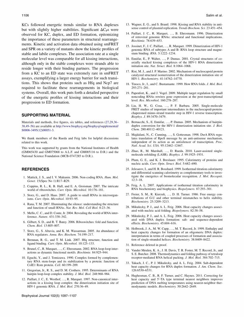
Kissing interactions of mutated hairpins usingsmFRET
smFRET was also used to measure the kinetics of kissinginteractions of the mutated hairpins described above. KCswere detected for HP1-U4G::HP2-A4C, HP1-C7A::HP2,and HP1-C1A::HP2 (Table S6 and Fig. 5). As shown inTable S6, the association rate constant is invariant at ~4 �106 M�1 s�1 for all hairpins. In contrast, the dissociationrate constants differ dramatically depending on the loopsequence. For HP1::HP2 and HP1-U4G::HP2:A4C, stableKCs were observed using ITC, SPR, and smFRET. Com-pared to the parent complex, the G,C mutant has one netextra hydrogen bond and slightly altered stacking potential,which decreased the koff by approximately threefold and re-sulted in a 0.6 kcal/mol stabilization. Attesting to the powerof smFRET, transient associations of HP1-C7A::HP2 andHP1-C1A::HP2 were detected, neither of which wasobserved by ITC or SPR. As shown in Fig. 5, KCs ofHP1-C7A::HP2 and HP1-C1A::HP2 were short-lived (koff¼6 5 1 and 10 5 1 s�1, respectively) because of themismatch within the KC. Both HP1-C7A::HP2 and HP1-C1A::HP2 KCs were destabilized by ~2 kcal/mol as a resultof this single base mismatch. When all three central base-pairs of the KC were disrupted (HP2-A(3-5)C::HP2), or
FIGURE 5 Representative single-moleculeFRET
trajectories (left) and histograms (right) from
selected mutants HP1-C7A::HP2 (A), HP1-C1A::
HP2 (B), and HP2-A4C::HP1-U4G (C), which
allowed the formation of a GC basepair by replacing
the AU basepair. Vertical arrows indicate the photo-
bleaching point for each trace.

RNA Kissing Interactions 1105
when the loop size was reduced from 7 nt to 5 nt (HP1-C1D,C7D::HP2), even transient formation of the KC wasabolished. These results indicate that under these experi-mental conditions, one mismatch is sufficient to greatlydestabilize the 7-basepair KC. These data also indicatethat dissociation kinetics ultimately dictate the stability ofthe KC, similar to what was observed by activation param-eters computed using SPR. These data support the notionthat discrimination between cognate and near-cognate inter-actions occurs after transient sampling of the interaction,which either accommodates into a stabilized state or rapidlydissociates, allowing it to test many possible interac-tion partners and avoid getting trapped in nonproductivecomplexes.
Energy landscapes for KCs and theirrearrangement to ED structures
Using the thermodynamic (DG) and kinetic (DGz) dataderived from the study of HP1::HP2 and HP2::HP3, a poten-tial energy surface for the rearrangement from hairpins toextended duplexes can be proposed (Fig. 6). This surfacecombines the insights derived from each of the biophysicalapproaches described above, including ultraviolet (UV)melting, ITC, SPR, and smFRET. Random-coiled strandsand free hairpins (Fig. 6, A and B, respectively) couldbe considered reactant states, whereas the KC and ED(Fig. 6, C and D, respectively) forms represented the low-energy products. These have been placed in coordinates ofincreasing intramolecular and intermolecular basepair inter-actions. Thermodynamics of transitions AD and AB weredetermined using UV melting analysis, whereas BC andBD were measured using ITC and SPR. Transition-stateenergetics for BC and CB was computed from SPR data.Energy wells have been drawn as symmetric Gaussian func-tions. That is a significant simplification, as the actual
energy landscape is expected to be much more ruggedthan depicted here. Nonetheless, significant insight canderive from viewing the data in this way.
KCs and ED states lie in the lowest energy wells on thissurface, whereas random-coiled strands and free hairpinsoccupy higher-energy regions. Data obtained from indepen-dent measurements when viewed as an ensemble correlatenicely within error in a thermodynamic cycle representingall possible transitions. KCs and EDs lie in comparableenergy wells (Table S2) despite the differences in thetwo structural contexts from which they were measured.Free hairpins associate to form a KC that crosses an~11 kcal/mol energy barrier, whereas ~21 kcal/mol energyis required for dissociation. These energetics yield a netstabilization of ~10 kcal/mol, identical in ITC and smFRETobservations. The smFRET data reveal that spontaneousrearrangement from KC to ED occurs with a probabilityof only ~0.005 (out of 600 trajectories) within 2.5 min,suggesting a large activation barrier for this transition.Thus, one of the roles proteins should play in these rear-rangements is to destabilize KCs. Using RNA-protein-binding energy to break the coaxial stacking of the KCwill facilitate the rearrangement by providing a driving-forceconversion to the kinetically inert ED state. Furthermore, thecolocalization of such a complex could dramatically reducethe entropic cost during strand exchange. On-going experi-ments are looking at further defining the energetic landscapeof these species and the role of protein cofactors in facili-tating the refolding of RNAs such as these model hairpinsto their ED conformation.
CONCLUSIONS
A detailed thermodynamic and kinetic analysis was per-formed using ITC, UV melting, SPR, and smFRET method-ologies for RNA KCs and their progression into the ED.
FIGURE 6 Putative potential energy surface for
KC and ED formation. Thermodynamic (DGIJ) and
activation energies (DGzIJ) for pathways associated
with KC formation and their conversion to EDs.
Thermodynamic data were obtained using ITC,
UV melting, and smFRET, and activation pa-
rameters were derived from kinetic data obtained
by SPR and smFRET. Reaction coordinates are
defined by the number of intramolecular (X) and
intermolecular (Y) basepairs present in the molec-
ular ensemble of a particular state. Stabilization
energies are plotted along the z axis. Four different
states are shown: unfolded strands (A), free hair-
pins (B), the KC (C), and the ED (D). The energy
of unfolded strands (A) was used as the reference
state (DGA ¼ 0).
Biophysical Journal 102(5) 1097–1107
1106 Salim et al.
KCs followed energetic trends similar to RNA duplexesbut with slightly higher stabilities. Significant DCPs wereobserved for KC, duplex, and ED formation, epitomizingthe importance of these parameters in structural rearrange-ments. Kinetic and activation data obtained using smFRETand SPR on a variety of mutants show the kinetic profiles ofstable and labile complexes. The association rate at a singlemolecular level was comparable for all kissing interactions,although only in the stable complexes were strands able toreside longer with their binding partners. The progressionfrom a KC to an ED state was extremely rare in smFRETassays, exemplifying a larger energy barrier for such transi-tions. This shows that proteins such as Hfq and Ncp7 arerequired to facilitate these rearrangements in biologicalsystems. Overall, this work puts forth a detailed perspectiveof the energetic profiles of kissing interactions and theirprogression to ED formation.
SUPPORTING MATERIAL
Materials and methods, five figures, six tables, and references (27,29,36–
38,49–56) are available at http://www.biophysj.org/biophysj/supplemental/
S0006-3495(12)00051-3.
We thank members of the Rueda and Feig labs for helpful discussions
related to this work.
This work was supported by grants from the National Institutes of Health
(GM065430 and GM075068 to A.L.F. and GM085116 to D.R.) and the
National Science Foundation (MCB-0747285 to D.R.).
REFERENCES
1. Mattick, J. S., and I. V. Makunin. 2006. Non-coding RNA. Hum. Mol.Genet. 15(Spec No 1):R17–R29.
2. Coppins, R. L., K. B. Hall, and E. A. Groisman. 2007. The intricateworld of riboswitches. Curr. Opin. Microbiol. 10:176–181.
3. Storz, G., and D. Haas. 2007. A guide to small RNAs in microorgan-isms. Curr. Opin. Microbiol. 10:93–95.
4. Rana, T. M. 2007. Illuminating the silence: understanding the structureand function of small RNAs. Nat. Rev. Mol. Cell Biol. 8:23–36.
5. Mello, C. C., and D. Conte, Jr. 2004. Revealing the world of RNA inter-ference. Nature. 431:338–342.
6. Gilbert, S. D., and R. T. Batey. 2006. Riboswitches: fold and function.Chem. Biol. 13:805–807.
7. Storz, G., S. Altuvia, and K. M. Wassarman. 2005. An abundance ofRNA regulators. Annu. Rev. Biochem. 74:199–217.
8. Brennan, R. G., and T. M. Link. 2007. Hfq structure, function andligand binding. Curr. Opin. Microbiol. 10:125–133.
9. Brunel, C., R. Marquet,., C. Ehresmann. 2002. RNA loop-loop inter-actions as dynamic functional motifs. Biochimie. 84:925–944.
10. Eguchi, Y., and J. Tomizawa. 1990. Complex formed by complemen-tary RNA stem-loops and its stabilization by a protein: function ofCoIE1 Rom protein. Cell. 60:199–209.
11. Gregorian, Jr., R. S., and D. M. Crothers. 1995. Determinants of RNAhairpin loop-loop complex stability. J. Mol. Biol. 248:968–984.
12. Paillart, J. C., E. Westhof, ., R. Marquet. 1997. Non-canonical inter-actions in a kissing loop complex: the dimerization initiation site ofHIV-1 genomic RNA. J. Mol. Biol. 270:36–49.
Biophysical Journal 102(5) 1097–1107
13. Wagner, E. G., and S. Brantl. 1998. Kissing and RNA stability in anti-sense control of plasmid replication. Trends Biochem. Sci. 23:451–454.
14. Paillart, J. C., R. Marquet, ., B. Ehresmann. 1996. Dimerizationof retroviral genomic RNAs: structural and functional implications.Biochimie. 78:639–653.
15. Jossinet, F., J. C. Paillart,., R. Marquet. 1999. Dimerization of HIV-1genomic RNA of subtypes A and B: RNA loop structure and magne-sium binding. RNA. 5:1222–1234.
16. Ennifar, E., P. Walter, ., P. Dumas. 2001. Crystal structures of co-axially stacked kissing complexes of the HIV-1 RNA dimerizationinitiation site. Nat. Struct. Biol. 8:1064–1068.
17. Rist, M. J., and J. P. Marino. 2002. Mechanism of nucleocapsid proteincatalyzed structural isomerization of the dimerization initiation site ofHIV-1. Biochemistry. 41:14762–14770.
18. Tinoco, Jr., I., and C. Bustamante. 1999. How RNA folds. J. Mol. Biol.293:271–281.
19. Papenfort, K., and J. Vogel. 2009. Multiple target regulation by smallnoncoding RNAs rewires gene expression at the post-transcriptionallevel. Res. Microbiol. 160:278–287.
20. Liu, H. W., G. Cosa, ., P. F. Barbara. 2005. Single-moleculeFRET studies of important intermediates in the nucleocapsid-protein-chaperoned minus-strand transfer step in HIV-1 reverse transcription.Biophys. J. 89:3470–3479.
21. Bernacchi, S., E. Ennifar, ., P. Dumas. 2005. Mechanism of hairpin-duplex conversion for the HIV-1 dimerization initiation site. J. Biol.Chem. 280:40112–40121.
22. Majdalani, N., C. Cunning, ., S. Gottesman. 1998. DsrA RNA regu-lates translation of RpoS message by an anti-antisense mechanism,independent of its action as an antisilencer of transcription. Proc.Natl. Acad. Sci. USA. 95:12462–12467.
23. Zhao, R., M. Marshall, ., D. Rueda. 2010. Laser-assisted single-molecule refolding (LASR). Biophys. J. 99:1925–1931.
24. Plum, G. E., and K. J. Breslauer. 1995. Calorimetry of proteins andnucleic acids. Curr. Opin. Struct. Biol. 5:682–690.
25. Jelesarov, I., and H. R. Bosshard. 1999. Isothermal titration calorimetryand differential scanning calorimetry as complementary tools to inves-tigate the energetics of biomolecular recognition. J. Mol. Recognit.12:3–18.
26. Feig, A. L. 2007. Applications of isothermal titration calorimetry inRNA biochemistry and biophysics. Biopolymers. 87:293–301.
27. Freier, S. M., R. Kierzek, ., D. H. Turner. 1986. Free energy con-tributions of G.U and other terminal mismatches to helix stability.Biochemistry. 25:3209–3213.
28. Mikulecky, P. J., and A. L. Feig. 2006. Heat capacity changes associ-ated with nucleic acid folding. Biopolymers. 82:38–58.
29. Mikulecky, P. J., and A. L. Feig. 2006. Heat capacity changes associ-ated with DNA duplex formation: salt- and sequence-dependenteffects. Biochemistry. 45:604–616.
30. Holbrook, J. A., M. W. Capp,., M. T. Record, Jr. 1999. Enthalpy andheat capacity changes for formation of an oligomeric DNA duplex:interpretation in terms of coupled processes of formation and associa-tion of single-stranded helices. Biochemistry. 38:8409–8422.
31. Reference deleted in proof.
32. Vander Meulen, K. A., J. H. Davis, T. R. Foster, M. T. Record, Jr., andS. E. Butcher. 2008. Thermodynamics and folding pathway of tetraloopreceptor-mediated RNA helical packing. J. Mol. Biol. 384:702–715.
33. Takach, J. C., P. J. Mikulecky, and A. L. Feig. 2004. Salt-dependentheat capacity changes for RNA duplex formation. J. Am. Chem. Soc.126:6530–6531.
34. Hughesman, C. B., R. F. Turner, and C. Haynes. 2011. Correcting forheat capacity and 50-TA type terminal nearest neighbors improvesprediction of DNA melting temperatures using nearest-neighbor ther-modynamic models. Biochemistry. 50:2642–2649.
RNA Kissing Interactions 1107
35. Hjalt, T., and E. G. Wagner. 1992. The effect of loop size in antisenseand target RNAs on the efficiency of antisense RNA control. NucleicAcids Res. 20:6723–6732.
36. Xia, T., J. SantaLucia, Jr., ., D. H. Turner. 1998. Thermodynamicparameters for an expanded nearest-neighbor model for formation ofRNA duplexes with Watson-Crick base pairs. Biochemistry.37:14719–14735.
37. SantaLucia, Jr., J. 2007. Physical principles and visual-OMP softwarefor optimal PCR design. Methods Mol. Biol. 402:3–34.
38. Reference deleted in proof.
39. Fiore, J. L., B. Kraemer,., D. J. Nesbitt. 2009. Enthalpy-driven RNAfolding: single-molecule thermodynamics of tetraloop-receptor tertiaryinteraction. Biochemistry. 48:2550–2558.
40. Hughesman, C. B., R. F. Turner, and C. A. Haynes. 2011. Role ofthe heat capacity change in understanding and modeling meltingthermodynamics of complementary duplexes containing standard andnucleobase-modified LNA. Biochemistry. 50:5354–5368.
41. Girard, F., F. Barbault, ., G. Lancelot. 1999. Dimer initiationsequence of HIV-1Lai genomic RNA: NMR solution structure of theextended duplex. J. Biomol. Struct. Dyn. 16:1145–1157.
42. Mujeeb, A., J. L. Clever, ., T. G. Parslow. 1998. Structure of thedimer initiation complex of HIV-1 genomic RNA. Nat. Struct. Biol.5:432–436.
43. Ennifar, E., M. Yusupov, ., P. Dumas. 1999. The crystal structure ofthe dimerization initiation site of genomic HIV-1 RNA reveals anextended duplex with two adenine bulges. Structure. 7:1439–1449.
44. Theilleux-Delalande, V., F. Girard, ., J. Paoletti. 2000. The HIV-1(Lai) RNA dimerization. Thermodynamic parameters associatedwith the transition from the kissing complex to the extended dimer.Eur. J. Biochem. 267:2711–2719.
45. Takahashi, K. I., S. Baba, ., G. Kawai. 2000. Structural requirementfor the two-step dimerization of human immunodeficiency virus type 1genome. RNA. 6:96–102.
46. Paillart, J. C., E. Skripkin,., R. Marquet. 1996. A loop-loop ‘‘kissing’’complex is the essential part of the dimer linkage of genomic HIV-1RNA. Proc. Natl. Acad. Sci. USA. 93:5572–5577.
47. Rist, M., and J. Marino. 2001. Association of an RNA kissing complexanalyzed using 2-aminopurine fluorescence. Nucleic Acids Res.29:2401–2408.
48. Zhao, R., and D. Rueda. 2009. RNA folding dynamics bysingle-molecule fluorescence resonance energy transfer. Methods.49:112–117.
49. Fasman, G. D. 1976. CRC Handbook of Biochemistry and MolecularBiology. CRC Press, Boca Raton, FL.
50. Iqbal, A., S. Arslan, ., D. M. Lilley. 2008. Orientation dependence influorescent energy transfer between Cy3 and Cy5 terminally attachedto double-stranded nucleic acids. Proc. Natl. Acad. Sci. USA.105:11176–11181.
51. McDowell, J. A., and D. H. Turner. 1996. Investigation of the structuralbasis for thermodynamic stabilities of tandem GU mismatches:solution structure of (rGAGGUCUC)2 by two-dimensional NMR andsimulated annealing. Biochemistry. 35:14077–14089.
52. Mizoue, L. S., and J. Tellinghuisen. 2004. The role of backlash in the‘‘first injection anomaly’’ in isothermal titration calorimetry. Anal.Biochem. 326:125–127.
53. Rueda, D., G. Bokinsky,., N. G. Walter. 2004. Single-molecule enzy-mology of RNA: essential functional groups impact catalysis froma distance. Proc. Natl. Acad. Sci. USA. 101:10066–10071.
54. Rueda, D., and N. G. Walter. 2006. Fluorescent energy transfer readoutof an aptazyme-based biosensor. Methods Mol. Biol. 335:289–310.
55. SantaLucia, Jr., J. 2000. The use of spectroscopic techniques in thestudy of DNA stability. In Spectrophotometry and Spectrofluorimetry.M. Gore, editor.; Oxford University Press, New York. 329–354.
56. Wiseman, T., S. Williston, ., L. N. Lin. 1989. Rapid measurement ofbinding constants and heats of binding using a new titration calorim-eter. Anal. Biochem. 179:131–137.
Biophysical Journal 102(5) 1097–1107