thesis submittea in partial fulfillment of the
TRANSCRIPT

THESIS
Submittea in partial fulfillment of the requirements
for the degree of Master of Arts in the Grad-
uate School of the University of Kansas.
-1916-

I I
' i \ '
LARVAL TRE!f...ATODES FROM KANSAS FRESH-WATER SNAILS
BY
EARL C:. 0' ROKE

INTRODUCTION.
This study of larval trematodes was undertaken
by the writer at the suggestion of Professor Bennet M.
Allen in an attempt to add to the knowledge of this
interesting group of parasites.
The snails used in the investigations were col-
lected during the summer and fall of 1915 in connection
with' the Kansas State Biological survey, and studies
were made of the behavior and habits of the living oer-
cariae before the specimens were preserved for further
morphological studies.
METHODS OF STUDY.
For convenience in transporting live snails, it
w~s found desirable to tie the specimens in cheesecloth
bags of twenty-five each, packing loosely in wet excel-
sior.
In the laboratory, the snails were isolated in
individual watch-glasses and kept covered with water.
normally the c ercariae emerged within two or three
days. Some of the infected snails were then preserved
entire and others were crushed for studies of living
sporocysts and rediae.

-2-
The fixatives used were a saturated corrosive
sublimate solution with one percent glacial acetic acid,
Zenker's fluid and four percent formalin. The latter
was discarded after trials proved tne superiority of the
other fixati vee. Corrosive sublimate was preferable for
freed cercariae.
Several different stains were used. Picro car-
mine was excellent for studies of the living ceraariae.
Specimens so stained co~ld sometimes be preserved by
drawing glycerine under the cover slip with filter paper.
For toto mounts, Mayer's baemalum gave the best results.
The sections were out from five to seven micra in thick-
ness and stained with iron alum haematoxylin. Good re-
sults were also obtained by staining with haemalum and
using eosin or ora.nge g. for counter stains.
The free cercariae could be made to expand by
heating slightly. When they were in this condition the
fixing fluid was poured on suddenly, fixing the speci-
mens in the best form for study.
The measurements !)jfi given in this pa.per were
taken usually from mounted specimens and are not as from
accurate as those11 living forms would be, because of
shrinkage of the specimens during the process of fixing
and mounting.

-3-
.AMPHISTOME CERCARIAE.
Three species of .Amphistome cercariae were found
in my collections. For the first of these I propose the
name Cercaria cortii.
Of twenty-three specimens of Planorbis trivolvie
collected at Cherryvale,,Kansas, October 16, one was in-
fected with Cercaria cortii. The snails were found
adhering to rocks at a depth of from six inches to a foot
in a large pond of clear water.
On October 17th, the snails wer'ejisolated in indiv-
idual watch-glass ea and kept covered with water, the
wa.ter being changed each day. Cercariae were noticed in
the dish as follows, the observations being made in the
morning at eight o'clock.
Oct. 18 6 cercariae. • 19 12 " All others encysted.
20 3 " All cercariae encysted. 21 0 " " ft ff
22 0 n II " .. 23 0 " " " " 24 0 " ff .. " 25 50 .. Five encysted • 26 6 " All others encysted. 26 Uov.6 O • All encysted.
nov. 2 All cysts still alive in water. ti 3 One new cercaria encysted " 4 One new cercaria encysting. " 5-7 No new developmen.te. • 8 Snail deal.
-4-
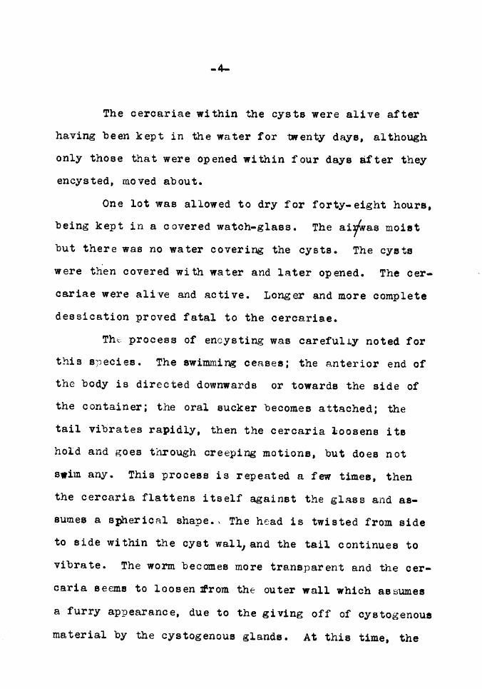
The cercariae within the cysts were alive after having been kept in the water for twenty days, although only those that were opened within four days after they encysted, moved about.
One lot was allowed to dry for forty-eight hours, being kept in a covered watch-glass. The ai'fwas moist but there was no water covering the cysts. The cysts were then covered with water and later opened. The cer-cariae were alive and active. Longer and more complete dessication proved fatal to the cercariae.
Tht:: process of encysting wa.s careful.Ly noted for this S})ecies. The swimming ceases; the anterior end of the body is directed downwards or towards the side of the container; the oral sucker becomes attached; the tail vibrates rapidly, then the cercaria loosens its hold and goes through creeping motions, but does not
s•im any. This process is repeated a few times, then the cercaria flattens itself against the glass and as-sumes a spherical shape., The head is twisted from side to side within the cyst wall, and the tail continues to vibrate. The worm becomes more transparent and the cer-
caria seems to loosen ~rom the outer wall which assumes
a furry appearance, due to the giving off of cystogenous
material by the cystogenous glands. At this time, the

_,_ cercaria begins peculiar rotating movements, which con-
tinue from an hour and twenty minutes to two hours and
ten minutes. The motion consists of a series of inter-
mittent movements. The time required for a cercaria to
make a complete turn, varied from one and three-fourths
minutes to two and a half minutes with from thirteen to
twenty- two separate movements in a r evolution.
The cyst wall be~omes thicker and more transparent.
Sometimes the tail loosens and swims away, while at
other times it may remain loosely attached to the cyst.
One tail vibrated constantly for eight hours after the
cercaria began to encyst. The tail as well as the body
gives off cystogenous material which is in the form of
a delicate sheath surrounding that organ and at a dis-
tance of one half the width of the tail from it.
The motions of the e1~cysting cercaria. become,
slower and slower and final~y cease as the process is
completed. When the cyst is fully formed, it is much
more transparent than the free swimming cercaria, and
the worm is coiled within the cyst with its anterior
a.nd posterior ends in contact. Slight spasmodic motions
within the cysts can be ~oted for three or four days
after encystment. Sometimes the cercaria breaks out of

-6-
its cyst by rupturing the wall 'nd forms a new cyst.
The cercariae usually excyeted on the bottom and sides
of the watch-glass nearest the window, and were shaped
like a de4p plano-oonvex lens. In a few cases where
encystment took place among masses of snail faeces and
not against the side of the container, the shape of the
cysts was ellipsoid.
Cercaria cortii is a rapid swim~er and its swim-
ming motions are strikingly regular. lt would often
propel itself in a straight line from on~ side to the
other of a Syracuse watch-glass. Creeping motions were
rather infrequent in open water but under the cover
slip this was the usual method of locomotion.
The measurements of this cercaria were taken
from preparations mounted in glycerine and glycerine
jelly after the specimens were ~illed by heating slightly,
and represent as accurately as possible the size of the
form in life.
Thie cercaria is elongate, oval, and wider at the
posterior than at the anterior end. It is capable of
assuming shapes varying from spherical to long and narrow.
The length of the body is .94 mm. and the length of the
tail is .87 mm.; a total length of l.81 mm. The width
of the acetabulum is .25 mm. and the width of the oral

...
-7-sucker is .08 mm. The edge of the oral sucker presents
a finely lobated appearance.
The mouth is situated within the oral sucker
nnd opens into an oral cavity .Ol mm. wide and .06 mm.
long. This is separated from the oesophagus by a aon-
striotion. The oesophagus narrows into a pharynx ab;..;ut
one third the diameter of the oral cavity. It is sur-
rounded by a band of longitudinal muscie fibers .027 mm.
in outside diameter. The width of the digestive diver-
ticula is .017 mm.
Fig. 2, a cross section through the region of the
eye-spate, shows the brain. The nerve cords could not
be traced any distance from this region.
The anterior one third of this cercaria is heavily
pigmented with dark pigment.
The oesophagus is long and narrow. The digestive
tract extends five-sevenths of the length of the body.
The two ey·a-spots are prominent and are .028 mm. in dia-
meter and .16 mm. apart. The tubes of the excretory
system extend from near the eyes to just in front of
the acetabulum.
The excretory tubes are made up of large flame
cells, irregularly joined together, and very closely
associated with the digestive tract. In the living
-8-
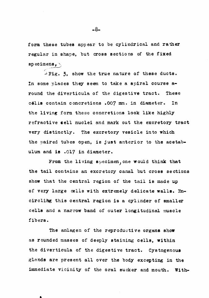
form these tubes appear to be cylindrical and rather
regular in shape, but cross sections of the fixed
spec imene 1 "'"-1 -·-----~
.J. Fig. 3, show the true nature of these ducts.
In some places they seem to take a spiral course a-
round the diverticula of the digestive tract. These
c&lls contain concretions .007 mm. in diameter. In
the living form these concretions look like highly
refractive cell nuclei and mark out the excretory tract
very distinctly. The excretory vesicle into which
the paired tubes open, is just anterior to the acetab-
ulum and is .017 in diameter.
From the living syecimenJone would think that
the tail contains an excretory canal but cross sections
show that the central region of the tail is made up
of very large aalls with extremely delicate walls. En-
circlihg this central region is a cylinder of smaller
cells and a narrow band of outer longitudinal muscle
fibers.
The anlagen of the reproductive organs show
as rounded masses of deeply staining cells, within
the diverticula of the digestive tract. Cystogenoue
glands are present all over the body excepting in the
immediate vicinity of the oral sucker and mouth. With-

-9-
in the cyatogenous glands are rod shaped grannulee of
the cyst forming material.
Of t'ltenty-three specimens of Planorbis tri vol vis one
collected at Lawrence Kansas October 10th, . w~,8 in-
fected with Cerca.ria diastrophora Cort. In general
appearance this form corresponds to Cercaria Cortii
but it is much smaller, measuring .48mm. long and .2
mm. wide. The tail is .58 mm. long. The oral sucker
is small measuring .06 mm. in width~while the acetae-
ulam is exceedingly large meaauring .15 JDJl· in out-
side diameter.
The oesophagus is very slender,..,opening into a
rounded bulbous pharynx three-fourths as broad as the
tip of the oral sucker. The ey~spo ts are prominent
and are oval rather than spherical, measuring .027rnm,
by.036 mm. They are separated by a distance equal to
the long axis of the eye.
The excretory system consisjte of paired ducts
opening into an excretory vesicle anterior to the ace-
tabulum. !h4s vessel also receives smaller ducte
from the region of the acetabulum. The excretory pore
opens on the dorsal surface.
This species stains deeply with haemalum except-
ing in the immediate vicinity of the oral sucker where

-10-
the cells seem to be devoid of nuclei. The tail is
made up of large central cells surrounded by a ring of
smaller cells. It contains no excretory canals.
The immature cercariae of this species are
blunt tailed and clumsy. No internal organs can be
distinguished excepting the eye-spots which are rudi-
mentary.
The rediae of this form are short and blunt
and are provided with anterior and posterior locomotor
projections. The digestive tract is short and blunt.
The cercariae within the rediae were all im.r::ature, due
perhaps to the fact that the snail was not fixed until
cercariae had ceased to emerg'e ·.: from it. The rediae
average .88 mm. long and .25 mm. wide.
Of twenty specimens of Planorbis trivol~is. oo•-lected at Lawrence Kansas, July 22nd, one%asi ti·&ed
with Carcaria inhabilis Cort. These snails were ob-
tained from the surface of Horseshoe Lake where they
were attached to twigs and floating objects.
The cercaria§ emerged { rom the snails that had
been isolated in a watch-glass and remained in the
immediate vicinity of the snail. Owing to their heavy
tails and unwieldy bodies their attempts at swimming I

-ll-
resulted in their floundering about. The creeping
motions also were very feeble. When the snail was
crushed rediae and cercariae in various stages of de-
velopment were found in the liver. A remarkable fact
in connection with this species was that the rediae
were smaller than the cercariae.
This apparent inconsistency may be explained
by c·omoaring the free cercariae with the sec ti one of
the infected snail liver, where free cercariae are to
be found completing their development in the digestive
gland near the periphery of that organ.
The length of this species is .76 mm. and the width is .23 mm. There are two prominent pigment
spots on the anterior dorsal surface of the body. The
digestive tract consists of a slender pharynx leading
from the mouth situated within the oral sucker, to the
di verticula of the intestine. The excretory system
consists of paired tubes opening into the excretory
pore just antericn· to the veDtral sucker. A mass of
deeply staining cells, the anlage of the reproductive or~an~,-. ~ is antericr to, and below the pore.
The rediae are elongated sacs. The immature
ones averaging .55 mm. long and .07 mm. wide. The
mature ones wre slightly longer and twice as wide.

-12-
The immature forms contain oµly germ balls while the
mature ones contain both germ balls and developing
cercariae that move about within the walls. No mature
cercariae were observed within the rediae of this
species.
DISTOME CERCARI.AE.
In my material are nine distoae cercariae be-
longing to five sub-groups.
Megalurous cercariae.
Of twenty specimens of "?lanorl1is tri vol vis
collected at Lawrence Kansas~ July 27th, one was in-
fected with Cercaria magnaceud~. This form is aft
active free-swimming form, positively phototropic and \ doee-not undergo creeping motions excepting when the
tail ·(::is detached from the body.
The behavior of this cercaria · was noted care-
fully. They would swim actively for a few minutes,
then come to rest pointing head downwards at an angle
of fifteen degrees from the vertical. In this position
the tail is blade shaped and flattened dorsoventrally.
Practically all of the cercariae would be either rest-
-13-
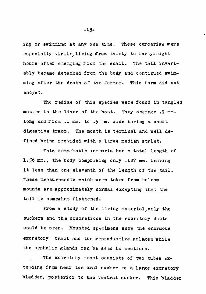
ing or swimming at any one time. These cercaria·e were
especially virile, living Jf'rom thirty to forty-eight
hours after emerging from the snail. The tail invari-
ably became detached from the body and continued swim-
ming after the death of the former. This form did not
encyst.
The rediae of this species were found in tangled
mas ~~es in the liver of the ho st. They a. verage .9 mm.
long and from .l mm. to .5 nm. wide having a short
digestive trao;:t. The mouth is terminal and well de-
fined being provided with a lnrge median stylet.
This remarkable cer ca.ria has a total length of
1.56 mm., the body comprising only .127 mm. leaving
it less than one eleventh of the length of the tail.
These measurements which were taken from balsam
mounts are approximately normal excepting that the
tail is somewhat flattened.
From a study of the living materialJonly the
suckers and the concretions in the excretory ducte
could be seen. Mounted specimens show the enormous
eccretory tract and the reproductive anlagen while
the cephalic glands cen be seen in sections.
The excretory tre.ct consists of two tubes ex-
tending from near the oral sucker to a la.rge excretory
bladder, posterior to the ventral sucker. Thie bladder

-14-
communicates with another in the anterior narrow part
of the tail. Opening into this secondary bladaer is
the median excretory duct of the tail which is ventral
in position.
The reproductive anlagen appear as two mas~es
of cells anterior and posterior to the ventral sucker.
No digestive tract could be traced.
The oral sucker ie .007 mm.. in outside diameter
while the ventral sucker is twice this size. The tail
is made up of lfirge parenchymoue cells and longitudi-
nal and cross muscle fibers. Owing to its delicate
structure it could not be embedded for sectioning. ·
Echinoetome cercariae.
Of one hundred sp~cimens of Physa gyrine collect-
ed at Lawrence Kansas, August 19th, one wa.e infected
with Cercaria fusiformie. This species is charactdr-
ized by a syrranetrical spindle shaped body with a long
dig es ti ve tract extending from the oral sucker to near
the crmdal end of the body. While this species ha.a all
of the characteristice of the Echinostcbme cercaria.e, no
spines are present anywhere. The region of the oral
sucker pr ea en ts a collared appearance. The total length

-15-
of this cercaria is .77 mm. and the width is .07 mm.
The oral sucker has a.n external diameter of .028 mm. and
an internal diameter of one-fourth th~t size. The ventral sucker is about the same eize and is ca.pe,ble
of being grea.tly extended or projected. Thie form
used the ventral suck er extensively in holding fast
to the substratum.
The digestive diverticula do not inclose the
ventre.l sucker as one would inf er from the dorsal aspect
but are do real to it in latera,l view. The dig es ti ve
tract consists of a. very narrow oesophagus... leading from
the mouth to thEJPharynx. Just ba.ck of the pharynx is
a median intestine extending as far as the anterior
end of the ventral sucker, where it bra.nches in to two
diverticula tha.t extend back to the posterior end of
the body. The oesopha,gus measures .003 mm. wide and
.03 mm. long. The width of the pharynx ie .018 mm. and that of the intestine .012 mm.
The excretory tract consists of narrow paired
tubes extending from the region of the pharynx to the
posterior end of the body. The excretory bladder and
the excretory duct in the anterior ·part of the tail
cou~d not be traced definitely but appeared ae more
transparent areas in the toto mount.

-16-
The anlage of the reproductive orgs.ns is a
dense mass of cells nea.r the posterior end of the body.
The posterior sucker is very muecular. The
figures eccompanying the plate show this sucker open,
ulosed,and extended.
The rediae are large and contain from six to
eight ma.ture cercariae together with a few large germ
balls. They .are distended in places by the cercariae
contained within. The average measurements of the
rediae are length l.91 mm. e.nd width .13 mm. to .33nm.
Gymnocephe .. loue ·eercariae.
Of fifty sp~cimens of Physa integra collected
at Chanute Kansas, October 17th, two were infected with
Cercaria cracilis. I propose this name because of the
ability of the species to draw itself out into a very
slender ehRpe. The infection was slight and these
studies were made entirely from the living material and
two mounted specimens.
This cercarie. is exceedingly active and is cap-
able of extending the body until it is as narrow ae
the tail. This fonn can also extend the ventral sucker
until it appee.rs prominent in a lateral view. In a
charac~eristic position this form is slender, heart-

-17-
shaped. The oral sucker is minute, the ventral sucker
being four times as large.
The oesophagus is slender and has a fold near
the anterior end. Just beneath the fold is the narrow
pharynx.
The diverticula of the digestive tract are broad,
encircling the ventral sucker. The excretory system
could not be made out, but paired rows of cells with
highly l!'~.raotive nuclei or possibly concretions mark
this tract. The tail is broad and large, being one-
third the width of the body and a little over twice
the length of' the same. The entire length of this
cercaria ie .53 mm.
The rediae are of unusual shape, tapering at
both ends with a difini te collar near the anterior end.
The digestive tract is slender extending about half
the length of the body. Within the rediae are germ
balis, developing cercariae, and mature cercariae. The
dimensions of the rediae are l.6 mm. long and .33 mm.
wide in the widest pla.ce.
Furcocercous cercariae.
Two 9ercent of large numbers of Physa gyrins, four
hundred and thirty-six in all, collected at Lawrence.

Kansas, during the months of July and August, from
Haskell pond and the lake at Lake View were parasi tis ed
with a. form for which I propose the name C ercaria in-
verse, because of its body being directed teil tore-
• most in swimming. The cercariae emerged freely from
the snails e.nd flitted a,bout rapidly in the water with
a peculiar vi bra. tile motion, directed tail foremost.
creeping movements were not observed excepting under
the cover slip, when the tail became severed from the
body. When the parasitiaed snails were crushed ,
rediae conte,in1ng cercaris,e in a.ll stages of develop-
ment from mere germ balls to mature forms, were prese:;.t.
The va.rious stages in the development of the cercariae
could be easily observed because of the characteristice
of the tails, which varied in length from mere stubs
and rounded lobes to the elocgate bifurcated tsile of
the mature forms.
The rediae were in a tangled mass in the liver.
The largest ones ·exhibited a. slightly wa.ving motion •
Under the cover slip o.ercariae emerged from the rediae,
usua.lly tail first, from the birth pore near the anteripr
end. It required about two minutes for e. cercaria. to
free its elf from the redia.. This form did not encyst,

-19-
but the cercariae soon died in the water, in no case living more than four hr)ure e.fter emerging from the
snail. Those liberated when the snail was crushed lived only from eight to twelve minutes. The number of this species emerging from R siri..gle snail wa.s estimated at five thousand.
inverse, C erce.ri E'. ;tr#//+ is a. sme.11 furco c ere ous form
correspond;ing as to size, shape ,and behavior to eer-caria douthi·tti described by Cort 1915 from Lymnaea· reflexa. This species however, contains no eye-spots a.nd is found in rediae instead of sporocysts. The length of the body is .16 7JJ1Jl• in well extended spec-imens and the width ·1e .045 mm. The unbranched pa.rt of the tail has a length of • 26 mm. e,nd a width of .027
mm. •• while the lobes are five-sixths as long as the main part of the tail and one-half as wide. These lobes taper to rather a sharp point. The openings in the suckers are about the same size, .Ol mm. but the out-side dimensions of the oral sucker etre greater than those of· the ventral sucker. The measurements are: oral sucker .042 mm. and ventral sucker .028 mm.
As in Cercaria douthitti, the region back of the center is filled with large cephalic glands. The ducts of these gle,nds extend f orwa,rd and open alongside the

-20-
oral suck er~ (Fig. 50), The excretory system of the
body region could not be traced; but a duct extends
through the main part of the tail and is joined by
ducts from each branch. .A:n excretory pore opens to the
exterior between the forks of the tail.
The anlage of the reproductive orgars is a mass
of small cells nee.r the posterior end of the body s.nd
ventral in position.
Six percent of large numbers of Physa gyrina
colle<ited at Lake View, Kansas. August 20th, were in-
f ected with a. form for which I propose the name Cer-,
CE'.ria echinocfluda. Col lee tions of saa.ils from the same
locality made October 12th, ahowed two percent to be
infected.
This cercarie. was very e.cti ve swimming both
forwa.rds e..nd backwards, but usually forwards with a
vibratile motion of the tail. The tail was loosely
attached to the body and was easily severed. When
this occurred the body died in about twenty minutes,
while the ·.tail lived for two hours, swimming about
actively. Six hours was the maximum time that theme

-21-
cerce.riae rema,ined e.li ve after emerging from the snail.
The body of this species is .31 mm. long and .125 mm. wide. The main part of the tail has a length of
.54 mm., while the branches are .18 nnn. long. Two large
eye-spote are present. They are made up of minute
pigment granules and mes.sure .02 nm. by .014 mm. in
dorsal view being longer in the trans verse directior1.
An excretory tube begins in the branches of the
tail and extends throughout the length of that organ
to the excretory pore in the posterior end of the body.
The excretory tract could not be made out in the body
region.
The branches of the tail are provided with a sort
of fin extending around the tip. Thie fin is beset with
minute s:pi nee.
A mouth is present in the oral sucker but there
is no dig es ti ve tract lea.ding from it.
The anlage. of the reproductive organs is ventral
in position ~md near the posterior end of the cercaria,.
A peculiar characteristic of this species wasthat,
in the fixed specimen,the tail is usually bent sharply at its junction with the ·body.
The rediae of this species average 2 mm. long and .11 mm. wide. They are filled with germ balls and

-22-
cerca.riae in various stages of development. The anterior
end is somewhat pointed, while the poaterior end is
rounded.
cerc8ris echinocauda is· similar to Oerca.ria
ocelle,te La Valette St. George es the measurements for
this species fQll within the wide range described for
eercarie. ocella ta. Both forms are provided with fin
like projections on the ta.il. The tail of cercaria
echinoca.ude. is not very contractile contrasting with
Cercaris: ocellata. Th oral sucker of cercaria
echinocauda is also much larger than that of Cerca.ri&'
ocellata. Cercaria echinocauda develops in rediae,
while Cerce.ria ocellata is found in aporocyste.
v Of thirteen speci\mene of Planorbis trivo~is
collected a.t Lawrence Kansas, October 7th, two were
infected with a cerca.ria for which I propose the name
Cercaria quiata, beca.uee of its often rema.ining motion-
less) floating in the water for brief periods of time.
Like Cercaria inversa this cercaria has a bifurcated
tail and swims by means of a rapid Vibratile motion of
this organ.
The tail is enormous in size comI>a.red to the

-23-
body and is not constricted off from the body in the
living specimen as is the case with other furcocercous
forms. The tail of this species ie never severed from
the body in thf .living form and only rarely did 1 t
become lost during mounting.
The cercarifl. can swim either forwards or back-
wards. The tips of the bi~urcated tail are fitted
with adhesive organs by means of which they a.tta.ch
themselves to the substratum or <lther cerca.ria.e. Both
oral and ventral suckers a.re sme.11 and of uniform size,
measuring .027 mm. in diameter.
The excretory sye~em consists of paired ducts
leading from the anterior end of the ·body to the jun_c-
tion of the bod)' and tail) where they amastomose and
extend on back to the excretory pore,which opens be-
tween the forks of the tail. No excretory vesicle is
present but the excretory tube is somewhat dilated
just anterior to the excretory pore.
The anlage of the reproductive system is pos-
terior to the ventral sucker. No digestive tract could
be traced.
The total length of the cercaria. is .8 mm. and
the width is .08 mm., the main part of the ta.il being
as wide a.s the body. Of the total length the body makes

-24-
up one-fifth, the unbranched tail two-fifths, and the
branched ta.il two-fif the.
The rediae of this species is long and cylin-
drical measuring 1.52 mm. by .2 mm. Each redia contains
many cercarie.e about one-fifth of which are mature. The
rediae were so tangled in the liver that it was almost
impossible to disoect them out entire.
xiphidiocercariae
~;
My material contains three species of Xiphidio.;;;
cerca.riae. Owing to the extreme difficulty of study-
ing these small forms my descriptions are in some places
incomplete. I am unable to make these species fit in
with previously described forms much as they resemble ,,
forms described by Cort and Lube.
For the first of these species, ·I propose the
name Cercaria haskelli from the locality where it was
found 1 .. Haskell Fond':. One of thirty- three specimens
of Physa. gyr ina. collected from this locality at
Lawrence1 Kansas,July 12th, was 4illf..'ected:.with this c species,
Cercaria haskelli is a rapid swimmer, the swim-

ming a.lternating with creeping movements. A marked
characteristic of this species was that it could extend
the te.il until 1 t was three or four times the length
GSf. ~e body. When this form was swimming, the body
would be contracted into a ball and the tail would
be very much extended.
The measurements for this species in an aver-
age state of contraction are: length .15 mm. and width
.05 mm. The tail measures four-fifths the length of
the body. A stylet protrudes from the region of the
oral sucker. This stylet measures .037 mm. long and
ha.a a width at its base of one-fifth of the lengt~~
It ti.apers to a. point and has no enlargements anywhere
along ite length.
The ore,l sucker is .04 mm. in diameter and
the ventral sucker measures .03 mm. In longitudinal
section, both suckers open into pouches wider than the
external openings. There are two layers of cuboidal
cells in the wall of the ventral sucker.
The muscular layers comprising the outer wa.ll
of the cerca.riE.l averHge .ou3 nnn. in thickness.
The oesophagus is exceedingly nar:row aYeraging
.0015 mm. in diameter. It is eurrounded by a ring of
deeply staining cells corresponding to the muscular

-26-
pharynx seen in other forms. The oesophagus broadens
into a medis.n digestive tract .03 mm. long a.nd .0125
mm. wide.
Anterior to and dorsa.l to the ventral sucker
are numerous unicellular glande. These are probably
stylet glands as their position is different from that , I
of the cephalic glands described for Cercaria inversa,
for instance. No ducts could be found leading form
these gla.nda.
The only excretory tract ~found coneiets of
irregulRr spaces lying against the dorsal wall of the
ventral sucker. In transverse section of the tail,
four large central cells can be set!·n surrounded by a
ring of muscle fibers. In some places the walls be-
tween these central cells are made up of smaller narrow
cells.
The a.nlRgen of the reproductive organs are
in two mas see dorsal to the ventral sucker. These
masses are connected by a narr-0w band of similar celis.
Cerca.ria. haskell.1 is found in sporooyste. They
are rounded elongate ea.cs containing germ balls, de-
veloping cercariae and mature cercariae. The eporo-
cysts are from .25 mm. to .42 mm. long and are from

-27-
one-third to one-half a.s wide ••
Of twenty-three specimens of Planorbie tri vol vie
collected at Cherryvale, Xansas, October 16th, five
were infected with Cercaria\ gregaria.. The cercs.riae \
emerged from the snails by thousands and had the pee~-
liar ha.bit of massing together in the water. They
would ~;.lose their tails and form such compact mas:.::;es
that they could not be separated without tearing the
tissues a.part. These masses conta.ined from fifty to
five hundred ·1ndi vi duals each. The cercarie.e reme.ined
alive in this condition for eight hours. Ho encyst-
ment was se~·n and very few cerca.riae remained for a.ny
length of time without joining with ontof the masses.
This is c.n exceedingly minute form measuring
only .37 mm. long including the tail which is half
the length of the body. The width is .03 mm. Both
of the suckers are the same size averaging .015 nun.
in outside diameter. This form has a stylet .006 mn.
long. On account of the minuteness of this form, the
internal structures could not be made out clearly.
The best results were obtained ly staining intravit-
ally with picro carmine. With this etainJpaired masses
of cephalic glands with their ducts could be made out.

-28-
The tail of this form cons is ts of large cells with
a definite single row of nuclii showing prominently
in the median line.
Sectioning the snails from which these cercariae
emerged failed to reveal either sporocysts or rediae.
Seventy-five percent of hundreds of Planorbis
trivolvis collected at Pratt Kansas, August 22nd, were
infected with a small Xiphidiocercariae which I pro-
pose to ca.ll Cer caria( k&nsiensi s. The body of this
form agerages .06 mm. wide and .09 mm. long. The tail
in a.n 2verage eta te of contraction is .064 mm. long.
The oral sucker is .024 mm. in diameter and the
ventral sucker is .028 mm. The openings in both suckers
are .007 mm. in diameter. There is a bicornuate groove
in the posterior end of the body in to which the ta.il
fits.
llo dig es ti ve tract could be tra.cedJ but a ring
of deeply staining cells me.rks the region of the pharynx.
Large unicellul.e.r cepha.lic glc:tnds a.re present.
They number about four on a side. No ducts could be
found leading from them.
no excret.ory tubes could be found but there
is a large excretory ·bla.dder .Ol mm. wide and .016
long, posterior to the ventra.l sucker. The long a.xis

-29-
of this bladder is in a transverse direction to the
long axis of the body.
The anle,gen of the reproductive orga.~e consist
of two mA.sses of cells dorsal to the ventral sucker.
The posterior mast is the larger.
Special studies were made of the etylet of Cer-
CHria. ka.nsiensis. It is embedded in the muscles of the
thici walled ore .. l sucker dorsal to the mouth opening,
and can be withdrawn into a hollow reaeptacle. Two
camera J.ucida sketches J figures 57 and 58 show the sty-
·l et extended and contracted. The stylet measures .02
:rrm. in length. At the base a.nd near the point it has
a width of one-sixth its length, but between these
points it is narrower.
Cercaria kansiensis is found in sporocyets
averFging .33 mm. long and about one third as wide.
In all cases where this form was found, the
infection was heavy, the liver of the snail being
filled with almost a solid mass of the sporocysts.
Estimates of the numbers of cercariae emerging from
Bny one snail ran from five thousand to eight thousand.
1:0TES.
The following notes while adding little to this
paper might be of aesistence to Anyone wishing to work
out life histories of trema,todea.

-30-
The collecting .. grounds from which my material
was taken covered a wide range of habitats from tem-
porary pools and pasture streams to artificial ponds
and permanent lr,kes.
Only two genera of snails were examined. In-fection was very rarely found in young snails and never
in snails collected from temporary pools or pasture
streams. Old ponds harboring fish, frogs, muskrats
and watei:.enakee, usually contained infected snails.
All of the furcccercous cercariae were found
in Physae col1ected from muddy permanent ponds. The
large 4mphistome cercariae and the xyphidio cercariae
were found in the genus Planorbis in the larger clear-
e~ lakes.
~akes of this type are inhabited by the species
of water life mentioned for the other type of ponds
and in ad di tio n are frequented by migratory birds.
The heaviest infection was found the latter
part of August at the State Fish Hatchery at Pratt,
where the number of snails parasi tis.ed ran as high
as ninety percent.
No cases of double inf ect1on were found.
Only one of ~he species studied, Cercaria oortii~
encysted under observation, and no cysts of any of the

-31-
other forms were found outside of the snail.
In on~y one case were there any cysts found
within the tissues of the host, one sp~:cimen of 1Han-
orbis tri vol vis showing a heavy infection in the di~-·
gestive gland. These cysts were spherical measuring
.05 mm. in diameter. No other infection besides these
cysts was present. There was no evidence to reveal
the identity of whatever cercaria the cysts may have
represented, but in this ca.se, Planorbis trivolvis
is evidently the secondary intermediate host of the
species. Cort has described the cysts of cercaria
trivolvis as having been found in the body cavities
of Planorbis trivolvis.
One characteristic of Cerc;:iria gregaria possibly
throws some light on the life history of the species.
That is the peculiA.r habit that the cercariae have of
losing their tails and forming compact masses in the
water :containing from fifty to five hundred living
individuals.
The tables accompanying the descriptions will
be found Useful for making summaries CGncerning the
amount and kind of infection.

-32-
Pond Index.
No.l,Lawrence, Kan. Haskell, east of dairy barn. " 2, " " " drainage ditch, south. " 3, " " Stubb's pdnd, NW campus. " 4, " " Stream, Woodland Park. " ?• " " Horseshoe, __ Lake, 6 mi. S E. n o, " " Stream N W Griesa nursery. " 7, Lake View East end of road through lake. " 8,Lawrence, Kan.Stream near E.A. Richards 6 mi. N w. " 9, " " " t mi. S W stream no. 8. " 10, " " Bismarck Grove North Lawrence~ " 11, " " Lake north of u. P. Depot. " 12, Lake View East of Depot. " 13, Pratt, Kan. State Fish Hatchery,Pond no. 22. ff 14, n M n If H A ff 39. " 15' " n " n " n " 41. " 16 ' " " " " " " " 51. " 17, " " " n " " " 77. n 18, " " n " n " ff 1. " 19, Lawrence, Kan. R.R. t mi. Seuth East Haskell. " 20, " " Pond on East 15 St. '' 21, Baldwin, " " in east edge of town. " 22, " " " South West of Depot. " 23, " " " Center of town. " 24, Ottawa, Kan. Stream East of Park near Library. " 25, " " Pond near Race Track. " 26, " " River West of W Bridge. " 27, Chanute, Kan, Santa Fe t mi. North of Depot. " 28, " " " " North of Frisco crossing. " 29, " " ff " Two mi. north, west side. " 30, Cherryvale, Kan. Lake in City limits. " 31, " " Pond t mi. north along Santa Fe. " 32, Abilene, Kan. Engle'e pond 7 mi. S w. 11 33, " " Pond t mi. east R. c. Lahr S W. " 34, " " Stream 4t mi. Svuth West.

-33-
Percentage of Infected Snails.
Date Pond Host No.snails No.infected Species
7~? 1 Physa gyrina 25 2 c. inversa 7 7 2 ti 20 1 c. haskelli 7~9 1 " 95 0 7 9 3 " 50 0 7/10 1 " 20 0 7/12 1 " 33 6 c. inversa 7/20 1 .. 130 0 7/21 4 " 23 0 7/22 1 " 30 1 c. inversa 7/24 g Planorbis trivolvis 15 1 c. magnacauda 7~1 Physa gyrina 6 0 7!jl 8 " 5 0 7~1 9 " 5 0 8 2 7 " 10 1 c. inversa 8~ 7 " 12 2 c. " 8 4 8 " 20 0 8/4 9 " 10 0 8/18 _, 10 " 10 0 8/18 11 " 12 0 8/19 1 " 100 l c. fusiformis 8/20 12 " 100 6 c. echinocauda 8/28 13 Planorbis trivolvis 16 4 c. kansieneis 8/28 14 " 30 13 " 8/28 15 " 20 1~ " 8/28 16 " 20 " 8/28 17 " 20 16 ff
8/29 18 " 29 13 n
8/29 17 ff 25 21 ff
2 c. inversa 8~0 16 " 50 45 c. kansiensis 8 31 17 tt 50 34 " 2 c. inhabilis ;; 8/31 16 " 90 75 c. kansiensis 1077 12 Physa•gyrina 50 1 c. echinocauda 10/7 5 Planorbis trivolvie 13 2 c. inhabilia 10/8 19 " 12 2 II
10/12 19 " 23 1 "

-34-
Percentage of Infected Snails.
Date Pond Host No.snails No.infected Species
10/19 21 Physa gyrina 50 0 16/19 22 " 50 2 c. haskelli 10/19 23 " 50 0 10/19 25 " 50 2 .. 10/19 26 " 50 0 10/19 27 " .50 0 10/19 28 Physa integra 50 2 c. gracilis 10/19 29 Physa gyrina 50 0 10/20 30 II 50 7 c. gregaria 10/21 30 .. 27 5 c. cortii 11/9 31 " 50 0 11/9 32 " 100 0 11/9 33 " 25 0

-35-
BIBLIOGRAPHY
Barker, Franklin D.
1915. Parasi tea of the American Muskrat.
Journal of Parasitology. Vol. l, ~o. 4.
Braun and Luhe
Cary, L. R.
Cort, W.W.
1910. Practical Paraeito~ogy.
1909. The life-history of Diplodiscus tem-
poratue Stafford. With especial reference
to the development of tne parthenogenetic
eggs. z·ool. Jahrb. a.ot. f. Anat. u. Ont.,
28: 59 5-659.
1915. Some Uorth American Larval Trematodes.
Ill. Biol. Monographs, Vol. l. :No. 4.
Goldschmidt, Richard.
Kerbert, C.
1902. Uber Bau und 'TIInbryonalentwicklung von
zoogonus mirus Les. Centralblatt, Vol.xXXll,
No. 12, 870-876.
1881. Beitrag zur Kenntniss der Trematoden.
Archiv. fur mikr. Anatomie, 19, 529-579·

-36-
von Lins tow.
1890. Uber den Bau un~ die Entwi~klung des
Distomum cylindraceum Zed. Archiv. fur mikr.
Anatomie. 36. 173-191. Nicoll, William
1906. Some new and little known Trematodes.
Ann. & Mag. N. Hist. Ser. 7, Vol. XVll.
514-526.

-37-
Explanation of 'lates
With the exception of Plate Vll all figures are
drawn with the camera lucida. The abbreviations are af-
ter the plan adopted by Cort.
Abbreviations Used.
ac acetabulum bp birth pore of redia br brain c concretions cg cystogenous glands cog cephalic glands de ducts of ccg. e eye-spot es esophagus ex excretory system exp excretory pore exd excretory duct exv excretory vessel gb genttball i intest~al caecum of cer-
caria ir intestine of redia la:: locomotor appendage of
redia
le large central cells of tail ml muscle layer o e oral suck er p pigmentat1on pb pharyngeal bulb pr pharynx of redia ra reproductive anlage s stylet sr s~ylet of redia sc cercariae in sporocysts sg s tyl et glands aw wall of sporocyst vs ventral sucker
-38-
.B'ig .l. Cercaria cortii X 72. Fig .2. Cross sec. of c.cortii tnrough brain x 144.
Fig.3. Crose sec. of c. cortii through R.A. x 144.
Fig.4. Cross sec. of tail of c. co rt ii x288. Fig.5. Longi. eec. of tail of C. cortii x 288. Fig.6. Cyst of c. cortii doraa,l view x 72. Fig.7. Cyst of c. cortii la tera.l view x 72.

-39-
Fig.8. Cercaria diastrophora X 144.
Fig.9. Cross sec. of C.diastrophora through oral
ce.vi ty X 288. Fig.10. Crose sec. of c. diaetrophora tnrough
pharynx X 144.
Fig. ll. Cross sec. of C. dia.atrophora through re-
productive anlage X 144.
Fig.12. Croes sec. of tail of c. diastrophora X 288. Fig.13. Immature redia of C. diastrophora x 144. Fig.14. Mature redia of u. diastrophora x 72. Fig.15. Longi. sec. through exc. pore of c. diaetro-
phora x 576.

-40-
Fig.16. Cercaria inhabilis Cort dorsal view X 44.
Fig.17. Immature Cercaric. inha.bilia X36
Fig.lb. Immature redia of c. inhabilis X 36.
Fig.19. Mature redia of c. inhabilie x36. Fig.20. Cross sec. of c. inhabilis through
intestinal caeca X 160.
Fig.21. Cross sec. of c. inhabilis through
excretory pore X 160.
Fig.22. Cross sec. of tail of c. inhabilie near
body X 160.
Fig .23. Cross sec. of ta.il of C. inhabilis near
posterior end X 160.
Fig.24. Cercaria gracilis X 160.
Fig.25. Redia:'Cbf Cercaria graci . .Lis X 36.

-41-
Fig.26. Cercaria fusiformis,dorsal view x 72. Fig.27. c. fusiform.is, ventral
Fig .28. c. fusiforrnis,ventral
Fig.29. c. fusiformis,ventral
Fig.30. Cercaria magnacauda
Fig.31.Body of C. magnacauda
Fig.32. Cercaria quieta
sucker
sucker
sucker
Fig .33 •.. Ventral sucker of C. qui eta
closed, x160.
open, x160.
lateral view
x 72.
x 260.
x·, 72. x 260
x
Fig.34. Mth. of Redia of c. mag. longi. sec. X 260.
Fig.35. Eth. of Hedia of C. mag. cross sec. X 260.
Fig.36. Redia of c. fusif ormis x 30. Fig.37. Redia of c. qui eta x 36. Fig.38. Redia of c. magnacaucta x 104.
160.

-42-
Fig.39. Cercaria echinocauda x 72.
Fig.40. Cercaria echinocauda x 72. Fig .41. Redia of i' v• echinocauda x 36. Fig.42. Longi. sec. through c.echinocauda x 160.
Fig.43. Cross sec. through C,echinocauda x 160. Fig.44. Tail of c. echino cauda, longi. sec. x 160.
Fig.45. Tail of c. echi no cauda, cross sec. x 160. Fig.46. Cercaria inversa x 72. Fig.47. Redia of c. in versa x 72. Fig.48. Cross sec. of tail of c. in versa x 144. Fig.4Cj. Cross sec. tnrough cepha.1.ic glands, c . in. x 144. Fig.50. Cross sec. through oral euck er, c. in. x 520. Fig.51. Longi. sec. through ex. pore, c. inversa x260.
-43-
Fig.52. Cercaria haskelli, dorsal view X 144.
Fig.53. c.· haskelli, longi. sec. X 160.
Fig.54. c. haskelli, cross sec. through taiL X 160.
Fig.55. Sporocysts of C. haskelli. X 72.
Fig.56. Cercaria kansiensis X 260.
Fig.57. Lateral view of stylet ,C.kan. withdrawn X 260.
Fig.58. Lat. viewof stylet extended, c. kan. X 260.
Fig.59. Sporocyst of c. kansiensis .x. 72.
Fig.60. Cercaria gregaria X 144.
Fig.61. Longi.aec.snail liver infected with C.kan. X 44.
Fig.62.Sporocyst c. kansiensis, cross sec. x 260.

-44-
Reconstructions of the cercariae described in
the preceding pages,all drawn to scale X 36. The num-
bers ref er to the index accompanying each plate. num-
bers 81- 56' refer to sporocysts and redi~correspond
ing to the cercariae.

-45-
I want to express my thank a to Dr. 'N. W. Cort
for his kindness in lending me slides, e~amining my
preparations, and aiding in the identification of
species. I also wish to thank Dr. H.A. Pilsbry for
identifying the snails used in the studies.
To Professor Bennet M. Allen. under whose direc-
tion the work has been done, I wish to express my ap-
preciation for hie interest 2nd constructive criti-
cisms.

-46-
With this thesis are submitted infected snails,
vials of cercariae, and seven plates corresponding to
index figures, l to 62.