to-day w4ith path2genesis only · a. examination of the cardio-vascular system a statistically...
TRANSCRIPT
\VASCULAR ORIGIN OF GLAUCOMA
A CONTRIBUTION TO THE THEORY OF THEVASCULAR ORIGIN OF GLAUCOMA*
BY
E. DIENSTBIER, J. BALiK and H. KAFKAPRAGUE
INTRODUCTIONIT appears to-day that, after nearly a hundred years of contro-versy, the solution of the problem of pathogenesis of glaucomahas entered its final phase. The literature concerning this themewas immense. Some of the authors approached the problem w4itha modern outlook, but Leber's classical theory, with which theirresults did not agree, could not be rejected. After a time theyformed a new conception based on their own observations.Though the vascular system was often mentioned, the old con-
ception of the path2genesis of glaucoma-remained unchanged.One thing was clear, namely, that primary glaucoma was not alocal disease of the eyes, but an eye complication of some organicor constitutional disturbance still unknown (Gordon 1938, Duke-Elder 1945).
It was apparent that the problem of the pathogenesis ofglaucoma could only be solved analytically. This method wasused in the study of glaucoma, published in 1946. Whenanalysing the\ origin of hypertension, the anatomical findings,clinical observations and the effects of treatment, the followingconclusions were made, glaucoma is the expression of stasis in thevenous system and the eye capillaries. It has its origin partlyin organic vascular changes with a more or less marked spasticfactor and partly in changes in function (vasoneurosis).When ocular hypertension was analysed, it was clear that the
problem was one of the intra-ocular fluid and blood circulation.These problems, which are so closely connected, are identical withthe question of the peripheral circulation and of fluid-balance.It is therefore necessary to observe the function of the peripheralarterial circulation in glaucoma. The disorders of the peripheralarterial circulation in glaucoma, as already mentioned, are not onlylocal but general., Some of the authors noticed a possible patho-genic connection with the circulation; on the contrary the majority
* Delivered at the XIVth Congress of the Ophthalmological Society of Czecho-slovakia on October 2, 1948. in Prague.
t Received for publication, May 9, 1949.From the first ophthalmic clinic, Director, Professor R. Kadlicky, and from the
second internal clinic, Director, Professor A. Vancura of the Charles University.Prague.
47
on June 19, 2020 by guest. Protected by copyright.
http://bjo.bmj.com
/B
r J Ophthalm
ol: first published as 10.1136/bjo.34.1.47 on 1 January 1950. Dow
nloaded from
4. DIENSTBLIER, J. BALIK at,zd HI. KAIEA
x-ere satisfied with the old conception. They believed that ocularhypertension caused all the disorders which appear in the evolu-tion of glauconma, including the circulator+- disorders. The wordglaucoma and ocular hypertension were synonymous. Nothingc(hanged this belief, not even the so-called glaucoma withoutlhy-pertension-the disease where the second main sign ofglaucoma is predominant, namely, the excavation of the papillaof the optic nerv,e without an increase in intra-ocular pressure.This form of glaucoma has a long history. It begins with Graefe's'amaurosis whit excavation," then the term glaucoma without
hlypertension and pseudoglaucoma and later the term incompletegolcucoma. It was necessary to change this term, because theexcavation wlhich could have its origin in hypertension, can exist\xithout it. Regarding the influence of eye hypertension it X-assaid that the hypertension might be only small or transitory(Donders). No proof could be obtained from anatomical find-ings, Schnabel (1892-1905), described the lacunar degeneration ofthe nerve-fibres in the optic nerve with glaucomatous excavation.This condition, which was similar to changes occurring in thebrain, was called " status lacunaris " byt Pierre Marie He des-cribed vacuoles formed in the optic nerve by the joining of thelaccunae, and causing depression of the papilla. The author him-self discovered that the causes of these disorders were angio-sclerotic retrobulbar lesions, namely lesions of Haller's arterialcircle. Thus Schnabel definitelv rejected the view of the pressure-origin of excavation. In spite of his clear proofs and the workof his followers, his opinion of the origin of glaucomic excavationis not generally accepted, even to-day (Elschnig, Ichikawa,Langrange, Beavieux, Gallanga, Magitot, etc.). Experimentalresults and clinical experience are not sufficient. Artificial hvper-tenision produiced by ligature of the veins did not give rise toexcavation even after five months. Clinical cases are well knownwhere the function of the eye restored by operation, deterioratesprogressively, even without hypertension. This might be con-sidered as evidence of disturbed tissue-nutrition.
REPORT OF THE METHODS USED AN!) OF OUR O1WN OBSERVATIONSXVheni estimating ocular hypertension it is seen that there are
problems which are in close connection with the peripheral bloodcirculation and fluid-balance. As already mentioned, these prob-lems led to our examination of patients suffering from glaucoma,with particular reference to the examination of the cardiovascularsyvstem. The colloid osmotic pressure of the plasma-proteins wasestimated, and the investigation completed by a series of testsleading to the explanation of the plasma-protein spectrum. The
48
on June 19, 2020 by guest. Protected by copyright.
http://bjo.bmj.com
/B
r J Ophthalm
ol: first published as 10.1136/bjo.34.1.47 on 1 January 1950. Dow
nloaded from
VASCULAR ORIGIN OF GLAUCOMA
blood cholesterol and urine were examined, and Wassermannreactions ascertained.A total of 97 patients were examined: 23 with acute inflam-
matory, 14 with chronic simple, 37 with chronic inflammatory and23 with secondary glaucoma.
A. Examination of the cardio-vascular systemA statistically important relation was found between vascular
hypertension and glaucoma - hypertension being found in ourseries more frequently than in patients of the same age using thehypertension table of Robertson and Brucer from Vancura. Inthe group of secondary glaucoma, deviations were not so striking.
Because even the highest diastolic readings are comparativelylow, hypertension due to loss of elasticity of the vascular wallcan be excluded. Systolic hypertension could be expected in ourpatients on account of their age, but radiological findings showsigns of arteriosclerosis. The ECG showed mostly left ventri-cular preponderance, which is usually caused by hypertension.Some findings, such as auricular fibrillation, prolonged P-Rinterval, infarctions and possibly bradycardia are signs ofcoronary sclerosis. \
In conclusion it may be said that glaucoma with hypertensionis quite evident in 95 cases where the examination of the cardio-vascular system was made. It is not possible to characterize thisco-existence more closely. But it can be stated with certainty thatthe patients of this group suffer from vascular disease.
It is necessary to complete the examination of the peripheralabnormalities of the vascular system by the determination of theoscillatory index and capillaroscopy. Before making the examina-tion a detailed family and personal history was taken for evidenceof cardiovascular disease.
Patients suffering from acute glaucoma often complained ofparaesthesia of the limbs, digiti mortui, cyanosis, intermittentclaudication, migraine, hypertension, diabetes, varicose veinsand their complications and urticaria, as seen from their personalhistories. These complaints confirm the presence of vasculardisease. This positive history was more frequent in chronicinflammatory glaucoma (42), than in acute inflammatory glaucoma(22), and in secondary glaucoma (12). Excitability is considereda sign of a labile vascular system by many authors. This wasfound in chronic inflammatory glaucoma 16 times, in acute inflam-matory 10 times, in secondary glaucoma 9 times, and in chronicsimple glaucoma only twice.
Oscillation was determined by means of the oscillotonometerin the usual way above the ankle, above and below the knee-joint,
49
on June 19, 2020 by guest. Protected by copyright.
http://bjo.bmj.com
/B
r J Ophthalm
ol: first published as 10.1136/bjo.34.1.47 on 1 January 1950. Dow
nloaded from
5() 1X . DIENSTBIER, J. B.XLIK anld H. KAFKA
and on the arrm. According to Prusilk the loss of the oscillatorvindex to 50 per cent. and more was regarded as a decrease fronmthe normal. Thle signs w-hich wrere reasonably supposed tobe of vascuilair origin, wvere verified by the symptomis olthe patients. A decrease in the oscillatory index was founjid ilnacute inflammatory glaucoma in 11 cases (12 normal values), inchronic simple glaucoma in 8 cases (6 normal values), in chronicinflammatory- in 1,5 cases (292) normal values) and in secondaryglaucoma in 4 cases (19 normal values). In 97 cases a distinctdecrease of values above the knee-joint was seen 4 times. Thisdecrease indicated the necessity for appropriate therapy.When making a critical evaluation of these facts two striking
points care seen: decreased oscillations in all groups exceptsecondars glaucoma, and the obvious predomiiniance of thisdecrease in chronic simple glaucoma.
Capillaroscopy: the capillaroscopic determinations were madeby the Zeiss capillaroscopic microscope. The capillaries of thenailrim, the flexor sides of the fore-arm, the outer part of the armabove the olecranon and the left infraclavicular region wereexamined. From the findings it was possible to estimate the con-dition of the capillaries.
Viso- TrallSitof i Arterio- N(rnralneurosis fortiis sclerosis
(Yl. ac. intl. 14 4chr. simple ... Iehr. infl. ... l HSCC. (. 1 4 1-2
\VNhenl makilng al microscopic examiniation of the capillariesnoine of the 23 cases of acute glaucoma gave normal findings. Thisgroup slhowed an absolute predominance of vasoneurosis. Inchronic simple glaucoma the relationship is quite different, givingevidence of arteriosclerosis. In chronic inflammatory glaucomaboth groups are the same and finally in secondary glaucoma themicroscopic findings of the capillaries are mostly normal.
In making a general estimation of the cardio-vascular findings,the most striking thing is the tendency to disease in ouir patients.The cardio-vascular findings show- that the patients w-ith glaucomaoften suffer from hypertension wx ith all its consequences andelectro-cardiographic findings. X-ray findings show the presenceof arterio-sclerosis, which is not surprising, considering the age ofthe patients. The oscillatory findings indicate anatomical and,considering the age, mostly arteriosclerotic changes in the peri-pheral arteries. The most common occurrence in chronic simpleglaucoma mai-y well illustrate the findings of those authors w-ho
on June 19, 2020 by guest. Protected by copyright.
http://bjo.bmj.com
/B
r J Ophthalm
ol: first published as 10.1136/bjo.34.1.47 on 1 January 1950. Dow
nloaded from
VTASCULAR ORIGIN OF GLAUCOMA
found sclerotic clhanges in the intra-ocular vessels. The -micro-scopic examination of the capillaries shows the same conditionin the smallest vessels. In general our cardio-vascular findingsare in agreement with the extensive literature.
B. Biochemical examinationThe protein spectrum was examined first to estimate the colloidal osmotic
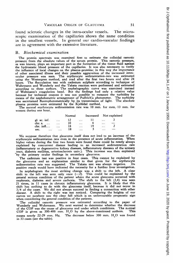
pressure from the absolute values of the serum protein. This osmotic pressure,as was known, plays an important part in the formation of the tissue fluid againstthe hydrostatic blood pressure of the capillaries. It was also necessary to verifythe influence of local changes on the plasma-proteins; in this way the importanceof other associated illness and their possible aggravation of the increased intra-ocular pressure was seen. The erythrocyte sedimentation-rate was estimatedusing the Westergren method, and read after the first two hours and after 24hours. The flocculation test with cadmium sulphate according to technique ofWuhrmann and Wunderly and the Takata reaction were performed and estimatedaccording to these authors. The nephelographic curve was examined insteadof Weltmann's coagulation band. But the findings had only a relative valuebecause for technical reasons it was not possible to measure the turbidity bymeans of the nephelometric arrangement of Pulfrich's photometer. The turbiditywas ascertained fluorophotometrically by its transmission of light. The absoluteplasma proteins were estimated by the Kjehldal method.The normal erythrocyte sedimentation rate was 10 mm. for men, 13 mm. for
women during one hour.
Normal Increased Not explainedgl. ac. inf. ... 12 ... 11 ... 3chr. s. ... ... 10 ... 4 ...chr. infl.... ... 18 ... 19 ... 2sec. ... ... 18 ... 5 ...
We suppose therefore that glaucoma itself does not lead to an increase of theerythrocyte sedimentation rate even in the presence of acute inflammation. Whenhigher values during the first two hours were found these could be nearly alwaysexplained by concurrent disease leading to an increased sedimentation rate(inflammatory or degenerative kidney diseases, inflammatory diseases of the urinarytract, diabetes mellitus, arteriosclerosis univ.). This increase was then explainedby the primary ocular findings in secondary glaucoma.The cadmium test was positive in four cases. This cannot be explained by
the glaucoma and an explanation similar to that given for the erythrocytesedimentation rate was suggested. The Takata test was always negative. Itspositive result would have indicated the necessity for a further liver investigation.
In nephelogram the most striking change was a shift to the left. A clearshift to the left was seen only once (-2). This could be explained by thegeneral serious condition of the patient where the acute glaucoma occurred withnephrosis, diabetes and severe cachexia. The shift to the left (3,4) was seen21 times, in 11 cases in chronic inflammatory glaucoma. It is likely that thisshift has nothing to do with the glaucoma itself, because it did not occur in3/4 of the cases. We did not always succeed in finding a connection with otherdisease. A shift to the right was not noticed. Comparing the heights of our
curves we nowhere saw the clear fall which is an unfavourable prognostic signwhen considering the general condition of the patients.The colloidal osmotic pressure was estimated according to the paper of
Wunderly and Wuhrmann. We next wanted to determine whether the decreaseof the COP was the cause of glaucoma and under which conditions. The normalCOP is given as 300-400 mm. H,O by the above-mentioned authors. Thismeans nearly 22-29 mm. Hg. The decrease below 300 mm. H wwas foundin 33 cases (see the table).
51
on June 19, 2020 by guest. Protected by copyright.
http://bjo.bmj.com
/B
r J Ophthalm
ol: first published as 10.1136/bjo.34.1.47 on 1 January 1950. Dow
nloaded from
E. DIENSTBIER, J. BALIK auid H. KAFKA
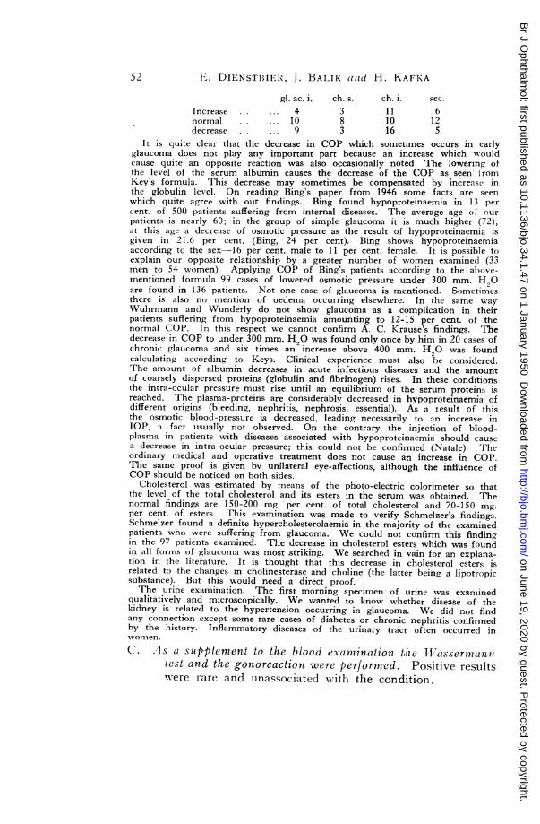
gl. ac. i. ch. s. ch. i. sec.Increase ... ... 4 3 11 6normal ... ... 10 8 10 12decrease ... ... 9 3 16 5
It is quite clear that the decrease in COP which sometimes occurs in earlyglaucoma does not play any important part because an increase which w ouldcause quite an opposite reaction was also occasionally noted The lowering ofthe level of the serum albumin causes the decrease of the COP as seen- iromKey's formula. This decrease may sometimes be compensated by increase inthe globulin level. On reading Bing's paper from 1946 some facts are seenwhich quite agree with our findings. Bing found hypoproteinaemia in 13 percent. of 500 patients suffering from internal diseases. The average age o.' ourpatients is nearly 60; in the group of simple glaucoma it is much higher (72);at this age a decrease of osmotic pressure as the result of hypoproteinaemia isgiven in 21.6 per cent. (Bing, 24 per cent). Bing shows hypoproteinaemiaaccording to the sex 16 per cent. male to 11 per cent. female. It is possible toexplain our opposite relationship by a greater number of women examined (33men to 54 women). Applying COP of Bing's patients according to the above-mentioned formula 99 cases of lowered osmotic pressure under 300 mm. H,0are found in 136 patients. Not one case of glaucoma is mentioned. Sometimesthere is also no mention of oedema occurring elsewhere. In the same wayWuhrmann and Wunderly do not show glaucoma as a complication in theirpatients suffering from hypoproteinaemia amounting to 12-15 per cent. of thenormal COP. In this respect we cannot confirm A. C. Krause's findings. Thedecrease in COP to under 300 mm. H20 was found only once by him in 20 cases ofchronic glaucoma and six times an increase above 400 mm. H.,O was foundcalculating according to Keys. Clinical experience must also be considered.The amount of albumin decreases in acute infectious diseases and the amountof coarsely dispersed proteins (globulin and fibrinogen) rises. In these conditionsthe intra-ocular pressure must rise until an equilibrium of the serum proteins isreached. The plasma-proteins are considerably decreased in hypoproteinaemia ofdifferent origins (bleeding, nephritis, nephrosis, essential). As a Iesult of thisthe osmotic blood-pressure is decreased, leading necessarily to an increase inIOP, a fact usually not observed. On the contrary the injection of blood-plasma in patients with diseases associated with hypoproteinaemia should causea decrease in intra-ocular pressure; this could not be confirmed (Natale). Theordinary medical and operative treatment does not cause an increase in COP.The same proof is given bv unilateral eye-affections, although the influence ofCOP should be noticed on both sides.
Cholesterol was estimated by means of the photo-electric colorimeter so thatthe level of the total cholesterol and its esters in the serum was obtained. Thenormal findings are 150-200 mg. per cent. of total cholesterol and 70-150 mg.per cent. of esters. This examination was made to verify Schmelzer's findings.Schmelzer found a definite hyoercholesterolaemia in the majority of the examinedpatients who were suffering from glaucoma. We could not confirm this findingin the 97 patients examined. The decrease in cholesterol esters which was foundin all forms of glaucoma was most striking. We searched in vain for an explana-tion in the literature. It is thought that this decrease in cholesterol esters isrelated to the changes in cholinesterase and choline (the latter being a lipotropicsubstance). But this would need a direct proof.The urine examination. The first morning specimen of urine was examined
qualitatively and microscopically. We wanted to know whether disease of thekidney is related to the hypertension occurring in glaucoma. We did not findany connection except some rare cases of diabetes or chronic nephritis confirmedby the history. Inflammatory diseases of the urinary tract often occurred inwvonen.C. As a supplement to the blood examination t,he llassermnan'u
test and the gonoreaction were performed. Positive resultswere rare and unassociated with the condition.
52
on June 19, 2020 by guest. Protected by copyright.
http://bjo.bmj.com
/B
r J Ophthalm
ol: first published as 10.1136/bjo.34.1.47 on 1 January 1950. Dow
nloaded from
VASCULAR OR1GIN OF GLAUCOMA
DiscussionThe examination of our patients suffering from glaucoma
showed that the pathological findings were to a large extentlesions of the cardio-vascular system. This confirmed our con-clusions about the functional and organic changes in the vascularsystem. These conclusions are in agreement with the findingsof many authors. When the rest of the war literature accessibleonly to-day is reviewed, and the literature since 1945, manyauthors are found to agree with the vascular origin of glaucoma.Magitot considers the increase in intra-ocular pressure to be dueto an increase in capillary permeability occurring as a result ofvascular disease. On the contrary, atrophy of the optic nerveindicates another process, which causes vascular obliteration andlacunar degeneration. This obliteration is probably an anatomicalobstruction which is the result of sclerotic narrowing of the lumenof the arteries. The spastic factor contributes in the early stagesas in an all vascular processes. Only later are the walls under theinfluence of obliterative degeneration. Redslob also considerslacunar degeneration of the optic nerve to be the main cause ofexcavation. Sjogren holds the same opinion. Weekers con-sidered that changes in the ocular vascular system were the causeof glaucoma. The lesions affect the uvea, retina and optic nerve.The three main symptoms of glaucoma correspond to these threelocalizations: ocular hypertension, loss of the visual field andatrophy with excavation of the papilla. These three symptomsappear generally together, but not always. Thus the author dealswith the expression of incomplete glaucoma (mono- and bisympto-matic glaucoma). Ge'rard agrees with Weekers. In the patho-genesis of glaucoma the vascular phenomena are primary-this issaid by Venco. Cristini considers that changes in the arteriolarvessels of the uvea are the cause of glaucoma. Morax in his con-tribution to the study of the pathogenesis of glaucoma considersthat the vascular-cihanges which are so constant in glaucoma arethe dominating influence.
In the second group there are authors who say that glaucomais caused by vascular disorders originating in the vegetativecentres. To this group belongs L. Hess, who declares that thevascular and circulatory changes in glaucoma,are angioneuroticchanges due to central irritation. An important part is played bythe diencephalic centre at the base of the brain (Karplus-Kreidl).MIorreau considers glaucoma to be an oculovascular disordercaused by a disorder in the equilibrium of the sympathetic andparasympathetic system under endocrine influence. Accordingto Marquenze glaucoma has its origin in vascular changes in theanterior and posterior segment of the bulbus oculi. These changes
53
on June 19, 2020 by guest. Protected by copyright.
http://bjo.bmj.com
/B
r J Ophthalm
ol: first published as 10.1136/bjo.34.1.47 on 1 January 1950. Dow
nloaded from
4 E. DILNSTBIER, J. BALIK and H. KAFKA
result frouin imlbalance of the vasomotor nerves. This imbalanceis sometimes caused by the thalamus and at other times by theendocrine glands. Zondek and Wolfsohn shox- that glaucoma 'sconnected with the di-encephalo-pituitarv system. Ma.clgitot, per-haps influeniced by these atithorities, especially bv Hess, concludes(1947) tlaat glaucomllat is not onI]lv a disease of the organ of sighltbut also a disease of the affect (which is far from his original con-ceptionl). The importance of the central nervous system (eithervegetative centres or centres of the autonomic nervous system) onthe increase in intra-ocular pressure is emphasized by Podljusak,Fradkin, Leviina, Archnagelskij. Lucena considers that generaland local vegetative dystonia is responsible for glaucoma.These papers stress the vascular pathogenesis of glaucoma, and
inention the participation of the organo-vegetative system andthe ductless glands - partly in isolation, and partly in connectionwith the autonomic nervous system. Indisputably the autonomicnervous system has an influence, especially througlh innervationof the carterial xxalls but this influence is inconstant andaccessory. Symnpathetic hypertension helps to promote anischa.emic crisis, just as parasympathetic hypertension discourageslocal angiospasm but is not able to neutralize it. The di-encephaliccentres play an illmpor-tant part in the general regulation andco-ordination of the various autonomic centres as observed in allthe higlher centres of the aautonomic svstemii. Thouglh their imnpor-tance is considerable they clo not always participate. As in allother lligher autonomic centres of the cerebrospinal sy-stem, aloss in function after the plhlase of transient damniping causes a free-ing of activitv of the lower centres whiclhl will take over autonomy.Failure may he to a certain extent compens.ated by the activity!of the lower centres. This was proved experimentally, and manyclinical observat-ions also confirm it, e.g., extensive destruction oftlhec di-encephalic sphere (bh tumour, inflarmmation, etc.) wereaissocia-te(d with i110 autonoml-ic symptols. In two cases Cuslingr-enmoved the whole infundibdblo-ttuberail area, but he saw no impor-tant sigins (Tinel).
It is texidentt that some authors explaining the influence ofemlotion Onl glaucomia failed to mlentioli the activity of importantintra-miural centres. There is a nexv danger that we shall nlotsucceed in discovering the patlhogenesis of glaucoma. The finalexplanation of the wlhole problemi will be again postponed. Siurelysthe autonomniic cenitres have ain influence, but these are the peri-pheral and intramurnal centres whi-ch form witlh the vessel togetheran anatomical and functional unit. OnlIr in this sense it is possibleto spealk about the influence of the autonomic system in glaucoma.
.\lany svndromes can appear as well as increased and decreased
54
on June 19, 2020 by guest. Protected by copyright.
http://bjo.bmj.com
/B
r J Ophthalm
ol: first published as 10.1136/bjo.34.1.47 on 1 January 1950. Dow
nloaded from
VASCULAR ORIGIN OF GLAUCOMA
irritation of the central regulative systems. Syndromes showingexcessive activity or functional inactivity of some peripheralautonomic centres do not depend on functional changes in thesympathetic or parasympathetic system. The changes in activityare brought about directly by disorders of the mural systems.This increased or decreased irritation can be constitutional, con-genital and hereditary. Other conditions associated with increasedirritation are certainly acquired. This acquired increase of irri-tation has different- causes: (1) repeated reflex excitation (approxi-mating to 'conditioned reflexes); (2) humoral, autotoxic causes,perhaps in connection with slight renal and hepatic insufficiency,intestinal intoxications, arthritic diathesis, etc.; (3) glandularcauses, e.g., Graves' disease, puberty, menopause or the men-strual cycle; (4) anaphylactic manifestations; (5) psychological dis-orders, especially emotion. These different factors nearly all leadto the activity and fixation of the different active or paralysingsubstances on the automatic nervous centres (Tinel).
It is the increased and decreased irritation-namely, dystoniaof the autonomic vascular intramural system-which enables usto explain the signs occurring in the course of glaucoma, the so-called inflammatory signs, especially in young subjects wherewe cannot expect structural vascular lesions. It is similar to thesimple vasomotor syndromes of other peripheral arteries,especially Raynaud's disease, which is a vasomotor neurosis inwhich spasni plays a primary part. Sclerotic vascular lesionsare found only in the later stages of the disease as a consequenceof prolonged vasomotor ischaemia. These lesions complicate andaggravate the syndrome, making the changes irreversible.According to Claude, it is one of the most striking examples of,a change from a functional, dynamic and paroxysmal lesion intoan organic, permanent and irreversible syndrome (Tinel). Thereare also cases where the spasm is secondary to a primary arteriallesion. In these cases there is a more marked narrowing of thevessels. It is generally known that angiospasm is associated witharteriosclerosis. The familial occurrence of inflammatory -glau-coma, the marked incidence of glaucoma in certain families, theappearance of acute crises and their spontaneous disappearance,the effect of temperature, the influence of emotional factors allsuggest a spastic factor. Some clinical manifestations are other-xvise quite unexplainable; e.g., the crisis of transitory amblyopia,the changing of scotomata and all the disorders which subsideafter disappearance of the inflammation. On the other hand,chronic simple glaucoma which tends to come later and to getprogressively worse is probably the result of a predominantlyanatomical lesion.
55
on June 19, 2020 by guest. Protected by copyright.
http://bjo.bmj.com
/B
r J Ophthalm
ol: first published as 10.1136/bjo.34.1.47 on 1 January 1950. Dow
nloaded from
1 . D)IENSTBIER, J. BLALiK (anzd H. KAFKA
From this point of view it is necessary to understand the influ-ence of the endocrine glands. Glaucoma rarely occurs as aIn early-or late complication of disorders in endocrine function. Whenglatucomat and endocrine disorders occur together, eithel- hvper-or hvpro-function of the endocrine glands may be present. Thedisorders of hvper- and hypo-function - i.e., the quantitativedisorder are not always the cause of enclocrine disorders, butmore often dvsfunction-a (1ualitative disorder. The influence ofthe intramural autonomic vascular apparatuses on chalnges ofirritability can be compared to the last drop ws-lich c iutses theglass to run over.
Finally, secondary glaucoma should be mentioned. Previouslyxx-hen workers have sought for the pathogenesis of glaucomla they-have considered the primarv and secondary forms as two distinctdiseases. WNe coinsider that this view is incorrect. The differ-ence is this In primary glaucoma the primary lesion is thevascular disease, functional or organic; but secondary glaucomais calUsed bv other ocular lesions. The mechainisnm of origin mayhe the same. In the peripheral arteries we found signs nlot onlyof vasoneurosis, but also of arteriosclerosis. But not all'secondary glaucomna is caused by- these disorders. In some casesthe influence on the vessels is direct, and after cessation of thisthe glaucomat spontaneously disappears. The most typicalinstance of tlis is acute serous iritis. The vascular disease isnot primiary in this case, as is confirmed bv the fact that theculatr function is not injured when the glaucoma lasts a long
time. Secondary glaucomnas show- only an increase in itintra-ocullar pressue(. This is a proof that hlpertensioni does not leadto the functional loss of the eve. On the contrary, there arediseatse.s +-hich cause ocular hypertension. These diseases, basedon axon-reflexes, can in time lead to a functional condition whichdevelops into an organic lesion with all its consequences.tiltimate loss of ocular function is Inot only due to changes inthe vessels of the optic nerv e, which lead to changes in theintegrity of the nerve fibres. Nutritional disturbances play aniimportant part. Thev have their origin in the damaged circu-laItion of the clhoroid. Glaucomra during thrombosis -enaecentralis retinae occupies a special place in this category- ofsecondarvy glaucoma. We know that it does not often occur.In the literature secondary glatucoma is Illentionedi as a compli-cation in 20 per cent. of cases, but is this a true secondarvglauconma'? It w-ould seem that the same vascular lesions whichleadl to thromiibosis cause glaucoma. The simultaneous appear-ance depends on the localisation of these lesions (retina, choroid).
56
on June 19, 2020 by guest. Protected by copyright.
http://bjo.bmj.com
/B
r J Ophthalm
ol: first published as 10.1136/bjo.34.1.47 on 1 January 1950. Dow
nloaded from
VASCULAR ORIGIN OF GLAUCOMA
The result of work on the estimation of acetylcholine, histamine, cholinesteraseand amino-oxydase in the aqueous humour and blood-serum in patients sufferingfrom glaucoma are not yet numerous, but indicate a vascular genesis. Bloomfieldfound a substance in the aqueous humour influencing the parasympathetic system17 times in 20 normal eyes.
In 7 cases acetylcholine was found, in 3 cases the substance was not acetylcholine.On the other hand the result was negative in 20 glaucomatous eyes. A feeblyactive parasympathicomimetic substance was found only 5 times. He concludesthat this cholinergic lack is related to glaucoma. A substance was found byHalbertsma in the aqueous humour of glaucomatous patients. This substance wassimilar to histamine, judging by its effect on the vessels of the guinea-pig.This agrees with the previous findings of Chopra and Ridley regarding epidemicdropsy.
Bruckner (1943) was the first to consider the problem of cholinesterase. Hewas also the first to find cholinesterase in the aqueous humour of man. He doesnot consider that cholinesterase has its origin in blood. Esterase in the vitreousprobably comes from the retina. In another work he determines the level ofesterase in isolated eye tissues. Rados (1943) was the first to estimate the levelof esterase in the blood of patients with g?aucoma. In glaucoma its level is notdifferent from the normal. On the contrary an increase in the serum level ofcholinesterase in some patients suffering from glaucoma was found by Galloisand Herschberg. Thomas, Verain, Cordier, Henry (1946) have inconsistent resultsin estimating the level of the blood cholinesterase. They did not find cholinesterasein the aqueous humour or in the plasmoid. But Vidal and Malbran (1946)found an increase in cholinesterase in the aqueous humour in patients sufferingfrom chronic glaucoma.Our conception of glaucoma has therefore changed very much
during the last few years. Two fundamental things are neces-sary: a careful diagnosis of glaucoma and the revision of ourtreatment. These are the problems with which we must concernourselves.*
* We wish here to express our sincere thanks to Prof. J. Horejsi,. the directorof the biochemical laboratory of the First Internal Clinic of Prof. K. Hynek, forenabling this work to be carried out and for his useful advice.
REFERENCES
BENNHOLD, H., KYLIN, E. and RUSZNYAK, ST. (1938).-Die Eiweisskbrper desBlutplasmas. Dresden.
BING, J. (1946).-ActaMed. Scaczd., 126, No. 4-5.BLOOMFIELD, S. (1947).-Ann. d'Ocul., 180, 444.BRUCKNER, R. (1943).-Ophlhalmologica, 105, 37 and 200.CRISTINI, G. (1947).-Anit. d Octul., 180, 530.DIENSTBIER, E. (1947).-Glaukom-prispevek ke studiu jeho podstaty, Praha.GALLOIS, J. and HERSCHBERG, A. D. (1947).-Arch. d'Ophtal., 7, 416.GERARD, R. (1947).-Arch.'d'Ophtal., 7, 519.HALBERTSMA, K. T. A. (1940).-Ophthalrtologica, 99, 443.HESS, L. (1948).-Arch. d'O.Phtal., 8, 197.LUCENA, T. (1947).-Ophthal. Lit., 1, Abs. 378.KOLDOVSKY, K. (1948).-Voj. zdrav. listy. c. 7-8.MAGITOT, A. (1938a).-Documenta Ophthal., 1.
(1947b).-Antn. d'Ocul., 180, 1 and 321.MARQUEZ, N. (1948). Ophthal. Lit., 2, Abs. 2160.MORAX, P. (1947).-Arch. d'Ophtal., 7, 532.MOREU, A. (1946).-Arch. d'Ophtal., 6, 344.NATALE, A. (1947).-Arch. d'Ophtal., 7, 129.RADOS, A. (1943).-Arch. Ophthal. (Chicago), 30, 371.REDSLOB, E. (1941).-Anzn. d'Ocul., 176, 323.SJOGREN, H. (1946).-Acta Ophthal., 24, 239.
57
on June 19, 2020 by guest. Protected by copyright.
http://bjo.bmj.com
/B
r J Ophthalm
ol: first published as 10.1136/bjo.34.1.47 on 1 January 1950. Dow
nloaded from

Book NOTICES
T Hu)\-AS, VF RA 1N, C0)u IE R -Iand H NRV (1947). -Arch. *1dOhttalI 7, 4 2S.VXENCO, (1948). A rclh.dl'Oplttil., 8, 200.VIDAI, and MA L1BRAN, G. L. (1947).-Apiin. d'Ocul., 180, IS3.WVFFRI Rs, R. (1942ai).-Ophlthlalnizologzcai, 104, 316.-(1943b). 0,lt/litXll;tlOo,)ttica, 105, 307.
(1947c).-An;,i. d)cul , 180, 10.V H't1xN1. . MAd 'AN 1)1 l x Cii. (1947a).-ScJl; ewz. nied. lVochL h. 77,
63.(1947b). lie l ,luteixveissk6rper des Mensclien. (Blasel).
/Zon H. and F S0llN, G. (1947).----AmrI. J. O/litha,l., 30, 596.
BOOK NOTICES
The Practice of Orthoptics. By G. H. GILES. I.B.O.A. (Hons.),F.S.M.C. Hammond, Hammiond and Co. Ltd., London. 1949.3O/-.
This book contains useful information with regard to the selectionand care of cases of strabismus suitable for orthoptic trea.tment.There is. however, a lack of appreciation of the scope of earlysurgical treatment.The author states in discussing the treatment of concomitant
convergent strabismus in children that " where the squint is alter-nating and the vision equal in both eyes, the squint can be safeilleft, provided due care and attention are paid to the refractive error.The patient reports periodically for exercises, but as a rule noorthoptic treatment is given uintil the child can appreciate what ismeant by " mental effort." This may be all very well if there issome contra-indication to operation on general grounds, but if sucha procedure is universally adopted it means that the squinting childis left to battle wkith the adverse psychological effect that hisphysical deformity may cause, and is allowed to consolidate thesecondary sensory and motor correspondences which subsequentlymay prove so difficult to remove. In the case of squint, as in anyother physical deformity, an attempt should be made to cure thecondition forthwith, not only cosmetically but also functionally.The chapter entitled " The selection of Orthoptic Cases of
Strabismus" is good and clearly set ouit, but it woould have beenbetter entitled " The selection of Cases of Strabismus for OrthopticTreatment."The section of the book dealing with practical lines of treatment
is good, and it is refreshing to find that Mr. Giles appreciates thevalue of simple forms of apparatus. It is a pity that the author,although realising that ocular palsies are outside the scope of thebook, has included two misleading diagrams (Figs. 16 and 21) whichare intended to show the actions of the extrinsic ocular muscles.It oulght to be recognised by now that the vertically acting recti
-S IS
on June 19, 2020 by guest. Protected by copyright.
http://bjo.bmj.com
/B
r J Ophthalm
ol: first published as 10.1136/bjo.34.1.47 on 1 January 1950. Dow
nloaded from