total cases of pmws and pcv2- associated disease at isu-vdl 1993 1994 1995 1996 1997 1998 1999 14 15...
TRANSCRIPT

Total Cases of PMWS and PCV2-Total Cases of PMWS and PCV2-Associated Disease at ISU-VDLAssociated Disease at ISU-VDL
Total Cases of PMWS and PCV2-Total Cases of PMWS and PCV2-Associated Disease at ISU-VDLAssociated Disease at ISU-VDL
19931993 19941994 19951995 19961996 19971997 19981998 19991999
1414 1515 1818 3737 130130 449*449*
* First full year using PCV IHC as routine diagnostic tool.* First full year using PCV IHC as routine diagnostic tool.
1111
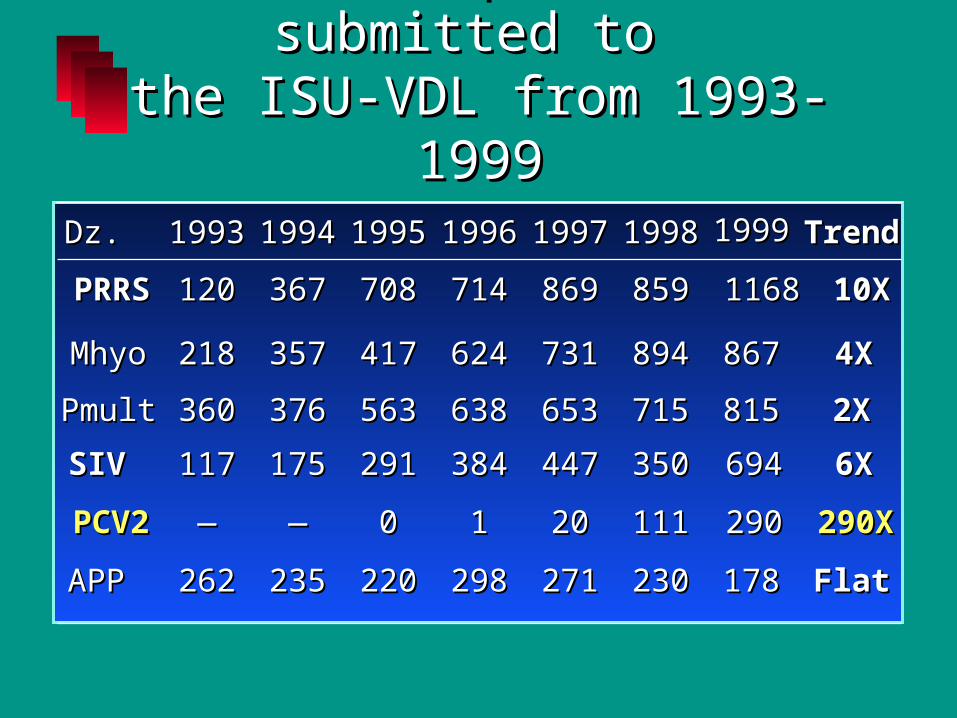
Cases of Cases of pneumoniapneumonia submitted to submitted to the ISU-VDL from 1993-1999the ISU-VDL from 1993-1999
PRRSPRRS
MhyoMhyo
PmultPmult
SIVSIV
APPAPP
PCV2PCV2
Dz.Dz.
120120
218218
360360
117117
262262
——
19931993
367367
357357
376376
175175
235235
——
19941994
708708
417417
563563
291291
220220
00
19951995
714714
624624
638638
384384
298298
11
19961996
869869
731731
653653
447447
271271
2020
19971997
10X10X
4X4X
2X2X
6X6X
FlatFlat
290X290X
TrendTrend
859859
894894
715715
350350
230230
111111
19981998
11681168
867867
815815
694694
178178
290290
19991999

PPostweaning ostweaning MMultisystemic ultisystemic WWasting asting SSyndrome (PMWS)yndrome (PMWS)
primarily in 10-20 week old pigs in the primarily in 10-20 week old pigs in the U.S.U.S.
pneumoniapneumonia wastingwasting anemia, paleness, +/- ictericanemia, paleness, +/- icteric +/- diarrhea+/- diarrhea 2-50% morbidity (usually < 10%)2-50% morbidity (usually < 10%) mortality of affected pigs may approach mortality of affected pigs may approach
100%100%

0%10%20%30%40%50%60%70%80%
Clinical Signs of PMWS

Age of Onset of PMWSAge of Onset of PMWS
-5-5
00
55
1010
1515
2020
2525
3030
3535
1 2 3 4 5 6 7 8 9 10 11 12 13 14 15 16 17 18 19 20
Age in WeeksAge in Weeks
Cas
esC
ases

Gross Lesions of PMWSGross Lesions of PMWSGross Lesions of PMWSGross Lesions of PMWS
LungsLungs• diffusely red-tan, noncollapsed, rubbery, with diffusely red-tan, noncollapsed, rubbery, with
variable amounts of cranioventral consolidationvariable amounts of cranioventral consolidation
Lymph nodesLymph nodes• diffusely enlarged and tandiffusely enlarged and tan
Other less common lesionsOther less common lesions• Icterus, gastric ulcers, skin lesions (PDNS), Icterus, gastric ulcers, skin lesions (PDNS),
enlarged kidneys, swollen or atrophied liverenlarged kidneys, swollen or atrophied liver
Gross lesions are rarely diagnostic - very Gross lesions are rarely diagnostic - very similar to PRRSsimilar to PRRS
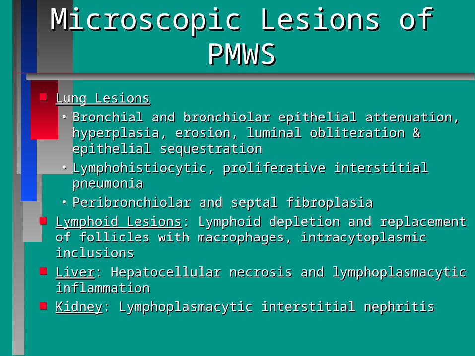
Microscopic Lesions of PMWSMicroscopic Lesions of PMWSMicroscopic Lesions of PMWSMicroscopic Lesions of PMWS Lung LesionsLung Lesions
• Bronchial and bronchiolar epithelial attenuation, Bronchial and bronchiolar epithelial attenuation, hyperplasia, erosion, luminal obliteration & epithelial hyperplasia, erosion, luminal obliteration & epithelial sequestrationsequestration
• Lymphohistiocytic, proliferative interstitial pneumoniaLymphohistiocytic, proliferative interstitial pneumonia• Peribronchiolar and septal fibroplasiaPeribronchiolar and septal fibroplasia
Lymphoid LesionsLymphoid Lesions: Lymphoid depletion and replacement : Lymphoid depletion and replacement of follicles with macrophages, intracytoplasmic inclusionsof follicles with macrophages, intracytoplasmic inclusions
LiverLiver: Hepatocellular necrosis and lymphoplasmacytic : Hepatocellular necrosis and lymphoplasmacytic inflammationinflammation
KidneyKidney: Lymphoplasmacytic interstitial nephritis: Lymphoplasmacytic interstitial nephritis
Lung LesionsLung Lesions• Bronchial and bronchiolar epithelial attenuation, Bronchial and bronchiolar epithelial attenuation,
hyperplasia, erosion, luminal obliteration & epithelial hyperplasia, erosion, luminal obliteration & epithelial sequestrationsequestration
• Lymphohistiocytic, proliferative interstitial pneumoniaLymphohistiocytic, proliferative interstitial pneumonia• Peribronchiolar and septal fibroplasiaPeribronchiolar and septal fibroplasia
Lymphoid LesionsLymphoid Lesions: Lymphoid depletion and replacement : Lymphoid depletion and replacement of follicles with macrophages, intracytoplasmic inclusionsof follicles with macrophages, intracytoplasmic inclusions
LiverLiver: Hepatocellular necrosis and lymphoplasmacytic : Hepatocellular necrosis and lymphoplasmacytic inflammationinflammation
KidneyKidney: Lymphoplasmacytic interstitial nephritis: Lymphoplasmacytic interstitial nephritis

Differential Diagnosis of Necrotizing Bronchitis and Bronchiolitis in SwineDifferential Diagnosis of Necrotizing Bronchitis and Bronchiolitis in Swine
SIV and PRCV• epithelial necrosis• epithelial attenuation• epithelial hyperplasia• mononuclear cuffing
SIV and PRCV• epithelial necrosis• epithelial attenuation• epithelial hyperplasia• mononuclear cuffing
PCV2• epithelial necrosis• epithelial attenuation• epithelial hyperplasia• mononuclear cuffing• peribronchiolar fibrosis• luminal obliteration• epithelial sequestration
PCV2• epithelial necrosis• epithelial attenuation• epithelial hyperplasia• mononuclear cuffing• peribronchiolar fibrosis• luminal obliteration• epithelial sequestration

Porcine Circovirus: DiagnosisPorcine Circovirus: Diagnosis Clinical signs and gross lesions very similar to PRRSClinical signs and gross lesions very similar to PRRS unique microscopic lesions and inclusion bodiesunique microscopic lesions and inclusion bodies immunohistochemistry (IHC), immunohistochemistry (IHC), in situin situ hybridization hybridization
(ISH) to confirm association of virus with lesion(ISH) to confirm association of virus with lesion virus isolation and serology available in some labsvirus isolation and serology available in some labs
• offered at ISU VDLoffered at ISU VDL
• ubiquitous virus so value and interpretation of ubiquitous virus so value and interpretation of serologic information is difficultserologic information is difficult
• attempt virus isolation in recurrent cases to have attempt virus isolation in recurrent cases to have virus available for potential autogenous vaccine virus available for potential autogenous vaccine productionproduction

PMWS Case ObservationsPMWS Case Observations PMWS concurrent infectionsPMWS concurrent infections
• confirm confirm PRRSVPRRSV in 60% of the cases, in 60% of the cases, – suspect PRRSV is involved in 80% of the casessuspect PRRSV is involved in 80% of the cases
• other common concurrent infectionsother common concurrent infections– SIV, HPS/SIV, HPS/S. suis, M. hyo.S. suis, M. hyo.
Mortality much higher in concurrent infection Mortality much higher in concurrent infection casescases• acute SIV + circovirus + acute SIV + circovirus + M. hyoM. hyo. pneumonia cases. pneumonia cases
– 2-10% mortality in finishers2-10% mortality in finishers• chronic PRDC due to PRRSV + M hyo + PMWSchronic PRDC due to PRRSV + M hyo + PMWS
– 10-15% mortality in finishers10-15% mortality in finishers

PMWS Case ObservationsPMWS Case Observations Duration of disease in a system variesDuration of disease in a system varies
• often a single batch problem in smaller, often a single batch problem in smaller, single-site herds that use internal gilt single-site herds that use internal gilt replacementsreplacements
• increasing number of cases where PMWS increasing number of cases where PMWS has become endemic in multiple building has become endemic in multiple building finishing sitesfinishing sites

Porcine Circovirus: PathogenesisPorcine Circovirus: Pathogenesis PMWS strains (PMWS strains (Type 2Type 2) differ from PK/15 cell ) differ from PK/15 cell
culture strains (culture strains (Type 1Type 1))• genetically types 1 and 2 are quite differentgenetically types 1 and 2 are quite different
– multiplex PCR can differentiatemultiplex PCR can differentiate• PCV1 is nonpathogenic in growing pigs (Allan et al.)PCV1 is nonpathogenic in growing pigs (Allan et al.)• evidence that “evidence that “shaker pig diseaseshaker pig disease” may be caused ” may be caused
by PCV1 or PCV2by PCV1 or PCV2 High seroprevalence of PCV2 but limited casesHigh seroprevalence of PCV2 but limited cases
• tested 27 high health herds with no clinical evidence tested 27 high health herds with no clinical evidence of PMWSof PMWS
– 100% or herds seropositive for PCV2100% or herds seropositive for PCV2– high percent of animals within a herd positivehigh percent of animals within a herd positive– passive antibody decay by 10-20 weekspassive antibody decay by 10-20 weeks

Experimental Reproduction of Experimental Reproduction of PMWSPMWS
Ellis et al., 1999Ellis et al., 1999• reproduced PMWS lesions in CDCD pigs inoculated reproduced PMWS lesions in CDCD pigs inoculated
with tissue filtrates; both PCV2 and PPV detectedwith tissue filtrates; both PCV2 and PPV detected Allan et al. 1999; and Kennedy et al. 2000Allan et al. 1999; and Kennedy et al. 2000
• inoculated colostrum-deprived pigs with PCV2 inoculated colostrum-deprived pigs with PCV2 alone, PPV alone, and PCV2+PPV; reproduced alone, PPV alone, and PCV2+PPV; reproduced severe disease and lesions typical of PMWS in severe disease and lesions typical of PMWS in dually-inoculated group whereas only mild lesions dually-inoculated group whereas only mild lesions and subclinical disease was reproduced in the and subclinical disease was reproduced in the PCV2 only groupPCV2 only group
Krakowka et al. 2000Krakowka et al. 2000• reproduced disease and PMWS lesions in reproduced disease and PMWS lesions in
gnotobiotic pigs co-infected with PCV2 and PPVgnotobiotic pigs co-infected with PCV2 and PPV

Experimental Experimental PCV2/PRRSV Co-InfectionPCV2/PRRSV Co-Infection
Experimental Experimental PCV2/PRRSV Co-InfectionPCV2/PRRSV Co-Infection
Field cases suggested interactionField cases suggested interaction Objective:Objective:
• Reproduce disease seen in field casesReproduce disease seen in field cases• Develop model to study the interactionDevelop model to study the interaction• Develop model for evaluating intervention Develop model for evaluating intervention
strategiesstrategies

Experimental DesignExperimental DesignExperimental DesignExperimental Design 4 groups of 3 week old CD/CD pigs
• Control 9 pigs• PCV2 19 pigs• PRRSV 13 pigs• PRRSV/PCV2 17 pigs
Intranasal inoculation of PRRSV and PCV2
Scheduled necropsies at 7,10,14,21,35,49 days post-inoculation
4 groups of 3 week old CD/CD pigs• Control 9 pigs• PCV2 19 pigs• PRRSV 13 pigs• PRRSV/PCV2 17 pigs
Intranasal inoculation of PRRSV and PCV2
Scheduled necropsies at 7,10,14,21,35,49 days post-inoculation

Clinical Respiratory Scores
0
1
2
3
4
5
6
7 10 11-14 15-21
Days
Control
PCV
PRRSV
PCV/PRRSV

Mean Gross Lung Scores
01020304050
60708090
100
7-10 11-14 15-21
Days
% L
un
g A
ffec
ted
Control
PCV
PRRSV
PCV/PRRSV

Summary of Findings: PCV2-Summary of Findings: PCV2-PRRSV Co-infectionPRRSV Co-infection
Summary of Findings: PCV2-Summary of Findings: PCV2-PRRSV Co-infectionPRRSV Co-infection
PCV2 GroupPCV2 Group• Minimal respiratory diseaseMinimal respiratory disease• Mortality of 40% between 10-30 DPI Mortality of 40% between 10-30 DPI • Mild necrotizing bronchiolitis, minimal Mild necrotizing bronchiolitis, minimal
pneumoniapneumonia• Lymphoid depletion, necrotizing hepatitisLymphoid depletion, necrotizing hepatitis
PRRSV groupPRRSV group• Moderate respiratory diseaseModerate respiratory disease• Mortality = 0%Mortality = 0%• Interstitial pneumonia, no airway lesionsInterstitial pneumonia, no airway lesions

Summary of Findings: Summary of Findings: PCV2-PRRSV Co-infectionPCV2-PRRSV Co-infection
Summary of Findings: Summary of Findings: PCV2-PRRSV Co-infectionPCV2-PRRSV Co-infection
PCV2/PRRSV groupPCV2/PRRSV group
• Severe respiratory diseaseSevere respiratory disease
• Mortality >90% between 10-21 DPIMortality >90% between 10-21 DPI
• Necrotizing and ulcerative Necrotizing and ulcerative bronchiolitisbronchiolitis
• Severe interstitial pneumoniaSevere interstitial pneumonia
• Lymphoid depletion, necrotizing Lymphoid depletion, necrotizing hepatitishepatitis

Experimental Evidence for PRRSV-Experimental Evidence for PRRSV-Induced Potentiation of PMWSInduced Potentiation of PMWS
Harms et al, 2000Harms et al, 2000• Reproduction of severe disease and lesions Reproduction of severe disease and lesions
typical of PMWS in CDCD pigs co-infected with typical of PMWS in CDCD pigs co-infected with PCV2 and PRRSVPCV2 and PRRSV
• Less severe disease, mortality, and lesions in Less severe disease, mortality, and lesions in PCV2 only groupPCV2 only group
• No evidence of PPV infectionNo evidence of PPV infection Allan et al., 2000Allan et al., 2000
• Experimental infection of CD piglets with PCV2 Experimental infection of CD piglets with PCV2 and PRRSV potentiates PCV2 replicationand PRRSV potentiates PCV2 replication

Activation of the Immune System May Lead to Activation of the Immune System May Lead to Increased Susceptibility to PCV2-induced PMWS?Increased Susceptibility to PCV2-induced PMWS?
Allan et al., 2000 Vet Rec; Krakowka et al., Vet Path, IN Allan et al., 2000 Vet Rec; Krakowka et al., Vet Path, IN PRESS. Activation of macrophages in lymphoid tissues PRESS. Activation of macrophages in lymphoid tissues of colostrum fed pigs may lead to potentiation of PCV2 of colostrum fed pigs may lead to potentiation of PCV2 replication and development of clinical disease and replication and development of clinical disease and lesions typical of PMWSlesions typical of PMWS
– 7/7 colostrum fed pigs inoculated with PCV2 and then 7/7 colostrum fed pigs inoculated with PCV2 and then immunized with keyhole limpet haemocyanin (in Freund’s immunized with keyhole limpet haemocyanin (in Freund’s incomplete adjuvant) developed clinical disease and lesions incomplete adjuvant) developed clinical disease and lesions typical of PMWS whereas subclinical disease and minimal typical of PMWS whereas subclinical disease and minimal lesions were reproduced in PCV2-inoculated pigs without lesions were reproduced in PCV2-inoculated pigs without immunizationimmunization
– 3/14 pigs vaccinated with M. hyo (2 dose) and APP (1 dose) 3/14 pigs vaccinated with M. hyo (2 dose) and APP (1 dose) developed PMWS whereas 0/14 unvaccinated pigs developed PMWS whereas 0/14 unvaccinated pigs developed PMWS developed PMWS

Control of PMWSControl of PMWS Confirm PMWS/circovirus by necropsy, histopath, IHC, Confirm PMWS/circovirus by necropsy, histopath, IHC,
ISHISH Identify farm, site, or system specific concurrent Identify farm, site, or system specific concurrent
infectionsinfections• eliminate PRRSV from the mix if possibleeliminate PRRSV from the mix if possible
– work to avoid simultaneous coinfectionwork to avoid simultaneous coinfection– focus on breeding herd stabilization and pig flowfocus on breeding herd stabilization and pig flow
• eliminate/control SIV with vaccinationeliminate/control SIV with vaccination• further explore role of PPV and use of PPV vaccinefurther explore role of PPV and use of PPV vaccine• minimize effects of minimize effects of M. hyoM. hyo. with vaccination and . with vaccination and
medicationmedication• treat bacterial coinfectionstreat bacterial coinfections• anti-inflammatories (aspirin, ketoprofen) usefulanti-inflammatories (aspirin, ketoprofen) useful

PMWS Summary and ConclusionsPMWS Summary and Conclusions
PMWS has become a major disease in PMWS has become a major disease in the U.S.the U.S.
Field and experimental evidence Field and experimental evidence supports a major role for PCV2 in supports a major role for PCV2 in PMWSPMWS
Field evidence also supports a role for Field evidence also supports a role for PCV2 in porcine respiratory disease PCV2 in porcine respiratory disease complexcomplex