translation termination depends on the sequential
TRANSCRIPT
4798–4813 Nucleic Acids Research, 2019, Vol. 47, No. 9 Published online 15 March 2019doi: 10.1093/nar/gkz177
Translation termination depends on the sequentialribosomal entry of eRF1 and eRF3Christian Beißel1, Bettina Neumann1, Simon Uhse1, Irene Hampe1, Prajwal Karki2 andHeike Krebber1,*
1Abteilung fur Molekulare Genetik, Institut fur Mikrobiologie und Genetik, Gottinger Zentrum fur MolekulareBiowissenschaften (GZMB), Georg-August Universitat Gottingen, Germany and 2Department of PhysicalBiochemistry, Max Planck Institute for Biophysical Chemistry, Gottingen, Germany
Received January 29, 2019; Revised February 28, 2019; Editorial Decision March 01, 2019; Accepted March 08, 2019
ABSTRACT
Translation termination requires eRF1 and eRF3 forpolypeptide- and tRNA-release on stop codons. Addi-tionally, Dbp5/DDX19 and Rli1/ABCE1 are required;however, their function in this process is currentlyunknown. Using a combination of in vivo and in vitroexperiments, we show that they regulate a stepwiseassembly of the termination complex. Rli1 and eRF3-GDP associate with the ribosome first. Subsequently,Dbp5-ATP delivers eRF1 to the stop codon and in thisway prevents a premature access of eRF3. Dbp5 dis-sociates upon placing eRF1 through ATP-hydrolysis.This in turn enables eRF1 to contact eRF3, as thebinding of Dbp5 and eRF3 to eRF1 is mutually ex-clusive. Defects in the Dbp5-guided eRF1 deliverylead to premature contact and premature dissoci-ation of eRF1 and eRF3 from the ribosome and tosubsequent stop codon readthrough. Thus, the step-wise Dbp5-controlled termination complex assem-bly is essential for regular translation terminationevents. Our data furthermore suggest a possible roleof Dbp5/DDX19 in alternative translation terminationevents, such as during stress response or in develop-mental processes, which classifies the helicase as apotential drug target for nonsense suppression ther-apy to treat cancer and neurodegenerative diseases.
INTRODUCTION
When a ribosome arrives at a stop codon on the mRNA,protein synthesis is terminated and the peptide is released(1). In eukaryotes, two essential release factors are wellknown to mediate translation termination. The eukaryoticrelease factor 1 (eRF1), in Saccharomyces cerevisiae en-coded by SUP45, is the only class I termination factor ineukaryotes that recognizes all three different stop codons
(UAG, UAA, UGA) and subsequently mediates the hy-drolysis of the peptidyl-tRNA in the ribosomal peptidyl-transferase center (PTC). In addition, the class II eukary-otic release factor 3 (eRF3), in S. cerevisiae encoded bySUP35, enhances translation termination efficiency with itsGTPase activity (2,3). Most termination models (Supple-mentary Figure S1A) anticipate that eRF1 and eRF3–GTPenter the ribosome together as a ternary complex once thestop codon is reached (1,2,4), as both factors strongly in-teract with each other via their C-terminal domains (5,6).Supposedly, successful stop codon recognition by eRF1 in-duces GTP-hydrolysis of eRF3, which in turn leads to aconformational rearrangement in eRF1 resulting in its ac-tive form, which positions its GGQ-motif in the PTC andmediates hydrolysis of the ester bond of the peptidyl-tRNA(7–9).
In light of these mostly in vitro studies, nothing seems tobe missing; however, novel factors essential for translationtermination in vivo were discovered and need to be incorpo-rated into a comprehensive model: The DEAD-box RNAhelicase Dbp5, encoded by RAT8 (human DDX19) (10),its stimulating co-factors Gle1 plus inositol hexakisphos-phate IP6 (11,12), the iron-sulfur containing ATP-bindingcassette protein Rli1 (human ABCE1) (13,14) and the initi-ation factor eIF3, including Hcr1 (15). Dbp5 and Gle1 arewell known for their function in mRNA-export through nu-clear pore complexes (NPCs) (16). Using its regulated AT-Pase cycle, Dbp5 remodels RNA–protein complexes at thecytoplasmic side of the NPC on emerging mRNAs (17).By dissociation of the export receptor Mex67-Mtr2 (humanTAP-p15) from the arriving mRNAs, its backsliding is pre-vented and directionality of the transport event established.Its co-factors Gle1 and IP6 stimulate ATP-hydrolysis lead-ing to RNP-release and binding of Dbp5-ADP to the NPC-protein Nup159 (human Nup214). Importantly, this bind-ing leads to ADP-release, a conformational change and thebinding of ATP (16,17). The ATPase activity of Dbp5 isalso essential for efficient translation termination (10,12).
*To whom correspondence should be addressed. Tel: +49 551 3933801; Fax: +49 551 3933804; Email: [email protected] address: Simon Uhse, Gregor Mendel Institute of Molecular Plant Biology, Austrian Academy of Sciences, Vienna Biocenter Campus (VBC), Vienna, Austria.
C© The Author(s) 2019. Published by Oxford University Press on behalf of Nucleic Acids Research.This is an Open Access article distributed under the terms of the Creative Commons Attribution License (http://creativecommons.org/licenses/by/4.0/), whichpermits unrestricted reuse, distribution, and reproduction in any medium, provided the original work is properly cited.
Dow
nloaded from https://academ
ic.oup.com/nar/article-abstract/47/9/4798/5381069 by M
PI Biophysical Chem
istry user on 17 July 2019
Nucleic Acids Research, 2019, Vol. 47, No. 9 4799
In addition to these functions, Dbp5 plays also a role in theexport of both ribosomal subunits (18). However, in con-trast, to mRNA export and translation termination, Dbp5acts independently of its ATPase activity in ribosome export(18).
Rli1 functions in biogenesis and nuclear export of pre-ribosomal subunits (19–21), translation initiation (22), ter-mination (13) and in particular in ribosome recycling (23).Rli1 is a soluble member of the ATP-binding cassette (ABC)protein superfamily that contains two nucleotide-bindingdomains (NBDs) and two N-terminal iron-sulfur clusters.A hinge domain connects both NBDs forming a cleft,which is open in the ADP-bound state, while ATP-bindinginduces its closure with a concomitant movement of theiron-sulfur domain allowing ATP-hydrolysis. This ATP-dependent tweezers-like motion converts chemical energyinto mechanical power, which is important for splitting theribosome into its ribosomal subunits (24). The protein ishighly conserved in eukaryotes and essential in all organ-isms tested (22). Interestingly, Rli1 acts ATP-hydrolysis in-dependent during translation termination (13,25). It wassuggested that Rli1 associates with the termination complexupon dissociation of eRF3–GDP, taking over its position tokeep eRF1 in its favourable position to facilitate peptidyl-tRNA hydrolysis (4,26).
The initiation factor eIF3 has recently been associatedwith translation termination, because mutations in its sub-units reduce the rate of stop codon readthrough (15). In-terestingly, deletion of the substoichiometric componentHCR1 shows an increased readthrough activity and thisphenotype was suppressed by high copy RLI1. A model wasproposed in which Hcr1 is not a bona fide translation ini-tiation factor, but rather acts in termination by promotingGDP–eRF3 ejection from the ribosomes (15).
So far, no translation termination model is available thatincludes all of these factors that support termination andmany results were obtained from in vitro assays with pu-rified components. Therefore, we analysed the process inS. cerevisiae in vivo and in vitro with all participating fac-tors and uncovered a sequential recruitment mechanism, inwhich Rli1 and eRF3 wait at the ribosome for the entry ofDbp5 that delivers eRF1 and at the same time shields it frompremature access of eRF3. Upon proper positioning Dbp5dissociates, allowing eRF3 to contact and stimulate eRF1activity. This stepwise entry of the termination factors andin particular the Dbp5 controlled eRF1–eRF3 interaction,prevents premature and inefficient translation termination.
MATERIALS AND METHODS
Yeast strains and plasmids
All S. cerevisiae strains, plasmids and oligonucleotides usedin this study are listed in the Expanded View SupplementaryTables S1, S2 and S3, respectively. For growth analyses, cellswere spotted in 10-fold serial dilutions onto selective agarplates and grown for 3 days at the indicated temperatures.
The strains, HKY1622 and HKY1623, were gener-ated by crossing HKY1271 (RLI1-GFP) and HKY446(sup45-2). Crossing the strains HKY445 (WT of HKY446)or HKY446 with the strain HKY1122 (Prt1-GFP) pro-duced the strain HKY1915 and the corresponding WT
HKY1914. The strain HKY1921 was generated by crossingthe strains HKY477 (rat8-2-myc) and HKY1907 (trp5Δ)and exchange of pHK629 with pHK693. For the genera-tion of plasmid pHK1292, the GFP ORF was amplifiedby polymerase chain reaction using the primers HK1194and HK1195 and inserted via XhoI and PstI sites intopHK887 (2μ Rli1-HA LEU2) replacing the HA-tag. To cre-ate pHK1474 and pHK1475, the RLI1-GFP ORF with pro-moter and terminator was amplified from pHK1292 withthe primers HK2136 and HK2137 and inserted via Gib-son assembly reaction into pHK86 and pHK87, respec-tively, which were linearized by SacI and SalI digestion.The GLE1 ORF was amplified from gDNA with HK1398and HK1399 and inserted via BamHI site into pHK825(CEN PADH13xMYC URA3) to generate pHK1323. Forthe generation of pHK1283, the SUP35 ORF was ampli-fied with the primers HK1109 and HK1110 and insertedvia EcoRI and XhoI sites into pGEX-4T-1. The SUP45ORF was amplified with the primers HK1144 and HK1156and inserted via NdeI and XhoI sites into pET28a to cre-ate pHK1280. The SUP45Δ1237-1311 ORF was amplifiedwith the primers HK1146 and HK1147 and inserted viaBamHI and XhoI sites into pET15b to generate pHK1278.To generate pHK1394, the GLE1 ORF was amplified withthe primers HK1613 and HK1614 and inserted via Gibsonassembly reaction into pETMBP1 1a that was linearized bySacI and NcoI digestion.
Co-immunoprecipitation experiments
In vivo interactions studies were carried out following theprotocols published previously (18,27). For immunoprecip-itation of GFP-tagged proteins, 10 �l slurry of GFP-Trap Abeads (Chromotek) and for TAP-tagged proteins, 20 �lslurry of IgG-Sepharaose beads (GE Healthcare) were usedper reaction and incubated with 200 (high abundant pro-teins) and up to 2000 �l (for low abundant proteins) of theclarified lysate for 3 h rotating at 4◦C. If indicated, the sam-ples were treated with 0.2 mg/ml RNase A (AppliChem) foradditional 30 min at 4◦C. Finally, the eluted proteins wereseparated on 10% SDS-polyacrylamide gels and analysedby western blotting.
Sucrose-density gradient fractionation
The experiments were essentially performed as describedpreviously (18) with the following modifications. For elon-gation factor mutants, no cycloheximide treatment was per-formed meaning that cells were directly harvested upon 1 htemperature shift, cycloheximide was omitted from the ly-sis buffer. For protein analyses, 15 OD260nm units of lysateswere loaded onto the top of linear 7–47% (w/v) sucrose gra-dients and centrifuged for 2 h and 40 min at 40 000 rpm and4◦C in a TH-641 rotor and Sorvall WX80 ultracentrifuge(Thermo Scientific). After gradient fractionation, proteinfractions were precipitated with 10% trichloroacetic acid,washed twice with 80% aceton and subjected to sodiumdodecylsulphate-polyacrylamide gel electrophoresis (SDS-PAGE) and western blotting.
Dow
nloaded from https://academ
ic.oup.com/nar/article-abstract/47/9/4798/5381069 by M
PI Biophysical Chem
istry user on 17 July 2019
4800 Nucleic Acids Research, 2019, Vol. 47, No. 9
Western blot analyses and quantification
Polyclonal rabbit antibodies against Dbp5 (dilution1:1000), eRF1 and eRF3 (kindly provided by D. Bedwell,dilution for both 1:1000), uL29 = Rpl35 and uS3 =Rps3 (kindly provided by M. Seedorf, dilution 1:5000and 1:10 000 respectively, or anti-Rps3 peptide antibody1:500), Aco1, Hem15, Por1 and Zwf1 (kindly providedby R. Lill, dilution 1:1000, 1:7000, 1:2000, and 1:4000,respectively), Asc1 (kindly provided by G. Braus, dilution1:2000) and Cdc28 (sc-28550; Santa Cruz, dilution 1:2000)were used. GFP-tagged proteins were detected with anti-GFP antibodies (sc-8334; Santa Cruz, dilution 1:1000),or GF28R (Pierce Protein Biology, 1:5000), or (ab183734;Abcam; 1:10 000), MYC-tagged proteins with an anti-MYC antibody (sc-789; Santa Cruz, dilution 1:750) andHA-tagged proteins with an anti-HA antibody (sc-57592and sc-7392; Santa Cruz, dilution 1:750) and GST-taggedproteins with an anti-GST antibody (sc-138; Santa Cruz,dilution 1:2000). Secondary anti-rabbit IgG (H+L)-HRPOand anti-mouse IgG (H+L)-HRPO (Dianova) antibodieswere used and detected with Amersham ECL PrimeWestern Blotting Detection Reagent (GE Healthcare)or WesternBright Chemilumineszenz Substrat Quantum(Biozym) and the FUSION-SL chemiluminescence detec-tion system (Peqlab). Quantification of western blot signalswas performed with the Bio1D software (Peqlab) or withImageStudio Lite (Li-COR Biosciences). For statisticalanalyses of co-immunoprecipitation studies, the intensity ofco-precipitated bands was related to that of the pull-downand finally, the ratio of the mutant or treated strains wascompared to the wild typical ratio. For quantification ofthe sucrose density gradient fractionation experiments, theintensity of each fraction was measured and the polysomalfractions were compared to the sum of the 80S, 60S, 40Sand non-ribosomal fractions.
In vitro binding studies
GST-Dbp5, GST-eRF1 and GST-eRF3�N65, a more sta-ble version of eRF3 that has a truncated C-terminus,were expressed in Escherichia coli Rosetta 2 cells, purifiedby affinity chromatography with GSTrap 4B GlutathioneSepharose (GE Healthcare) and stored at −80◦C in elutionbuffer (for GST-Dbp5: 50 mM Tris pH 7.5, 150 mM NaCl,30 mM Glutathione reduced) (for GST-eRF1 and GST-eRF3: 20 mM HEPES pH 7.5, 500 mM NaCl, 5% glycerol,4 mM �-MeOH, 30 mM Glutathione reduced). To obtainuntagged Dbp5, eRF1 and eRF3, the purified GST-taggedproteins were cleaved with PreScission protease overnightat 4◦C. Afterwards, GST was removed by performing a sec-ond affinity chromatography with GSTrap 4B GlutathioneSepharose (GE Healthcare). The proteins were further pu-rified by running the collected flow through over a gel filtra-tion chromatography. Purified GST–Dbp5 and Dbp5 werestored in 50 mM Tris pH 7.5, 150 mM NaCl, 10% glycerol at−80◦C, whereas GST-eRF1, eRF1, GST- eRF3 and eRF3were stored in 50 mM NaCl pH 7.5, 20 mM HEPES, 5%glycerol and 4 mM �-MeOH at 80◦C.
For binding studies either purified proteins as indicatedabove were used or Rosetta II (DE3) cells were trans-formed with pET15b-HIS6-SUP45 delta 25 (eRF1 lacking
25aa at the C-terminus), pET28a-HIS6-SUP45, pGEX4T1-GST-SUP35, pGEX-6P-1-GST-RLI1 and pGEX6P1-GST.Overexpression was induced by growing the cells for 3 daysin auto-inducing media (LB media with 0.5% (v/v) glycerol,0.05% (v/v) Ggucose and 0.2% (v/v) lactose plus 25 mMK2HPO4, 25 mM NaH2PO4, 50 mM NH4Cl and 0.5 mMNa2SO4) at 16◦C. Cells were harvested and resuspended inlysis buffer (50 mM Tris–HCl, pH 7.5, 150 mM NaCl, 2 mMMgCl, 5% (v/v) glycerol, 1 mM Dithiothreitol (DTT), 0.2%(v/v) NP-40 and ethylenediaminetetraacetic acid (EDTA)-free protease inhibitor mix (Roche)). After cell lysis by soni-cation and centrifugation, the supernatant was used for fur-ther analysis. The used GSTrap 4B Glutathione Sepharose(GE Healthcare) beads were pre-incubated with 3% Albu-min Fraction V (Roche) and mixed with lysis buffer. Next,the GST tagged proteins or only GST were incubated with15 �l slurry of GSTrap for 2 h at 4◦C. After several washingsteps with buffer (excluding protease inhibitor), the beadswere incubated with the protein lysates for additional 2 hat 4◦C. For the competition assay, the eRF1 lysate was firstincubated with GST-Dbp5 bound to the GSTrap for 20 minat 4◦C, before adding purified eRF3�N65 in the indicatedamounts and further incubation for 1 h at 4◦C. After fivewashing steps with lysis buffer, the proteins that were boundto the beads were analysed by SDS-PAGE and western blot.
Prt1-GFP and Nip1-GFP were purified from yeast cellsbefore the in vitro binding assay was carried out with recom-binantly expressed GST-Rli1.
Readthrough assay
The dual reporter �-galactosidase luciferase assay was basi-cally performed as described previously (10,28). Briefly, allanalysed yeast strains were transformed with the reporterplasmids pHK607 or pHK608, respectively. Yeast cells weregrown at 25◦C to mid-log phase, shifted for 30 min to 37◦Cand afterward divided: 20 OD600 were used for the luciferaseassay and 50 ml that were split for triplicates for the �-galactosidase assay,
The luciferase assay was performed with the ‘Beetle-JuiceLuciferase Assay Firefly’ kit (p.j.k GmbH) according to themanufacturer’s protocol. For that, cell pellets were lysedwith 300 �l glass beads in a FastPrep-24 machine (MPBiomedical) in 400 �l lysis buffer (77 mM K2HPO4, 23mM KH2PO4, 0.2% Triton X-100, 1 mM DTT) supple-mented with Complete, EDTA-free protease inhibitor cock-tail (Roche). The lysates were centrifuged twice for 5 min at21 000 × g and 4◦C. Afterwards, 20 �l of the cleared lysatewas transferred in triplicate into a white 96-well plate, sup-plemented with always 50 �l substrate and measured withthe luminometer (Victor X3 2030 multilabel reader fromPerkin Elmer).
For the �-galactosidase assay, all cell pellets were resus-pended in 300 �l of Z-buffer (100 mM phosphate bufferpH 7.0, 10 mM KCl, 1 mM MgSO4) and lysed with thefreeze-and-thaw method (1 min in liquid nitrogen, 2 min in37◦C water bath, four times repeated). Afterwards, 700 �lof Z-buffer with 1 mM DTT and 160 �l of the substrateONPG (4 mg/ml of o-Nitrophenyl-�-D-galactopyranosidin Z-buffer) were added and incubated at 30◦C until colourchanges into yellow. The reaction was stopped by the addi-
Dow
nloaded from https://academ
ic.oup.com/nar/article-abstract/47/9/4798/5381069 by M
PI Biophysical Chem
istry user on 17 July 2019
Nucleic Acids Research, 2019, Vol. 47, No. 9 4801
tion of 1 M Na2CO3 and the OD420nm was measured witha spectrophotometer (UV-1601 from Shimadzu). The rela-tive readthrough activity was calculated from the ratio of lu-ciferase to �-galactosidase activity measured with the stopcodon-containing reporter related to the ratio from the in-frame control.
Statistical analysis
Quantification was performed for at least three independentexperiments. Quantification of co-immunoprecipitation ex-periments shown in Figures 1D, 2C, E, 3C, E, 4B, C, E, G6D, G and I were analysed for significance by Student’s two-tailed, two-sample, unequal variance t-test. In all cases, eachdataset was normalized and only compared to wild-type.The mean ± standard deviations are displayed. The samewas done for Figures 1G, 2H and 6B, with the exceptionthat the differences were analysed by a Student’s one-tailed,two-sample, unequal variance t-test. In Figures 1G and 6B,the signal intensity of the analysed proteins in the polysomalfraction of the tef2-9 strain was subtracted from the wild-type signals in the polysoms of the respective protein andnormalized to the Asc1/uS3 protein content that reflectedthe amount of polysomes. All data were finally analysed forsignificance by Student’s two tailed, two-sample, unequalvariance t-test. Significance P-values below 0.05 were indi-cated by asterisks (*P < 0.05; **P < 0.01; ***P < 0.001).
RESULTS
Binding of Dbp5, but not Rli1, to the termination complexdepends on eRF1
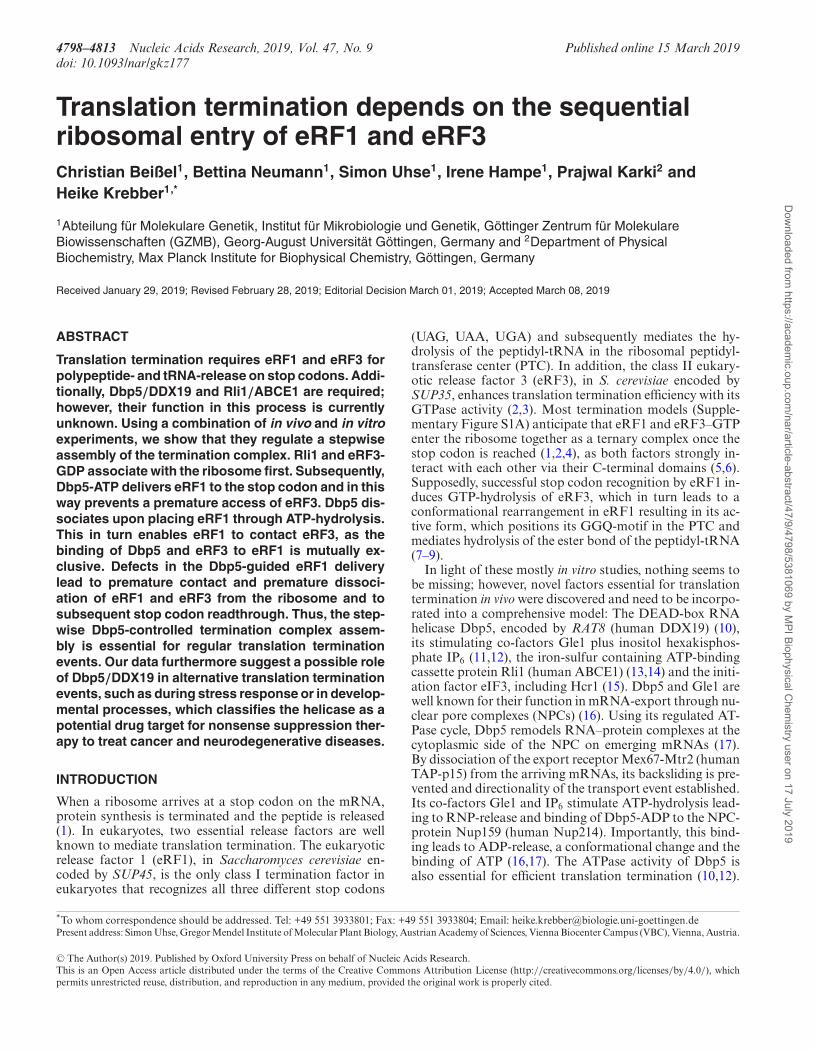
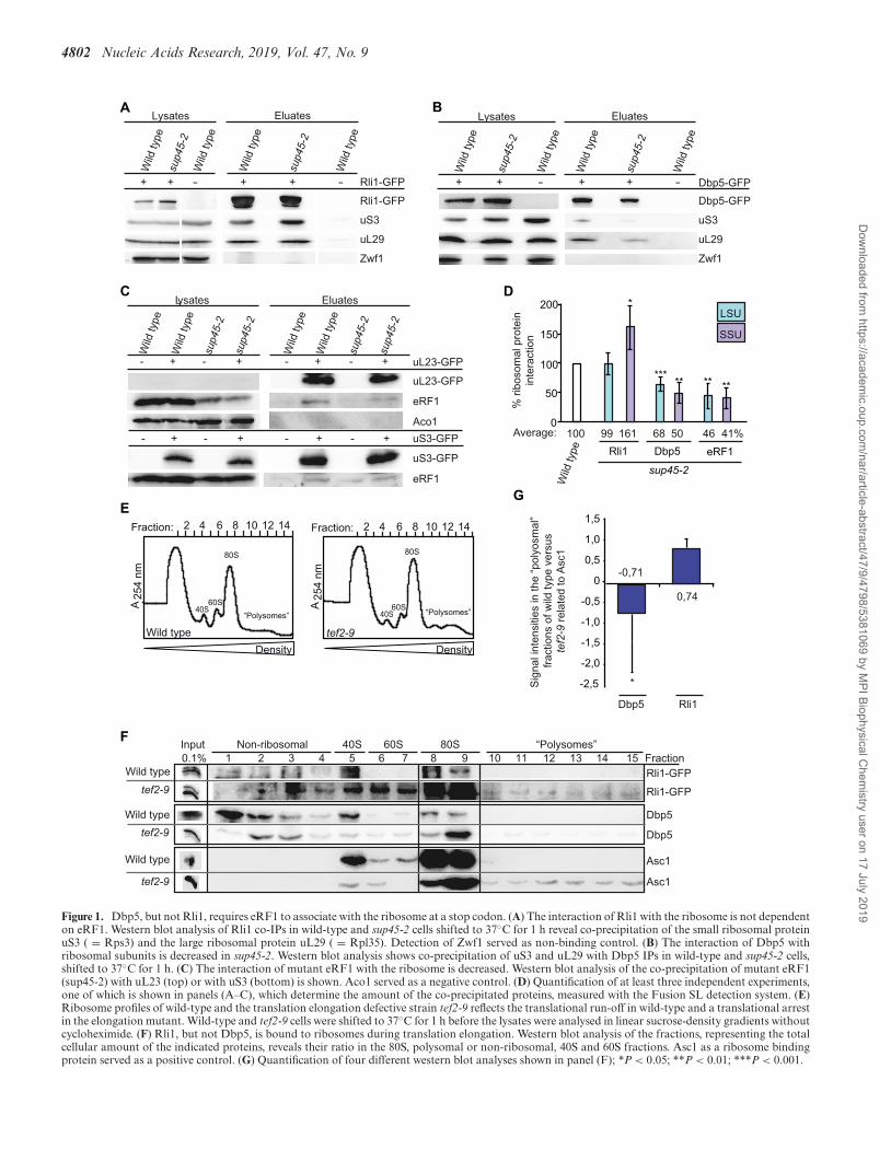
Previous studies of the translation termination process havenot revealed, how and when Rli1 and Dbp5 are recruitedto the terminating ribosome. Crystal structure and in vitroanalyses with purified proteins proposed that Rli1 andeRF3 bind to the same position on the terminating ribo-some so that their association seemed to be mutually ex-clusive and it was suggested that Rli1 might take over thebinding site of eRF3 after its dissociation to catalyse thesubsequent ribosome recycling (4,29) (Supplementary Fig-ure S1B). However, a potentially earlier association of Rli1with the ribosome before ribosome recycling has not beeninvestigated. The same is unclear for Dbp5. The helicasemight already be associated with elongating ribosomes or itcould be recruited at a later step together with eRF1 uponarrival of the ribosome at the stop codon. To address thesequestions, we analysed the ribosomal association of Rli1and Dbp5 in the temperature-sensitive eRF1 mutant sup45-2 that is defective in translation termination and in ribo-some binding. Upon a temperature shift to 37◦C, ribosomebinding of the mutated eRF1 protein sup45-2 is disturbed,resulting in stop codon recognition defects and subsequentreadthrough activity in which near-cognate tRNAs are in-corporated at the termination codon and translation elon-gation continues to the next stop codon (30). If Rli1 wouldenter the termination process after eRF1 has entered, onewould expect to see a reduced binding of Rli1 to ribosomesin that mutant. However, co-immunoprecipitation experi-ments (co-IPs) show that the binding of Rli1 to the largeribosomal protein uL29 (yeast Rpl35) is unchanged and
its association with the small ribosomal protein uS3 (yeastRps3) is even increased in sup45-2 cells (Figure 1A and D).These results indicate that Rli1 binds ribosomal particleswithout functional eRF1 and that it is possibly associatedwith ribosomes before eRF1 enters. The increased bindingof Rli1 to the 40S subunit in sup45-2 might represent an en-hanced presence of 43S pre-initiation complexes, which arestabilized by Rli1 (22). In contrast to Rli1, the interactionof Dbp5 and the mutated eRF1 protein with both riboso-mal proteins is reduced in sup45-2 (Figure 1B–D) suggestingthat Dbp5 requires functional eRF1 for its association withterminating ribosomes. This reduced ribosomal associationmight be due to the fact that mutant eRF1 is rather unstableand only ∼50% of the protein amount is detectable in lysateson western blots (Figure 1C). Thus, while the association ofRli1 with the ribosome rather increases in sup45-2 cells, theassociation of Dbp5 and mutant eRF1 decreases. These re-sults might indicate that Rli1 associates with the ribosomebefore Dbp5 and eRF1.
To verify this sequential recruitment, we analysedwhether Rli1, but not Dbp5 associates with ribosomes ar-rested in translation elongation and found that this is indeedthe case (Figure 1E–G). Mutations in eEF1A (such as intef2-9) lead to defects in translation elongation (31), whichis reflected in polysomal profiles of sucrose-density gradi-ent fractionations that were prepared without the usual ad-dition of cycloheximide. Under such conditions, wild-typecells continue elongation, leading to a complete polysomerun-off, while elongation factor mutants stall ribosomesduring elongation on the mRNA, thereby preventing theirarrival at the stop codon (Figure 1E) (32). Western blotanalyses of the corresponding protein fractions show thathigh amounts of Rli1 are present in the mono- and polyso-mal factions in tef2-9 cells, similar to the ribosome-boundprotein Asc1 (Figure 1G). This finding is in agreement withstructural analyses of the human homolog of Rli1, ABCE1at the ribosome, which show that the protein binds to theintersubunit space of the ribosome where aEF1 also asso-ciates (29), suggesting that their binding is mutually exclu-sive. However, as stalled ribosomes have free A-sites whenthe elongation factor is inactivated as in the tef2-9 mutant,different factors can stochastically go there, among themRli1. These findings suggest that Rli1 can bind to the ribo-some as soon as the A-site is free and thus might be the firsttermination factor that enters the ribosome, which is clearlyearlier than anticipated. In contrast to that, Dbp5 is almostabsent in the polysome fractions of tef2-9 cells (Figure 1Fand G), indicating that Dbp5 is recruited to ribosomes onlyafter translation elongation. Thus, our results suggest thatthe ribosomal association of Dbp5 not only requires a freeA-site like this is the case for Rli1, because in contrast toRli1 Dbp5 is not associated with ribosomes in an elonga-tion mutant, but Dbp5 recruitment also seems to dependon a stop codon and functional eRF1.
Dbp5 and Rli1 interact with each other during translation ter-mination
Although both Rli1 and Dbp5 were identified as transla-tion termination factors (10,13), it is unclear whether theyinteract with each other. To answer that question, we co-
Dow
nloaded from https://academ
ic.oup.com/nar/article-abstract/47/9/4798/5381069 by M
PI Biophysical Chem
istry user on 17 July 2019
4802 Nucleic Acids Research, 2019, Vol. 47, No. 9
A B
C
E
D
G
F
Figure 1. Dbp5, but not Rli1, requires eRF1 to associate with the ribosome at a stop codon. (A) The interaction of Rli1 with the ribosome is not dependenton eRF1. Western blot analysis of Rli1 co-IPs in wild-type and sup45-2 cells shifted to 37◦C for 1 h reveal co-precipitation of the small ribosomal proteinuS3 ( = Rps3) and the large ribosomal protein uL29 ( = Rpl35). Detection of Zwf1 served as non-binding control. (B) The interaction of Dbp5 withribosomal subunits is decreased in sup45-2. Western blot analysis shows co-precipitation of uS3 and uL29 with Dbp5 IPs in wild-type and sup45-2 cells,shifted to 37◦C for 1 h. (C) The interaction of mutant eRF1 with the ribosome is decreased. Western blot analysis of the co-precipitation of mutant eRF1(sup45-2) with uL23 (top) or with uS3 (bottom) is shown. Aco1 served as a negative control. (D) Quantification of at least three independent experiments,one of which is shown in panels (A–C), which determine the amount of the co-precipitated proteins, measured with the Fusion SL detection system. (E)Ribosome profiles of wild-type and the translation elongation defective strain tef2-9 reflects the translational run-off in wild-type and a translational arrestin the elongation mutant. Wild-type and tef2-9 cells were shifted to 37◦C for 1 h before the lysates were analysed in linear sucrose-density gradients withoutcycloheximide. (F) Rli1, but not Dbp5, is bound to ribosomes during translation elongation. Western blot analysis of the fractions, representing the totalcellular amount of the indicated proteins, reveals their ratio in the 80S, polysomal or non-ribosomal, 40S and 60S fractions. Asc1 as a ribosome bindingprotein served as a positive control. (G) Quantification of four different western blot analyses shown in panel (F); *P < 0.05; **P < 0.01; ***P < 0.001.
Dow
nloaded from https://academ
ic.oup.com/nar/article-abstract/47/9/4798/5381069 by M
PI Biophysical Chem
istry user on 17 July 2019

Nucleic Acids Research, 2019, Vol. 47, No. 9 4803
D E
GH
F
CBA
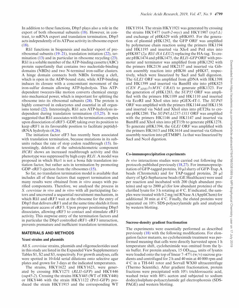
Figure 2. Rli1 supports the recruitment of Dbp5 and eRF1 to the ribosome. (A) Dbp5 interacts RNA-independently with Rli1. Immunoprecipitation ofTAP-Dbp5 in the presence of RNase A shows co-precipitation of Rli1-HA in western blot analysis. Detection of eRF1 served as positive and of Por1 asnegative control. (B) The interaction between Rli1 and Dbp5 is decreased in sup45-2, shifted to 37◦C for 1 h. Western blot analyses of Rli1-IPs revealless co-precipitation of Dbp5, but no reduction of the ribosomal protein uS3 in sup45-2 compared to wild-type. Cdc28 served as negative control. (C)Quantification of four different experiments shown in panel (B). (D) Inhibition of translation elongation leads to a reduced interaction between Rli1and Dbp5. Western blot analyses of co-IPs of Dbp5 with Rli1 upon treatment with 0.5 mg/ml cycloheximide (CHX) for 30 min are shown. Hem15 wasdetected as non-binding control. (E) Quantification of three different experiments shown in panel (D). (F) Overexpression of RLI1 partially rescues thegrowth defects of sup45-2, while the wild-type growth is not influenced. Serial dilutions of the indicated strains are shown upon growth on selective platesfor 3 days at 35◦C. (G) Overexpression of RLI1 suppresses the binding defect of eRF1 to the ribosome in sup45-2 cells. Co-IPs of eRF1 with uS3-GFP areshown in the indicated strains with or without high copy (HC) RLI1. (H) Quantification of four different experiments shown in panel (G); *P < 0.05; **P< 0.01; ***P < 0.001.
precipitated Rli1 with Dbp5 in vivo (Figure 2A) and viceversa Dbp5 as well as its co-factor Gle1 with Rli1 (Supple-mentary Figure S1B) unrevealing a physical interaction be-tween Dbp5 and Rli1 that might be direct or mediated byother proteins. To verify that this interaction actually occursduring translation termination and not for instance dur-ing pre-ribosomal subunit export from the nucleus in whichRli1 and Dbp5 are both involved (18,19,21), we comparedtheir interaction in sup45-2 cells in which the terminationprocess is inhibited and Dbp5 does not enter the ribosome.
Our results show that in sup45-2 the interaction of Rli1 andDbp5 is indeed reduced to more than half, while its inter-action to the ribosome is not decreased (Figure 2B and C).Moreover, we treated wild-type cells with the antibiotic cy-cloheximide, which inhibits translation elongation and thus,prevents ribosomes from arriving at stop codons, reflectedin the ribosomal profiles. Also in this case the prevention oftranslation termination leads to a significantly decreased as-sociation of Rli1 and Dbp5 (Figure 2D and E). These resultssuggest that an interaction of Dbp5 and Rli1 takes place
Dow
nloaded from https://academ
ic.oup.com/nar/article-abstract/47/9/4798/5381069 by M
PI Biophysical Chem
istry user on 17 July 2019
4804 Nucleic Acids Research, 2019, Vol. 47, No. 9
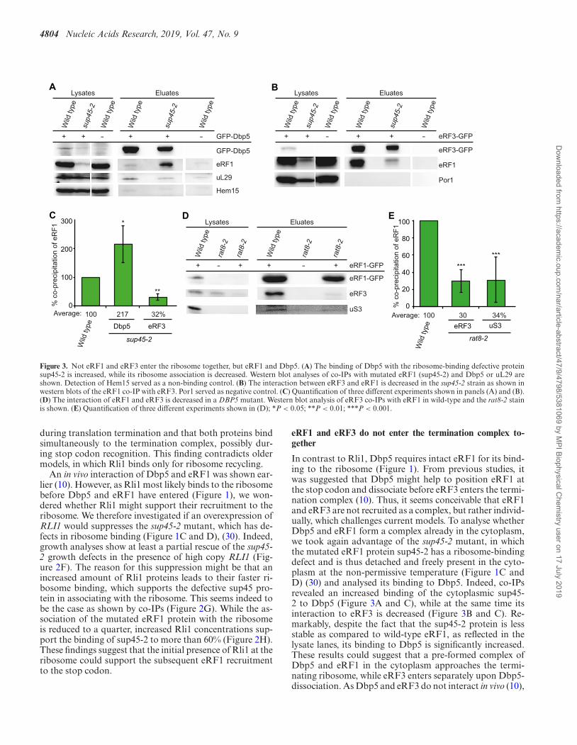
A B
C D E
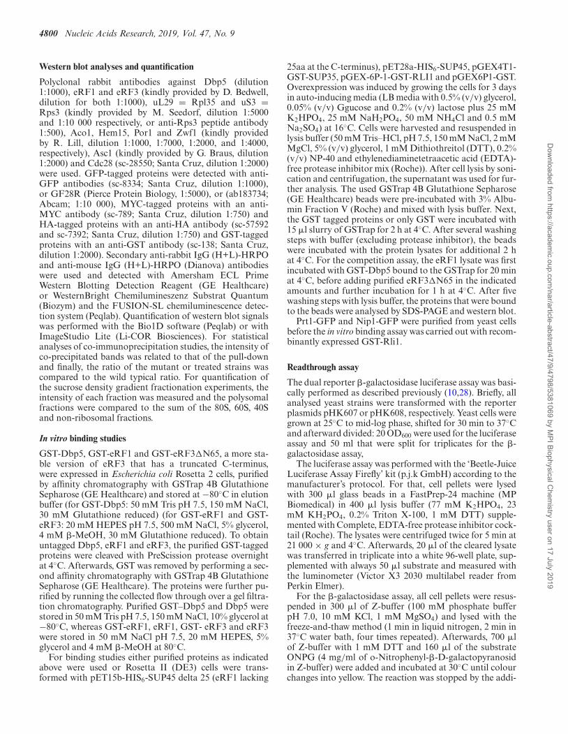
Figure 3. Not eRF1 and eRF3 enter the ribosome together, but eRF1 and Dbp5. (A) The binding of Dbp5 with the ribosome-binding defective proteinsup45-2 is increased, while its ribosome association is decreased. Western blot analyses of co-IPs with mutated eRF1 (sup45-2) and Dbp5 or uL29 areshown. Detection of Hem15 served as a non-binding control. (B) The interaction between eRF3 and eRF1 is decreased in the sup45-2 strain as shown inwestern blots of the eRF1 co-IP with eRF3. Por1 served as negative control. (C) Quantification of three different experiments shown in panels (A) and (B).(D) The interaction of eRF1 and eRF3 is decreased in a DBP5 mutant. Western blot analysis of eRF3 co-IPs with eRF1 in wild-type and the rat8-2 stainis shown. (E) Quantification of three different experiments shown in (D); *P < 0.05; **P < 0.01; ***P < 0.001.
during translation termination and that both proteins bindsimultaneously to the termination complex, possibly dur-ing stop codon recognition. This finding contradicts oldermodels, in which Rli1 binds only for ribosome recycling.
An in vivo interaction of Dbp5 and eRF1 was shown ear-lier (10). However, as Rli1 most likely binds to the ribosomebefore Dbp5 and eRF1 have entered (Figure 1), we won-dered whether Rli1 might support their recruitment to theribosome. We therefore investigated if an overexpression ofRLI1 would suppresses the sup45-2 mutant, which has de-fects in ribosome binding (Figure 1C and D), (30). Indeed,growth analyses show at least a partial rescue of the sup45-2 growth defects in the presence of high copy RLI1 (Fig-ure 2F). The reason for this suppression might be that anincreased amount of Rli1 proteins leads to their faster ri-bosome binding, which supports the defective sup45 pro-tein in associating with the ribosome. This seems indeed tobe the case as shown by co-IPs (Figure 2G). While the as-sociation of the mutated eRF1 protein with the ribosomeis reduced to a quarter, increased Rli1 concentrations sup-port the binding of sup45-2 to more than 60% (Figure 2H).These findings suggest that the initial presence of Rli1 at theribosome could support the subsequent eRF1 recruitmentto the stop codon.
eRF1 and eRF3 do not enter the termination complex to-gether
In contrast to Rli1, Dbp5 requires intact eRF1 for its bind-ing to the ribosome (Figure 1). From previous studies, itwas suggested that Dbp5 might help to position eRF1 atthe stop codon and dissociate before eRF3 enters the termi-nation complex (10). Thus, it seems conceivable that eRF1and eRF3 are not recruited as a complex, but rather individ-ually, which challenges current models. To analyse whetherDbp5 and eRF1 form a complex already in the cytoplasm,we took again advantage of the sup45-2 mutant, in whichthe mutated eRF1 protein sup45-2 has a ribosome-bindingdefect and is thus detached and freely present in the cyto-plasm at the non-permissive temperature (Figure 1C andD) (30) and analysed its binding to Dbp5. Indeed, co-IPsrevealed an increased binding of the cytoplasmic sup45-2 to Dbp5 (Figure 3A and C), while at the same time itsinteraction to eRF3 is decreased (Figure 3B and C). Re-markably, despite the fact that the sup45-2 protein is lessstable as compared to wild-type eRF1, as reflected in thelysate lanes, its binding to Dbp5 is significantly increased.These results could suggest that a pre-formed complex ofDbp5 and eRF1 in the cytoplasm approaches the termi-nating ribosome, while eRF3 enters separately upon Dbp5-dissociation. As Dbp5 and eRF3 do not interact in vivo (10),
Dow
nloaded from https://academ
ic.oup.com/nar/article-abstract/47/9/4798/5381069 by M
PI Biophysical Chem
istry user on 17 July 2019

Nucleic Acids Research, 2019, Vol. 47, No. 9 4805
A B C
D E F
G H
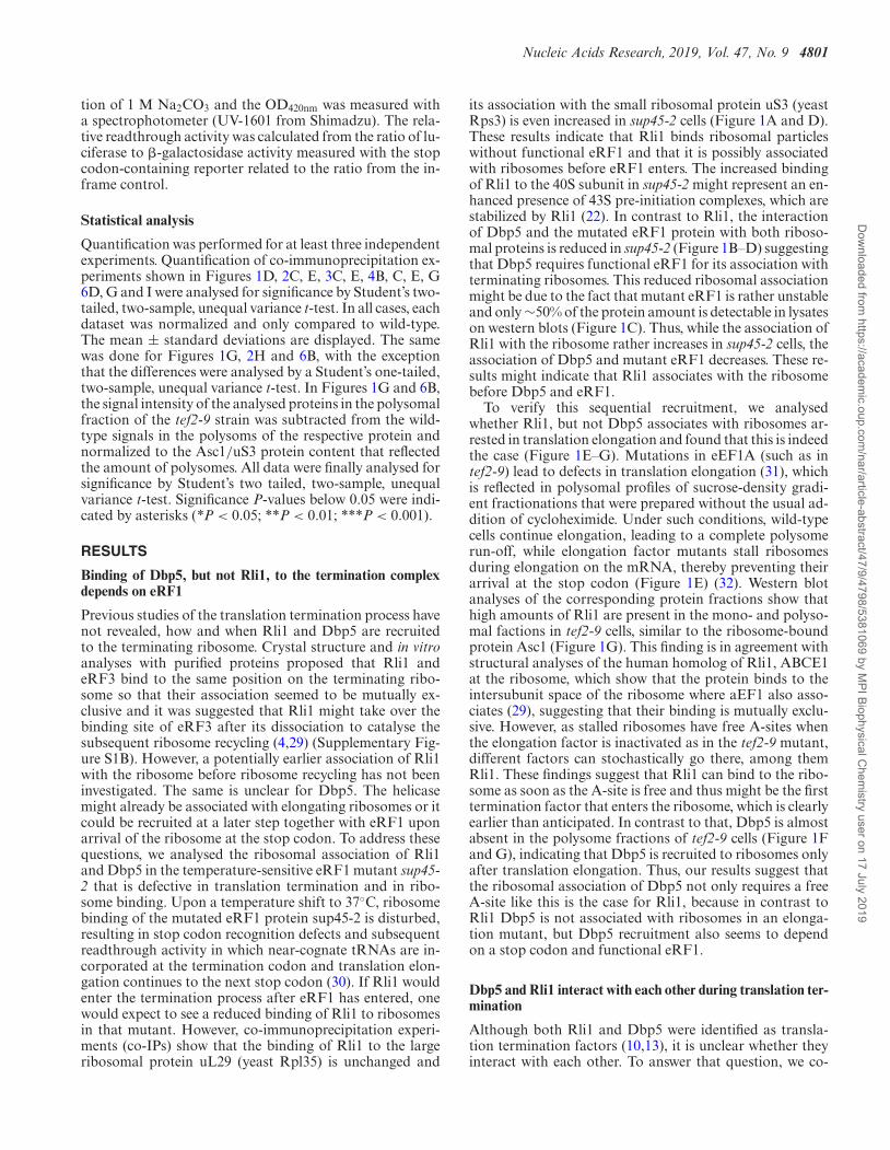
Figure 4. Nup159 recycles Dbp5-ATP also for translation termination. (A) Scheme of the reporter plasmids used in the dual reporter �-galactosidaseluciferase assay. The lacZ gene, expressing �-galactosidase and the luc gene, expressing luciferase is either separated by the stop codon UAG or in frame. Inthe upper case, luciferase will only be expressed in case the stop codon is readthrough. The in-frame reporter serves as control to monitor basal expressionlevels and relate it to the stop codon containing construct. (B) Mutants of NUP159 show increased readthrough of the stop codon. The average readthroughactivity of at least three independent experiments is shown after shift of all indicated strains to 37◦C for 30 min. (C) High copy DBP5 rescues the increasedstop codon readthrough of rat7ΔN. All strains were shifted to 37◦C for 30 min. (D) The interaction of Dbp5 and eRF1 is disturbed in the recycling defectivemutant rat7ΔN. Western blot analysis of a co-IP with Dbp5 and eRF1 is shown. Aco1 served as a negative control. (E) Quantification of three differentexperiments shown in panel (D). (F) The interaction of eRF1 and eRF3 and the ribosome is diminished in rat7ΔN cells. Western blot analysis of a co-IPwith eRF1-GFP and eRF3 or the ribosome bound protein Asc1 is shown. (G) Quantification of three different experiments shown in panel (F). (H) Dbp5and eRF1 directly interact in the presence of a non-hydrolysable ATP-analogue. An in vitro binding study with recombinant proteins in which GST-taggedDbp5 or eRF3 were used in pull-down experiments in the presence of His-eRF1 and if indicated 1 mM AMP–PNP is shown in western blot analysis. GSTalone served as a non-binding control; *P < 0.05; **P < 0.01; ***P < 0.001.
a complex formation between the three termination factorsis unlikely or their potential contact is very short.
To further investigate whether Dbp5 might indeed delivereRF1 to the ribosome and eRF1 and eRF3 do not enter theribosome together, we analysed if mutations in DBP5 wouldlead to a reduced binding of eRF1 to eRF3 and to the ribo-some. For this purpose, we used the rat8-2 strain that pro-duces instable Dbp5 protein due to a leucine to proline ex-change at position 267 (33) (Figure 6H). Indeed, in vivo in-teraction studies of eRF1 and eRF3 in rat8-2 mutants reveala ∼70% reduction of eRF1 binding to the ribosome and toeRF3 upon a 1 h temperature shift to 37◦C (Figure 3D andE). The reduced eRF1 and eRF3 interaction was also de-
tected earlier and seems to happen immediately, already af-ter a 20 min temperature shift of the rat8-2 strain. However,the ribosomal binding of eRF1 was less obviously decreasedafter this short shifting time (10). But the longer 1 h shiftproduces a clear ribosome binding defect of eRF1 (Figure3D and E). Together, our findings suggest that Dbp5 mightdeliver eRF1 to the ribosome without eRF3.
Nup159 recycles Dbp5–ADP for export and translation ter-mination
The ATPase activity of Dbp5 is essential not only formRNA transport, but also for translation termination
Dow
nloaded from https://academ
ic.oup.com/nar/article-abstract/47/9/4798/5381069 by M
PI Biophysical Chem
istry user on 17 July 2019

4806 Nucleic Acids Research, 2019, Vol. 47, No. 9
(10,16,17). During termination, we suggest that the helicasemight deliver eRF1 and it seems possible that it could useits ATPase-dependent activity to position eRF1 properlyon the stop codon. In particular, because we have shownearlier that its ATPase activity is necessary not only for itsfunction in mRNA export, but also for its role in transla-tion termination (10). In both cases, upon ATP-hydrolysis,the enzyme needs to be recycled. During mRNA export,the nucleoporin Nup159/Rat7 is the ADP-release factor ofDbp5 (17). Thus, it is conceivable that recycling from ter-mination also occurs at the NPC via Nup159, rather thanat the ribosome. In particular, because Dbp5 must returnto the cytoplasm to capture a new molecule of eRF1 asthey first interact in the soluble fraction of the cytosol (Fig-ure 1). Indeed, readthrough experiments with a dual �-galactosidase luciferase reporter system show an increasedreadthrough activity in different nup159 mutants, very sim-ilar to the dbp5 mutant rat8-2 (Figure 4A and B), (10).As both nup159 mutants, rat7-1 and rat7ΔN, the latter ofwhich specifically lacks the interaction domain for Dbp5(34), exhibit increased readthough activities as comparedto wild-type (Figure 4B), Nup159 seems to be the recyclingfactor for Dbp5 not only for mRNA export, but also fortranslation termination. In support of this model, we foundthat overexpression of DBP5 leads to a rescue of the highreadthrough activity in rat7ΔN cells (Figure 4C) indicatingthat less recycling by Nup159 is needed when more Dbp5–ATP is present.
These results suggest that Nup159 recycles Dbp5–ADPalso upon its action in translation termination, which isquite attractive, because in this way Dbp5 might couple twoimportant cellular processes––nuclear mRNA export andtranslation. When the translation rate is low, Dbp5 is freeto increasingly act in mRNA export to raise the mRNAamount in the cytoplasm and vice versa, high translationrates could reduce mRNA export. To verify a dependence ofthe eRF1–Dbp5 interaction on Nup159, because their con-tact should only be established when Dbp5 is ATP-bound,we performed co-IPs with these proteins in rat7ΔN. Indeed,while the interaction of Dbp5 and eRF1 was clearly de-tectable in wild-type, it was significantly reduced in rat7ΔN(Figure 4D and E), supporting a model in which Dbp5 isrecycled at the NPC with ATP. These findings further sug-gest that Dbp5–ATP can bind eRF1, while Dbp5–ADPmight release the termination factor. As Dbp5 could notbe re-charged in rat7ΔN and thus cannot deliver eRF1 tothe ribosome anymore, the interaction of eRF1 with eRF3should also be decreased in this mutant, which is indeed thecase as shown by co-IPs (Figure 4F and G).
The switch in eRF1 binding and release through ATP-hydrolysis of Dbp5 was further investigated in in vitro ex-periments with purified recombinantly expressed proteins.We show that Dbp5 only binds to eRF1 in the presence ofthe non-hydrolysable ATP-analogue AMP–PNP, whereaseRF3 interacts also ATP-independently with eRF1 (Figure4H). Together, these in vivo and in vitro studies support amodel in which eRF1 associates with Dbp5–ATP in the cy-toplasm and dissociates from Dbp5–ADP at the ribosome,where Dbp5 possibly uses its ATP-dependent helicase ac-tivity to place eRF1 properly on the stop codon. Moreover
it becomes evident that eRF1 and Dbp5 form a complex inthe cytoplasm, from which eRF3 is absent.
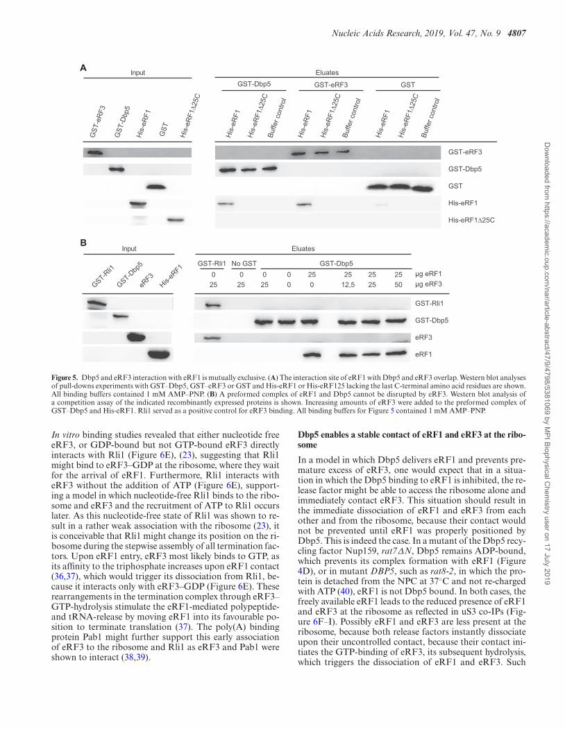
Binding of Dbp5 and eRF3 to eRF1 is mutually exclusive
As Dbp5 and eRF1 enter termination complexes together,and Dbp5 does not interact with eRF3 (10), it seems possi-ble that the binding of Dbp5 and eRF3 with eRF1 is mutu-ally exclusive. It was shown that the eRF3-interaction do-main of eRF1 comprises the last 25 amino acid residues ofits C-terminus (5). In vitro binding studies with recombi-nant proteins were carried out to investigate whether thisdomain is also the Dbp5-interaction domain. Indeed, whilefull-length eRF1 interacts with both, eRF3 and Dbp5, nointeraction was detectable with eRF1�25C (Figure 5A), in-dicating that both termination factors share the same bind-ing site on eRF1. Interestingly, although also the middledomain of eRF1 was reported to contribute to the eRF1–eRF3 interaction (35), we found that the deletion of the C-terminal domain is sufficient to abrogate the interaction ofeRF1 with eRF3 and Dbp5 in vitro. Moreover, a preformedinteraction of Dbp5 and eRF1 was not disrupted by theaddition of increasing amounts of eRF3 in a competitionassay (Figure 5B). Intriguingly, these findings suggest in-deed a sequential and mutually exclusive binding of Dbp5and eRF3 to eRF1 with a first complex formation betweenDbp5 and eRF1. Thus, a model is possible, in which duringthe progress of termination, Dbp5–ATP prohibits the ac-cess of eRF3 to eRF1 until eRF1 was placed properly in theribosomal A-site. Such a mechanism would prevent a pre-mature access of eRF3 and a consequent premature GTP-hydrolysis. Because as soon as eRF3 contacts its guanineexchange factor eRF1 at the ribosome, eRF3 binds GTP,which is subsequently hydrolysed, resulting in the immedi-ate dissociation of eRF3 from the ribosome (36,37). Thesuggested sequential entry of the termination factors wouldhave the advantage that the contact of eRF3 with eRF1 iscontrolled, which will prevent premature GTP-hydrolysis ofeRF3 and its subsequent premature dissociation before thestop codon is successfully recognized.
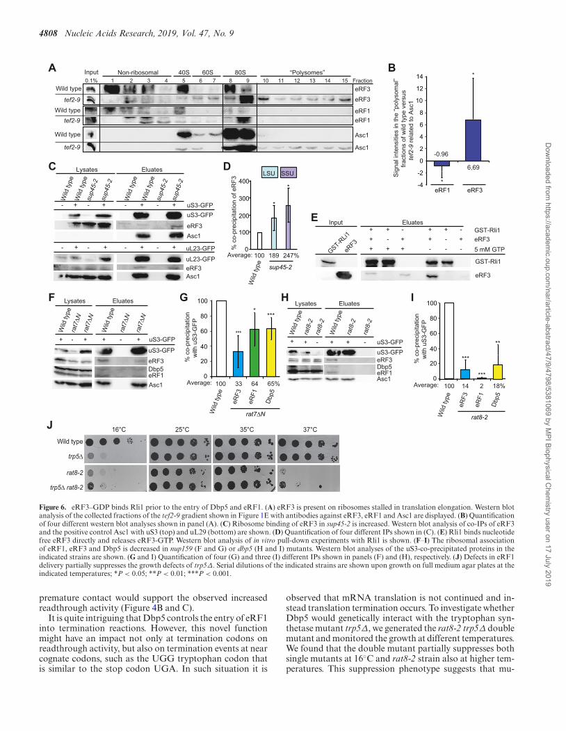
eRF3 binds to the ribosome prior to eRF1
Protection of eRF1 from premature eRF3 access wouldonly be necessary if eRF3 would already be present at the ri-bosome when eRF1 enters. Therefore, we analysed its ribo-somal association in the tef2-9 mutant that arrests in trans-lation elongation as shown in Figure 1E and F. Strikingly,eRF3, but almost no eRF1 is detectable in the polysomalfractions of this elongation mutant, suggesting that eRF3can independently bind ribosomes before they arrive at astop codon and is therefore already present when eRF1 en-ters (Figure 6A and B). These findings are supported byco-immunoprecipitation analyses of eRF3 with ribosomalproteins in the mutant sup45-2. In the situation in whichthis mutant eRF1 protein accumulates with Dbp5 in the cy-toplasm (Figure 1), the binding of eRF3 to the ribosomalprotein uS3 is not reduced, but rather increases as its eRF1mediated GTP-hydrolysis and release is prevented (Figure6C and D).
Because Rli1 is also present at that early time point, weinvestigated a potential direct interaction of eRF3 and Rli1.
Dow
nloaded from https://academ
ic.oup.com/nar/article-abstract/47/9/4798/5381069 by M
PI Biophysical Chem
istry user on 17 July 2019
Nucleic Acids Research, 2019, Vol. 47, No. 9 4807
A
B
Figure 5. Dbp5 and eRF3 interaction with eRF1 is mutually exclusive. (A) The interaction site of eRF1 with Dbp5 and eRF3 overlap. Western blot analysesof pull-downs experiments with GST–Dbp5, GST–eRF3 or GST and His-eRF1 or His-eRF125 lacking the last C-terminal amino acid residues are shown.All binding buffers contained 1 mM AMP–PNP. (B) A preformed complex of eRF1 and Dbp5 cannot be disrupted by eRF3. Western blot analysis ofa competition assay of the indicated recombinantly expressed proteins is shown. Increasing amounts of eRF3 were added to the preformed complex ofGST–Dbp5 and His-eRF1. Rli1 served as a positive control for eRF3 binding. All binding buffers for Figure 5 contained 1 mM AMP–PNP.
In vitro binding studies revealed that either nucleotide freeeRF3, or GDP-bound but not GTP-bound eRF3 directlyinteracts with Rli1 (Figure 6E), (23), suggesting that Rli1might bind to eRF3–GDP at the ribosome, where they waitfor the arrival of eRF1. Furthermore, Rli1 interacts witheRF3 without the addition of ATP (Figure 6E), support-ing a model in which nucleotide-free Rli1 binds to the ribo-some and eRF3 and the recruitment of ATP to Rli1 occurslater. As this nucleotide-free state of Rli1 was shown to re-sult in a rather weak association with the ribosome (23), itis conceivable that Rli1 might change its position on the ri-bosome during the stepwise assembly of all termination fac-tors. Upon eRF1 entry, eRF3 most likely binds to GTP, asits affinity to the triphosphate increases upon eRF1 contact(36,37), which would trigger its dissociation from Rli1, be-cause it interacts only with eRF3–GDP (Figure 6E). Theserearrangements in the termination complex through eRF3–GTP-hydrolysis stimulate the eRF1-mediated polypeptide-and tRNA-release by moving eRF1 into its favourable po-sition to terminate translation (37). The poly(A) bindingprotein Pab1 might further support this early associationof eRF3 to the ribosome and Rli1 as eRF3 and Pab1 wereshown to interact (38,39).
Dbp5 enables a stable contact of eRF1 and eRF3 at the ribo-some
In a model in which Dbp5 delivers eRF1 and prevents pre-mature excess of eRF3, one would expect that in a situa-tion in which the Dbp5 binding to eRF1 is inhibited, the re-lease factor might be able to access the ribosome alone andimmediately contact eRF3. This situation should result inthe immediate dissociation of eRF1 and eRF3 from eachother and from the ribosome, because their contact wouldnot be prevented until eRF1 was properly positioned byDbp5. This is indeed the case. In a mutant of the Dbp5 recy-cling factor Nup159, rat7ΔN, Dbp5 remains ADP-bound,which prevents its complex formation with eRF1 (Figure4D), or in mutant DBP5, such as rat8-2, in which the pro-tein is detached from the NPC at 37◦C and not re-chargedwith ATP (40), eRF1 is not Dbp5 bound. In both cases, thefreely available eRF1 leads to the reduced presence of eRF1and eRF3 at the ribosome as reflected in uS3 co-IPs (Fig-ure 6F–I). Possibly eRF1 and eRF3 are less present at theribosome, because both release factors instantly dissociateupon their uncontrolled contact, because their contact ini-tiates the GTP-binding of eRF3, its subsequent hydrolysis,which triggers the dissociation of eRF1 and eRF3. Such
Dow
nloaded from https://academ
ic.oup.com/nar/article-abstract/47/9/4798/5381069 by M
PI Biophysical Chem
istry user on 17 July 2019
4808 Nucleic Acids Research, 2019, Vol. 47, No. 9
Figure 6. eRF3–GDP binds Rli1 prior to the entry of Dbp5 and eRF1. (A) eRF3 is present on ribosomes stalled in translation elongation. Western blotanalysis of the collected fractions of the tef2-9 gradient shown in Figure 1E with antibodies against eRF3, eRF1 and Asc1 are displayed. (B) Quantificationof four different western blot analyses shown in panel (A). (C) Ribosome binding of eRF3 in sup45-2 is increased. Western blot analysis of co-IPs of eRF3and the positive control Asc1 with uS3 (top) and uL29 (bottom) are shown. (D) Quantification of four different IPs shown in (C). (E) Rli1 binds nucleotidefree eRF3 directly and releases eRF3-GTP. Western blot analysis of in vitro pull-down experiments with Rli1 is shown. (F–I) The ribosomal associationof eRF1, eRF3 and Dbp5 is decreased in nup159 (F and G) or dbp5 (H and I) mutants. Western blot analyses of the uS3-co-precipitated proteins in theindicated strains are shown. (G and I) Quantification of four (G) and three (I) different IPs shown in panels (F) and (H), respectively. (J) Defects in eRF1delivery partially suppresses the growth defects of trp5Δ. Serial dilutions of the indicated strains are shown upon growth on full medium agar plates at theindicated temperatures; *P < 0.05; **P < 0.01; ***P < 0.001.
premature contact would support the observed increasedreadthrough activity (Figure 4B and C).
It is quite intriguing that Dbp5 controls the entry of eRF1into termination reactions. However, this novel functionmight have an impact not only at termination codons onreadthrough activity, but also on termination events at nearcognate codons, such as the UGG tryptophan codon thatis similar to the stop codon UGA. In such situation it is
observed that mRNA translation is not continued and in-stead translation termination occurs. To investigate whetherDbp5 would genetically interact with the tryptophan syn-thetase mutant trp5Δ, we generated the rat8-2 trp5Δ doublemutant and monitored the growth at different temperatures.We found that the double mutant partially suppresses bothsingle mutants at 16◦C and rat8-2 strain also at higher tem-peratures. This suppression phenotype suggests that mu-
Dow
nloaded from https://academ
ic.oup.com/nar/article-abstract/47/9/4798/5381069 by M
PI Biophysical Chem
istry user on 17 July 2019
Nucleic Acids Research, 2019, Vol. 47, No. 9 4809
tations in DBP5 indeed affect termination events at nearcognate codons and the deleterious effect of the trp5 dele-tion stems from inefficient decoding of UGG codons, andincreased eRF1-catalysed mistermination events on suchcodons. This effect appears suppressed in rat8-2 mutantswhere the delivery of eRF1 to such codons would be re-duced (Figure 6J).
eIF3 enters translation termination after the Dbp5-mediateddelivery of eRF1
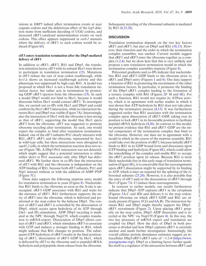
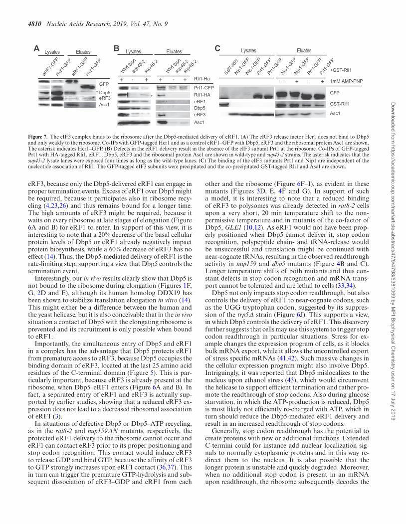
In addition to eRF1, eRF3, Rli1 and Dbp5, the transla-tion initiation factor, eIF3 with its subunit Hcr1 were shownto participate in translation termination (15). Mutationsin eIF3 reduce the rate of stop codon readthrough, whilehcr1Δ shows an increased readthrough activity and thisphenotype was suppressed by high copy Rli1. A model wasproposed in which Hcr1 is not a bona fide translation ini-tiation factor, but rather acts in termination by promot-ing GDP–eRF3 ejection from the ribosomes (15). In sucha model Dbp5 would not bind to Hcr1, because it woulddissociate before Hcr1 would contact eRF3. To investigatethis, we carried out co-IPs with Hcr1 and Dbp5 and couldconfirm the Hcr1 eRF3 interaction, while no interaction be-tween Hcr1 and Dbp5 was visible (Figure 7A). Interestingly,also the interaction of Hcr1 with the ribosome is less strongas that of eRF1, supporting the model that Hcr1 ejectseRF3 from the ribosome. eIF3 was suggested to promoteribosome recycling after termination. As such, one wouldexpect the complex to bind after translation termination.Indeed, one of the eIF3 subunits Prt1 clearly interacts withRli1, eRF3, eRF1 and the ribosome, represented by Asc1in wild-type cells, but these interactions were abrogated insup45-2 cells, in which the termination reaction does not oc-cur (Figure 7B). A Dbp5 Prt1 interaction was not detected,which might suggest that a potential interaction is eitherrather short or Prt1 associates only after Dbp5 has deliv-ered eRF1. We further show in co-IPs that the interactionof eIF3 with Rli1 and the ribosome is independent on theATP-binding of Rli1, because both eIF3 subunits, Prt1 andNip1 interact without or with the addition of AMP–PNP(Figure 7C).
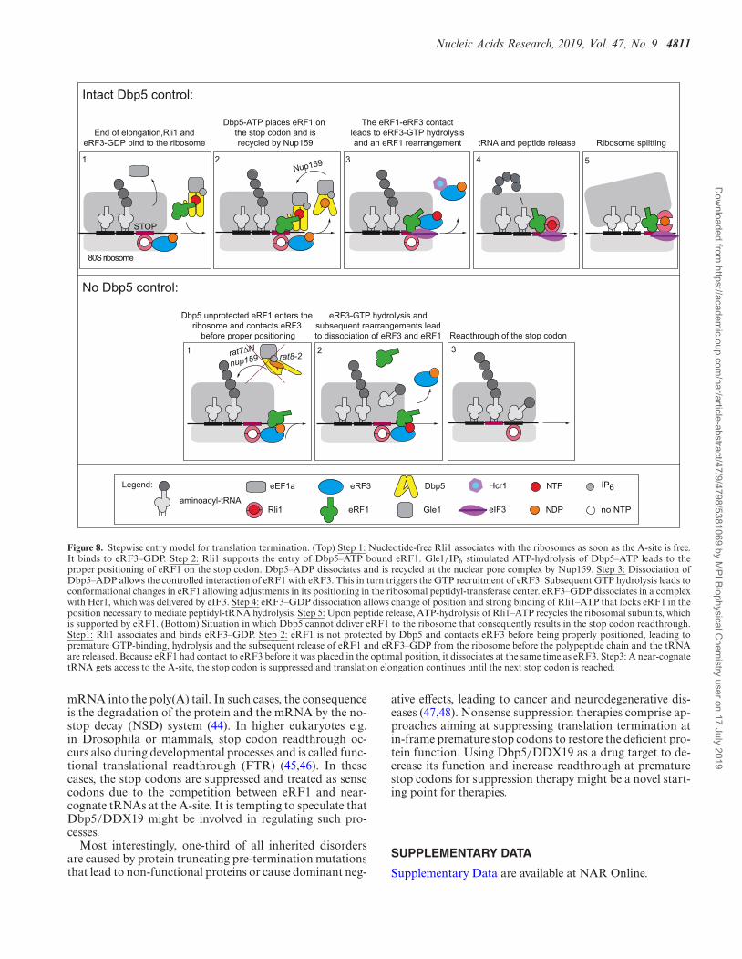
These data support the following stepwise entry modelfor translation termination in yeast (Figure 8). Nucleotide-free Rli1 binds to the ribosome as soon as the A-site is un-occupied. eRF3–GDP associates with Rli1 and waits forthe entrance of eRF1. Rli1 furthermore supports the en-try of eRF1 into the ribosome, which is delivered and po-sitioned at the stop codon by the helicase Dbp5. The con-tact of eRF3 and eRF1 is controlled by the dissociation ofDbp5, which occurs upon hydrolysis of its ATP throughGle1- and IP6-stimulation. ADP–Dbp5 recycling is medi-ated at the NPC through Nup159, which couples transla-tion to mRNA-export. Dissociation of Dbp5 allows con-tact of eRF1 and eRF3, which stimulates eRF3 to associatewith GTP and induces a stronger binding to Rli1, whichmight indicate that Rli1 changes its position. The subse-quent GTP-hydrolysis of eRF3 results in the final position-ing of eRF1, dissociation of eRF3–GDP by Hcr1, whichis delivered by eIF3 to the ribosome and to peptidyl-tRNAhydrolysis and polypeptide chain release from the ribosome.
Subsequent recycling of the ribosomal subunits is mediatedby Rli1 (4,23,26).
DISCUSSION
Translation termination depends on the two key factorseRF1 and eRF3, but also on Dbp5 and Rli1 (10,13). How-ever, their function and the order in which the terminationcomplex assembles, was unclear. Current models suggestthat eRF1 and eRF3 enter the ribosome together as a com-plex (1,2,4), but we show here that this is very unlikely andpropose a new translation termination model in which thetermination complex assembles stepwise (Figure 8).
Polysomal gradients and co-IPs indicate that nucleotide-free Rli1 and eRF3–GDP binds to the ribosome prior toeRF1 and Dbp5 entry (Figures 1 and 6). Our data supporta function of Rli1 in promoting the recruitment of the othertermination factors. In particular, it promotes the bindingof the Dbp5–eRF1 complex leading to the formation ofa ternary complex with Rli1 (Figures 2F–H and 6E). Forsuch a function, Rli1 would not require its ATPase activ-ity, which is in agreement with earlier studies in which itwas shown that ATP-hydrolysis by Rli1 does not take placeduring the termination process (4,13). It was furthermoresuggested earlier that Rli1 associates with the terminationcomplex upon dissociation of eRF3–GDP, taking over itsposition to lock eRF1 in its favourable position to facilitatepeptidyl-tRNA hydrolysis (4,26). In contrast to this model,we present evidence that Rli1 and eRF3–GDP are the ini-tial components of the termination complex that bind tothe ribosome. However, our data are in agreement with amodel in which in the course of the stepwise assembly, Rli1could take over the position of eRF3, because initially eRF3binds to Rli1 in its GDP bound form and dissociates uponGTP binding and hydrolysis (Figure 6E), which could allowthe remodelling of the complex and enable Rli1 to occupythe eRF3 position upon its release. Because Rli1 is mostlikely nucleotide-free in this early stage of translation termi-nation (Figure 6E), it is conceivable that the rearrangementsupon eRF3 dissociation might be supported by its bindingto ATP, which is later on required for the splitting of the ri-bosomal subunits (23,24). However, it is also possible thatthe entry of eIF3 and/or the dissociation of eRF3 throughHcr1 (Figure 7A–C) induce these rearrangements.
In contrast to earlier models, our results furthermoreindicate that Dbp5–ATP captures eRF1 in the cytoplasm(Figures 3A,C and 4D) and delivers it to the stop codon-bound ribosome on which eRF3–GDP and Rli1 are al-ready present (Figures 1F,G and 6A,B). The interaction be-tween Rli1 and Dbp5 might thereby support the Dbp5–eRF1 recruitment (Figure 2). Upon placing eRF1 prop-erly on the stop codon, Dbp5–ADP dissociates and is re-cycled at the NPC via Nup159 (Figure 4). In this way, thetwo key processes of mRNA export and translation arecoupled via Dbp5. How the duty of Dbp5 in both pro-cesses is divided and how Dbp5 captures eRF1 is currentlyunclear and needs further investigation. Interestingly, theoverall cellular protein abundance estimates suggest an in-tracellular Dbp5:eRF1:eRF3 ratio of ∼1:2:4 (https://www.yeastgenome.org/). Dbp5 as a limiting factor further quali-fies itself as a regulator of the interaction between eRF1 and
Dow
nloaded from https://academ
ic.oup.com/nar/article-abstract/47/9/4798/5381069 by M
PI Biophysical Chem
istry user on 17 July 2019
4810 Nucleic Acids Research, 2019, Vol. 47, No. 9
Figure 7. The eIF3 complex binds to the ribosome after the Dbp5-mediated delivery of eRF1. (A) The eRF3 release factor Hcr1 does not bind to Dbp5and only weakly to the ribosome. Co-IPs with GFP-tagged Hcr1 and as a control eRF1–GFP with Dbp5, eRF3 and the ribosomal protein Asc1 are shown.The asterisk indicates Hcr1–GFP. (B) Defects in the eRF1 delivery result in the absence of the eIF3 subunit Prt1 at the ribosome. Co-IPs of GFP-taggedPrt1 with HA-tagged Rli1, eRF1, Dbp5, eRF3 and the ribosomal protein Asc1 are shown in wild-type and sup45-2 strains. The asterisk indicates that thesup45-2 lysate lanes were exposed four times as long as the wild-type lanes. (C) The binding of the eIF3 subunits Prt1 and Nip1 are independent of thenucleotide association of Rli1. The GFP-tagged eIF3 subunits were precipitated and the co-precipitated GST-tagged Rli1 and Asc1 are shown.
eRF3, because only the Dbp5-delivered eRF1 can engage inproper termination events. Excess of eRF1 over Dbp5 mightbe required, because it participates also in ribosome recy-cling (4,23,26) and thus remains bound for a longer time.The high amounts of eRF3 might be required, because itwaits on every ribosome at late stages of elongation (Figure6A and B) for eRF1 to enter. In support of this view, it isinteresting to note that a 20% decrease of the basal cellularprotein levels of Dbp5 or eRF1 already negatively impactprotein biosynthesis, while a 60% decrease of eRF3 has noeffect (14). Thus, the Dbp5-mediated delivery of eRF1 is therate-limiting step, supporting a view that Dbp5 controls thetermination event.
Interestingly, our in vivo results clearly show that Dbp5 isnot bound to the ribosome during elongation (Figures 1F,G, 2D and E), although its human homolog DDX19 hasbeen shown to stabilize translation elongation in vitro (14).This might either be a difference between the human andthe yeast helicase, but it is also conceivable that in the in vivosituation a contact of Dbp5 with the elongating ribosome isprevented and its recruitment is only possible when boundto eRF1.
Importantly, the simultaneous entry of Dbp5 and eRF1in a complex has the advantage that Dbp5 protects eRF1from premature access to eRF3, because Dbp5 occupies thebinding domain of eRF3, located at the last 25 amino acidresidues of the C-terminal domain (Figure 5). This is par-ticularly important, because eRF3 is already present at theribosome, when Dbp5–eRF1 enters (Figure 6A and B). Infact, a separated entry of eRF1 and eRF3 is actually sup-ported by earlier studies, showing that a reduced eRF3 ex-pression does not lead to a decreased ribosomal associationof eRF1 (3).
In situations of defective Dbp5 or Dbp5–ATP recycling,as in the rat8-2 and nup159ΔN mutants, respectively, theprotected eRF1 delivery to the ribosome cannot occur andeRF1 can contact eRF3 prior to its proper positioning andstop codon recognition. This contact would induce eRF3to release GDP and bind GTP, because the affinity of eRF3to GTP strongly increases upon eRF1 contact (36,37). Thisin turn can trigger the premature GTP-hydrolysis and sub-sequent dissociation of eRF3–GDP and eRF1 from each
other and the ribosome (Figure 6F–I), as evident in thesemutants (Figures 3D, E, 4F and G). In support of sucha model, it is interesting to note that a reduced bindingof eRF3 to polysomes was already detected in rat8-2 cellsupon a very short, 20 min temperature shift to the non-permissive temperature and in mutants of the co-factor ofDbp5, GLE1 (10,12). As eRF1 would not have been prop-erly positioned when Dbp5 cannot deliver it, stop codonrecognition, polypeptide chain- and tRNA-release wouldbe unsuccessful and translation might be continued withnear-cognate tRNAs, resulting in the observed readthroughactivity in nup159 and dbp5 mutants (Figure 4B and C).Longer temperature shifts of both mutants and thus con-stant defects in stop codon recognition and mRNA trans-port cannot be tolerated and are lethal to cells (33,34).
Dbp5 not only impacts stop codon readthrough, but alsocontrols the delivery of eRF1 to near-cognate codons, suchas the UGG tryptophan codon, suggested by its suppres-sion of the trp5Δ strain (Figure 6J). This supports a view,in which Dbp5 controls the delivery of eRF1. This discoveryfurther suggests that cells may use this system to trigger stopcodon readthrough in particular situations. Stress for ex-ample changes the expression program of cells, as it blocksbulk mRNA export, while it allows the uncontrolled exportof stress specific mRNAs (41,42). Such massive changes inthe cellular expression program might also involve Dbp5.Intriguingly, it was reported that Dbp5 mislocalizes to thenucleus upon ethanol stress (43), which would circumventthe helicase to support efficient termination and rather pro-mote the readthrough of stop codons. Also during glucosestarvation, in which the ATP-production is reduced, Dbp5is most likely not efficiently re-charged with ATP, which inturn should reduce the Dbp5-mediated eRF1 delivery andresult in an increased readthrough of stop codons.
Generally, stop codon readthrough has the potential tocreate proteins with new or additional functions. ExtendedC-termini could for instance add nuclear localization sig-nals to normally cytoplasmic proteins and in this way re-direct them to the nucleus. It is also possible that thelonger protein is unstable and quickly degraded. Moreover,when no additional stop codon is present in an mRNAupon readthrough, the ribosome subsequently decodes the
Dow
nloaded from https://academ
ic.oup.com/nar/article-abstract/47/9/4798/5381069 by M
PI Biophysical Chem
istry user on 17 July 2019
Nucleic Acids Research, 2019, Vol. 47, No. 9 4811
Figure 8. Stepwise entry model for translation termination. (Top) Step 1: Nucleotide-free Rli1 associates with the ribosomes as soon as the A-site is free.It binds to eRF3–GDP. Step 2: Rli1 supports the entry of Dbp5–ATP bound eRF1. Gle1/IP6 stimulated ATP-hydrolysis of Dbp5–ATP leads to theproper positioning of eRF1 on the stop codon. Dbp5–ADP dissociates and is recycled at the nuclear pore complex by Nup159. Step 3: Dissociation ofDbp5–ADP allows the controlled interaction of eRF1 with eRF3. This in turn triggers the GTP recruitment of eRF3. Subsequent GTP hydrolysis leads toconformational changes in eRF1 allowing adjustments in its positioning in the ribosomal peptidyl-transferase center. eRF3–GDP dissociates in a complexwith Hcr1, which was delivered by eIF3. Step 4: eRF3–GDP dissociation allows change of position and strong binding of Rli1–ATP that locks eRF1 in theposition necessary to mediate peptidyl-tRNA hydrolysis. Step 5: Upon peptide release, ATP-hydrolysis of Rli1–ATP recycles the ribosomal subunits, whichis supported by eRF1. (Bottom) Situation in which Dbp5 cannot deliver eRF1 to the ribosome that consequently results in the stop codon readthrough.Step1: Rli1 associates and binds eRF3–GDP. Step 2: eRF1 is not protected by Dbp5 and contacts eRF3 before being properly positioned, leading topremature GTP-binding, hydrolysis and the subsequent release of eRF1 and eRF3–GDP from the ribosome before the polypeptide chain and the tRNAare released. Because eRF1 had contact to eRF3 before it was placed in the optimal position, it dissociates at the same time as eRF3. Step3: A near-cognatetRNA gets access to the A-site, the stop codon is suppressed and translation elongation continues until the next stop codon is reached.
mRNA into the poly(A) tail. In such cases, the consequenceis the degradation of the protein and the mRNA by the no-stop decay (NSD) system (44). In higher eukaryotes e.g.in Drosophila or mammals, stop codon readthrough oc-curs also during developmental processes and is called func-tional translational readthrough (FTR) (45,46). In thesecases, the stop codons are suppressed and treated as sensecodons due to the competition between eRF1 and near-cognate tRNAs at the A-site. It is tempting to speculate thatDbp5/DDX19 might be involved in regulating such pro-cesses.
Most interestingly, one-third of all inherited disordersare caused by protein truncating pre-termination mutationsthat lead to non-functional proteins or cause dominant neg-
ative effects, leading to cancer and neurodegenerative dis-eases (47,48). Nonsense suppression therapies comprise ap-proaches aiming at suppressing translation termination atin-frame premature stop codons to restore the deficient pro-tein function. Using Dbp5/DDX19 as a drug target to de-crease its function and increase readthrough at prematurestop codons for suppression therapy might be a novel start-ing point for therapies.
SUPPLEMENTARY DATA
Supplementary Data are available at NAR Online.
Dow
nloaded from https://academ
ic.oup.com/nar/article-abstract/47/9/4798/5381069 by M
PI Biophysical Chem
istry user on 17 July 2019
4812 Nucleic Acids Research, 2019, Vol. 47, No. 9
ACKNOWLEDGEMENTS
We thank L. Soldner, S. Niehus, M. Urh and S. Uzuntasfor technical assistance. We thank D. Bedwell, G. Braus, R.Lill, M. Seedorf and O. Valerius for sharing antibodies andC. Cole, T. Kinzy, R.Lill, P. Silver, G. Stahl and F. Winstonfor providing strains and/or plasmids.
FUNDING
Deutsche Forschungsgemeinschaft (DFG) [SFB860to H.K.]. Funding for open access charge: DeutscheForschungsgemeinschaft [SFB 860].Conflict of interest statement. None declared.
REFERENCES1. Jackson,R.J., Hellen,C.U. and Pestova,T.V. (2012) Termination and
post-termination events in eukaryotic translation. Adv. Protein Chem.Struct. Biol., 86, 45–93.
2. Alkalaeva,E.Z., Pisarev,A.V., Frolova,L.Y., Kisselev,L.L. andPestova,T.V. (2006) In vitro reconstitution of eukaryotic translationreveals cooperativity between release factors eRF1 and eRF3. Cell,125, 1125–1136.
3. Salas-Marco,J. and Bedwell,D.M. (2004) GTP hydrolysis by eRF3facilitates stop codon decoding during eukaryotic translationtermination. Mol. Cell. Biol., 24, 7769–7778.
4. Shoemaker,C.J. and Green,R. (2011) Kinetic analysis reveals theordered coupling of translation termination and ribosome recyclingin yeast. Proc. Natl. Acad. Sci. USA, 108, E1392–E1398.
5. Eurwilaichitr,L., Graves,F.M., Stansfield,I. and Tuite,M.F. (1999)The C-terminus of eRF1 defines a functionally important domain fortranslation termination in Saccharomyces cerevisiae. Mol. Microbiol.,32, 485–496.
6. Merkulova,T.I., Frolova,L.Y., Lazar,M., Camonis,J. andKisselev,L.L. (1999) C-terminal domains of human translationtermination factors eRF1 and eRF3 mediate their in vivo interaction.FEBS Lett., 443, 41–47.
7. Cheng,Z., Saito,K., Pisarev,A.V., Wada,M., Pisareva,V.P.,Pestova,T.V., Gajda,M., Round,A., Kong,C., Lim,M. et al. (2009)Structural insights into eRF3 and stop codon recognition by eRF1.Genes Dev., 23, 1106–1118.
8. des Georges,A., Hashem,Y., Unbehaun,A., Grassucci,R.A.,Taylor,D., Hellen,C.U., Pestova,T.V. and Frank,J. (2014) Structure ofthe mammalian ribosomal pre-termination complex associated witheRF1.eRF3.GDPNP. Nucleic Acids Res., 42, 3409–3418.
9. Taylor,D., Unbehaun,A., Li,W., Das,S., Lei,J., Liao,H.Y.,Grassucci,R.A., Pestova,T.V. and Frank,J. (2012) Cryo-EM structureof the mammalian eukaryotic release factor eRF1-eRF3-associatedtermination complex. Proc. Natl. Acad. Sci. USA, 109, 18413–18418.
10. Gross,T., Siepmann,A., Sturm,D., Windgassen,M., Scarcelli,J.J.,Seedorf,M., Cole,C.N. and Krebber,H. (2007) The DEAD-box RNAhelicase Dbp5 functions in translation termination. Science , 315,646–649.
11. Alcazar-Roman,A.R., Bolger,T.A. and Wente,S.R. (2010) Control ofmRNA export and translation termination by inositolhexakisphosphate requires specific interaction with Gle1. J. Biol.Chem., 285, 16683–16692.
12. Bolger,T.A., Folkmann,A.W., Tran,E.J. and Wente,S.R. (2008) ThemRNA export factor Gle1 and inositol hexakisphosphate regulatedistinct stages of translation. Cell, 134, 624–633.
13. Khoshnevis,S., Gross,T., Rotte,C., Baierlein,C., Ficner,R. andKrebber,H. (2010) The iron-sulphur protein RNase L inhibitorfunctions in translation termination. EMBO Rep., 11, 214–219.
14. Mikhailova,T., Shuvalova,E., Ivanov,A., Susorov,D., Shuvalov,A.,Kolosov,P.M. and Alkalaeva,E. (2017) RNA helicase DDX19stabilizes ribosomal elongation and termination complexes. NucleicAcids Res., 45, 1307–1318.
15. Beznoskova,P., Cuchalova,L., Wagner,S., Shoemaker,C.J.,Gunisova,S., von der Haar,T. and Valasek,L.S. (2013) Translationinitiation factors eIF3 and HCR1 control translation termination andstop codon read-through in yeast cells. PLoS Genet., 9, e1003962.
16. Tieg,B. and Krebber,H. (2013) Dbp5 - From nuclear export totranslation. Biochim. Biophys. Acta, 1829, 791–798.
17. Noble,K.N., Tran,E.J., Alcazar-Roman,A.R., Hodge,C.A., Cole,C.N.and Wente,S.R. (2011) The Dbp5 cycle at the nuclear pore complexduring mRNA export II: nucleotide cycling and mRNP remodelingby Dbp5 are controlled by Nup159 and Gle1. Genes Dev., 25,1065–1077.
18. Neumann,B., Wu,H., Hackmann,A. and Krebber,H. (2016) Nuclearexport of Pre-Ribosomal subunits requires Dbp5, but not as anRNA-Helicase as for mRNA export. PLoS One, 11, e0149571.
19. Kispal,G., Sipos,K., Lange,H., Fekete,Z., Bedekovics,T., Janaky,T.,Bassler,J., Aguilar Netz,D.J., Balk,J., Rotte,C. et al. (2005) Biogenesisof cytosolic ribosomes requires the essential iron-sulphur proteinRli1p and mitochondria. EMBO J., 24, 589–598.
20. Strunk,B.S., Novak,M.N., Young,C.L. and Karbstein,K. (2012) Atranslation-like cycle is a quality control checkpoint for maturing 40Sribosome subunits. Cell, 150, 111–121.
21. Yarunin,A., Panse,V.G., Petfalski,E., Dez,C., Tollervey,D. andHurt,E.C. (2005) Functional link between ribosome formation andbiogenesis of iron-sulfur proteins. EMBO J., 24, 580–588.
22. Dong,J., Lai,R., Nielsen,K., Fekete,C.A., Qiu,H. andHinnebusch,A.G. (2004) The essential ATP-binding cassette proteinRLI1 functions in translation by promoting preinitiation complexassembly. J. Biol. Chem., 279, 42157–42168.
23. Pisarev,A.V., Skabkin,M.A., Pisareva,V.P., Skabkina,O.V.,Rakotondrafara,A.M., Hentze,M.W., Hellen,C.U. and Pestova,T.V.(2010) The role of ABCE1 in eukaryotic posttermination ribosomalrecycling. Mol. Cell, 37, 196–210.
24. Barthelme,D., Dinkelaker,S., Albers,S.V., Londei,P., Ermler,U. andTampe,R. (2011) Ribosome recycling depends on a mechanistic linkbetween the FeS cluster domain and a conformational switch of thetwin-ATPase ABCE1. Proc. Natl. Acad. Sci. USA, 108, 3228–3233.
25. Shoemaker,C.J., Eyler,D.E. and Green,R. (2010) Dom34:Hbs1promotes subunit dissociation and peptidyl-tRNA drop-off to initiateno-go decay. Science, 330, 369–372.
26. Preis,A., Heuer,A., Barrio-Garcia,C., Hauser,A., Eyler,D.E.,Berninghausen,O., Green,R., Becker,T. and Beckmann,R. (2014)Cryoelectron microscopic structures of eukaryotic translationtermination complexes containing eRF1-eRF3 or eRF1-ABCE1. CellRep., 8, 59–65.
27. Baierlein,C., Hackmann,A., Gross,T., Henker,L., Hinz,F. andKrebber,H. (2013) Monosome formation during translation initiationrequires the serine/arginine-rich protein Npl3. Mol. Cell. Biol., 33,4811–4823.
28. Bidou,L., Stahl,G., Hatin,I., Namy,O., Rousset,J.P. andFarabaugh,P.J. (2000) Nonsense-mediated decay mutants do notaffect programmed -1 frameshifting. RNA , 6, 952–961.
29. Becker,T., Franckenberg,S., Wickles,S., Shoemaker,C.J., Anger,A.M.,Armache,J.P., Sieber,H., Ungewickell,C., Berninghausen,O.,Daberkow,I. et al. (2012) Structural basis of highly conservedribosome recycling in eukaryotes and archaea. Nature, 482, 501–506.
30. Stansfield,I., Kushnirov,V.V., Jones,K.M. and Tuite,M.F. (1997) Aconditional-lethal translation termination defect in a sup45 mutant ofthe yeast Saccharomyces cerevisiae. Eur J Biochem., 245, 557–563.
31. Dinman,J.D. and Kinzy,T.G. (1997) Translational misreading:mutations in translation elongation factor 1alpha differentially affectprogrammed ribosomal frameshifting and drug sensitivity. RNA , 3,870–881.
32. Anand,M., Chakraburtty,K., Marton,M.J., Hinnebusch,A.G. andKinzy,T.G. (2003) Functional interactions between yeast translationeukaryotic elongation factor (eEF) 1A and eEF3. J. Biol. Chem., 278,6985–6991.
33. Snay-Hodge,C.A., Colot,H.V., Goldstein,A.L. and Cole,C.N. (1998)Dbp5p/Rat8p is a yeast nuclear pore-associated DEAD-box proteinessential for RNA export. EMBO J., 17, 2663–2676.
34. Hodge,C.A., Colot,H.V., Stafford,P. and Cole,C.N. (1999)Rat8p/Dbp5p is a shuttling transport factor that interacts withRat7p/Nup159p and Gle1p and suppresses the mRNA export defectof xpo1-1 cells. EMBO J., 18, 5778–5788.
35. Kononenko,A.V., Mitkevich,V.A., Dubovaya,V.I., Kolosov,P.M.,Makarov,A.A. and Kisselev,L.L. (2008) Role of the individualdomains of translation termination factor eRF1 in GTP binding toeRF3. Proteins, 70, 388–393.
Dow
nloaded from https://academ
ic.oup.com/nar/article-abstract/47/9/4798/5381069 by M
PI Biophysical Chem
istry user on 17 July 2019
Nucleic Acids Research, 2019, Vol. 47, No. 9 4813
36. Frolova,L., Le Goff,X., Zhouravleva,G., Davydova,E., Philippe,M.and Kisselev,L. (1996) Eukaryotic polypeptide chain release factoreRF3 is an eRF1- and ribosome-dependent guanosinetriphosphatase. RNA , 2, 334–341.
37. Pisareva,V.P., Pisarev,A.V., Hellen,C.U., Rodnina,M.V. andPestova,T.V. (2006) Kinetic analysis of interaction of eukaryoticrelease factor 3 with guanine nucleotides. J. Biol. Chem., 281,40224–40235.
38. Jacobson,A. (2005) The end justifies the means. Nat. Struct. Mol.Biol., 12, 474–475.
39. Uchida,N., Hoshino,S., Imataka,H., Sonenberg,N. and Katada,T.(2002) A novel role of the mammalian GSPT/eRF3 associating withpoly(A)-binding protein in Cap/Poly(A)-dependent translation. J.Biol. Chem., 277, 50286–50292.
40. Scarcelli,J.J., Viggiano,S., Hodge,C.A., Heath,C.V., Amberg,D.C. andCole,C.N. (2008) Synthetic genetic array analysis in Saccharomycescerevisiae provides evidence for an interaction between RAT8/DBP5and genes encoding P-body components. Genetics, 179, 1945–1955.
41. Zander,G., Hackmann,A., Bender,L., Becker,D., Lingner,T.,Salinas,G. and Krebber,H. (2016) mRNA quality control is bypassedfor immediate export of stress-responsive transcripts. Nature, 540,593–596.
42. Zander,G. and Krebber,H. (2017) Quick or quality? How mRNAescapes nuclear quality control during stress. RNA Biol., 14,1642–1648.
43. Takemura,R., Inoue,Y. and Izawa,S. (2004) Stress response in yeastmRNA export factor: reversible changes in Rat8p localization arecaused by ethanol stress but not heat shock. J. Cell Sci., 117,4189–4197.
44. Shoemaker,C.J. and Green,R. (2012) Translation drives mRNAquality control. Nat. Struct. Mol. Biol., 19, 594–601.
45. Schueren,F. and Thoms,S. (2016) Functional translationalReadthrough: A systems biology perspective. PLoS Genet., 12,e1006196.
46. von der Haar,T. and Tuite,M.F. (2007) Regulated translationalbypass of stop codons in yeast. Trends Microbiol., 15, 78–86.
47. Bordeira-Carrico,R., Pego,A.P., Santos,M. and Oliveira,C. (2012)Cancer syndromes and therapy by stop-codon readthrough. TrendsMol. Med., 18, 667–678.
48. Keeling,K.M., Xue,X., Gunn,G. and Bedwell,D.M. (2014)Therapeutics based on stop codon readthrough. Annu. Rev. GenomicsHum. Genet., 15, 371–394.
Dow
nloaded from https://academ
ic.oup.com/nar/article-abstract/47/9/4798/5381069 by M
PI Biophysical Chem
istry user on 17 July 2019