trophysiological results suggest
TRANSCRIPT

trophysiological results suggest that thiseffect may be mediated by way of a di-rect pathway from the raphe to theSPN's. Although other mechanisms suchas disfacilitation or disynaptic inhibitioninvolving a spinal interneuron cannot bedefinitively excluded, the weights of thecombined evidence suggest that the med-ullary raphe inhibits the SPN's by a di-rect pathway.
JOHN B. CABOTJ. MARTIN WILD, DAVID H. COHEN
Department of Physiology, School ofMedicine, University of Virginia,Charlottesville 22901
References and Notes
1. S. D. Anderson, A. I. Basbaum, H. L. Fields,Brain Res. 123, 363 (1977); A. I. Basbaum, C. H.Clanton, H. L. Fields, Proc. Natl. Acad. Sci.U.S.A. 73, 4685 (1976); J. E. Beall, R. F. Mar-tin, A. E. Applebaum, W. D. Willis, Brain Res.114, 328 (1976); H. L. Fields, A. I. Basbaum, C.H. Clanton, S. D. Anderson, ibid. 126, 441(1977); G. Guilbaud, J. L. Oliveras, G. Giesler,Jr., J. M. Besson, ibid., p. 355; W. D. Willis, L.H. Haber, R. F. Martin, J. Neurophysiol. 40,968 (1977).
2. A. Dahlstrom and K. Fuxe, Acta Physiol.Scand. Suppl. 232, 1 (1964); ibid. 247, 5 (1965).
3. J. A. Zivin, J. L. Reid, J. M. Saavedra, I. J. Ko-pin, Brain Res. 99, 293 (1975).
4. A. J. Smolen and L. L. Ross, ibid. 139, 153(1978).
5. J. R. Adair, B. L. Hamilton, K. A. Scappaticci,C. J. Helke, R. A. Gillis, ibid. 128, 141 (1977); R.S. Alexander, J. Neurophysiol. 9, 205 (1946); J.H. Coote and V. H. Macleod, J. Physiol. (Lon-don) 241, 477 (1974); P. M. Gootman and M. I.Cohen, Am. J. Physiol. 219, 897 (1970); Exp.Brain Res. 13, 1 (1971); J. L. Henry and F. R.Calaresu, ibid. 20, 485 (1974); F. Kirchner, A.Sato, H. Weidinger, Pfluegers Arch. 326, 324(1971); F. Kirchner, I. Wyszogrodski, C. Po-losa, ibid. 357, 349 (1975); M. C. Koss and S. C.Wang, Am. J. Physiol. 222, 900 (1972); S. K.Manchanda, R. Bhattarai, U. Nayar, Indian J.Physiol. Pharmacol. 19, 105 (1975); M. Mon-nier, Arch. Int. Physiol. 49, 455 (1939); R. J.Neumayr, B. D. Hare, D. N. Franz, Life Sci. 14,793 (1974); J. Pdrszasz, T. Barankay, J. Szol-csanyi, K. Gibiszer-P6rszasz, K. Madarasz,Acad. Sci. Hung. 22, 29 (1962); D. W. Snyderand G. L. Gebber, Am. J. Physiol. 225, 1129(1973); S. C. Wang and S. W. Ranson, J. Comp.Neurol. 71, 437 (1939); D. G. Ward and C. G.Gunn, Brain Res. 107, 407 (1976).
6. J. H. Coote and V. H. Macleod, J. Physiol.(London) 241, 453 (1974).
7. A. Carlsson, B. Falck, K. Fuxe, N.-A. Hillarp,Acta Physiol. Scand. 60, 112 (1964).
8. J. H. LaVail, K. R. Winston, A. Tish, BrainRes. 58, 470 (1973).
9. W. M. Cowan, D I. Gottlieb, A. E. Hendrick-son, J. L. Price, T. A. Woolsey, ibid. 37, 21(1972).
10. R. P. Fink and L. Heimer, ibid. 4, 369 (1967).11. Nomenclature according to H. J. Karten and W.
Hodos, A Stereotaxic Atlas of the Brain of thePigeon (Columba livia) (Johns Hopkins Press,Baltimore, 1967).
12. A cytoarchitectonic analysis of the medullaryraphe is not available for the pigeon; however,our preliminary evaluation of the complex sug-gests an organization similar to that of the cat[E. Taber, A. Brodal, F. Walberg, J. Comp.Neurol. 114, 161 (1960)].
13. H. G. J. M. Kuypers and V. A. Maisky, Neu-rosci. Lett. 1, 9 (1975).
14. J. B. Cabot and D. H. Cohen, Brain Res. 131, 89(1977).
15. J. F. Huber, J. Comp. Neurol. 65, 43 (1936); R.L. Macdonald and D. H. Cohen, ibid. 140, 343(1970); T. Terni, Arch. Ital. Anat. Embriol. 20,433 (1923).
16. A. I. Basbaum, C. H. Clanton, H. L. Fields, J.Comp. Neurol. 178, 209 (1978).
17. Stimuli were 0.5-msec, cathodal pulses deliv-ered by a constant-current stimulator throughNo. 00 stainless steel insect pins insulated togive tip exposures of =100 Am. Electrode local-ization was verified histologically from 50-,umcelloidin sections stained with cresyl echt violet.Threshold was at stimulus intensities of 30 to 60
,uA with supramaximal responses at 50 to 100MA. The SPN's were identified on the basis ofcollision [I. Darian-Smith, G. Phillips, R. D.Ryan, J. Physiol. (London) 168, 129 (1963)] of aspontaneous discharge with an antidromic dis-charge elicited by bipolar stimulation of the pre-ganglionic axons in sympathetic ganglion 14 [J.B. Cabot and D. H. Cohen, Brain Res. 131, 73(1977)]. Recording electrodes were 4M NaClmicropipettes with tip resistances of 6 to 10megohms.
18. The collision test was applied in all cases, andraphe recording sites were verified histologi-cally.
19. Blood pressure and heart rate were recorded byconventional methods (14). Stimulating elec-trodes were located in the raphe while the ani-mals were anesthetized with ether; the animalswere allowed to recover for 2 to 2.5 hours before
Earlier this year, Guzman (1, 2) re-
ported his remarkable rediscovery of pe-rennial teosinte, thought extinct in thewild since 1921 (3), at two sites in south-ern Jalisco, Mexico. Subsequently, bothsites were visited by three of us (H.H.I.,J.F.D., and R.G.M.), and specimens,seeds, and rhizomes were collected andinitial analyses were made. This reportconfirms Guzman's conclusion regard-ing the Ciudad Guzman population-thatit is, indeed, conspecific with the tetra-ploid (2n = 40) Zea perennis (Hitchcock)Reeves and Mangelsdorf, originally dis-covered in this area by Hitchcock in1910. However, the plants from thesecond location, Cerro de San Miguel,though similar in many ways, are a
clearly distinct diploid taxon, here de-scribed for the first time:
Zea diploperennis Iltis, Doebley & Guz-man. sp. nov.
Similis a Zea perennis sed robustior, culmis1-2 cm diam., rhizomatibus perennibus di-morphis (gracilioris non nisi 5-15 cm x 5-10mm, brevioris crassis, tuberosis 1-4 cm x 9-15 mm), uterque cum internodiis brevibus 2-6mm longis, foliis multo majoribus (40-80 x 4-5 cm), inflorescentiis d cum 3-13 ramis, ro-bustioribus et 6-15 cm longis. Typus: Iltis,Doebley & Guzmdn 450.
Robust, erect, maizelike, looselyclump-forming perennial, with five toten, or more, primary culms from one
rhizome system; rhizomes of two inter-grading sorts, (i) cordlike long shoots, 5to 15 cm long, 5 to 10 mm in diameter,these with many dense short (2 to 6 mm)internodes, scaleless when mature, usu-
ally vertical or strongly ascending andchanging abruptly into the much thickerculms, or less often horizontal and pro-
0036-8075/79/01 12-0186$00.50/0 Copyright K 1979 AAAS
stimulation. Stimulus trains were delivered for 3or 5 seconds at frequencies of 25, 50, or 100 Hz.Threshold was at intensities of 25 to 50 uA withsupramaximal responses at 30 to 100 MLA.
20. J. B. Cabot, J. M. Wild, D. H. Cohen, in prepa-ration.
21. D. H. Cohen, in Limbic and Autonomic Ner-vous Systems Research, L. V. DiCara, Ed. (Ple-num, New York, 1974), p. 223; D. H. Cohen andD. M. Goff, Physiol. Psychol. 6, 127 (1978).
22. We thank D. Hannum, D. Goff, and N. Richard-son for technical assistance. This research wassupported by NSF grant BNS-75-20537 toD.H.C. J.B.C. and J.M.W. were supported byfellowships from the Alfred P. Sloan Foundationto the Neuroscience Program at the Universityof Virginia.
11 April 1978
ducing one to several culms from shortlateral shoots, or (ii) thick and tuberous,ovoid to obovoid short shoots 1 to 4 cmlong, 9 to 15 mm in diameter, each ofthese produced horizontally from thelowest two or three nodes of the primaryculms, clothed when young with tri-angular, strongly convergent-veined,overlapping, connivent scales, at timesgrowing upward (into a long shoot?) andproducing a culm, or sometimes remain-ing dormant to eventually produce one tofour lateral short or long shoots (orboth).Primary culms 10 to 25 dm tall, 1 to 2
cm in diameter, unbranched (or with oneto three inconspicuous lateral branches),the nodes, internodes, and leaf sheathsglabrous throughout except for a more
or less dense fringe of long hairs on up-per sheath margin and auricles of the up-per leaves; ligule a thin membrane 1 to 2mm long, the collar prominent; leafblades linear-lanceolate, the major cen-
tral or lower ones 40 to 80 cm long, 4 to 5cm wide, subcordate, glabrous, or sub-glabrous, except for a few marginal longhairs near base.Male inflorescences with (2 to) 3 to 13divergent to nodding branches; these
6 to 15 cm long, 12 to 20 mm wide, thecentral one barely exceeding the others;branching axis 1 to 4 cm long; spikeletsin sessile or pedicellate pairs (pedicels1.5 to 3 mm long), crowded and over-
lapping (for example, 14 spikelet pairs in4 cm); the branch internodes short (2 to 6mm); the branch rachis about 1 mm
wide, in cross-section triangular with cil-iate edges; spikelets 8.5 to 11.5 mmlong, about 3 mm wide; outer glumesvery thin and translucent, often purple-
SCIENCE, VOL. 203, 12 JANUARY 1979
Zea diploperennis (Gramineae): A New Teosinte from Mexico
Abstract. A perennial teosinte or "wild maize" endemic to the Cerro de San Mi-guel, Sierra de Manantlan, Jalisco, Mexico differs from Zea perennis by dimorphicrhizomes, robust habit, and a larger number of longer, laxer tassel branches. Thefact that it is a diploid (2n = 20) has taxonomic and agronomic significance. Theseeds are used locally forfood.
186
on
Aug
ust 5
, 201
0 w
ww
.sci
ence
mag
.org
Dow
nloa
ded
from

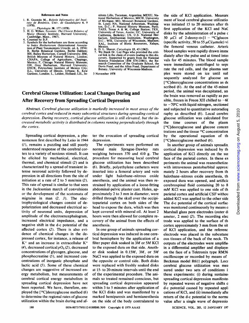
Fig. I (left). Root system of Zea diploperennis. Left plant showing a cordlike rhizome (note short internodes); right plant has two sectionedtuberous short rhizomes, which are covered with scale leaves; the arrow points to the stump of previous years' culm. Fig. 2 (right). Com-parative growth of the two perennial teosinte species in a uniform environment (University of Guadalajara greenhouse), with Zea perennis on theleft, Z. diploperennis on the right, and R. Guzmin M. in the middle. The plants were grown from rhizomes collected in the wild, and were 9months in cultivation. Plants growing in the wild would be somewhat shorter.
inged, strongly green-nerved, theierves usually clustered marginally near
:he apically ciliate prominent lateralwings; outer glume strongly enclosing in-rier glume.Female spikes sessile or often bornen long peduncles, frequently tipped by;hort male racemes; fruit cases 5 to 10?er spike, trapezoidal-cylindric, 6 to 8.2m,m on the long side, 2.5 to 4.5 mm on
:he short side, 4 to 5 mm in diameter;when mature light sepia to grayish brown;peckled with dark brown or nearly)lack; weight of 100 mature fruit cases
7.12 g.
Chromosomes number: 2n = 20; mei-)sis regular with ten bivalents (4).MEXICO: JALISCO: many, often
lense, colonies, mostly among tall;rasses and herbs (Dahlia coccinea,rhalictrum), in deep soft soil, often on
-dge of (or in) small streams, and some-
Jimes on edge of (but not in) maize fields)r in grazed pastures, on what was
,ormerly open Pinus-Quercus (ellip-ica ?)-Carpinus caroliniana forest: at)ase of rocky north-northeast-facing up-
3ermost slopes of Cerro de San Miguel(east end of Sierra de Manantlan), justnorth of and below saddle (crest) at LaVentana (104°13"W, 19'31'45"N), near an
[ndian hut surrounded by five giganticYucca (elephantipes?) trees, 20 km duesouth of El Chante, 7 km east-northeast2 JANUARY 1979
of El Durazno (Municipio de Cuautitlan).altitude 2250 to 2400 m, 22 September1978, H. H. Iltis, R. Guzman M., J.Doebley, and A. Lasseigne No. 450.The holotype is in the Herbario de la
Universidad de Guadalajara (Zapopan);isotypes (to be distributed) in B, BH,BM, CHAPA, ENCB, F, GH, ILL,K, L, LIL, MEXU, MICH, MO, NA,P, TAES, TEX, UC, US, WIS, XAL(5).
Another collection from the same pop-
ulation was distributed as Zea perennis[the location data given on this label (seebelow) and by Guzman (1) are not quitecorrect]:
Campos cultivos de mafz cerca del bosquefrio de pino, Cerro de la Ventana San Miguel,.15 km al E de la comunidad indigenia deCuzalapa, Municipio de Cuautitldn, Jalisco,1700 m alt., 15 December 1977, R. GuzmdnM. 777 [in ARIZ, Universidad AutonomaGuadalajara, Universidad Guadalajara, Zapo-pan, MICH (5)].
This collection included mature seedswhich will be distributed with the typematerial.Common name: "Chapule," "Maiz
Chapule," or "Milpilla."It is of interest that the local people re-
port grinding up and mixing the kernelswith maize for use as food in hard times.
Similar to Zea perennis, Z. diplope-rennis differs by its dimorphic rhizomes
with much shorter internodes (Fig. 1),those of Z. perennis being usually 1 to 3cm long; by its more open root systemwhich is not densely sod-forming, by thelarger number of, and longer and laxertassel branches (Fig. 2); by wider andlonger leaves; and by its considerablymore robust habit (Fig. 2).The implications of this discovery are
considerable. (i) Being morphologicallyprimitive, this diploid wild maize couldgive clues to the evolution of Zea, andspecifically to the origin of the supposed-ly autotetraploid Z. perennis (6), itsprobable descendant. (ii) Since it is adiploid perennial, and interfertile withmaize, as shown by F, hybrids, grownfrom field-collected seeds at the Univer-sidad de Guadalajara, this new speciesshould provide geneticists and maizebreeders with a potentially valuablesource of germ plasm, and may lead tothe development of perennial maize.
HUGH H. ILTISJOHN F. DOEBLEY
Department of Botany, University ofWisconsin-Madison, Madison 53706
RAFAEL GUZMAN M.Instituto de Botdnica, Apartado 139,Universidad de Guadalajara,Zapopan, Jalisco, Mexico
BATIA PAZYDepartment of Botany, HebrewUniversity, Jerusalem, Israel
187
on
Aug
ust 5
, 201
0 w
ww
.sci
ence
mag
.org
Dow
nloa
ded
from
References and Notes
1. R. Guzman M., Boletfn Informativo del Insti-tuto de Botdnica, Univ. de Guadalajara 6, 9(1978).
2. __ , Phytologia 38, 177 (1978).3. H. G. Wilkes, Teosinte: The Closest Relative of
Maize (Bussey Institute, Harvard University,Cambridge, Mass., 1967).
4. Counted by B.P.5. Symbols for the herbariums are those according
to Index Herbariorum (International Associa-tion of Plant Taxonomists Utrecht, ed. 6, 1974):B, Berlin Botanical Gardens, Berlin Dahlem;BH, Bailey Hortorium, Cornell University; BM,British Museum of Natural History, London;CHAPA, College of Agriculture, Chapingo,Mexico; F, Chicago Natural History Museum;GH, Gray Herbarium, Harvard University;ENCB, Polytechnic Institute of Mexico City;ILL, University of Illinois, Urbana; K, KewGardens, London; L, Leiden, Holland; LIL, In-
stituto Lillo, Tucuman, Argentina; MEXU, Na-tional Herbarium of Mexico; MICH, Universityof Michigan; MO, Missouri Botanical Gardens,St. Louis; NA, National Arboretum, Washing-ton, D.C.; P, Natural History Museum, Paris;TAES, Texas A & M, College Station; TEX,University of Texas, Austin; UC, University ofCalifornia, Berkeley; US, U.S. National Her-barium, Smithsonian Institution, Washington,D.C.; WIS, University of Wisconsin, Madison;XAL, Institute of Biotic Resources, Xalapa,Mexico.
6. D. L. Shaver, Caryologia 15, 43 (1962).7. We thank Dr. Luz Puga who provided the criti-
cal link in the chain of events leading to this dis-covery. Supported by grants from the NationalScience Foundation (BM S74-21861), the Re-search Committee of the Graduate School, theDavis Fund, and the Allen Fund, Department ofBotany, University of Wisconsin-Madison.
3 November 1978
Cerebral Glucose Utilization: Local Changes During andAfter Recovery from Spreading Cortical Depression
Abstract. Cerebral glucose utilization is markedly increased in most areas of thecerebral cortex and reduced in many subcortical structures during spreading corticaldepression. During recovery, cortical glucose utilization is still elevated, but the in-creased metabolic activity is distributed in columns running perpendicularly throughthe cortex.
Spreading cortical depression, a phe-nomenon first described by Ledo in 1944(1), remains a puzzling and still poorlyunderstood response of the cerebral cor-
tex to a variety of noxious stimuli. It canbe elicited by mechanical, electrical,thermal, and chemical stimuli (2) and ischaracterized by a spread of transient in-tense neuronal activity followed by de-pression in all directions from the site ofinitiation at a rate of 2 to 5 mm/min (2).This rate of spread is similar to that seen
in the Jacksonian march of convulsionsor the development of the scotomata ofmigraine in man (2, 3). The elec-trophysiological changes consist of de-polarization and decreased electrical ac-
tivity of neuronal units, depression ofamplitude of the electroencephalogram,increased electrical impedance, and a
negative shift in the d-c potential of theaffected cortex (2). There is also evi-dence of chemical changes in the de-pressed cortex, for instance, a release ofK+ and an increase in extracellular K+(4), decreased cortical P02 (2), decreasedconcentrations of glycogen, glucose, andphosphocreatine (5), and increased con-
centrations of inorganic phosphate andlactic acid (5). Some of these chemicalchanges are suggestive of increased en-
ergy metabolism, but measurements ofcerebral cortical energy metabolism inspreading cortical depression have notbeen reported. We have, therefore, em-
ployed the [14C]deoxyglucose method (6)to determine the regional rates of glucoseutilization within the brain during and af-
ter the evocation of spreading corticaldepression.The experiments were performed on
normal male Sprague-Dawley ratsweighing between 370 and 410 g. Theprocedure for measuring local cerebralglucose utilization has been described(6). Briefly, polyethylene catheters were
inserted into a femoral artery and veinunder light halothane-nitrous oxideanesthesia, and the animal was then re-
strained by application of a loose-fittingabdominal-pelvic plaster cast. Holes, ap-
proximately 2 to 3 mm in diameter, were
drilled through the skull over the occipi-toparietal cortex on both sides of thehead to expose the dura, which was thenkept covered with mineral oil. At least 2
hours were then allowed for complete re-
covery of the animal from the effects ofanesthesia.
In one group of animals spreading cor-
tical depression was induced in one cere-bral hemisphere by the application of a
filter paper disk soaked in 3M or 5M KClto the exposed dura on that side. Anoth-er disk soaked in 0.15M, 3M, or 5MNaCl was applied to the exposed dura onthe opposite or control side. Both diskswere replaced with freshly soaked disksat 15- to 20-minute intervals until the endof the experimental procedure. The ani-mals so treated remained conscious, butspreading cortical depression appearedwithin 3 to 5 minutes after application ofthe KCl disks and was manifested by a
marked hemiparesis and hemianesthesiaon the side of the body contralateral to
0036-8075/79/01 12-0188$00.50/0 Copyright K) 1979 AAAS
the side of KCl application. Measurement of local cerebral glucose utilizatiowas initiated 15 to 20 minutes after thfirst application of the KCl and NaCdisks by the administration of a pulse c50 ,uCi of 2-deoxy-D-[1 - 14C]glucos(specific activity, 50 to 55 ,uCi/,mole) vithe femoral venous catheter. ArteriCblood samples were rapidly drawn immediately after the pulse and at timed intelvals for 45 minutes. The blood samplewere immediately centrifuged to sepErate the red cells, and the plasma sawrples were stored on ice until sutsequently analyzed for glucose an[14C]deoxyglucose concentrations as described (6). At the end of the 45-minutperiod, the animal was decapitated, anthe brain was removed as rapidly as possible, frozen in Freon XII chilled to -6(to -70°C with liquid nitrogen, sectionedand subjected to quantitative autoradioEraphy as described (6). Local cerebreglucose utilization was calculated frorthe time courses of the plasm[14C]deoxyglucose and glucose concentrations and the tissue 14C concentrationby the operational equation of th[14C]deoxyglucose method (6).
In another group of animals spreadin,cortical depression was induced by thapplication of KCl directly on the suiface of the parietal cortex. In these eNperiments the animal was reanesthetizewith intravenous pentobarbital approximately 2 hours after recovery from thhalothane-nitrous oxide anesthesia, thexposed dura was opened, and artificiEcerebrospinal fluid containing 20 to 8mM KCI was applied to one side of thparietal cortex and artificial CSF withouadded KCl was applied to the other sideThe d-c potential of the cortical surfac,was monitored continuously by means aMarshall glass pore electrodes (outer diameter, 2 mm) (2). The recording electrode was applied to the surface of thcortex approximately 3 mm from the sitof KCl application, and the referencelectrode was placed in the subcutaneous tissues of the back of the neck. Thoutputs of the electrodes were amplifie,in a differential amplifier and displayeon the face of a Tektronix type RM56oscilloscope or recorded by means ofBeckman model R611 polygraph. LocEcerebral glucose utilization was measured under two sets of conditions ithese experiments: (i) during sustaineispreading cortical depression manifesteiby repeated waves of negative shifts cd-c potential caused by repeated applications of KCl, and (ii) immediately aftereturn of the d-c potential to the normEvalue after a single wave of depressio
SCIENCE, VOL. 203, 12 JANUARY 197188
on
Aug
ust 5
, 201
0 w
ww
.sci
ence
mag
.org
Dow
nloa
ded
from