tunicate 13 5 10 32 36 - repository.unhas.ac.id
TRANSCRIPT

53
Tunicate Aplidium conicum, Bioorganic & Medicinal Chemistry Letters,
13(2003): 4481–4483.
Ajuru, M.G., Williams, L.F. dan Ajuru, G., 2017, Qualitative and Quantitative
Phytochemical Screening of Some Plants Used in Ethnomedicine in the
Niger Delta Region of Nigerian, Journal of Food and Nutrition Sciences,
5(5): 198-205.
Alexander, J.W., 2009, History of the Medical Use of Silver, Surgical Infections,
10(3): 289–292.
Alqadi, M.K., Noqtah, O.A.A., Alzoubi, F.Y., Alzouby, J. dan Aljarrah, K., 2014,
pH Effect on the Aggregation of Silver Nanoparticles Synthesized by
Chemical Reduction, Materials Science-Poland, 32(1): 07-111.
Apriandanu, D., Wahyuni, S., Hadisaputro, S. dan Harjono, 2013, Sintesis
Nanopartikel Perak Menggunakan Metode Poliol dengan Agen Stabilisator
Polivinilalkohol (PVA), Jurnal MIPA, 36(2): 157-168.
Aravinthan, A., Govarthanan, M. dan Selvam, K., 2015, Unroot Mediated
Synthesis and Characterizationof Silver Nanoparticles and Evaluation of Its
Antibacterial and Rat Splenocyte Cytotoxic Effects, International Journal of
Nanomedicine,10: 1977–1983.
Arfan, A.R., 2017, Sintesis Nanopartikel Perak Menggunakan Ekstrak Hidroid
Aglaophenia Cupressina lamouroux sebagai Bioreduktor dan Uji
Potensinya sebagai Antibakteri, Skripsi tidak diterbitkan, Jurusan Kimia,
FMIPA, Universitas Hasanudin, Makassar.
Ariyanta, H.A., Wahyuni, S. dan Priatmoko, S., 2014, Preparasi Nanopartikel
Perak dengan Metode Reduksi dan Aplikasinya sebagai Antibakteri Penyebab
Infeksi, Indonesian Journal of Chemical Science, 3(1): 1-6.
Armah, Z., 2014, Sintesis dan Karakterisasi Nanopartikel Perak Menggunakan
Daun Gedi Abelmoschus manihot L. untuk Sensor Kadar Glukosa Darah,
Tesis tidak diterbitkan, Jurusan Kimia, FMIPA, Universitas Hasanudin,
Makassar.
Asmathunisha, N. dan Kathiresan, K., 2013, A Review on Biosynthesis of
Nanoparticles by Marine Organisms, Colloids and Surfaces B:Biointerfaces,
103: 283–287.
Astuti, S.M., Sakinah, M.A.M., Andayani, R.B.M. dan Risch, A., 2011,
Determination of Saponin Compound from Anredera cordifolia (Ten)
Steenis Plant (Binahong) to Potential Treatment for Several Diseases,
Journal of Agricultural Science, 3(4): 224-232.
Backhouse, G., 2012, Pyura sp, Invertebrates of Coral Sea.

54
Bandyopandhyay, A.K., 2008, Nano Material, New Age International Ltd., New
Delhi.
Bargah, R.K., 2015, Preliminary Test of Phytochemical Screening of Crude
Ethanolic and Aqueous Ecxtract of Moringa pterygosperm Gaertn, Journal
of Pharmacognosy and Phytochemistry, 4(1): 7-9.
Bettelheim, K.A., 2000, Role of non O157 VTEC, J. Appl. Symp. Microbiol.
Suppl, 88:38-50. Buzea, C., Blandino, I.I.P. dan Robbie, K., 2007, Nanomaterial and
Nanoparticles: Sources and Toxicity, Biointerphases, 2: 170-172.
Canham, G.R. dan Overton, T., 2000, Descriptive Inorganic Chemistry, W.H.
Freeman and Company, New York.
Carter, G.R dan Wise, D.J., 2004, Essentials of Veterinary Bacteriology and
Mycology Sixth Edition, Iowa State Press, Iowa, USA.
Chandran, S.P., Chaudhary, M., Pasricha, R., Ahmad, A. dan Sastry, M., 2006,
“Synthesis of Gold Nanotriangles and Silver Nanoparticles Using Aloe vera
Plant Extract,” Biotechnology Progress, 22(2): 577–583.
Chauhan, R., Kumar, A. dan Abraham, J.A., 2013, Biological
Approach to the Synthesis of Silver Nanoparticles with
Streptomyces sp. JAR1 and its Antimicrobial Activity, Sci Pharm, 81:607-21.
Chou, K.S. dan Lu, Y.C., 2008, High Concentration Nanoscale Silver Colloidal
Solution and Preparing Process Thereof, Patent Application Publication,
6:47-67. Coulter, B., 2008, Delsa Nano Series, (Online)
(http://www.dafratec.com/pdf/catalogo, diakses 10 Maret 2018).
Cowan, S.T., 1984, Manual for the Identification of Medical Bacteria, 2nd Ed,
Cambridge University Press, USA. Daniel, S.C.G.K., Nehru, K. dan Sivakumar, M., 2012, Rapid Biosynthesis of
Silver Nanoparticles Using Eichornia crassipes and its Antibacterial
Activity, Curr. Nanosci., 8: 125–129.
Das, R., Nath, S.S., Chakdar, D. dan Gope, G., 2009, Preparation of Silver
Nanoparticles and Their Characterization Synthesis of Silver Nanoparticles,
Journal of Materials,1–9.
DeLeo, F.R., Otto, M., Kreiswirth, B.N. dan Chambers, H.F., 2010, Community-
Associated Meticillin-Resistant Staphylococcus aureus, Laboratory of

55
Human Bacterial Pathogenesis, Rocky Mountain Laboratories, National
Institute of Allergy and Infectious Diseases, National Institutes of Health,
Hamilton.
Dewi, A.K., 2013, Isolasi, Identifikasi dan Uji Sensitivitas Staphylococcus aureus
terhadap Amoxicillin dari Sampel Susu Kambing Peranakan Ettawa (PE)
Penderita Mastitis di Wilayah Girimulyo, Kulonprogo, Yogyakarta, Jurnal
Sain Veteriner, 31(2): 138-150.
Duran, N., Marcato, P.D., De Conti, R., Alves O.L., Costab, F.T.M. dan
Brocchib, M., 2010, Potential Use of Silver Nanoparticles on Pathogenic
Bacteria, their Toxicity and Possible Mechanisms of Action, J Braz Chem
Soc, 21(6): 949–959.
Edhaya, N.B. dan Prakash, S., 2013, Biological Synthesis of Gold Nanoparticles
Using Marine Algae Gracilaria corticata and its Application as A Potent
Antimicrobial and Antioxidant Agent, Asian J. Pharm Clin Res, 6(2):
179–182. Elgorban, A.M., El-Samawaty, A.E.M., Yassin, M.A., Sayed, S.R., Adil, S.F.,
Elhindi, K.M. dan Khan, M.B.M., 2016, Antifungal Silver Nanoparticles:
Synthesis, Characterization and Biological Evaluation, Biotechnology &
Biotechnological Equipment, 30(1): 56-62.
El-Shishtawy, R.M., Asiri, A.M. dan Al-Otaibi, M.M., 2011, Synthesis and
Spectroscopic Studies of Stable Aqueous Dispersion of Silver
Nanoparticles, Spectrochim Acta Part A Mol Biomol Spectrosc,79(5):
1505–1510. Feldheim, D.L. dan Foss, C.A.Jr., 2002, Metal Nanoparticles: Sinthesis,
Characterization and Appalications, Marcel Dekker Inc., Switzerland. Firdhouse, M.J. dan Lalitha, P., 2015, Biosynthesis of Silver Nanoparticles and its
Applications, Journal of Nanotechnology, 1-18.
Gong, P., Li, H., He, X., Wang, K., Hu, J., Zhang, S. dan Yang, X., 2007,
Preparation and Antibacterial Activity of Fe3O4 and Ag Nanoparticles,
Nanotechnology, 18(28): 604-611. Gopinath, V., Ali, D.M., Priyadarshini, S., Priyadharsshini, N.M., Thajuddin, N.
dan Velusamy, P., 2012, Biosynthesis of Silver Nanoparticles from Tribulus
terrestris and its Antimicrobial Activity: A Novel Biological Approach,
Colloids and Surfaces B: Biointerfaces, 96:69–74.
Haerudin, A., Pujilestari, T. dan Atika, V., 2017, Pengaruh Jenis Pelarut terhadap
Hasil Ekstraksi Rumput Laut Gracilaria sp. sebagai Zat Warna Alam pada
Kain Batik Katun dan Sutera, Dinamika Kerajinan dan Batik , 34(2): 83-92.
Hamed, M.R. dan Givianrad, M.H., 2015, Biosynthesis of Silver Nanoparticles
Using Marine Sponge Haliclona, Oriental Journal of Chemistry, 31(4):

56
1961–1967.
Handayani, W., Bakir, Imawan, C. dan Purbaningsih, S., 2010, Potensi Ekstrak
Beberapa Jenis Tumbuhan sebagai Agen Pereduksi untuk Biosintesis
Nanopartikel Perak, Seminar Nasional Biologi, 558-567.
Haryani, Y., Kartika, G.F., Yuharmen, Putri, E.M., Alchalish, D.T. dan Melanie,
Y., 2016, Pemanfaatan Ekstrak Air Rimpang Jahe Merah (Zingiber
officinale Linn. Var. Rubrum) pada Biosintesis Sederhana Nanopartikel
Perak, Chimica et Natura Acta, 4(3): 151-155.
Haryono, A., Sondari, D., Harmani, S.B. dan Randy, M., 2008, Sintesa
Nanopartikel Perak dan Potensi Aplikasinya, Jurnal Riset Industri, 2(3):
156-163.
Horikoshi, S. dan Serpone, N., 2013, Introduction to Nanoparticles, Microwaves
in Nanoparticle Synthesis First Edition, Wiley-VCH Verlag GmbH & Co.
KgaA, Canada.
Hosokawa, M., Nishino, J. Dan Kanno, Y., 2007, Nanoparticle Technology
Handbook 1st Edition, Jordan Hill, Oxford.
Hussain, M.A., 2014, One Pot Light Assisted Green Synthesis, Storage and
Antimicrobial Activity of Dextran Stabilized Silver Nanoparticles,
J. Nanobiotechnol, 12(53):1-6.
Inbakandan, D., Sivaleela, G., Peter, D.M., Kiurbagaran, R., Venkatesan, R. dan
Khan, S.A., 2012, Marine Sponge Extract Assisted Biosynthesis of Silver
Nanoparticles, Materials Letters, 87: 66–68. Jawetz, E., Melnick, J.L. dan Adelberg, E.A., 2001, Mikrobiologi Kedokteran
Edisi 2, Penerbit Buku Kedokteran, Jakarta. Jawetz, E., Melnick, J.L. dan Adelberg, E.A., 2005, Mikrobiologi Kedokteran
Buku 1, Salemba Medika, Jakarta. Kashiwagi, T., 2005, Flammability of Nanocomposites–Effects of the Shape of
Nanoparticles, New Applications of Mineral Fillers, Royal Society of
Chemistry, United Kingdom.
Kathiresan, K., Manivannan, S., Nabeel. M.A. dan Dhivya, B., 2009,
Studies on Silver Nanoparticles Synthesized by A Marine
Fungus, Penicillium fellutanum Isolated From Coastal Mangrove Sediment. Colloids Surf B, 71(1):133–137.
Kawashima, Y., Yamamoto, H., Takeuchi, H. Dan Kuno, Y., 2000, Mucoadhesive
DL-lactide/glycolide Copolymer Nanospheres Coated with Chitosan to

57
Improve Oral Delivery of Elcatonin, Pharmaceutical Development and
Technology, 5(1): 77-85.
Keat, C.L., Aziz, A., Eid, A.M. dan Elmarguzi, N.A., 2015, Biosynthesis of
Nanoparticles and Silver Nanoparticles, Bioresources and Bioprocessing,
15(2): 47–57.
Khan, Z., Singh, T., Ijaz, J., Yousif, A., Al-thabaiti, S.A. dan El-mossalamy, E.H.,
2013, Colloids and Surfaces B: Biointerfaces Starch-directed Green
Synthesis, Characterization and Morphology of Silver Nanoparticles,
Colloids and Surfaces B: Biointerfaces, 102: 578–584.
Kholoud, M.M., 2009, Synthesis and Applicationsof Silver Nanoparticles,
Arabian Journal of Chemistry, 3:135–140.
Kott, P., 1989, Form and Function In the Sea Ascidiacea, Bulletin Of Marine
Science, 45: 253-276.
Kumar, V. dan Yadaf, S.K., 2009, Plant-Mediated Synthesis of Silver and Gold
Nanopartcles and their Applications, Journal Chemical Technology and
Biotechnology, 84:151-157.
La Tapa, F., Suryanto, E. dan Momuat, L.I., 2016, Biosintesis Nanopartikel Perak
Menggunakan Ekstrak Empelur Batang Sagu Baruk (Arenga microcarpha)
dan Aktivitas Antioksidannya, Chem. Prog., 9(1): 9-15.
Lambert, G., 2004, Relaxing and Fixing Ascidian for Taxonomy, (Online),
(www.depts-washington.edulascidian.htm., diakses 1 Maret2018). Lavanya, M., 2013, Synthesis, Characterization and Evaluation of Antimicrobial
Efficacyof Silver Nanoparticles Using Paederia foetida L. Leaf Extract, Int
Res J Biol Sci, 2(3):28-34. Lay, B.W., 1994, Analisis Mikroba di Laboratorium, Edisi 1, Raja Grafindo
Persada, Jakarta. Leela, A. dan Vivekanandan, M., 2008, Tapping the Unexploited Plant Resources
for the Synthesis of Silver Nanopartikel, African Journal of Biotechnology,
7(17): 3162-3165. Lembang, E.Y., 2014, Sintesis Nanopartikel Perak dengan Metode Reduksi
Menggunakan Bioreduktor Ekstrak Daun Ketapang (Terminalia catappa),
Skripsi tidak diterbitkan, Jurusan Kimia, FMIPA, Universitas Hasanudin,
Makassar.
Li, Mahendra, Q., Lyon, S., Brunet, D.Y., Liga, L., Li, M.V. dan Alvarez, P.J.J.,
2008, Antimicrobial Nanomaterials for Water Disinfection and Microbial
Control: Potential Applications and Implications, Water Res, 42(1):
4591–4602.

58
Litaay, M., Grace, C., Risco, G.B. dan Zaraswati, D., 2015, Bioaktivitas Simbion
Tunikata Polycarpa aurata sebagai Antimikroba, Seminar Nasional Biologi
ke XXIII PBI, Jayapura.
Lowenstam, H.A., 1989, Spicular Morphology and Mineralogy In Some Pyuridae
(Ascidiacea), Bulletin of Marine Science,45(2): 243-252.
Lowry, F.D., 1998, Staphylococcus aureus Infection, N Engl J Med, 339:520-532.
Madhuri, S., Maheshwar, S., Sunil, P. dan Oza, G., 2012, Nanotechnology:
Concepts and Applications, CRC Press, USA.
Makarov, V.V., Love, A.J., Sinitsyna, O.V., Makarova S.S. dan Yaminsky, I.V.,
2014, ”Green”Nanotechnologies: Synthesis of Metal Nanoparticles Using
Plants, Acta Naturae, 6(1)20: 35-44.
Malik, P., Shankar, R., Malik, V., Sharma, N.dan Mukherjee, T.K., 2014, Green
Chemistry Based Benign Routes for Nanoparticle Synthesis, J Nanopar.
Manivasagan, P., 2014, Actinobacteria Mediated Synthesis of
Nanoparticles and their Biological Properties, Crit Rev Microbiol, 28:1-13.
Maryani, D., Firdaus, M.L. dan Nurhamidah, 2017, Biosintesis
Nanopartikel Perak Menggunakan Ekstrak Buah Passiflora flavicarva
(Markisa) untuk Mendeteksi Logam Berat, Alotrop Jurnal Pendidikan dan
Ilmu Kimia, 1(1): 49-54.
Masakke, Y., Sulfikar dan Rasyid, M., 2015, Biosintesis Partikel-nano Perak
Menggunakan Ekstrak Metanol Daun Manggis (Garcinia mangostana L.), Jurnal Sainsmat, 4(1): 28-41.
Mishra, P., 2009, Isolation, Spectroscopic Characterization and Molecular
Modeling Studies of Mixture of Curcuma longa, Ginger and Seeds of
Fenugreek, International Journal of Pharm Tech Research,1(1): 79-95. Mittal, A.K., Thanki, K., Jain, S. dan Banerjee, U.C., 2016, Comparative Studies
of Anticancer and Antimicrobial Potential of Bioinspired Silver and Silver-
Selenium Nanoparticles, Applied Nanomedicine, 1(1): 1-6. Mo, Y., Tang, Y., Wang, S., Lin, J. dan Zhang, H., 2015, Green Synthesis of
Silver Nanoparticles Using Eucalyptus Leaf Extract, Mater. Lett., (144):
165-167. Mock, J.J., 2002, Shape Effects In Plasmon Resonance of Individual Colloidal
Silver Nanoparticles, Journal of Chemical Physics,116: 6755-6759.

59
Mohanpuria, P., Rana, N. dan Yadav, S., 2008, Biosynthesis of Nanoparticles:
Technological Concepts and Future Applications, J. Nanopart Res, 10(3):
507–517.
Mpila, D., Fatimawali, F. dan Wiyono, W., 2012, Uji Aktivitas Antibakteri
Ekstrak Etanol Daun Mayana (Coleus atropurpureys [L] Benth) terhadap
Staphylococcus aureus, Escherichia coli dan Pseudomonas aeruginosa
secara In-Vitro, Pharmacon, 1(1): 13–21. Muharni, Fitrya dan Farida, S., 2017, Uji Aktivitas Antibakteri Ekstrak Etanol
Tanaman Obat Suku Musi di Kabupaten Musi Banyuasin, Sumatera Selatan,
Jurnal Kefarmasian Indonesia, 7(2): 127-135. Nagarajan, R. dan Horton, T.A., 2008, Nanoparticles: Synthesis, Stabilization,
Passivation and Functionalization, American Chemical Society,
Washington DC. Natarajan, K., Selvaraj, S. dan Ramachandra, M.V., 2010, Microbial Production
of Silver Nanoparticles, Digest Journal of Nanomaterials and Biostructures,
(5):135-140.
Norouzi, H., Larijani, K. dan Behmadi, H., 2016, Silver Nanoparticles Green
Synthesis Using Aqueous Extract Citrus reticulate var page, Oriental
Journal Of Chemistry, 32(4): 2199-2203.
Nurwantoro dan Abbas, S., 2001, Mikrobiologi Pangan Hewani Nabati, Kanisius,
Yogyakarta. Panda, S.S., 2014, Synthesis of Silver Nanoparticles From Plant Extract and Its
Application In Cancer Treatment: A Review, Int. J. Plant Animal Environ
Sci, 4(3):137-145.
Panigrahi, S., Kundu, S., Ghosh, S. K., Nath, S. dan Pal, T., 2004, General
Method of Synthesis for Metal Nanoparticles, Journal of Nanoparticle
Research, 6: 411-414.
Paryati, S.P.Y., 2002, Patogenesis Mastitis Subklinis pada Sapi Perah yang
Disebabkan oleh Staphylococcus aureus, Makalah Pengantar Falsafah
Sains, Institute Pertanian Bogor.
Phanjom, P. dan Ahmed, G., 2017, Effect of Different Physicochemical
Conditions on the Synthesis of Silver Nanoparticles Using Fungal Cell
Filtrate of Aspergillus oryzae (MTCC No. 1846) and their Antibacterial
Effect, Advances in Natural Sciences: Nanoscience and Nanotechnology,
8(2017):1-13.
Prabhu, S. dan Poulose, E., 2012, Silver Nanoparticles: Mechanism of
Antimicrobial Action, Synthesis, Medical Applications and Toxicity Effects,
Int Nano Lett, 2(1):1–10.

60
Purnomo, S.R., Rupiasih, N.N. dan Sumadiyasa, M., 2017, Sintesis Nanopartikel
Perak dengan Metode Biologi Menggunakan Ekstrak Tanaman Sambiloto
(Andrographis paniculata Ness), Buletin Fisika, 18(1): 6–11.
Suarsa, I.W. dan Suarya, P., 2011, Optimasi Jenis Pelarut dalam Ekstraksi Zat
Warna Aalm dan Batang Pisang Kapok (Musa paradiasiaca L. cv kepok)
dan Batang Pisang Susu (Musa paradiasiaca L. cv susu), Jurnal Kimia 5,
5(1), 72–80.
Qi, W.H. dan Wang, M.P., 2004, Size and Shape Dependent Melting Temperature
of Metallic Nanoparticles, Mater Chem Phys, 88: 280–284.
Rajeshkumar, S., Kannan, C. dan Annadurai, G., 2012, Green Synthesis of Silver
Nanoparticles Using Marine Brown Algae Turbinaria conoides and its
Antibacterial Activity, Int. J. Pharm. Bio. Sci., 3(4): 502–510.
Ravani, S., 2011, Green Synthesis of Metal Nanoparticles Using Plants, Green
Chem, 13(10): 2638–2650.
Rawle, A., 2010, Basic Principles of Particle Size Analysis, Technical paper of
Malvern Instruments, Worcestershire, United Kingdom. Rismana, E., Kusumaningrum, S., Olivia B.P., Rosidah, I. dan Marhamah, 2012,
Sintesis dan Karakterisasi Nanopartikel Kitosan Ekstrak Kulit Buah
Manggis (Garcinia mangostana), Jurnal Sains dan Teknologi Indonesia,
14(3): 189-196. Ristian, I., 2013, Kajian Pengaruh Konsentrasi Perak Nitrat (AgNO3) terhadap
Ukuran Nanopartikel Perak, Skripsi tidak diterbitkan, Jurusan Kimia,
FMIPA, UNNES, Semarang. Rius, M. dan Peter, R.T., 2011, Article A Revision of the Pyura stolonifera
Species Complex (Tunicata, Ascidiacea), with a Description of a New
Species from Australia, Zootax, 2754: 27–40.
Roduner, E., 2006, Size Matters: Why Nanomaterials Are Different, Chem Soc
Rev, 35(7): 583–592.
Rohdiana, D., Arief, D.Z. dan Budiman, A., 2013, Aktivitas Penghambatan
Pertumbuhan Bakteri Escherichia coli oleh Berbagai Jenis Teh dan
Seduhannya, Jurnal Penelitian Teh dan Kina, 16(1): 37-44.
Rupper, E., 2004, Invertebrate Zoology: A Functional Evolutionary Approach,
Cole Pub. Co, Brooks.
Ruppert, E.E., Fox, R.S. dan Barnes, R.D., 2004, Invertebrate Zoology: A
Functional Evolutionary Approach, Thomson Brooks, USA.

61
Sanghi, R. dan Verma, P., 2010, pH Dependant Fungal Proteins In
the ‘Green’ Synthesis of Gold Nanoparticles, Adv Mat Lett, 1(3): 193-9.
Santhi, K. Dan Sengottuvel, R., 2016, Qualitative and Quantitative Phytochemical
Analysis of Moringaconcanensis Nimmo, International Journal of Current
Microbiology and Applied Sciences, 5(1): 633-640.
Sardiani, N., 2015, Potensi Tunikata Rhopalea sp sebagai Sumber Inokulum
Bakteri Endosimbion Penghasil Antibakteri, Skripsi tidak diterbitkan,
Jurusan Biologi, FMIPA,Universitas Hasanuddin, Makassar.
Sari, D.K., Lestari, R.S.D. dan Rahmat, A., 2016, Biosintesis
Nano/Mikro Partikel Perak dari Rumput Laut (Eucheuma cottonii) Berbantu
Gelombang Ultrasonik, Seminar Nasional Sains dan Teknologi, 1-10.
Satalkar, P., Elger, B.S. dan Shaw, D.M., 2015, Defining Nano, Nanotechnology
and Nanomedicine: Why Should It Matter?, Science and Engineering
Ethics.
Seabra, A.B., Haddad, P. dan Duran, N., 2013, Biogenic Synthesis of
Nanostructured Iron Compounds: Applications and Perspective, IET
Nanobiotechnol, 7: 90-99.
Shankar, S.S., Rai, A., Ahmad, A. dan Sastry, M., 2004, Rapid Synthesis of Au,
Agand Bimetalic Au core-Ag Shell Nanoparticles Using Neem (Azadirachta
indica) Leaf Broth. Journal of Colloid and Interface Science, 275(2):
496-502.
Sharma, V.K., Yngard, R.A. dan Lin, Y., 2009, Silver Nanoparticles: Green
Synthesis and Their Antimicrobial Activities, Advances in Colloid and
Interface Science, 145(1–2): 83–96.
Singh, M.J., 2016, Green Nano Actinobacteriology An Interdisciplinary Study,
(Online ), (http://dx.doi.org/10.5772/61308, diakses 12 Maret 2018).
Singh, P. dan Balaji R.R., 2011, Biological Synthesis of
Nanoparticle from Trichodermah harzianum, Asian J Exp Biol Sci, 2(4):600-5.
Singh, P.,Kim, Y.J., Singh, H.,Wang, C. dan Hwang, K.H., 2015, Biosynthesis, Characterization and Antimicrobial Applications of Silver
Nanoparticles, International Journal of Nanomedicine, 10: 2567–2577.
Suwignyo, S., 2005, Avertebrata Air Jilid 1, Swadaya, Jakarta.
62
Syahrurachman, A., Chatim, A., Soebandrio, A. dan Karuniawati, A., 1993, Buku
Ajar Mikrobiologi Kedokteran, Edisi Revisi, Binarupa Aksara.
Thamer, N.A. dan Almashhedy, L.A., 2014, Green synthesis Optimization and
Characterization of Silver Nanoparticles Using Aqueous Extract of Crocus
sativus L., Int J Pharm Bio Sci, 5(4): 759-70.
Todar, K., 1998, Bacteriology 330 Lecture Topics: Staphylococcus, University of
Wisconsin Department of Bacteriology, Wisconsin, USA.
Todar, K., 2002, Staphylococcus Bacteriology at UW-Bacteriology 330:1-7.
Umayaparvathi, S., Arumugam, M., Meenakshi, S. dan Balasubramanian, T.,
2013, Biosynthesis of Silver Nanoparticles Using Oyster Saccostrea
Cucullata (Born, 1778): Study of in-Vitro Antimicrobial Activity,
International Journal of Science and Nature, 4(1): 199–203.
Vanaja, M., Rajeshkumar, S., Paulkumar, K., Gnanajobitha, G., Malarkodi, C. dan
Annadurai, G., 2013, Phytosynthesis and Characterization of Silver
Nanoparticles Using Stem Extract of Coleus aromaticus, International
Journal of Materials and Biomaterials Applications, 3(1): 1-4.
Vidhya, A. dan Balagurunathan, R., 2013, Isolation and Screening
of Alkalophilic Actinobacteria, Streptomyces for Biosynthesis
and Characterization of Silver Nanoparticles, Int J Novel Trends Pharm Sci, 3(1): 7-14.
Wahyudi, T. dan Rismayani, S., 2008, Aplikasi Nanoteknologi pada Bidang
Tekstil, Arena Tekstil, 23(2): 52-109.
Wahyudi, T., Sugiyana, D. dan Helmy, Q., 2011, Sintesis Nanopartikel Perak dan
Uji Aktivitasnya terhadap Bakteri E. coli dan S. aureus, Arena Tekstil,
26(1): 1-60.
Wiley, B.J., Im, S.H., Li, Z.Y., McLellan, J., Siekkinen, A. dan Xia, Y., 2006,
Maneuveringthe Surface Plasmon Resonance of Silver Nanostructure
through Shape-Controlled Synthesis, J. Phys. Chem. B.,110:15666-15675.
Willems dan Wildenberg, V.D., 2005, Roadmap Report on Nanoparticle, W & W
Españas, Spain.
Yeo, S.Y., Lee, H.J. dan Jeong, S.H., 2003, Preparation of Nanocomposite Fiber
for Permanent Antibacterial Effect, J. Mater. Sci., 38: 2143-2147.
Yuwono, 2012, Metichillin Resistant Staphylococcus Aureus (MRSA), Skripsi
tidak diterbitkan, Fakultas Kedokteran Universitas Sriwijaya, Palembang.

63
Zarina, A., 2014, Green Approach for Synthesis of Silver
Nanoparticles from Marine Streptomyces-MS 26 and their
Antibiotic Efficacy, J Pharm Sci Res, 6(10): 321-7. Zeng, L. dan Swalla, B.J., 2005, Molecular Phylogeny of the Protochordates:
Chordate Evolution, Cannadian Journal of Zoology, 83: 24-33.
Zhang, X., Yan, S., Tyagi, R.D. dan Surampalli, R.Y., 2011, Synthesis of
Nanoparticles by Microorganisms and Their Application in Enhancing
Microbiological Reaction Rates, Chemosphere, 82: 489–494.

64
Lampiran 1. Diagram Alur Penelitian
Ekstraksi
Optimasi pH dan
Komposisi Larutan
Sintesis Nanopartikel Perak
Karakterisasi Uji Antibakteri
Spektrofotometer
UV-Vis PSA XRD FTIR
Uji Fitokimia

65
Lampiran 2. Bagan Kerja Preparasi Sampel dan Ekstraksi Sampel
- Dibilas dengan air bersih dan ditiriskan
- Dibilas dengan alkohol 70%
- Dikeringkan dibawah sinar matahari
- Dikeringkan dengan menggunakan freeze dryer
- Dihaluskan dengan menggunakan mesin penggiling
- Ditimbang sebanyak 5 gram
- Direbus dengan akuabides 100 mL hingga suhu
70-90 oC
- Didinginkan
- Disaring dengan menggunakan kertas saring
Whatmann No. 41
Sampel
Ekstrak Air dari Pyura sp

66
Lampiran 3. Bagan Kerja Optimasi pH
Ekstrak Air dari Pyura sp
- Dipipet sebanyak 1 mL
- Ditambahkan ke dalam Erlenmeyer yang berisi 40 mL
AgNO3 1 mM
- Ditambahkan NaOH 0,1 M hingga diperoleh pH 7, 8, 9,
10, 11, 12 dan 13.
- Diaduk selama 2 jam
- Dianalisis dengan spektrofotometer UV-Vis
pH Optimum

67
Lampiran 4. Bagan Kerja Optimasi Komposisi Larutan
- Dipipet sebanyak
1 mL ke dalam
Erlenmeyer yang
berisi 30 mL
AgNO3 1 mM
- Diatur pH larutan
pada pH optimum
- Diaduk selama 2
jam
- Dipipet sebanyak
1 mL ke dalam
Erlenmeyer yang
berisi 40 mL
AgNO3 1 mM
- Diatur pH larutan
pada pH optimum
- Diaduk selama 2
jam
- Dipipet sebanyak
2.5 mL ke dalam
Erlenmeyer yang
berisi 25 mL
AgNO3 1 mM
- Diatur pH larutan
pada pH optimum
- Diaduk selama 2
jam
- Dipipet sebanyak
1.5 mL ke dalam
Erlenmeyer yang
berisi 30 mL
AgNO3 1 mM
- Diatur pH larutan
pada pH optimum
- Diaduk selama 2
jam
AgNPs 1:10 AgNPs 1:20 AgNPs 1:30 AgNPs 1:40
- Analisis Absorbansi dan panjang gelombang
maksimum dengan spektrofotometer UV-Vis
Komposisi Optimum
Ekstrak air dari Pyura sp

68
Lampiran 5. Bagan Kerja Uji Fitokimia
Catatan : 1. Terbentuknya endapan hijau menandakan adanya tannin
2. Terbentuknya endapan berwarna kuning (penambahan timbal asetat)
dan endapan berwarna jingga (penambahan asam sulfat)
menandakan adanya flavonoid
3. Terbentuknya busa yang banyak menandakan adanya saponin
Catatan: 1. Terjadinya perubahan warna dari ungu menjadi biru atau hijau
menandakan adanya steroid
2. Terbentuknya warna merah pada lapisan antarmuka (interface)
menujukkan adanya terpenoid
Ekstrak air dari Pyura sp
- diambil
sebanyak 2 mL
- dicampur
dengan 2 mL
akuades
- ditambahkan
beberapa tetes
FeCl3
Hasil
- diambil
sebanyak 2,5 mL
- ditambahkan
beberapa tetes
akuades
- dikocok dengan
kencang
Hasil
Uji Tanin Uji Saponin Uji Flavonoid
- dimasukkan ke dua tabung
reaksi
- tabung reaksi pertama
ditambahkan beberapa tetes
Pb(CH3COO)2
- tabung reaksi kedua
ditambahkan beberapa tetes
H2SO4
Hasil
Ekstrak air dari Pyura sp
- diambil sebanyak 5 mL
- ditambahkan 2 mL anhidrida
asetat
- ditambahkan 2 mL H2SO4
secara perlahan
Hasil
Uji Steroid Uji Terpenoid
- diambil sebanyak 5 mL
- ditambahkan 2 mL CHCl3
- ditambahkan 3 mL H2SO4 secara
perlahan hingga terbentuk lapisan
antarmuka (interface)
Hasil

69
Catatan : Terbentuknya endapan berwana oranye pada saat penambahan reagen
Dragendorff dan endapan berwarna krim kekuning-kuningan pada saat
penambahan reagen Mayer menandakan adanya alkaloid.
Ekstrak air dari Pyura sp
- diambil sebanyak 3 mL
- ditambahkan 3 mL HCl 1 % lalu
diaduk di atas penangas
- dimasukkan ke dalam dua tabung
reaksi berbeda masing-masing
sebanyak 1 mL
Uji
Alkaloid
- ditambahkan beberapa
tetes reagen Dragendorff
- ditambahkan beberapa
tetes reagen Mayer
Hasil Hasil

70
Lampiran 6. Bagan Kerja Sintesis Nanopartikel Perak
- Disentrifuse pada kecepatan 10000 rpm selama 30 menit
pada suhu 4oC sebanyak 2-3 kali
- Disuspensikan dengan akuabides
- Disentrifuse kembali pada 8000 rpm selama 15 menit
- Dikeringkan dengan metode freeze dried pada suhu 4oC
Nanopartikel Perak
Larutan AgNO3 1 mM
- Dipipet sebanyak 200 mL kedalam erlenmeyer
- Diteteskan sebanyak 5 mL ekstrak air dari Pyura sp kedalam
erlenmeyer (1:40)
- Ditambahkan NaOH 0,1 M hingga pH 8
- Distirrer selama 2 jam
- Diamati perubahan warna yang terjadi
Koloid Nanopartikel Perak

71
Lampiran 7. Bagan Kerja Karakterisasi Nanopartikel
,
Koloid Nanopartikel Perak
Spektrofotometri UV-Vis
Konfirmasi terbentuknya
nanopartikel serta
Distribusi ukuran
nanopartikel
- dikarakterisasi
Analisis PSA
- Dipipet sebanyak 4 mL
kedalam kuvet
- Dilakukan pengukuran
pada panjang gelombang
185 – 700 nm
- Dipipet sebanyak 3 mL
kedalam kuvet kuarsa
- Dilakukan pengukuran
ukuran partikel
Gugus fungsi
- Nanopartikel perak sebanyak 2
mg dicampur dengan 100 mg KBr
kemudian dibuat pelet
- Pelet yang telah dibuat dianalisis
dengan FTIR
Nanopartikel Perak

72
Lampiran 8. Bagan Kerja Uji Bioaktivitas Antibakteri
- Ditimbang sebanyak 5 gram dimasukkan kedalam gelas kimia
- Dilarutkan dengan 250 mL akuades di dalam labu erlenmeyer
- Dihomogenkan dengan stirrer diatas penangas air hingga mendidih dan
diatur pH nya menjadi pH 7.
- Nutrien agar dimasukkan ke dalam tabung reaksi sebanyak ± 5 mL
- Disterilkan dalam autoklaf pada suhu 121oC tekanan 2 atm selama 15
menit
- Didinginkan hingga suhu mencapai 40 – 45oC pada kemiringan 30o
- Bakteri Escherichia coli sebanyak 2-3 ose di gores ke dalam medium
nutrient agar miring secara aseptis
- Diinkubasi dalam incubator pada suhu 37oC selama 18-24 jam
- Diswab merata pada permukaan media Mueller Hinton Agar (MHA)
yang terdapat pada cawan petri
- Didiamkan selama 5 menit
-
- Nanopartikel perak ditimbang sebanyak 7.2 mg kemudian dilarutkan
dengan 2 mL akuades steril.
- Kertas cakram dicelupkan kedalam larutan nanopartikel, AgNO3,
ekstrak air dari Pyura sp, ampisilin (kontrol positif) dan akuades
(kontrol negatif) selama 15 menit.
- Diletakkan diatas media MHA yang telah berisi bakteri
- Diinkubasi pada suhu 37oC selama 2×24 jam
- Diamati dan diukur diameter hambat yang terbentuk
Catatan: Diberikan perlakuan yang sama berdasarkan bagan kerja diatas untuk
bakteri Staphylococus aureus.
Nutrien Agar
Media Uji Bioaktivitas Bakteri
Daya Hambat Bakteri
Nutrien Agar Miring
Bakteri Uji

73
Lampiran 9. Dokumentasi Penelitian
Gambar 5. Proses sintesis
nanopartikel perak
Gambar 1. Sampel Pyura sp
sebelum dihaluskan
Gambar 2. Sampel Pyura sp
setelah dihaluskan
Gambar 3. Proses ekstraksi Gambar 4. Ekstrak air dari Pyura sp
Gambar 6. Nanopartikel perak

74
Lampiran 10. Hasil Uji Fitokimia
Gambar 7. Uji Tanin Gambar 8. Flavonoid 1 Gambar 8. Flavonoid 2
Gambar 9. Uji Saponin Gambar 10. Uji Steroid Gambar 10. Uji Terpenoid
Gambar 11. Uji Alkaloid 1 Gambar 12. Uji Alkaloid 2

75
Lampiran 11.Spektrum FT-IR Ekstrak Air dari Pyura sp

76
Lampiran 12.Spektrum FT-IR Nanopartikel Perak

77
Lampiran 13. Hasil Analisis XRD

78
Lampiran 14. Data JCPDS Nanopartikel Perak
79
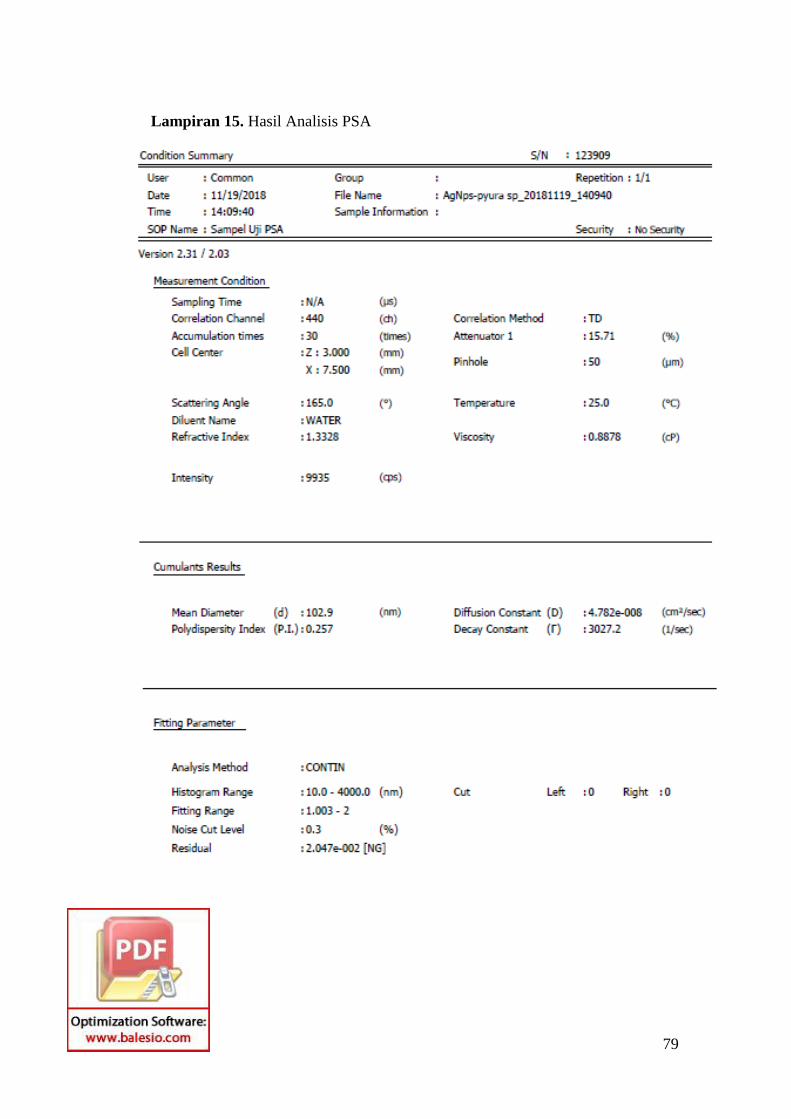
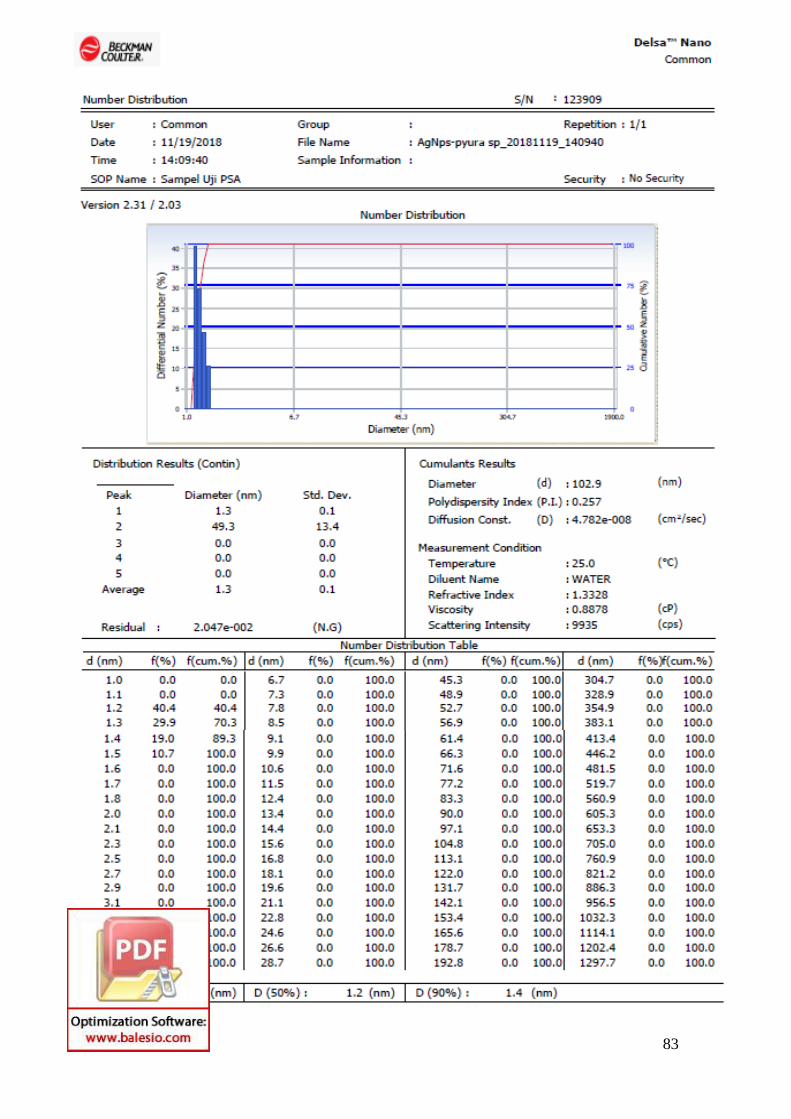
Lampiran 15. Hasil Analisis PSA

80

81

82
83

84

85

86
Lampiran 16. Pengamtan Uji Aktivias Antibakteri
/
24 jam
Gambar 14. Pengamatan 24 jam
Gambar 13. Pengamatan 18 jam
Gambar 15. Pengamatan 48 jam

87
Lampiran 17. Hasil Uji Aktivias Antibakteri
Sampel
E. Coli S. Aureus
18 Jam 24 Jam 48 Jam 18 Jam 24 Jam 48 Jam
NPAg 11,85 11,8 9,5 12,525 12,875 11,425
AgNO3 10,1 11,5 10,15 8,95 11,2 7,65
Ekstak Pyura 8,5 9,1 9,55 7,45 7,95 7,425
Kontrol (+) 25,375 24,225 17,775 24,9 24,3 23,2
Kontrol (-) 0 0 0 0 0 0