unc radiology malawi: partnership boosts global …
TRANSCRIPT
Inside this Issue:
UNC RADIOLOGY MALAWI: PARTNERSHIPBOOSTS GLOBAL HEALTH IMAGING CARE
Former Diagnostic Radiology Program Director (2011-2015) Dr. Bob Dixon has interviewed countless residency-bound medical students. As interviewees relate experiences in medicine that have most impacted them, one topic repeatedly surfaces – participation in global medical missions. As program directors like Dixon know all too well, such opportunities diminish once aspiring physicians enter their practicum-intensive residency years. A strong believer in physician service, Dixon finds it incumbent for program directors to identify outlets for global health involvement for residents where possible.
In 2013, the American Board of Radiology (ABR)’s restructuring of its core Diagnostic Radiology training and examination process presented such an outlet. After 36 months of Diagnostic Radiology training, eligible residents may now take their first initial qualifying (“core”) exam, rather than taking it after completing their four-year residency. The final 12 months of residency allows 4th-year residents who’ve successfully demonstrated their core knowledge of diagnostic radiology to take on additional learning and experience. In the past three+ years, Dixon has encouraged those with global health interests to explore settings where they can apply specialty-specific skills and knowledge to highly under-resourced clinical settings in critical need of radiologist support.
As Dixon notes: “Radiology is a cornerstone service for diagnosing and treating pathology in healthcare. We are fortunate that the ABR examination restructuring now allows 4th-year residents who’ve tested successfully on their core specialty knowledge to explore not only additional learning opportunities in their final training year, but hands-on application in global health settings.”
Dixon's efforts to identify global health initiatives for interested 4th-year residents aligned well several years ago with those of Joy Renner, Director of the UNC Department of Allied Health Sciences (DAHS)’s Division of Radiologic Science. Renner was looking around for similar opportunities suited to her radiologic technologist students' skill set. Through teaming up their School of Medicine (SOM) trainees, the two program directors established an ideal means
for UNC’s future radiologists and technologists to partner and offer mutually supportive services in global areas greatly lacking in imaging care.
In September 2013, Dixon’s and Renner’s visions turned actuality when the UNC Radiology Malawi program was formed to extend the mission of the SOM’s UNC Project Malawi to imaging care. Under the coordination of DAHS faculty member Melissa Culp, the DAHS-Department of Radiology partnership program facilitates global health imaging care support opportunities via sending trainees on medical mission trips to Lilongwe, Malawi, at Kamuzu Central Hospital (KCH). Along with other universities, the UNC Radiology Malawi program is a major partner with RAD-AID International, a non-profit focusing on sustainable global health radiology initiatives.
As Culp observed: “Bob Dixon and Joy Renner agree that the complementary skill sets of their respective trainees can create a positive, effective force in global health radiology. The Division of Radiologic Science and the Department of Radiology are uniting to achieve more than either could do alone.”
Newsletter of the Department of Radiology, School of Medicine at the
University of North Carolina at Chapel Hill
and the University of North Carolina Hospitals
Cover Story . . . . . . . . . . . . . . . . 1-2
Awards & Recognition . . . . . . . . 3-6
Spotlight on Education . . . . . . . . . . . 7
Alumnus Profile . . . . . . . . . . . . . 8 Chairman's Corner. . . . . . . . . . . . . 9
New Faculty/Employees . . . . . . 10-12
New Residents/Fellows . . . . . . . . 13-17
Faculty Notes . . . . . . . . . . . . . . . . 17
Faculty Publications . . . . . . . 18-19
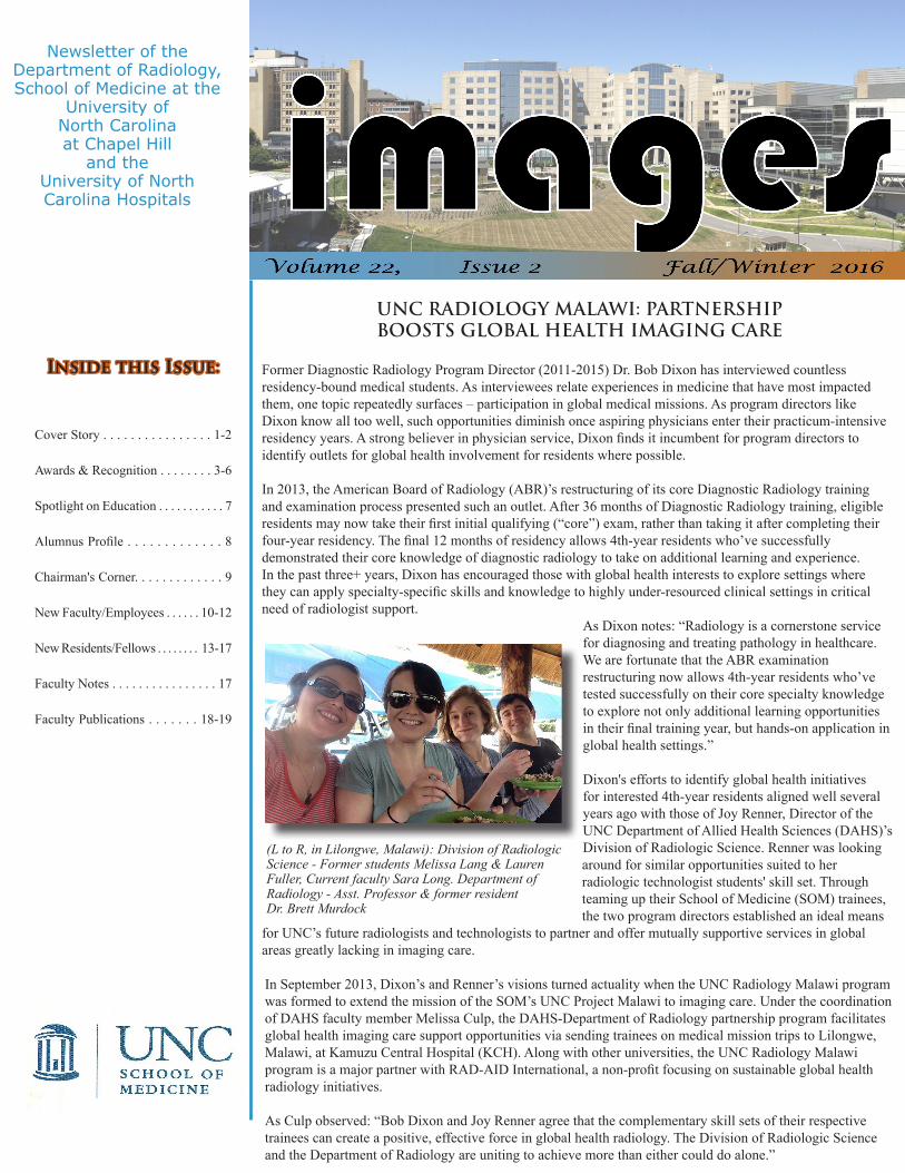
(L to R, in Lilongwe, Malawi): Division of Radiologic Science - Former students Melissa Lang & Lauren Fuller, Current faculty Sara Long. Department of Radiology - Asst. Professor & former resident Dr. Brett Murdock
2
routinely in diagnostic consultation for studies with clinical teams and individual patients. It is not often a radiologist gets the opportunity to explain imaging findings and their implications to the most important people involved, the patients themselves.”
Murdock partnered with Division of Radiologic Science trainees to develop and deliver a lecture series for Malawian technologist students that enhanced their program’s curriculum. He also developed a tremendous bond with the medical officers of the radiology department at KCH. These medical officers are physicians without residency specialty being apprenticed in radiology.
“I developed a great deal of respect for these colleagues seeing firsthand their commitment to excellence in patient care despite any challenges faced. These are great people doing great work. Because of this inspirational team, I am dedicated to this partnership and will continue to be part of it.”
Now an Assistant Professor of Radiology (Cardio-thoracic Imaging) at UNC, Murdock continues promoting global health radiologist support through working with Diagnostic Radiology Program Director Dr. Sheri Jordan. For a program of more than 30 residents, in the short-term, Jordan wants to establish a structured global health radiology curriculum. She will join Murdock next year in working with in-country leadership at KCH, as well as other SOM clinicians working with UNC Project Malawi.
Jordan notes: “Across the institution, UNC's global health programs are sources of great interest and pride. It is both a privilege and honor to add UNC Radiology Malawi to our senior electives list. Recurring resident conferences and emphasis during the resident recruitment season are both succeeding!”
As Dixon observes of UNC Radiology Malawi’s impact on trainees: “This initiative is such an effective way to broaden the mission of UNC Project Malawi. When diagnostic radiology and imaging technology trainees come together as two UNC entities complementing one another’s practice, it’s fantastic to expand upon an established [SOM] global health initiative.”
As Vascular-Interventional Radiology (VIR) Fellowship Program Director, Dixon also promotes participation in UNC Radiology Malawi amongst the Department’s subspecialty residents. In March 2017, current VIR Fellow Dr. Aaron Kline will join the next team headed to Lilongwe. While there, Kline will assist a VIR needs assessment, as well as clinical coordination, between UNC’s Surgery and Radiology teams at KCH. Kline’s selection as one of only six SOM trainees named as a UNC Office of International Activities (OIA) Fall 2016 Fellow will help fund his trip.
Former residents Drs. Melissa Davis (2010-2014) and Brett Murdock (2012-2015) both worked at KCH with UNC Radiology Malawi as 4th-year residents. Two months prior to completing residency, Davis traveled to Lilongwe for two weeks in March 2014. A year later, Murdock joined the UNC Radiology Malawi team at KCH in March 2015.
Equipped with a comprehensive level of diagnostic knowledge and skills in her final residency months, Davis encountered a clinical setting with limited resources in Lilongwe. Imaging care at KCH was administered with no PACS system, contrast was used sparingly or not at all, and team members were fortunate that the available US, CT and X-ray equipment was functioning at the time. While working with UNC Radiology Malawi, Davis also conducted a field assessment alongside UNC and KCH clinicians.
She recalls: “As I concluded my Diagnostic Radiology training, my two weeks in Malawi allowed me to reinforce lean and effective practice in using supplies and equipment when administering patient care. Employing useful skills as a 4th-year resident showed me how meaningful my day-to-day contributions could be. As a 1st- or 2nd-year resident, I might’ve easily been overwhelmed in the same situation.”
Now a faculty member at Yale University, Davis promotes opportunities for residents with global health interests whom she is mentoring and helped to launch a resident-led chapter of RAD-AID in her department. The Yale chapter is nurturing projects in Tanzania, Cameroon, and Jamaica. In December 2016, Davis is traveling to Jamaica to continue these efforts.
As with healthcare in any setting, professionals in Lilongwe face some challenges in the radiology department, but Murdock found that the team approach to patient care overcame many obstacles. He found that the consultant radiologist, Dr. Mzumara, was personable and warm. Under Mzumara’s supervision, Murdock participated in clinical activities from an ultrasound evaluation of a child with hydrocephalus to an ultrasound-guided biopsy of a large chest mass, where a surgical procedure would have likely been the only other option.
Of the department's equipment, only one of three radiography units and two sonography rooms were operational. The CT scanner was non-functioning during the visit at the time. Though this scanner was down, a backlog of studies had accumulated. Murdock assisted the team by rendering reports to make a difference in workflow.
Murdock notes, “A key takeaway from this experience was observing the positives of how a health system and radiology work together in Malawi. I was involved
continued from page 1 cover story
imagesis the newsletter of the
Department of Radiology School of Medicine of the University of North Carolina at
Chapel Hill and the University
of North Carolina HospitalsPublished by the Department of Radiology, School of Medicine,
The University of North Carolina.CB# 7510
Old Clinic BuildingChapel Hill, NC
27599-7510Phone: (919) 966-4238
www.med.unc.edu/radiology
Department Chair Matthew A. Mauro, MD
Excecutive Vice Chair Paul L. Molina, MD
Associate Chair for Administration
Robert Collichio, MPA
Residency Program Director
Sheri Jordan, MD
EditorLaurie Birdsong
Graphic LayoutElizabeth Bowen
3
awards & recognition
JKT Lee Receives Society of Computed Body Tomography and Magnetic Resonance Gold Medal
The Department would like to congratulate Immediate Past Chair and Professor Dr. Joseph KT Lee for receiving the Society of Computed Body Tomography and Magnetic Resonance (SCBT-MR)’s Gold Medal. Lee was amongst three recipients awarded the Society’s highest honor at its 39th annual meeting in September 2016 in Salt Lake City, Utah. This is the third group of Gold Medal recipients since the Society began this award in 2014. Two radiologists were awarded the Society’s Gold Medal in both 2014 and 2015.
The SCBT-MR’s Gold Medal is amongst other peer awards that Lee has received in recent years in recognition of his career achievements, including: 1) the American Roentgen Ray Society (ARRS)' Gold Medal for Distinguished Service to Radiology (April 2013); and 2) the Asian Oceanian Society of Radiology (AOSR)’s Gold Medal (October 2008). He has also received Washington University’s School of Medicine Alumni Achievement Award (April 2013) and is an Honorary Member of both the Korean Society of Radiology (2010) and the Japanese Society of Radiology (2012). Within the Department, graduating Diagnostic Radiology residents chose Lee in 1992 and 2008 for the annual Charles A. Bream Award for Teaching Excellence.
Over the past two decades, Lee has served as SCBT-MR’s Annual Course Faculty. He also served on its Executive Board for over seven years, culminating in his 1993-1994 Presidency. The Gold Medal Award recognizes Lee's pioneering contributions in the field of Body Computed Tomography and Magnetic Resonance Imaging through his seminal textbooks and numerous peer-reviewed, high-impact journal articles.
Lee noted: “I am especially honored to receive the award in the same year as Drs. Morton Bosniak and Melvin Korobkin. Their respective work has become the standard of practice in renal and adrenal imaging worldwide. In the Society’s early days, members [now called Fellows] were all leading experts in body CT and MR. Annual meetings allowed them to exchange cutting-edge research ideas and teach the practicing radiologists state-of-the art advances in these two technologies. It is heart-warming to see that this trend continues even after Society membership has expanded in recent years.”
Castillo Assumes 2016-2017 ARRS PresidencyAt its April 2016 annual meeting in Los Angeles, Dr. Mauricio Castillo assumed his new post as the 2016-2017 President of the American Roentgen Ray Society (ARRS). Castillo became the ARRS' 116th President and was named President-Elect at the Society’s 2015 annual meeting.
Castillo noted: "I am thrilled and honored to have been named ARRS' new president. In the coming year, I offer guidance to the Society in preserving its reputation as the radiologist’s most trusted source of information and establishing new partnerships around the globe. In leading one of radiology’s foremost professional organizations, I’m thankful to have served in top leadership posts such as [2013-2014] American Society of Neuroradiology (ASNR) President. However, as an academic radiologist, UNC Department of Radiology has long served as the sole entity that allows me every day to identify issues that I most want to advocate for in our field.”
Over his career, Castillo has served in multiple capacities within the Society, amongst them: Vice President (2014-2015); ARRS Fund Board of Trustees (2014-present); Gold Medal Committee & Restructuring Task Force (2014-present); Nominating Committee (2014-present); Finance and Budget Committee (2014-present); International Committee Chair (2011-2015); Executive Council member (2008-present); Categorical Course on Neuroradiology - Co-director (2007); Instructional Courses Committee - Chair & Associate Chair (2003-2011); and Neuroradiology Instructional Course Subcommittee - Chair (2000-2006).

4
awards & recognition
Lin Receives Two NIH Awards to Study Early Brain DevelopmentThe Department would like to recognize Dr. Weili Lin for receiving two major National Institutes of Health (NIH) awards in summer 2016. In August 2016, Lin was awarded R21 funding ($418K) by the NIH’s National Institute of Neurological Disorders and Stroke (NINDS) for his study entitled, “Characterizing morphological and hemodynamic characteristics of human brain perivascular spaces with aging using 7T MRI.” In September 2016, the National Institute of Mental Health (NIMH) awarded Lin U01 collaborative research funding ($4m+) to conduct a 3.5+-year, multi-institutional study entitled, “UNC/UMN Baby Connectome Project (BCP).”
Over a two-year period (8/15/2016 – 7/31/2018), Lin’s R21 study seeks to advance the non-invasive, high resolution approaches currently feasible in imaging cerebral perivascular spaces (PVS). Suggested by evidence as beneficial to the brain’s lymphatic function, PVS nonetheless remain difficult to detect in a non-enlarged state. Given enlarged PVS are commonly seen in aging-related neurological disorders, Lin will employ 7-Tesla imaging to assess PVS physiology and pathophysiology in healthy young adults. If non-invasive approaches in younger individuals can be developed to examine PVS appearance and functional status, Lin’s team can advance its study of neurological disorder progression via PVS function in early brain development.
Over the coming 3.5+-years (9/1/2016 – 5/31/2020), Lin will lead a UNC Biomedical Research and Imaging Center (BRIC) team in collaborating with University of Minnesota (UMN) investigators to conduct his U01 research. The NIMH has funded this study to extend the Lifespan Human Connectome Project (L-HCP), an endeavor to map human brain circuitry in the developing, adult and aging human brains. Via non-invasive, magnetic resonance imaging of child participants under age five, the study aims to establish structural and functional brain connectivity contributing to early-life, healthy brain development.
Through conducting MRI scans over almost four years on 500 typically developing children ages five and under, UMN’s and Lin’s BRIC research teams will collect both longitudinal and cross-sectional data for analysis. Coupled with parent reports and directly assessing cognitive and behavioral development in study participants, their collaborative aim is to provide a more comprehensive understanding of how emerging patterns of brain connectivity shape early-life behavioral development.
Lin noted: “We’re very excited that R21 funding allows us to leverage the BRIC’s impressive imaging capabilities that shed new light on the roles of human perivascular systems in aging. The 7T MR scanner enables acquiring ultra-high resolution images with an adequate signal-to-noise ratio, and results may help us better understand if dysfunctional perivascular systems are related to Alzheimer’s disease. In contrast, with U01 funding, we will conduct one of the most comprehensive studies for characterizing early brain development. Although the study only focuses on normal developing children, results could be utilized better to discern subtle abnormalities in patients with neurodevelopmental disorders.”
Nyante Awarded NC TraCS KL2 Funding to Improve Registry-Based Research The Department would like to congratulate Dr. Sarah Nyante for being chosen in June 2016 as a North Carolina Translational and Clinical Sciences Institute (NC TraCS) KL2 Program 2016-2017 Scholar. Dr. Nyante’s one-year, annually renewable KL2 scholarship provides 75%
salary support and will support her research entitled, “Data Quality in Registry-Based Linkage: Implementing Methods to Reduce Missing Data and Bias.”
Since her December 2014 faculty appointment, Dr. Nyante’s investigative efforts as a Carolina Mammography Registry (CMR) Co-Investigator have advanced the understanding of the relationship between breast imaging and breast cancer etiology via her study of the relationship between mammographic findings and breast cancer prognosis.
As a 2016-2017 KL2 Scholar, Dr. Nyante will her narrow her scope of research to examining the completeness and accuracy of the CMR’s breast cancer diagnosis data. Her study’s aim is to identify the best analytic approach to reducing bias due to high proportions of missing data that can occur when information is drawn from disease registries rather than collected actively by a specific study. By characterizing potential sources of case non-reporting and missing data, Dr. Nyante’s study aims to improve registry-based studies of breast cancer prognosis.
“The research I complete with this award will strengthen CMR analyses related to breast cancer prognosis by allowing us to develop the best methods to deal with missing data in this particular population, increasing the validity of our research.”
5
awards & recognition
Shen Receives R01 Funding to Study Pelvic Organ Delineation in Treating Prostate Cancer
The Department would like to recognize Dr. Dinggang Shen for receiving National Institutes of Health/National Cancer Institute (NIH/NCI) R01 funding for his project entitled, “Automatic Pelvic Organ Delineation in Prostate Cancer Treatment” in August 2016. This latest NIH award comes at a time that Shen is serving as PI or MPI of multiple R01-funded investigations sponsored by the
National Institute on Aging, the National Institute of Biomedical Imaging and Bioengineering, the National Institute on Mental Health, and the National Institute of Dental and Craniofacial Research. Over five years, Shen’s study aims to produce a set of novel machine learning tools for more accurate, reliable and efficient imaging in delineating important pelvic organs (eg, prostate, bladder, and rectum) in prostate cancer radiotherapy. For the imaging modalities (eg, planning CT, treatment CT/CBCT and MRI) used in for prostate cancer radiotherapy, Shen and other study investigators will develop tools that accurately segment planning CT images and incorporate patient-specific information from that patient’s previous treatment images to progressively improve treatment through CT/CBCT segmentation. They will additionally develop a tool for learning MRI-specific features for prostate MRI segmentation, as well as a novel method for collaborative MRI and planning CT segmentation. Shen notes: “Once all are developed, we will validate these methods on real patients in our clinical workflow.
Although these machine-learning tools are designed for radiotherapy of prostate cancer, they can also be easily extended to other new organs and new modalities after appropriate updating and training.”
Henderson Receives R21 to Study Breast Density Law's Impact on Supplemental Screening
The Department is pleased to recognize Dr. Louise Henderson for receiving a National Institutes of Health/National Cancer Institute R21 in August 2016. Entitled, “Evaluating the Effect of the Breast Density Legislation on Supplemental Screening,” this two-year study will examine how the North Carolina breast density notification law, enacted in January 2014, impacts breast cancer screening behavior among women in North Carolina. Using existing data collected as part of the Carolina Mammography Registry (CMR), Dr. Henderson and her team will determine how radiology facilities across the state are interpreting and implementing the law pre- and post-passage of the legislation. North Carolina was the 12th state to pass breast density notification legislation. It requires mammography providers to inform all women screened for breast cancer of their breast density classification and its potential impact on imaging capabilities. Research has shown that dense breast tissue reduces the sensitivity of screening mammography and may increase the risk of breast cancer. With its longitudinal structure and comprehensive breast cancer screening information, the CMR is an ideal data source for determining the impact of this legislation on community facilities throughout the state. Dr. Henderson anticipates that the use of supplemental screening among women with dense breasts will increase after 2014 and that this use will differ by patient, radiologist, and facility characteristics. A state-wide survey will also be employed to gain insights into the availability of supplemental screening services and patient educational resources for women found to have dense breast tissue. Henderson noted: “This project is just one example of how population-based data on healthcare use allows for rapid examination of health services questions which impact patient care and possibly downstream outcomes and cost.”
6
spotlight on educationawards & recognition
Li Receives Two NIH Awards for 4D Cortical Surface Analysis Tool Development
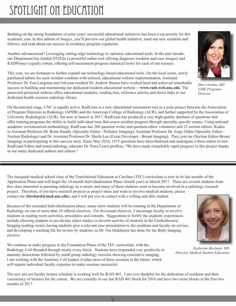
The Department would like to recognize Dr. Gang Li for his receipt of two National Institutes of Health (NIH) awards in spring and summer 2016. In April 2016, the National Institute of Mental Health (NIMH) awarded Li R21 funding ($418K) as PI of a two-year project (04/01/2016-03/31/2018) entitled, “Constructing 4-dimensional Infant Cortical Surface Atlases” (MPI: Dr. Dinggang Shen). In August 2016, Li received a second NIMH award as recipient of K01 funding (FY '16 - $139,573), annually renewable up to four years, as PI of a research training project entitled, “4D Cortical Surface Analysis Tools for Study of Early Brain Development in Typical and High-Risk Infants.”
Over a two-year period, Li’s R21 funding will support developing 4D cortical surface-based brain atlases for studying the dynamic developing cortex of infants. The early postnatal population is critical to the study of neurodevelopmental origins and abnormal trajectories of neuropsychiatric disorders. However, cortical surface atlases used for adults and neonates cannot be used in neuroimaging the dynamic, rapidly developing infant cortex due to dramatic cortical differences. If successful, at the study’s conclusion, Li’s team will have created the first longitudinal 4D infant cortical surface atlases for accurate neuroimaging mapping of dynamic infant brain development. Such tools will allow the neuroimaging research community to more comprehensively study the highly-folded cortex across all age groups.
As an early career UNC Biomedical Research and Imaging Center (BRIC) researcher, Li’s K01 training supports his advancement toward becoming an independent, pediatric neuroimaging investigator. Over a four-year period, his grant-supported training will teach him to apply infant MR imaging techniques to accurately characterize early brain development. Through acquiring skills in infant neuroimaging mapping, Li’s training aims are to produce a unique suite of infant-specific, 4D cortical surface-based neuroimaging analysis tools applicable to typically developing and high-risk (schizophrenia) infants. If successful, at the grant period’s conclusion, Li will have produced infant neuroimaging mapping tools enabling researchers to identify early biomarkers of schizophrenia risk and to develop preemptive intervention strategies.
"With these two awards, I am able to significantly enhance and extend my research on MR imaging studies of early brain development, thus greatly helping me to be a recognized expert int he field."
Wu Awarded K01 Funding to Develop Neuroimaging Tools for Early Alzheimer's Diagnosis
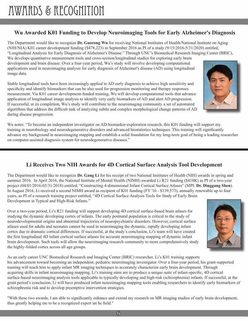
The Department would like to recognize Dr. Guorong Wu for receiving National Institutes of Health/National Institute on Aging (NIH/NIA) K01 career development funding ($478,223) in September 2016 as PI of a study (9/15/2016-5/31/2020) entitled, “Longitudinal Analysis for Early Diagnosis of Alzheimer's Disease.” Through UNC’s Biomedical Research Imaging Center (BRIC), Wu develops quantitative measurement tools and cross-section/longitudinal studies for exploring early brain development and brain disease. Over a four-year period, Wu’s study will involve developing computational applications used in neuroimaging analysis for early diagnosis of Alzheimer's disease (AD) using longitudinal image data.
Stable longitudinal tools have been increasingly applied to AD early diagnosis to achieve high sensitivity and specificity and identify biomarkers that can be also used for progression monitoring and therapy responses measurement. Via K01 career development-funded training, Wu will develop computational tools that advance application of longitudinal image analysis to identify very early biomarkers of AD and alert AD progression. If successful, at its completion, Wu’s study will contribute to the neuroimaging community a set of automated algorithms that address the difficult task of analyzing subtle and complex morphological change patterns of AD during disease progression.
Wu notes: “To become an independent investigator on AD-biomarker-exploration research, this K01 funding will support my training in neurobiology and neurodegenerative disorders and advanced biostatistics techniques. This training will significantly advance my background in neuroimaging mapping and establish a solid foundation for my long-term goal of being a leading researcher on computer-assisted diagnosis system for neurodegenerative diseases.”
7
spotlight on education
Building on the strong foundation of prior years’ successful educational initiatives has been a top priority for this academic year. In this edition of Images, you’ll preview our global health initiative, meet our new residents and fellows, and read about our success in residency program expansion.
Another advancement? Leveraging cutting-edge technology to optimize educational tools. In the past decade, our Department has funded STATdx (a powerful online tool offering diagnosis modules and case images) and RADPrimer (equally robust, offering self-assessment progress statistical tools) for each of our trainees.
This year, we are fortunate to further expand our technology-based educational tools. On the local scene, newly purchased tablets for each resident combine with tailored, educational website implementation. Assistant Professor Dr. Eun Langman and 3rd-year resident Dr. Andrew Barnes have worked hard and achieved remarkable success in building and maintaining our dedicated resident educational website -- www.rads.web.unc.edu. The password-protected website offers educational modules, reading lists, reference articles and direct links to our dedicated health sciences radiology library.
On the national stage, UNC is equally active. RadExam is a new educational assessment tool as a joint project between the Association of Program Directors in Radiology (APDR) and the American College of Radiology (ACR), and further supported by the Association of University Radiologists (AUR). Set now to launch in 2017, RadExam has produced a very high-quality database of questions that offer training programs the ability to build individual tests that assess resident progress through specialty-specific exams. Using national academic crowdsourced methodology, RadExam has 260 question writer and question editor volunteers and 22 section editors. Kudos to Assistant Professor Dr. Brian Handly (Specialty Editor - Pediatric Imaging); Assistant Professor Dr. Jorge Oldan (Specialty Editor - Nuclear Radiology) and Dr. Assistant Professor Dr. Sheila Lee (Exam Developer - Breast Imaging). They join me (Section Editor-Breast Imaging) in participating in this success story. Since May 2016, 1675 questions have been finalized and undergone a three-editor review. RadExam Editor and noted radiology educator Dr. Petra Lewis proffers, "We have made remarkably rapid progress in this project thanks to our many dedicated authors and editors."
Sheri Jordan, MDGME Programs Director
The inaugural medical school class of the Translational Education at Carolina (TEC) curriculum is now in its last months of the Application Phase and will begin the 14-month Individualization Phase (fourth year) in March 2017. There are several students from this class interested in pursuing radiology as a career, and many of these students want to become involved in a radiology research project. Therefore, if you have research projects or project ideas and want to involve medical students, please contact me ([email protected]), and I will put you in contact with a willing and able student.
Because of the extended Individualization phase, many more students will be rotating in the Department of Radiology on one of more than 10 offered electives. For divisional electives, I encourage faculty to involve students in reading room activities, procedures and consults. Suggestions to fortify the students' experiences include allowing students to pre-dictate select studies (a favorite activity of students in the Cardiothoracic Imaging reading room), having students give a relevant case presentation to the residents and faculty on service, and developing a teaching file for review by students, as Dr. Nia Mukherjee has done for the Body Imaging elective.
We continue to make progress in the Foundation Phase of the TEC curriculum, with the Radiology Coil threaded through nearly every block. Students have responded very positively to anatomy dissections followed by small group radiology sessions showing correlative imaging. I am working with the Anatomy Coil leaders to plan more of these sessions in the future, which will require individual faculty expertise to make sessions successful.
The new pre-set faculty lecture schedule is working well for RAD 401. I am ever thankful for the dedication of residents and their consistency of lectures for the course. We are currently in our last RAD 401 block for 2016 and have two more blocks in the first two months of 2017.
Katherine Birchard, MDDirector, Medical Student Education
8
Chairman's cornerThe Department has a proud tradition of North Carolina Radiological Society (NCRS) representation amongst both current members and alumni. In recent years alone, multiple faculty have served as NCRS President, amongst them: 1) Drs. Sheri Jordan (2016-2017); 2) Charles Burke (2015-2016); Valerie Jewells (2009-2010); and Paul Molina (2005-2006). Advancing their specialty through education, research and didactics makes academic radiologists likely advocates for promoting the NCRS' mission. Similarly, private practice radiologists who’ve led within their physician group are compelled to advocate for their specialty’s interests as active NCRS members. Durham Radiology Associates (DRA) President Dr. Hal Safrit is one such Diagnostic Radiology alumnus (1987) whose leadership within his practice extends to promoting the NCRS' mission alongside most its active, long-time members. As a former NCRS President (2004-2005) and now a Councilor representing this state chapter of the national American College of Radiology (ACR), Safrit notes of his long-time NCRS involvement:
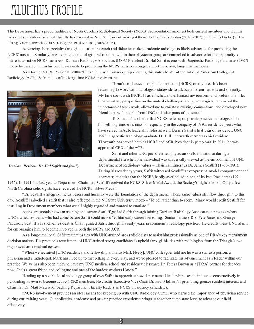
“I can’t emphasize enough the impact of [NCRS] on my life. It’s been rewarding to work with radiologists statewide to advocate for our patients and specialty. My time spent with [NCRS] has enriched and enhanced my personal and professional life, broadened my perspective on the mutual challenges facing radiologists, reinforced the importance of team work, allowed me to maintain existing connections, and developed new friendships with people from UNC and other parts of the state.” To Safrit, it’s an honor that NCRS relies upon private practice radiologists like himself to promote its mission, especially in the company of 1980s residency peers who have served in ACR leadership roles as well. During Safrit’s first year of residency, UNC 1983 Diagnostic Radiology graduate Dr. Bill Thorwarth served as chief resident. Thorwarth has served both as NCRS and ACR President in past years. In 2014, he was appointed CEO of the ACR. Safrit and other UNC peers learned physician skills and service during a departmental era when one individual was universally viewed as the embodiment of UNC Department of Radiology values – Chairman Emeritus Dr. James Scatliff (1966-1991). During his residency years, Safrit witnessed Scatliff’s ever-present, model comportment and character, qualities that the NCRS hardly overlooked in one of its Past Presidents (1974-
1975). In 1991, his last year as Department Chairman, Scatliff received the NCRS' Silver Medal Award, the Society’s highest honor. Only a few North Carolina radiologists have received the NCRS' Silver Medal. “Dr. Scatliff’s integrity, inclusiveness and humility were the foundation of the department. Those same values still flow through it to this day. Scatliff embodied a spirit that is also reflected in the NC State University motto – ‘To be, rather than to seem.’ Many would credit Scatliff for instilling in Department members what we all highly regarded and wanted to emulate.” At the crossroads between training and career, Scatliff guided Safrit through joining Durham Radiology Associates, a practice where UNC-trained residents who had come before Safrit could now offer him early career mentoring. Senior partners Drs. Pete Jones and George Paddison, Scatliff’s first chief resident as Chair, guided Safrit through his early years in community radiology practice. He credits these UNC alums for encouraging him to become involved in both the NCRS and ACR. As a long-time local, Safrit maintains ties with UNC-trained area radiologists to assist him professionally as one of DRA’s key recruitment decision makers. His practice’s recruitment of UNC-trained strong candidates is upheld through his ties with radiologists from the Triangle’s two major academic medical centers. “When we recruited [UNC residency and fellowship alumnus Mark Neely], UNC colleagues told me he was a star as a person, a physician and a radiologist. Mark has lived up to that billing in every way, and we’re pleased to facilitate his advancement as a leader within our practice. We’ve has also been lucky to have my UNC medical school and residency classmate Dr. Teresa Brown as a [DRA] partner for decades now. She’s a great friend and colleague and one of the hardest workers I know.” Heading up a sizable local radiology group allows Safrit to appreciate how departmental leadership uses its influence constructively in persuading its own to become active NCRS members. He credits Executive Vice Chair Dr. Paul Molina for promoting greater resident interest, and Chairman Dr. Matt Mauro for backing Department faculty leaders as NCRS presidency candidates. “NCRS involvement provides an ideal means for keeping up with UNC Radiology alumni who learned the importance of physician service during our training years. Our collective academic and private practice experience brings us together at the state level to advance our field effectively.”
alumnus profile
Durham Resident Dr. Hal Safrit and family

9
I’ve served as UNC Department of Radiology Chair for almost a decade now and as UNC Faculty Physicians (UNCFP) CEO for the past two years. In both roles, I’m reminded that strong operational governance at the institutional and departmental levels is critical for attracting academic physicians and researchers most capable of sustaining the UNC School of Medicine (SOM)’s excellence in clinical care, research and education.
Since early 2015, the Department of Radiology has appointed 12 new clinical faculty members. As an SOM chairman, I’m proud foremost that my department attracts academic radiologists who are ready to work with faculty peers within their own discipline, as well as with those across a top-tier institution like UNC. As the institutional physician most responsible for sustaining UNC HCS' needed clinical workforce, I’m also proud that our Department’s own are amongst more than 1700 UNC faculty physicians who provide clinical services to North Carolina’s ever-growing patient population.
In the past few years, opening a second UNC Hospitals campus in Hillsborough and expanding its number of outpatient clinics in the area has caused UNC HCS to need more providers where it serves the most patients in central North Carolina. Along with recruiting more clinical faculty, an academic medical center recruits more physicians in training when an expanded network takes on a greater patient caseload. In October 2016, the American Council on Graduate Medical Education (ACGME) approved a permanent complement increase to our Diagnostic Radiology program, from 32 to 36 residency positions. Effective July 2017, this approval allows us to add to the ranks of this program just as the Department starts up its Interventional Radiology residency program next year. With two programs incrementally adding to the network’s overall base of physicians, the Department looks forward in the coming years to strengthening diagnostic and interventional clinical services offered at UNC HCS.
I also look back on 2016 as a period of increased research strength in basic science. Our recruitment of three basic scientists this year brings this base of faculty to an unprecedented 16 members. As we grow these ranks, I’m amazed by what our Department-affiliated UNC basic science entities – the Carolina Mammography Registry (CMR) and UNC’s Biomedical Research Imaging Center (BRIC) – continue to secure in major research funding.
Since early 2016, new federal and institutional funding has leveraged the health services research, quality improvement and disease surveillance activities the CMR conducts in areas such as breast cancer screening. In June 2016, CMR Co-Investigator Dr. Sarah Nyante was awarded a North Carolina Translational and Clinical Sciences (NC TraCS) Institute KL2 Program 2016-2017 Scholarship to further the investigative contributions she’s made in her two years on faculty. Two months later, CMR Principal Investigator Dr. Louise Henderson received NIH National Cancer Institute (NCI) R21 funding to examine the effect of new state law on supplemental mammography screening behavior across North Carolina.
The Department's three new basic scientists in 2016 are all BRIC faculty appointees. Their recruitment brings investigative support to a University entity that has been awarded $5.1m+ in National Institutes of Health (NIH)-affiliated funding for eight studies since July 2016 alone. In 2016, the NIH awards received by its investigators have highlighted the BRIC’s funding successes over the course of the year. In April, National Institute of Mental Health (NIMH) awarded Dr. Gang Li two-year R21 funding. Between June and August 2016, Dr. Weili Lin received National Institute of Neurological Disorders and Stroke (NINDS) two-year R21 funding, as well as National Institute of Mental Health (NIMH) 3.5+-year multi-institutional, collaborative U01 funding. In September, Dr. Guorong Wu was awarded National Institute on Aging (NIA) four-year K01 career development funding. As faculty growth allows BRIC to guide junior faculty toward becoming independent investigators, we look forward to watching the Center's continued success in securing major funding to study early brain development and progression of neurodegenerative disease via applied imaging.
In fall 2015, the Department reorganized oversight of its clinical services and basic research realms into three distinctive vice chairmanship roles -- Diagnostic Service, Interventional Service and Basic Research. Our steady growth in faculty over a year’s time is one indicator that such well-defined leadership appeals to aspiring academic radiologists and basic researchers. As we head into 2017, the sought-after faculty we continue to recruit as an SOM department will remain one of our key ways of upholding the mission of the institution.
Chairman's corner

10
new FacultyThe Department was pleased to appoint Dr. Eran Dayan as Assistant Professor in September 2016, working under the direction of Biomedical Research Imaging Center (BRIC) Director Dr. Weili Lin. In his new role, Dr. Dayan's work will involve statistical modeling and data analysis related to network connectivity in health and disease. Dr. Dayan's BRIC-affiliated lab will test the prospects of using brain network connectivity as a biomarker in various neurological conditions. His lab will also try to delineate the principles governing changes in network connectivity along early development, as well as to develop methods that externally interact with network connectivity states using non-invasive brain stimulation.
Prior to UNC, Dayan received his BA in Behavioral Sciences at the Ben-Gurion University in Beer-Sheva, Israel, in September 2000. In November 2003, he received his MA in Cognitive Psychology at the Hebrew University of Jerusalem in Israel. In February 2010, he received his PhD in Neuroscience in the Departments of Neurobiology, Computer Science and Applied Mathematics from Weizmann Institute of Science in Israel. From 2010 to 2016, Dayan worked as both a Postdoctoral Fellow and Research Fellow at the National Institutes of Health (NIH)’s National Institute of Neurological Disorders and Stroke (NINDS) in Bethesda, MD. In these roles, his research focused on interactions between large-scale brain systems, using a combination of neuroimaging and non-invasive brain stimulation.
Dayan notes: “The time I have spent at NINDS has taught me how to identify questions which are both mechanistically and clinically important. Indeed, this is also the main drive behind my current work.”
The Department was pleased to appoint Dr. Brett Murdock as Clinical Assistant Professor of Radiology in August 2016, working in Cardiothoracic Imaging under the direction of Division Chief Dr. Paul Molina. Dr. Murdock trained as a fellow in his respective subspecialty for a year prior to his faculty appointment.
Murdock received his MD from Georgetown University School of Medicine (Washington, DC) in 2010. He stayed in the District another year and completed his internship (Surgery) at Washington Hospital Center. He then trained in Diagnostic Radiology for a year at Monmouth Medical Center (Long Branch, NJ) before transferring to UNC to finish his residency in the Department in June 2015.
Murdock is currently applying for a seed grant for a study on the use of chest tomosynthesis for cardiac-gated coronary calcium scoring, as well as submitting a study comparing the efficacy and cost effectiveness of transthoracic versus transbronchial lung biopsies. Dr. Murdock is also the UNC Radiology Malawi faculty liaison for the School of Medicine’s UNC Project Malawi. He’s currently developing a resident Global Health curriculum and will next travel to Lilongwe, Malawi, in March 2017.
“I have relished the opportunity to join such a strong departmental division that emphasizes optimal patient care and close collaborative efforts with patient care teams. It’s also exciting as a new faculty member to have the flexibility to
work with a School of Medicine initiative that allows UNC Radiology to provide much-needed imaging services in a global setting lacking in healthcare resources.”
The Department was pleased to appoint Dr. Katrina McGinty as Clinical Assistant Professor of Radiology in August 2016, working in Abdominal Imaging under the direction of Division Chief Dr. Jeffrey Neitlich. McGinty received her MD from New York Medical College (Valhalla, NY) in May 2005. A year later, she completed her internship (Transitional Year) at Saint Vincent's Hospital (New York City, NY). She then completed her Diagnostic Radiology residency at North Shore University Hospital (Manhasset, NY) in 2010, thereafter training in Oncologic Imaging for a year at Memorial Sloan Kettering Cancer Center (New York City, NY). Prior to UNC, Dr. McGinty spent five years on clinical faculty at Geisinger Medical Center’s Department of Radiology (Danville, PA). "I'm excited to be joining the radiology department and have a chance to work with such wonderful faculty. I am strongly interested in oncologic imaging and treatment response criteria and appreciate the clinical and research opportunities presented by having such an excellent cancer center as part of our facility. Most of all, I'm looking forward to working with and teaching our residents!"
11
new FacultyThe Department was pleased to appoint Dr. David Mauro as Clinical Assistant Professor in August 2016, working in Vascular-Interventional Radiology under the direction of Division Chief Dr. Charles Burke.
Dr. Mauro received his MD from UNC in May 2009. A year later, he completed his Preliminary Year (Surgery) at the University of Virginia (UVA) Health System (Charlottesville, VA). He remained at UVA for his Diagnostic Radiology residency, completing his training in 2014. Post-residency, he served for a year as a Clinical Instructor at UVA Health System in both the Divisions of Body Imaging and Non-Invasive Cardiovascular Imaging. From 2015 to 2016, he further subspecialized in Interventional Radiology at Mt. Sinai Hospital (New York City, NY). Mauro’s secondary faculty appointment is in the Department of Radiology. His primary faculty appointment is in UNC’s Department of Surgery, reporting to Dr. Melina Kibbe, Professor and Chair of the Department of Surgery.
“I am excited to return to Chapel Hill and UNC Hospitals. I look forward to the teaching opportunities with our residents and fellows. I hope to collaborate with other departments to provide excellent patient care and further research within my field.”
The Department was pleased to appoint Dr. Xiaopeng Zong as Research Assistant Professor in May 2016, working under the direction of Biomedical Research Imaging Center (BRIC) Director Dr. Weili Lin. Zong’s appointment directly follows three years of working as a BRIC Research Associate.
Dr. Zong received his BS in Physics and his BE in Electrical and Information Engineering in July 1998 and July 1999, respectively, from the University of Science and Technology of China in Hefei, China. In December 2007, he received his PhD in Condensed Matter Physics from Iowa State University in Ames, IA.
Prior to UNC, Zong held several research associate positions over seven years (2009-2016) in the Departments of Radiology at both the University of Pittsburgh and Michigan State University. In his earlier post-graduate years, Zong’s research background also included prior experience at Baylor College of Medicine’s Department of Neuroscience carrying out functional MRI study of neural plasticity in stroke patients. Additionally, he worked in Iowa State University’s Department of Physics and Astronomy, focusing on NMR studies of condensed matter physics.
Since joining Dr. Lin’s team, Zong has overseen the BRIC’s 7T MR Scanner operations and has also held responsibility for developing protocols for the Center’s 3T, 7T and 9.4T scanners. As Zong explores areas at the BRIC suited to his skills and knowledge, in the longer term he will conduct independent research studies related to developments of new MRI methods for improved understanding, prognosis and treatment of neurological diseases, such as dementia, stroke, and radiation-therapy induced brain injury.
"BRIC provides a highly diverse and supportive research environment that allows me to follow my passion in MR physics and develop independent and collaborative research projects."
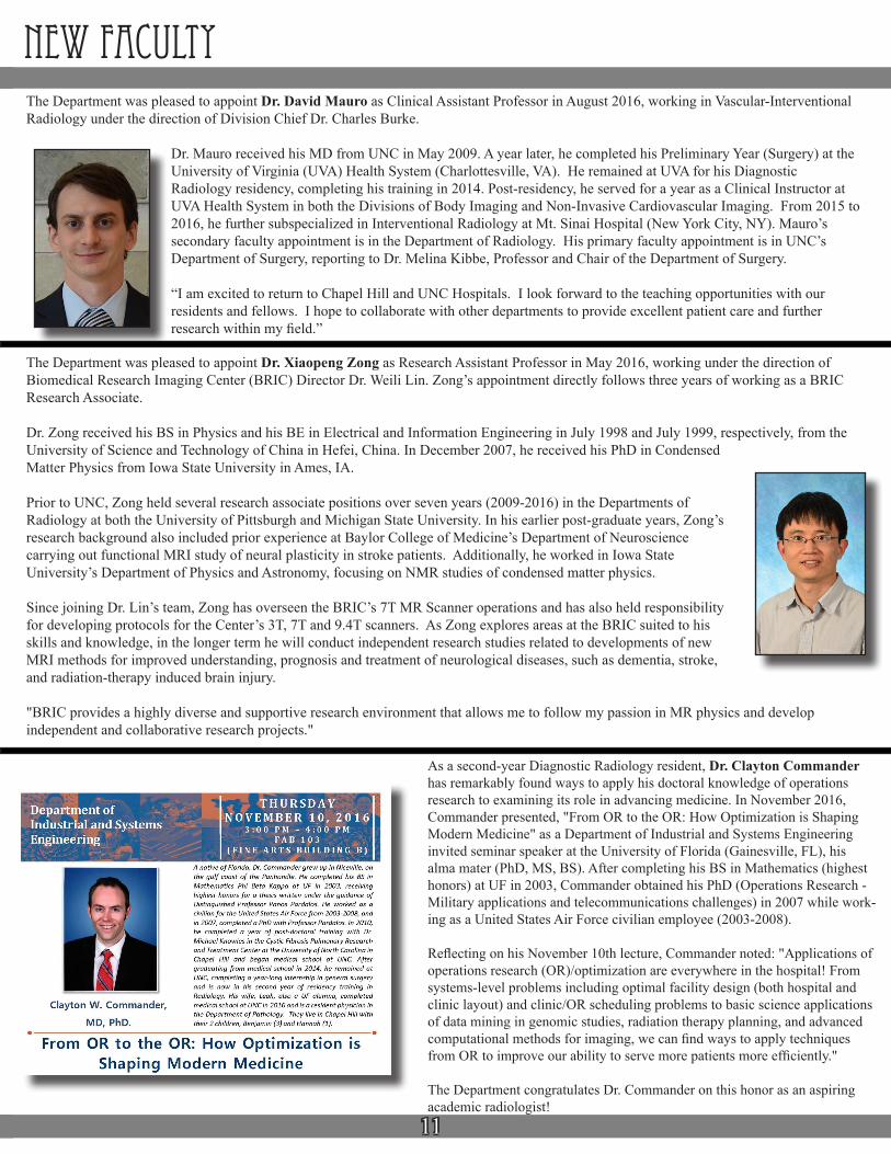
As a second-year Diagnostic Radiology resident, Dr. Clayton Commander has remarkably found ways to apply his doctoral knowledge of operations research to examining its role in advancing medicine. In November 2016, Commander presented, "From OR to the OR: How Optimization is Shaping Modern Medicine" as a Department of Industrial and Systems Engineering invited seminar speaker at the University of Florida (Gainesville, FL), his alma mater (PhD, MS, BS). After completing his BS in Mathematics (highest honors) at UF in 2003, Commander obtained his PhD (Operations Research - Military applications and telecommunications challenges) in 2007 while work-ing as a United States Air Force civilian employee (2003-2008).
Reflecting on his November 10th lecture, Commander noted: "Applications of operations research (OR)/optimization are everywhere in the hospital! From systems-level problems including optimal facility design (both hospital and clinic layout) and clinic/OR scheduling problems to basic science applications of data mining in genomic studies, radiation therapy planning, and advanced computational methods for imaging, we can find ways to apply techniques from OR to improve our ability to serve more patients more efficiently."
The Department congratulates Dr. Commander on this honor as an aspiring academic radiologist!
12
new EMPLOYEES
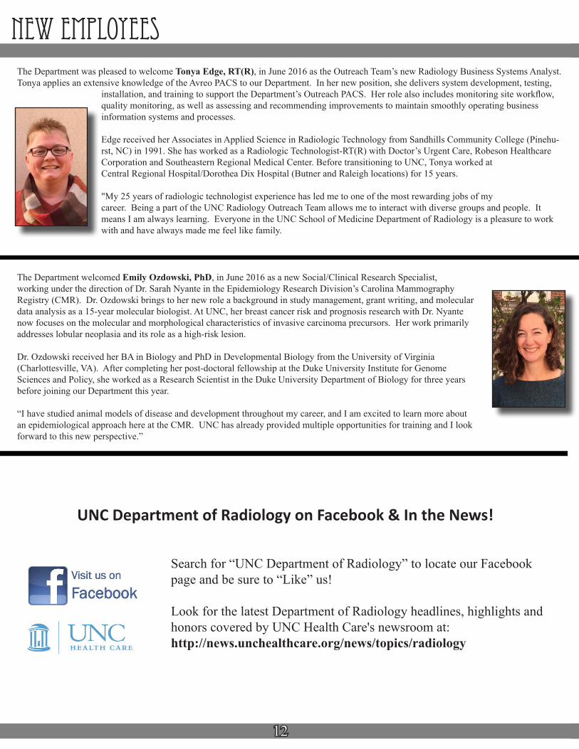
The Department welcomed Emily Ozdowski, PhD, in June 2016 as a new Social/Clinical Research Specialist, working under the direction of Dr. Sarah Nyante in the Epidemiology Research Division’s Carolina Mammography Registry (CMR). Dr. Ozdowski brings to her new role a background in study management, grant writing, and molecular data analysis as a 15-year molecular biologist. At UNC, her breast cancer risk and prognosis research with Dr. Nyante now focuses on the molecular and morphological characteristics of invasive carcinoma precursors. Her work primarily addresses lobular neoplasia and its role as a high-risk lesion.
Dr. Ozdowski received her BA in Biology and PhD in Developmental Biology from the University of Virginia (Charlottesville, VA). After completing her post-doctoral fellowship at the Duke University Institute for Genome Sciences and Policy, she worked as a Research Scientist in the Duke University Department of Biology for three years before joining our Department this year.
“I have studied animal models of disease and development throughout my career, and I am excited to learn more about an epidemiological approach here at the CMR. UNC has already provided multiple opportunities for training and I look forward to this new perspective.”
The Department was pleased to welcome Tonya Edge, RT(R), in June 2016 as the Outreach Team’s new Radiology Business Systems Analyst. Tonya applies an extensive knowledge of the Avreo PACS to our Department. In her new position, she delivers system development, testing,
installation, and training to support the Department’s Outreach PACS. Her role also includes monitoring site workflow, quality monitoring, as well as assessing and recommending improvements to maintain smoothly operating business information systems and processes.
Edge received her Associates in Applied Science in Radiologic Technology from Sandhills Community College (Pinehu-rst, NC) in 1991. She has worked as a Radiologic Technologist-RT(R) with Doctor’s Urgent Care, Robeson Healthcare Corporation and Southeastern Regional Medical Center. Before transitioning to UNC, Tonya worked at Central Regional Hospital/Dorothea Dix Hospital (Butner and Raleigh locations) for 15 years.
"My 25 years of radiologic technologist experience has led me to one of the most rewarding jobs of my career. Being a part of the UNC Radiology Outreach Team allows me to interact with diverse groups and people. It means I am always learning. Everyone in the UNC School of Medicine Department of Radiology is a pleasure to work with and have always made me feel like family.
Search for “UNC Department of Radiology” to locate our Facebook page and be sure to “Like” us!
Look for the latest Department of Radiology headlines, highlights and honors covered by UNC Health Care's newsroom at: http://news.unchealthcare.org/news/topics/radiology
UNC Department of Radiology on Facebook & In the News!

13
new ResidentsJohn Campbell Undergraduate: Wofford College (Spartanburg, SC) Medical School: Medical University of South Carolina (MUSC) (Charleston, SC)Preliminary Year (Medicine): University of North Carolina Hospitals
“My fiancé and I were part of the couples match in Family Medicine and Radiology, respectively. We wanted to go to superior programs in the Southeast, and UNC was clearly one of them. When I interviewed in November, it was evident the residents were happy with the program and its direction. The collegiality and comradery between residents and faculty was amazing, as evidenced by many of them sporting mustaches for [men’s health awareness fundraiser] ‘Movember.’ After coming to UNC and starting radiology residency, I can honestly say program is continuously striving for improvement and superiority.”
Michael CelliniUndergraduate: University of Georgia (Athens, GA)Medical School: Philadelphia College of Osteopathic Medicine (Philadelphia, PA)Preliminary Year (Surgery): Lenox Hill Hospital (New York, NY)
“While attending my radiology interview at UNC, it became instantly clear that this is where I wanted to train. The faculty and staff are positive, helpful, and overall happy to be at UNC. There is a wide variety of pathology and volume that I feel will prepare me well for my future career. I also love how the beach is just a short drive away!”
Adanna EmekauwaUndergraduate: UNC-Greensboro (Greensboro, NC)Dual degree: MD/MBA: East Carolina University/Brody School of Medicine (Greenville, NC) Preliminary Year (Transitional): St. Mary Mercy Hospital (Livonia, MI)
“What I love most about the Diagnostic Radiology program is the encouraging educational environment here at UNC. The attendings are not only some of the most accomplished people within their respective fields, but they are incredibly down to earth and personable. This makes UNC an ideal place to train under great professional and personal role models like Dr. Jordan and Dr. Dixon.”
Hamilton HoweUndergraduate: Wofford College (Spartanburg, SC)Medical School: University of South Carolina School of Medicine (Columbia, SC)Preliminary Year (Transitional): St. Joseph Mercy Ann Arbor (Ann Arbor, MI)
“During my fourth year of medical school, I rotated at UNC in Interventional Radiology and was impressed with the diversity of procedures. I also had the opportunity to see the diagnostic side of radiology and was equally impressed with UNC’s case volume and diversity. My wife and I see the Triangle as a highly desirable place to live, and it was easy choice for me to make in pursuing UNC for residency.”

14
new residents
Justin RodriguezUndergraduate: Wake Forest University (Winston-Salem, NC)Medical School: Sidney Kimmel Medical College-Thomas Jefferson University (Philadelphia, PA)Preliminary Year (Medicine): Carolinas Medical Center (Charlotte, NC)
“UNC was an easy choice for residency. The program had an outstanding reputation. I felt confident that I would get excellent training here and graduate a competent radiologist. I spent time here doing an away rotation as a fourth year medical student and found it an easy fit, both with the fellow residents as well as the faculty. The Chapel Hill area is a great place to live and is close to my family. It also didn't hurt that my fiancée was already a UNC resident.”
Benson Langdon Undergraduate: University of South Carolina (Columbia, SC)Medical School & Preliminary Year (Medicine): Medical University of South Carolina (MUSC) (Charleston, SC)
“Before my interview day, I had heard good things about Chapel Hill and UNC Radiology. When I came to visit, I was really impressed with the quality of the attendings and the camaraderie of the residents. Everyone made me feel at home. I knew that I would not only get great training here, but that I would also really enjoy my time training for four years at UNC.”
Israel SaramagoUndergraduate: Rutgers University - Livingston College (Piscataway Township, NJ)Medical School / Preliminary Year (Medicine): Rutgers - New Jersey Medical School (Newark, NJ)
“Several factors influenced why I chose UNC as my #1 pick for residency. I had a great interview day in an ideal location and atmosphere. Meeting Drs. Mauro, Birchard, Yu and Dixon also made my experience with faculty very positive. At UNC, I saw a resident-run program comprised of responsible individuals. They quickly picked up on calling me by my preferred nickname and were genuinely nice. I had a feeling that this environment would help me become a great radiologist.”
Brooks WilsonUndergraduate: Clemson University (Clemson, SC)Medical School: Medical University of South Carolina (MUSC) (Charleston, SC)Preliminary Year (Transitional): Naval Medical Center (San Diego, CA)
“Having served as a Naval Flight Surgeon in Hawaii, I was looking to train closer to home. After interviewing throughout the Southeast, I felt the best connection with the faculty and residents in the UNC Department of Radiology. The strong reputation for preparation as a radiologist and a desire to one day practice in North Carolina solidified the decision to train at UNC. My wife, a UNC grad, could not have been happier!”
15
new fellows
Lee Bell - Breast ImagingMedical School: University of Tennessee (Memphis, TN)Preliminary (Medicine): University of Tennessee (Memphis, TN)Diagnostic Radiology Residency: UNC-Chapel Hill
“I stayed here because all of the breast faculty members are great teachers, I knew I was going to see a wide range of breast disease, and I would be able to perform lots of breast-related procedures. I also wanted to stay because my family and I like the Chapel Hill area and it is a great place to raise kids.”
Jonathan Yu – Musculoskeletal Imaging Medical School: Chicago College of Osteopathic Medicine-Midwestern University. (Downers Ridge, IL)Preliminary Year (Medicine): Advocate Lutheran General Hospital (Park Ridge, IL)Diagnostic Radiology Residency: University of Illinois - Chicago
“I chose UNC for my Musculoskeletal Imaging fellowship because the Department of Radiology has a nationally recognized reputation, excellent faculty and diverse pathology ranging from oncology to high-end, sports-related injuries. During a year, I knew I could develop a proficient skill set through a very active procedural rotation as a future academic radiologist. This year, I have enjoyed traveling and exploring the Carolinas, as well as visiting family in Atlanta.”
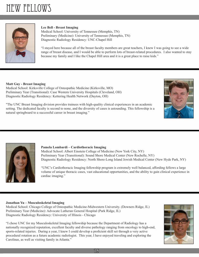
Matt Guy - Breast ImagingMedical School: Kirksville College of Osteopathic Medicine (Kirksville, MO)Preliminary Year (Transitional): Case Western University Hospitals (Cleveland, OH)Diagnostic Radiology Residency: Kettering Health Network (Dayton, OH)
"The UNC Breast Imaging division provides trainees with high-quality clinical experiences in an academic setting. The dedicated faculty is second to none, and the diversity of cases is astounding. This fellowship is a natural springboard to a successful career in breast imaging.”
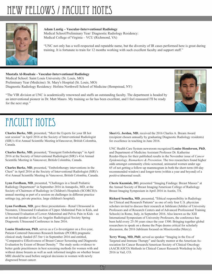
Pamela Lombardi – Cardiothoracic ImagingMedical School: Albert Einstein College of Medicine (New York City, NY)Preliminary Year (Transitional): Sound Shore Medical Center (New Rochelle, NY)Diagnostic Radiology Residency: North Shore-Long Island Jewish Medical Center (New Hyde Park, NY)
“UNC’s Cardiothoracic Imaging fellowship program is extremely well balanced, affording fellows a large volume of unique thoracic cases, vast educational opportunities, and the ability to gain clinical experience in cardiac imaging.”

16
new fellows
Brian Boyd – NeuroradiologyMedical School: Washington University School of Medicine (St. Louis, MO)Preliminary Year (OB/GYN): Naval Medical Center - San DiegoDiagnostic Radiology Residency: UNC-Chapel Hill
“I’ve made many transitions during my post-graduate medical training, but until I started residency at UNC, none had felt like home. There is a camaraderie within the Department of Radiology that I’ve seen matched by none. Add that to a solid caseload, variety of pathologies, and a sense of belonging and I think anyone can see why I stayed!”
Christopher Atkinson – Neuroradiology Medical School: Uniformed Services University of the Health Sciences (Bethesda, MD)Preliminary Year (Transitional) & Diagnostic Radiology Residency:San Antonio Military Medical Center (San Antonio, TX)
“I chose UNC because I wanted an education at a well-respected institution where I could train to be an excellent clinical radiologist. I also chose Chapel Hill because it is a great place to raise a family and I spend much of my free time outside of work with my three-year-old boy and six-year-old girl.”
John Duncan - Neuroradiology Medical School: St. George's University School of Medicine (Grenada, West Indies)Preliminary Year: Nassau University Medical Center (East Meadow, NY)Diagnostic Radiology Residency: St. Barnabas Medical Center (Livingston, NJ)
“In evaluating fellowship programs, I was seeking an institution where I would see a large volume of cases with wide-ranging pathology. I looked for the opportunity to work with some of the most well-respected faculty in their discipline in hopes of bettering my skills as a future subspecialty radiologist. I found these qualities at UNC. Chapel Hill also offers some of the most beautiful landscapes for outdoor activities, especially some of the country’s best golf courses.”
Joseph Fuller - NeuroradiologyMedical School: University of Miami Miller School of MedicineInternship: Swedish Medical Center (Seattle, WA) Diagnostic Radiology Residency: University of Washington (Seattle, WA)
"I chose UNC for my subspecialty training in Neuroradiology because of the institution’s tradiation of excel-lence, the accomplished and approachable Neuroradiology staff and the exposure to a myriad of interesting cases that funnel through this esteemed academic center."
Aaron Kline – Vascular-Interventional RadiologyMedical School: University of Florida (Gainesville, FL)Preliminary Year (Transitional): University of Tennessee - ChattanoogaDiagnostic Radiology Residency: University of Florida (Gainesville, FL)
“The main reason I chose UNC-Chapel Hill for fellowship was because of the people. The people in the VIR section are kind, hard-working and collegial. Additionally, there is a diverse case mix, leadership that is involved with the [American College of Radiology] and [American Board of Radiology], and active STET research.”
17
new fellows / faculty notes
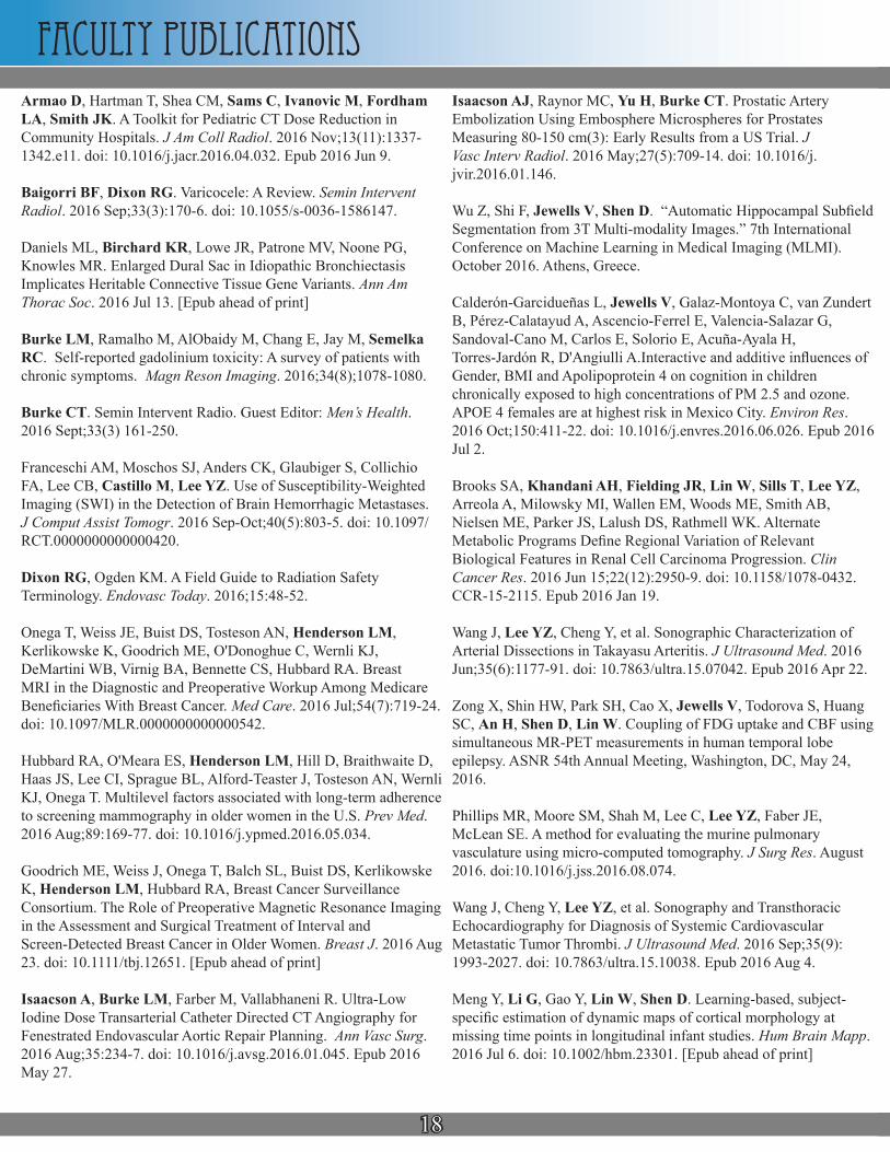
Mustafa Al-Roubaie - Vascular-Interventional RadiologyMedical School: Saint Louis University (St. Louis, MO)Preliminary Year (Medicine): St. Mary's Hospital (St. Louis, MO) Diagnostic Radiology Residency: Hofstra Northwell School of Medicine (Hempstead, NY)
“The VIR division at UNC is academically renowned and staffs an outstanding faculty. The department is headed by an interventional pioneer in Dr. Matt Mauro. My training so far has been excellent, and I feel reassured I'll be ready for the next step.”
Adam Lustig – Vascular-Interventional RadiologyMedical School/Preliminary Year/ Diagnostic Radiology Residency: Medical College of Virginia – VCU (Richmond, VA)
“UNC not only has a well-respected and reputable name, but the diversity of IR cases performed here is great during training. It is fortunate to train for 12 months working with such excellent faculty and support staff.”
faculty notes Charles Burke, MD, presented, “Meet the Experts for your IR hot seat session” in April 2016 at the Society of Interventional Radiologist (SIR)’s 41st Annual Scientific Meeting inVancouver, British Colombia, Canada.
Charles Burke, MD, presented, “Emergent Embolotherapy” in April 2016 at the Society of Interventional Radiologist (SIR)’s 41st Annual Scientific Meeting in Vancouver, British Colombia, Canada.
Charles Burke, MD, presented, “Embolotherapy interventions in the Chest” in April 2016 at the Society of Interventional Radiologist (SIR)’s 41st Annual Scientific Meeting in Vancouver, British Colombia, Canada.
Lynn Fordham, MD, presented, "Challenges in a Small Pediatric Radiology Department" in September 2016 in Annapolis, MD, at the Society of Chairmen of Radiology in Children's Hospitals (SCORCH)'s annual meeting as part of a session on challenges in different practice settings (eg, private practice, large children's hospital).
Lynn Fordham, MD, gave three presentations - Renal Ultrasound in Neonates, Ultrasound Evaluation of Upper Abdominal Pain in Kids, and Ultrasound Evaluation of Lower Abdominal and Pelvic Pain in Kids – as an invited speaker at the Los Angeles Radiological Society Spring Diagnostic Ultrasound Conference in June 2016.
Louise Henderson, PhD, serves as a Co-Investigator on a five-year, Patient-Centered Outcomes Research Institute (PCORI) pragmatic clinical study funded ($7.5m+) in September 2016 and entitled, “Comparative Effectiveness of Breast Cancer Screening and Diagnostic Evaluation by Extent of Breast Density.” The study seeks evidence to better guide practitioners in best screening methods for women with and without dense breasts as well as seek informed insights on whether breast MRI should be used before surgical decisions in women with newly diagnosed breast cancer.
Sheri G. Jordan, MD, received the 2016 Charles A. Bream Award (recipient chosen annually by graduating Diagnostic Radiology residents) for excellence in teaching in June 2016.
UNC Health Care System newsroom recognized Louise Henderson, PhD, and Department of Medicine Assistant Professor Dr. Katherine Reeder-Hayes for their published results in the November issue of Cancer Epidemiology, Biomarkers & Prevention. The two researchers found higher odds amongst community clinic-screened, uninsured women under age 65 of not getting a follow-up mammogram in both the short-term (60-day recommended window) and longer-term (within a year and beyond) of a positive/abnormal result.
Cherie Kuzmiak, DO, presented “Imaging Findings: Breast Masses” at the Annual Society of Breast Imaging/American College of Radiology Breast Imaging Symposium in April 2016 in Austin, TX.
Richard Semelka, MD, presented, "Ethical responsibility in Radiology for Clinical and Research Patients" as one of only four U.S. physician scholars invited to discuss their research at Jubileum (Jubilee of University Professors and of Research Centers and of Advanced Professional Training Schools) in Rome, Italy, in September 2016. Also known as the XIII International Symposium of University Professors, the conference has been held every 25-30 years since the year 1300. Bringing together global researchers to speak on a theme the Pope deems critical for scholarly discussion, the 2016 Jubileum focused on Misericordia (Mercy).
Terry Wong, MD, PhD, served as speaker “Imaging in the Era of Targeted and Immune Therapy” and faculty mentor at the American As-sociation for Cancer Research/American Society of Clinical Oncology (AACR/ASCO) Methods in Clinical Cancer Research Workshop in July 2016 in Vail, CO.
18
Faculty publicationsArmao D, Hartman T, Shea CM, Sams C, Ivanovic M, Fordham LA, Smith JK. A Toolkit for Pediatric CT Dose Reduction in Community Hospitals. J Am Coll Radiol. 2016 Nov;13(11):1337-1342.e11. doi: 10.1016/j.jacr.2016.04.032. Epub 2016 Jun 9.
Baigorri BF, Dixon RG. Varicocele: A Review. Semin Intervent Radiol. 2016 Sep;33(3):170-6. doi: 10.1055/s-0036-1586147.
Daniels ML, Birchard KR, Lowe JR, Patrone MV, Noone PG, Knowles MR. Enlarged Dural Sac in Idiopathic Bronchiectasis Implicates Heritable Connective Tissue Gene Variants. Ann Am Thorac Soc. 2016 Jul 13. [Epub ahead of print]
Burke LM, Ramalho M, AlObaidy M, Chang E, Jay M, Semelka RC. Self-reported gadolinium toxicity: A survey of patients with chronic symptoms. Magn Reson Imaging. 2016;34(8);1078-1080.
Burke CT. Semin Intervent Radio. Guest Editor: Men’s Health. 2016 Sept;33(3) 161-250.
Franceschi AM, Moschos SJ, Anders CK, Glaubiger S, Collichio FA, Lee CB, Castillo M, Lee YZ. Use of Susceptibility-Weighted Imaging (SWI) in the Detection of Brain Hemorrhagic Metastases. J Comput Assist Tomogr. 2016 Sep-Oct;40(5):803-5. doi: 10.1097/RCT.0000000000000420.
Dixon RG, Ogden KM. A Field Guide to Radiation Safety Terminology. Endovasc Today. 2016;15:48-52.
Onega T, Weiss JE, Buist DS, Tosteson AN, Henderson LM, Kerlikowske K, Goodrich ME, O'Donoghue C, Wernli KJ, DeMartini WB, Virnig BA, Bennette CS, Hubbard RA. Breast MRI in the Diagnostic and Preoperative Workup Among Medicare Beneficiaries With Breast Cancer. Med Care. 2016 Jul;54(7):719-24. doi: 10.1097/MLR.0000000000000542.
Hubbard RA, O'Meara ES, Henderson LM, Hill D, Braithwaite D, Haas JS, Lee CI, Sprague BL, Alford-Teaster J, Tosteson AN, Wernli KJ, Onega T. Multilevel factors associated with long-term adherence to screening mammography in older women in the U.S. Prev Med. 2016 Aug;89:169-77. doi: 10.1016/j.ypmed.2016.05.034.
Goodrich ME, Weiss J, Onega T, Balch SL, Buist DS, Kerlikowske K, Henderson LM, Hubbard RA, Breast Cancer Surveillance Consortium. The Role of Preoperative Magnetic Resonance Imaging in the Assessment and Surgical Treatment of Interval and Screen-Detected Breast Cancer in Older Women. Breast J. 2016 Aug 23. doi: 10.1111/tbj.12651. [Epub ahead of print]
Isaacson A, Burke LM, Farber M, Vallabhaneni R. Ultra-Low Iodine Dose Transarterial Catheter Directed CT Angiography for Fenestrated Endovascular Aortic Repair Planning. Ann Vasc Surg. 2016 Aug;35:234-7. doi: 10.1016/j.avsg.2016.01.045. Epub 2016 May 27.
Isaacson AJ, Raynor MC, Yu H, Burke CT. Prostatic Artery Embolization Using Embosphere Microspheres for Prostates Measuring 80-150 cm(3): Early Results from a US Trial. J Vasc Interv Radiol. 2016 May;27(5):709-14. doi: 10.1016/j.jvir.2016.01.146.
Wu Z, Shi F, Jewells V, Shen D. “Automatic Hippocampal Subfield Segmentation from 3T Multi-modality Images.” 7th International Conference on Machine Learning in Medical Imaging (MLMI). October 2016. Athens, Greece.
Calderón-Garcidueñas L, Jewells V, Galaz-Montoya C, van Zundert B, Pérez-Calatayud A, Ascencio-Ferrel E, Valencia-Salazar G, Sandoval-Cano M, Carlos E, Solorio E, Acuña-Ayala H, Torres-Jardón R, D'Angiulli A.Interactive and additive influences of Gender, BMI and Apolipoprotein 4 on cognition in children chronically exposed to high concentrations of PM 2.5 and ozone. APOE 4 females are at highest risk in Mexico City. Environ Res. 2016 Oct;150:411-22. doi: 10.1016/j.envres.2016.06.026. Epub 2016 Jul 2.
Brooks SA, Khandani AH, Fielding JR, Lin W, Sills T, Lee YZ, Arreola A, Milowsky MI, Wallen EM, Woods ME, Smith AB, Nielsen ME, Parker JS, Lalush DS, Rathmell WK. Alternate Metabolic Programs Define Regional Variation of Relevant Biological Features in Renal Cell Carcinoma Progression. Clin Cancer Res. 2016 Jun 15;22(12):2950-9. doi: 10.1158/1078-0432.CCR-15-2115. Epub 2016 Jan 19.
Wang J, Lee YZ, Cheng Y, et al. Sonographic Characterization of Arterial Dissections in Takayasu Arteritis. J Ultrasound Med. 2016 Jun;35(6):1177-91. doi: 10.7863/ultra.15.07042. Epub 2016 Apr 22.
Zong X, Shin HW, Park SH, Cao X, Jewells V, Todorova S, Huang SC, An H, Shen D, Lin W. Coupling of FDG uptake and CBF using simultaneous MR-PET measurements in human temporal lobe epilepsy. ASNR 54th Annual Meeting, Washington, DC, May 24, 2016.
Phillips MR, Moore SM, Shah M, Lee C, Lee YZ, Faber JE, McLean SE. A method for evaluating the murine pulmonary vasculature using micro-computed tomography. J Surg Res. August 2016. doi:10.1016/j.jss.2016.08.074.
Wang J, Cheng Y, Lee YZ, et al. Sonography and Transthoracic Echocardiography for Diagnosis of Systemic Cardiovascular Metastatic Tumor Thrombi. J Ultrasound Med. 2016 Sep;35(9):1993-2027. doi: 10.7863/ultra.15.10038. Epub 2016 Aug 4.
Meng Y, Li G, Gao Y, Lin W, Shen D. Learning-based, subject-specific estimation of dynamic maps of cortical morphology at missing time points in longitudinal infant studies. Hum Brain Mapp. 2016 Jul 6. doi: 10.1002/hbm.23301. [Epub ahead of print]
19
Faculty publicationsJiang W, Li G, Liu H, Shi F, Wang T, Shen C, Shen H, Hu D, Wang W, Shen D. Reduced cortical thickness and increased surface area in antisocial personality disorder. Neuroscience. 2016 Sep 4. pii: S0306-4522(16)30434-1. doi: 10.1016/j.neuroscience.2016.08.052. [Epub ahead of print]
Chen Y, Dhar R, Heitsch L, Ford A, Fernandez-Cadenas I, Carrera C, Montaner J, Lin W, Shen D, An H, Lee JM. Automated quantification of cerebral edema following hemispheric infarction: Application of a machine-learning algorithm to evaluate CSF shifts on serial head CTs. Neuroimage Clin. 2016 Sep 26;12:673-680. eCollection 2016.
Oldan JD, Hawkins AS, Chin BB. (18)F Sodium Fluoride PET/CT in Patients with Prostate Cancer: Quantification of Normal Tissues, Benign Degenerative Lesions, and Malignant Lesions. World J Nucl Med. 2016 May-Aug;15(2):102-8. doi: 10.4103/1450-1147.172301.
Oldan JD, Chin BB. FDG PET/CT Imaging of Prostate Carcinosarcoma. Clin Nucl Med. 2016 Aug;41(8):629-31.
Oldan JD, Patel PS. Positron Emission Tomography/Computed Tomography for Gynecologic Malignancies. Obstet Gynecol Surv. 2016 Sep;71(9):545-56.
Bazyar S, Ramalho J, Eldeniz C, An H, Lee YZ. Comparison of Cerebral Blood Volume and Plasma Volume in Untreated Intracranial Tumors. PLoS One. 2016 Sep 1;11(9):e0161807. doi: 10.1371/journal.pone.0161807. eCollection 2016.
Semelka RC, Commander CW, Jay M, Burke LM, Ramalho M. Presumed Gadolinium Toxicity in Subjects With Normal Renal Function: A Report of 4 Cases. Invest Radiol. 2016 Aug 19.
Semelka RC, Ramalho J, Vakharia A, AlObaidy M, Burke LM, Jay M, Ramalho M. Gadolinium deposition disease: initial description of a disease that has been around for a while. Magn Reson Imaging. 2016 Aug 13.
Semelka RC, Busireddy KKR, Burke LM, Mari-Bonmati L, Ramalho M, et al. Radiologist income, receipts, and academic performance: An Analysis of Many Nations. Acta Radiol. 2016 Feb 27.; PMID: 26924837.
Wu H, Chen G, Jin Y, Shen D, Yap PT. Embarrassingly Parallel Acceleration of Global Tractography via Dynamic Domain Partitioning. Front Neuroinform. 2016 Jul 13;10:25. doi: 10.3389/fninf.2016.00025. eCollection 2016.
Huang L, Jin Y, Gao Y, Thung KH, Shen D; Alzheimer's Disease Neuroimaging Initiative. Longitudinal clinical score prediction in Alzheimer's disease with soft-split sparse regression based random forest. Neurobiol Aging. 2016 Oct;46:180-91. doi: 10.1016/j.neurobiolaging.2016.07.005. Epub 2016 Jul 15.
Qiao L, Zhang H, Kim M, Teng S, Zhang L, Shen D. Estimating functional brain networks by incorporating a modularity prior. Neuroimage. 2016 Nov 1;141:399-407. doi: 10.1016/j.neuroimage.2016.07.058. Epub 2016 Jul 30.
Huang L, Jin Y, Gao Y, Thung KH, Shen D; Alzheimer's Disease Neuroimaging Initiative. Longitudinal clinical score prediction in Alzheimer's disease with soft-split sparse regression-based random forest. Neurobiol Aging. 2016 Oct;46:180-91. doi: 10.1016/j.neurobiolaging.2016.07.005. Epub 2016 Jul 15.
Wu H, Chen G, Jin Y, Shen D, Yap PT. Embarrassingly Parallel Acceleration of Global Tractography via Dynamic Domain Partitioning. Front Neuroinform. 2016 Jul 13;10:25. doi: 10.3389/fninf.2016.00025. eCollection 2016.
Jiang W, Shi F, Liao J, Liu H, Wang T, Shen C, Shen H, Hu D, Wang W, Shen D. Disrupted functional connectome in antisocial personality disorder. Brain Imaging Behav. 2016 Aug 19. [Epub ahead of print]
Zhang Y, Shi F, Wu G, Wang L, Yap PT, Shen D. Consistent Spatial-Temporal Longitudinal Atlas Construction for Developing Infant Brains. IEEE Trans Med Imaging. 2016 Jul 7. [Epub ahead of print]
Zhang H, Chen X, Shi F, Li G, Kim M, Giannakopoulos P, Haller S, Shen D. Topographic Information based High-Order Functional Connectivity and its Application in Abnormality Detection for Mild Cognitive Impairment. J Alzheimers Dis. 2016 Aug 19. [Epub ahead of print]
Zhao F, Qiao L, Shi F, Yap PT, Shen D. Feature fusion via hierarchical supervised local CCA for diagnosis of autism spectrum disorder. Brain Imaging Behav. 2016 Aug 17. [Epub ahead of print]
Yerubandi V, Ronald J, Howard BA, Suhocki PV, James OG, Wong TZ, Kim CY. Patient and tumor characteristics predictive of an elevated hepatopulmonary shunt fraction before radio-embolization of hepatic tumors. Nucl Med Commun. 2016 Sep;37(9):939-46.
Wu G, Shen D, Sabuncu M. Machine Learning and Medical Imaging. Academic Press. 1st Edition August 2016. Cambridge, MA.
Yap PT, Zhang Y, Shen D. Multi-Tissue Decomposition of Diffusion MRI Signals via l0 Sparse-Group Estimation. IEEE Trans Image Process. 2016 Jul 7. [Epub ahead of print]
Yu H, Isaacson AJ, Burke CT. Review of Current Literature for Prostatic Artery Embolization. Semin Intervent Radiol. 2016 Sep;33(3):231-5. doi: 10.1055/s-0036-1586141.
Faculty publications, con't

images newsletter Department of RadiologyCB 7510, Old Clinic BuildingThe University of North CarolinaChapel Hill, NC 27599-7510
Address Service Requested
Final thoughts
This is the last issue of Images in print!! UNC Department of Radiology will be rolling out its first e-newsletter by end of year and looks forward
to keeping its readers informed via email starting early 2017.
If we have not written you already to confirm your email, please submit it at the below link on the Department’s website.
Your email will strictly be used to distribute a more summary e-newsletter to you in quarterly editions.
http://www.med.unc.edu/radiology/Dept-info/department-news/alumni-address-update** Please update other contact information at this link as needed **